research papers 250 doi:10.1107/S2052252514013001 IUCrJ (2014). 1, 250–260 IUCrJ ISSN 2052-2525 BIOLOGY j MEDICINE Received 10 February 2014 Accepted 4 June 2014 Edited by J. L. Smith, University of Michigan, USA ‡ Current address: Liverpool John Moores University, Egerton Court, 2 Rodney Street, Liverpool L1 2UA, England. Keywords: N-myristoyltransferase; inhibitor; ligand binding; Leishmania; drug discovery PDB references: LmNMT–MyrCoA, 4cgp; LmNMT–MyrCoA–6KV, 4cgo; LmNMT– MyrCoA–7AH, 4cgn; LmNMT–MyrCoA–A6K, 4cgl; LmNMT–MyrCoA–CWZ, 4cgm Supporting information: this article has supporting information at www.iucrj.org Diverse modes of binding in structures of Leishmania major N-myristoyltransferase with selective inhibitors James A. Brannigan, a Shirley M. Roberts, a Andrew S. Bell, b Jennie A. Hutton, b Michael R. Hodgkinson, c Edward W. Tate, b Robin J. Leatherbarrow, b ‡ Deborah F. Smith c and Anthony J. Wilkinson a * a Structural Biology Laboratory, Department of Chemistry, University of York, York YO10 5DD, England, b Department of Chemistry, Imperial College London, South Kensington Campus, London SW7 2AZ, England, and c Centre for Immunology and Infection, Department of Biology, University of York, York YO10 5DD, England. *Correspondence e-mail: [email protected]The leishmaniases are a spectrum of global diseases of poverty associated with immune dysfunction and are the cause of high morbidity. Despite the long history of these diseases, no effective vaccine is available and the currently used drugs are variously compromised by moderate efficacy, complex side effects and the emergence of resistance. It is therefore widely accepted that new therapies are needed. N-Myristoyltransferase (NMT) has been validated pre-clinically as a target for the treatment of fungal and parasitic infections. In a previously reported high-throughput screening program, a number of hit compounds with activity against NMT from Leishmania donovani have been identified. Here, high-resolution crystal structures of representative compounds from four hit series in ternary complexes with myristoyl-CoA and NMT from the closely related L. major are reported. The structures reveal that the inhibitors associate with the peptide-binding groove at a site adjacent to the bound myristoyl-CoA and the catalytic -carboxylate of Leu421. Each inhibitor makes extensive apolar contacts as well as a small number of polar contacts with the protein. Remarkably, the compounds exploit different features of the peptide-binding groove and collectively occupy a substantial volume of this pocket, suggesting that there is potential for the design of chimaeric inhibitors with significantly enhanced binding. Despite the high conservation of the active sites of the parasite and human NMTs, the inhibitors act selectively over the host enzyme. The role of conformational flexibility in the side chain of Tyr217 in conferring selectivity is discussed. 1. Introduction The leishmaniases, caused by species of the kinetoplastid parasite Leishmania, are a spectrum of diseases associated with immune dysfunction, with 350 million people at risk in 98 countries where these diseases are endemic (Alvar et al., 2012). Clinical symptoms range from the disfiguring skin lesions of cutaneous leishmaniasis (CL) to the often fatal visceral leishmaniasis (VL) characterized by prolonged fever, enlarged spleen and liver and progressive anaemia. These symptoms are exacerbated in children and the immuno- compromised, such as those diagnosed as human immuno- deficiency virus (HIV) positive, and HIV–VL co-infection is an increasing problem (Alvar et al., 2008). Almost all clinically symptomatic VL patients die within months if untreated. There are currently no anti-leishmanial vaccines licensed for use in humans. The principal drugs used to treat visceral leishmaniasis have been pentavalent
antimonials, but these compounds have toxic side effects and
their effectiveness is threatened by the emergence of drug
resistance, especially in the Indian subcontinent. The recog-
nized alternatives, miltefosine, amphotericin B and paromo-
mycin, suffer from various drawbacks including lack of an oral
formulation, prolonged treatment times, high costs of treat-
ment and toxicity. As a result, the development of new
therapies for treating leishmaniasis is an international priority.
Previous studies have identified myristoyl-CoA:protein
N-myristoyltransferase (NMT) as a promising candidate for
drug development against pathogenic protozoan parasites
(Price et al., 2003; Panethymitaki et al., 2006; Bowyer et al.,
2007, 2008; Brannigan et al., 2010; Frearson et al., 2010).
In eukaryotic cells, NMT catalyses the transfer of the
14-carbon saturated fatty acid myristate from myristoyl-CoA
(MyrCoA) to the amino-terminal glycine of a subset of
proteins. This predominately co-translational modification
contributes to the targeting of substrate proteins to membrane
locations as well as facilitating protein–protein interactions
(Resh et al., 2012). N-Myristoylation by NMT proceeds via
an ordered bi-bi reaction mechanism (Fig. 1a): binding of
MyrCoA generates small conformational changes that enable
docking of the substrate protein and deprotonation of its
�-amino group by the �-carboxylate of the C-terminal residue
acting as a base (Rudnick et al., 1991; Bhatnagar et al., 1994).
The myristate group is then transferred to the N-terminal
glycine of the substrate in a
nucleophilic addition–elimination
reaction with the formation of an
amide bond (Fig. 1a). There
follows stepwise release of first
the free CoA and then the
N-myristoylated protein
(Rudnick et al., 1991; Bhatnagar
et al., 1999). NMTs have been
well characterized in Saccharo-
myces cerevisiae (Duronio et al.,
1989) and human cells (Ducker
et al., 2005) and are essential for
viability in pathogenic fungi
(Lodge et al., 1994).
Comparative sequence and
biochemical analyses demon-
strated high conservation of the
MyrCoA binding sites in the two
human isoforms of the enzyme,
HsNMT1 and HsNMT2, and in
the fungal NMTs, but divergent
peptide-binding specificities
(Johnson et al., 1994). This led
to the development of peptide-
based and peptidomimetic inhi-
bitors that showed selectivity
against the NMT from Candida
albicans relative to human NMT
(Lodge et al., 1997, 1998). As a
consequence, NMT was the target
of antifungal drug-development
programmes in the pharmaceu-
tical industry, with the focus on
selective inhibitors acting at the
peptide-binding pocket. In the
preliminary stages of these
programs, high selectivity and
specificity were achieved around
benzothiazole (Pfizer, unpublished
work) and benzofuran (Roche;
Masubuchi et al., 2001, 2003)
scaffolds. However, the best
leads proved to be specific for
research papers
IUCrJ (2014). 1, 250–260 James A. Brannigan et al. � N-Myristoyltransferase 251
Figure 1The reaction catalysed by N-myristoyltransferase. (a) Top, scheme showing the ordered binding ofmyristoyl-CoA (MyrCoA) and the substrate protein followed by the ordered release of CoASH and themyristoylated product. Bottom, schematic of a step in the reaction mechanism showing the active sitefollowing binding of substrates and abstraction by the �-carboxylate of the C-terminal residue Leu421 ofa proton from the �-amino group of Gly1 of the substrate protein. Thr303 and Asn169 form polarinteractions with the amino group which attacks the carbonyl C atom of MyrCoA. The reactionintermediate is stabilized by interactions with an oxyanion hole formed by the amides of Leu167 andPhe168. This mechanism is adapted from Farazi et al. (2001). (b) Ribbon (left) and electrostatic surface(right) representations of LmNMT with bound MyrCoA and compound 7AH. The ligands bind in anextended cleft running across the molecule that is partially covered by the Ab loop. In the left-hand imagethe ligands are displayed as cylinders and coloured by atom type; in the right-hand image MyrCoA and7AH are shown as green and light green spheres, respectively.
C. albicans and unlikely to give rise to the types of broad-
spectrum drugs (ideally also active against Aspergillus and
Cryptococcus spp.) that would enable them to compete with
current antifungal drugs. Cross-species activity is not essential
in drug development for parasitic infections, which are readily
diagnosed according to clinical, molecular and epidemiolo-
gical indicators. Although there is no conclusive evidence for
toxic effects arising from inhibition of either human NMT,
selectivity for the appropriate parasitic NMT is highly desir-
able.
These considerations suggested NMT as a suitable target
for developing chemotherapeutics against infectious parasites
(Tate et al., 2014) to treat diseases such as malaria (caused by
Plasmodium spp.), leishmaniasis (Leishmania spp.) or African
sleeping sickness (Trypanosoma brucei). To substantiate this
hypothesis, the NMTs of L. major and L. donovani (which
cause CL and VL, respectively), P. falciparum and T. brucei
(Price et al., 2003; Panethymitaki et al., 2006; Bowyer et al.,
2007, 2008; Brannigan et al., 2010) were characterized and
shown to be essential for the viability of these species using
targeted gene disruption, RNAi techniques and chemical
biology approaches (Price et al., 2003, 2010; Wright et al.,
2014). The validity of NMT as a drug target was demonstrated
by the use of high-throughput screening to produce a small-
molecule inhibitor of T. brucei NMT that killed bloodstream
parasites in vivo with high sensitivity and specificity (Frearson
et al., 2010). Most recently, inhibitors of NMT from Plasmo-
dium have been developed (Goncalves, Brannigan, Whalley et
al., 2012; Yu et al., 2012; Rackham et al., 2013, 2014) and were
shown to disrupt the formation of critical subcellular struc-
tures, leading to rapid parasite cell death (Wright et al., 2014).
A complementary high-throughput screen of over 150 000
compounds from the Pfizer Global Diverse Representative
Set against protozoan parasite NMT proteins (Bell et al., 2012)
identified a number of submicromolar inhibitors of L. dono-
vani NMT which also displayed selectivity over the host
(human) enzyme. Since the published in vivo active inhibitors
of T. brucei NMT are reported to have single-digit nanomolar
activity in enzyme and cellular assays, we envisaged that a
further 100-fold to 1000-fold improvement in enzyme affinity
would be required to produce a useful clinical candidate using
a structure-guided approach.
We have resynthesized selected samples of the high-
throughput screen hits (or a close analogue in one case) and
now report the binding modes of four distinct Leishmania-
selective small-molecule inhibitors in crystal structures of
ternary complexes with L. major NMT and MyrCoA co-
substrate. Analysis of the crystal structures has identified key
binding-site residues and strategies to modify the inhibitors to
achieve the desired increase in enzyme affinity and selectivity
over the human NMTs.
2. Materials and methods
2.1. Protein preparation and crystallization
Protein expression and purification was essentially as
described for LdNMT (Brannigan et al., 2010) using clone
LmNMT_SGC:B1 that encodes an N-terminal histidine tag
and a cleavage site for TEV protease (MHHHHHHSSGRE-
NLYFQG) followed by residues 5–421 of LmNMT (Frearson
et al., 2010). Protein at 10 mg ml�1 was incubated at 4�C
overnight with a 1/20th volume of cofactor MyrCoA (10 mM
in 50% DMSO) and crystallized by vapour diffusion using a
mother liquor consisting of 30% PEG 1500, 0.2 M NaCl, 0.1 M
sodium cacodylate pH 5.5. For co-crystallization of 7AH,
ligand at a final concentration of 1 mM was incubated with
LmNMT and MyrCoA as above (32% PEG 1500, 0.2 M NaCl,
0.1 M sodium cacodylate pH 5.6). For the other ligands,
crystal-soaking experiments proved to be more reliable and
convenient. Ligand compounds (25 mM stocks in 50%
DMSO) were added to a stabilization solution (33% PEG
1500, 0.22 M NaCl, 0.11 M sodium cacodylate pH 5.5) to give a
final ligand concentration of 2.5 mM. Ligand solution was used
to replace liquid in crystallization drops containing LmNMT–
MyrCoA crystals by careful pipetting, repeated three times to
completely wash away the original drop solution and left to
soak for 20 h.
2.2. Data collection and refinement
X-ray diffraction data were collected on synchrotron
beamlines at the Diamond Light Source and were processed
using XDS (Kabsch, 2010) and SCALA (Evans, 2006) imple-
mented within xia2 (Winter, 2010). Data-collection and
refinement statistics are summarized in Table 2. The protein
coordinates from PDB entry 3h5z (Frearson et al., 2010) were
used directly for refinement using the maximum-likelihood
methods implemented in REFMAC (Murshudov et al., 2011).
Cycles of refinement using anisotropic temperature factors
were interspersed with model building and adjustment using
Coot (Emsley et al., 2010). The complete chain can be traced
for the protein, with the exception of the N-terminal residues
preceding Ala11 (numbering as in the full-length native
LmNMT protein). These residues are not defined in the
electron-density maps and they are assumed to be disordered.
The final refined protein structure model displays good
geometry, with 97% of the residues in the preferred region
of the Ramachandran plot and only 0.3% (corresponding to
amino-acid residue His347) as outliers. The coordinates and
structure-factor files have been deposited in the Protein Data
Bank under accession codes 4cgp (LmNMT–MyrCoA), 4cgo
(LmNMT–MyrCoA–6KV), 4cgn (LmNMT–MyrCoA–7AH),
4cgl (LmNMT–MyrCoA–A6K) and 4cgm (LmNMT–
MyrCoA–CWZ).
3. Results and discussion
A high-throughput screen against L. donovani NMT (Bell
et al., 2012) identified a number of compound classes with
submicromolar inhibition of the parasite NMT and selectivity
against human NMT. Four chemically distinct scaffolds
† Protein Data Bank three-letter compound codes, PF code as designated by Bell et al. (2012) and IMP code designation for the resynthesized compound (Imperial College,London). ‡ Molecular weight (g mol�1). § Ligand efficiency LE = 1.4(�log LdNMT IC50)/N, where N is the number of non-H atoms (Hopkins et al., 2014).
proximity to the �-carboxylate of the C-terminal residue
Leu421, which plays a catalytic role. The CoA moiety adopts a
compact structure, with the adenine ring surrounded by the
pantetheine and fatty-acyl species. Compound 7AH binds to
the C-terminal lobe that has been shown in the NMTs from
S. cerevisiae (Bhatnagar et al., 1998; Farazi et al., 2001) and
C. albicans (Sogabe et al., 2002) to form the binding site for
peptide substrates or peptidomimetic inhibitors (Fig. 1b).
The five LmNMT structures presented here are closely
similar, with pairwise values for the root-mean-square devia-
tion (r.m.s.d.) in C� coordinate positions in the range 0.2–0.6 A
(Supplementary Fig. S1a). The MyrCoA binding sites are
closely similar and the mode of co-substrate binding is iden-
tical, as shown in Supplementary Fig. S1(b). The most notable
structural changes accompanying binding of the inhibitors
take place in the acidic loop containing residues Glu82-Asp-
Asp-Asp85 (the Ab loop), which has been termed a lid
that closes over the active site upon substrate binding (Fig. 1),
and the glycine-rich segment Gly393-Ala-Gly-Asp-Gly397
situated on the lower surface of the binding site. Comparison
of the binary complexes of LmNMT with MyrCoA and of
LdNMT with a nonhydrolysable analogue of the co-substrate
(PDB entry 2wuu; Brannigan et al., 2010) gives an r.m.s.d.
value of 1.0 A for 402 equivalent C� positions.
3.2. Binding of the thienopyrimidine compound 6KV
This inhibitor is a close analogue of the original high-
throughput screen hit, the only difference being the deletion
of the methyl substituent on the thienopyrimidine ring
(Table 1). Its biological profile is similar to the original hit
compound, with an IC50 value for LdNMT of 0.25 mM and
modest selectivity (approximately tenfold) against human
NMT isoform 1 (HsNMT1). Unexpectedly, two molecules of
compound 6KV occupy the peptide-binding groove of
LmNMT (Fig. 2a, Supplementary Fig. S2a). These will be
referred to as the proximal (P) and distal (D) ligands based on
research papers
254 James A. Brannigan et al. � N-Myristoyltransferase IUCrJ (2014). 1, 250–260
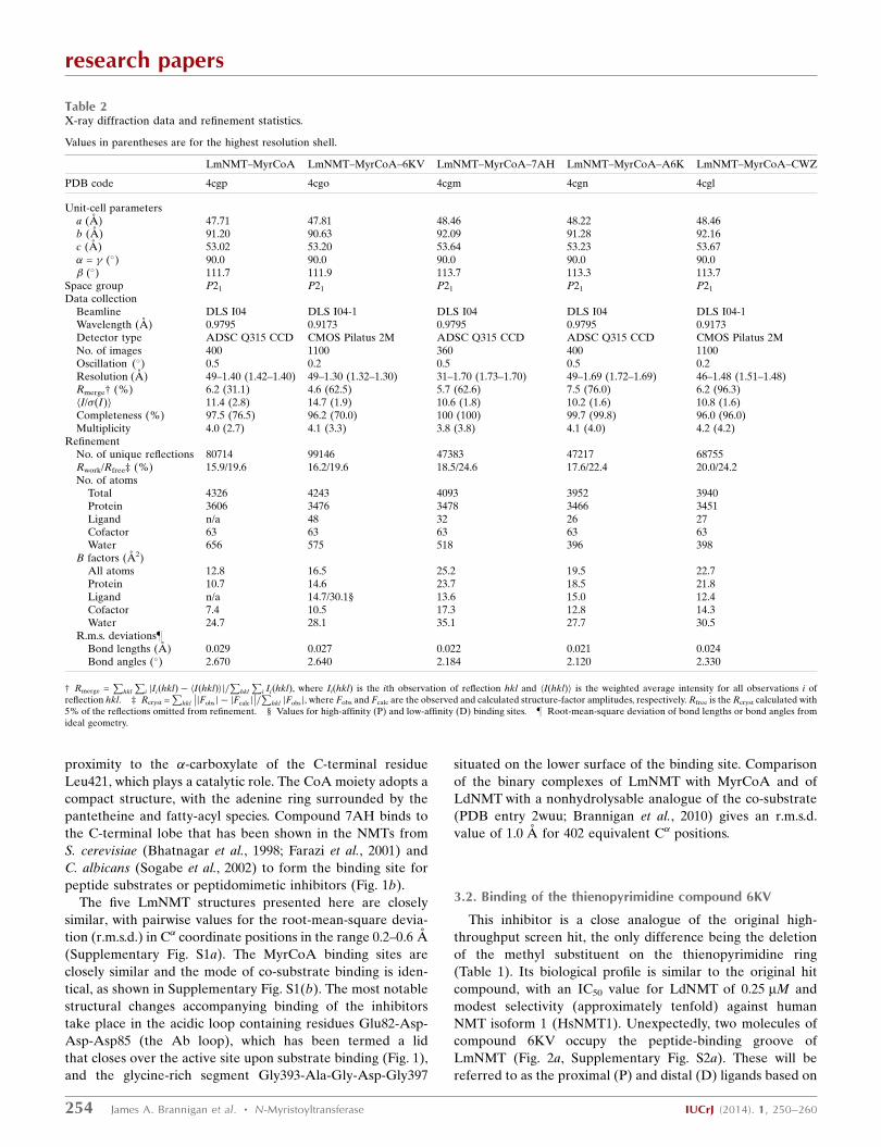
Table 2X-ray diffraction data and refinement statistics.
Values in parentheses are for the highest resolution shell.
Pi IiðhklÞ, where Ii(hkl) is the ith observation of reflection hkl and hI(hkl)i is the weighted average intensity for all observations i of
reflection hkl. ‡ Rcryst =P
hkl
��jFobsj � jFcalcj
��=P
hkl jFobsj, where Fobs and Fcalc are the observed and calculated structure-factor amplitudes, respectively. Rfree is the Rcryst calculated with5% of the reflections omitted from refinement. § Values for high-affinity (P) and low-affinity (D) binding sites. } Root-mean-square deviation of bond lengths or bond angles fromideal geometry.
their positions relative to the MyrCoA ligand (Fig. 3a,
Table 3).
The proximal ligand is expected to be the higher affinity
binding ligand based on its fit to the electron-density maps
(Fig. 2a, Supplementary Fig. S3a) and its lower mean atomic
temperature (B) factor. As is evident, the thienopyrimidine
rings pack together. The two ligands bury 390 and 400 A2,
respectively, of their surface area through contacts with the
protein and a further 80 A2 through contacts with each other
(Table 3). The two molecules are related to one another by
a rotation about an approximately
vertical axis, as viewed in Fig. 2(a),
followed by a translation in the vertical
direction. The piperidine ring in the
distal inhibitor molecule is poorly
defined in the electron-density maps.
The mean temperature factor of the
atoms of the N-methylpiperidine moiety
is 25 A2 higher than the mean value for
the other atoms of the distal ligand. This
suggests that there is rotation about
the N4—C11 bond and that the distal
piperidine ring adopts an ensemble of
conformations. The presence of the
ligand(s) induces ordering of a loop
implicated in substrate binding which
contains the conserved residue Gly397.
Val81, Phe90 and His219 (which is
modelled in two conformations) form
significant interactions with both ligand
molecules. Asp83, Phe88, Phe232 and
Asp396 are prominent in binding the
D ligand molecule, with Gly205 and
Tyr217 forming significant interactions
exclusively with the P ligand.
There are few obvious polar inter-
actions between the protein and either
inhibitor molecule. There is a direct
hydrogen bond between the phenolic
hydroxyl of Tyr345 (2.7 A) and the ring
N atom N4 of the pyrimidine ring. The
N-methylpiperidine ring is protonated
so it will form an ionic interaction with
the carboxylate of Leu421, from which
it is separated by 3.2 A. The same N
atom forms a hydrogen bond (2.9 A) to
a well ordered water molecule (W1),
which may additionally form polar
interactions with the C-terminal
carboxylate and the side chains of resi-
dues Tyr80, Tyr92 and Asn167. The
pendant nitrile group in the proximal
ligand is oriented towards the MyrCoA
(nitrile N to cofactor S distance of
4.2 A) and is surrounded by Val81,
Ala204, Gly205 and a number of solvent
molecules. For the distal ligand, this
moiety is projecting into a pocket
circumscribed by Phe90, Phe232,
Ser330, Leu341, Ala343, Tyr345 and
Val374, with the potential for a
hydrogen bond between the N atom of
research papers
IUCrJ (2014). 1, 250–260 James A. Brannigan et al. � N-Myristoyltransferase 255
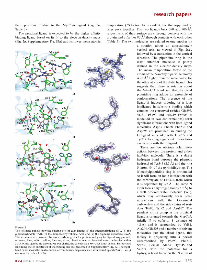
Figure 2The left-hand panels show the binding site for each ligand: (a) the thienopyrimidine 6KV, (b) thepiperidinylindole 7AH, (c) the aminoacylpyrrolidine A6K and (d) the biphenyl derivative CWZ.The structures are coloured by atom; carbon, green for protein and grey for ligand; oxygen, red;nitrogen, blue; sulfur, yellow; fluorine, silver; chlorine, mauve. Selected water molecules within3.5 A of the ligands are also shown. For clarity, the co-substrate MyrCoA is not shown. Stereoviews(including the co-substrate) of the binding site are presented in Supplementary Fig. S2. The right-hand panel shows the final refined electron-density map associated with bound ligand (2mFo�DFc)contoured at a level of 1�.
the nitrile and the side-chain amino group of Asn376
(3.2 A).
3.3. Binding of the piperidinylindole compound 7AH
Compound 7AH inhibits LdNMT with an IC50 value of
0.3 mM with excellent (�200-fold) selectivity against HsNMT1
(Table 1). When bound to LmNMT, 7AH effectively wraps
around the side chain of Phe90, with its phenyl and piperidine
rings packing onto opposite faces of the aromatic ring of the
side chain; meanwhile, the amide linker between the indole
and fluorophenyl species packs against the edge of the Phe90
ring (Fig. 2b, Supplementary Fig. S2b). 510 A2 of its 570 A2 of
surface area is buried by interaction with the protein. The
faces of the indole ring itself pack
against the edges of the aromatic rings
of the side chains of Tyr217 and Tyr345.
In this structure, there is a noticeable
ordering of the Ab loop containing the
acidic segment Glu82-Asp-Asp-Asp85
and there is the possibility of a polar
interaction with fluorine. Since this
halogen atom rarely acts as a hydrogen-
bond acceptor, we postulate a dipole–
dipole interaction with a backbone
amide group.
The piperidine ring forms an ion pair
(2.8 A) with the �-carboxylate of
Leu421 and is additionally solvated by
two water molecules which include W1
and form a local network of polar
interactions extending to residues
Tyr80, Tyr92, Asn167 and Thr203.
Elsewhere, the carbonyl of the amide of
the ligand forms hydrogen bonds to the
side-chain hydroxyl of Tyr345 (2.7 A)
and the amide amino group of Asn376
(3.2 A), the amide carbonyl of which
forms a strong hydrogen bond to N� of
His219, which is well ordered in this
complex. This local network of
hydrogen bonding is completed by a
water molecule which forms hydrogen
bonds to the ligand carbonyl and the
side chains of residues Tyr345 and
Asn376.
3.4. Binding of the amino-acylpyrrolidine compound A6K
This ligand was identified through
analogue screening of the primary high-
throughput screening hit and has the
highest IC50 value (0.08 mM for
LdNMT) among the four inhibitors
presented here. Its IC50 against the
human enzyme is 5 mM, giving a selec-
tivity factor of �80 (Table 1).
Compound A6K is seen bound as a
single diastereomer (SRR) and strik-
ingly has a compact structure when
bound to LmNMT (Fig. 2c, Supple-
mentary Fig. S2c). Adoption of this type
of conformation has been referred to as
research papers
256 James A. Brannigan et al. � N-Myristoyltransferase IUCrJ (2014). 1, 250–260
Figure 3Binding of ligands and MyrCoA cofactor. Molecules are in cylinder representation in all panels. Theatoms are coloured by element (oxygen, red; nitrogen, blue; sulfur, yellow; phosphorus, magenta;fluorine, silver; chlorine, mauve) with the C atoms coloured by inhibitor: (a) 6KV, grey; (b) 7AH,coral; (c) A6K, green; (d) CWZ, pink; (e) 646 (PDB entry 2wsa), ice blue. In ( f ) the MyrCoA andthe inhibitors in (a)–(d) are overlaid. (g, h) Two views of the inhibitor ligands overlaid. For clarity,only the proximal 6KV molecule is shown in the overlaps.
hydrophobic collapse (Wiley & Rich, 1993). The first chloro-
phenyl group packs between Tyr217 and Tyr345 and above
Val378, with the plane of its ring perpendicular to the plane of
the pyrrolidine ring. The second of the two chlorophenyl rings
is folded back over the pyrrolidine ring, with which it lies in an
approximately parallel plane. It makes extensive apolar
interactions with Val81 and Phe90.
The exocyclic hydroxymethyl group displaces a water
molecule (W3), the position of which is conserved in the other
ligand complexes, and makes two strong interactions with the
hydroxyl group of Tyr326 (2.6 A) and an O atom of the
�-carboxylate of Leu421 (2.6 A) as part of a local network of
polar interactions that include the carbonyl O atom of Met420.
The folding of the inhibitor projects the primary amino group
in the direction of the MyrCoA co-substrate. Thr203 makes
bridging contacts to this primary amine group as well as to
the carbonyl O atom of the ligand through hydrogen bonds
formed to its main-chain carbonyl and side-chain hydroxyl
groups, respectively. The primary amine of the inhibitor forms
additional polar interactions with the side-chain amide
carbonyl of Asn167 and a water molecule (W2); the latter also
forms hydrogen bonds to the main-chain amide and carbonyl
groups of Thr203.
The chloro substituents of the two aromatic rings project
away from the protein core. One Cl atom (Cl1) is surrounded
by four water molecules, two of which are within 3.3 A. It is
3.9 A from the Tyr217 hydroxyl, and could contribute a rela-
tively weak binding interaction, given the potential for organic
chlorines to interact with protein Lewis bases via halogen
bonding (Sirimulla et al., 2013).
3.5. Binding of the biphenyl-derivative compound CWZ
This inhibitor has an IC50 value of 0.9 mM for LdNMT and
45 mM for HsNMT1, giving a selectivity factor of 50. The
relatively weak binding of this compound is reflected in the
Relative to the other three inhibitors described here, it also
has a low ligand efficiency value (Hopkins et al., 2014) of 0.29
(Table 1). The binding of this ligand has the effect of ordering
the acidic Glu82–Asp85 region of the Ab loop and there is a
single orientation of residue His219, which has lost its inter-
action with Asn376.
This ligand makes extensive and largely apolar interactions
with the protein, with 630 of its 700 A2 of accessible surface
area buried by interactions with protein residues. In a similar
manner to compound 7AH, compound CWZ wraps around
the side-chain aromatic group of Phe90, while the biphenyl
species packs between the side chains of Tyr217 and Tyr345
(Fig. 2d, Supplementary Fig. S2d). There is a dearth of polar
protein–inhibitor interactions. The only obvious strong
hydrogen bond is formed between the N atom of the thiazole
moiety and the hydroxyl of Ser330, where the contact distance
is 2.7 A. At the other end of the molecule, there are two
possible orientations of the pyridine ring arising from rotation
about the C1—C2 bond. In the orientation shown, the pyri-
dine N atom is situated 3.5 A from a water molecule and 3.7 A
from the S atom of the MyrCoA co-substrate; in the other
orientation it would be 3.7 A from the phenolic hydroxyl of
Tyr345.
3.6. Comparison of binding with the inhibitor DDD85646
DDD85646 (646) is a potent inhibitor of NMT from
T. brucei (IC50 = 2 nM) that was evolved from a compound
discovered in a high-throughput screen (Frearson et al., 2010).
This compound has low selectivity, inhibiting HsNMT1 with an
IC50 of 12 nM and LdNMT with an IC50 value of below 6 nM.
As shown in Fig. 3(e), Supplementary Fig. S2(e) and Table 3,
when bound to LmNMT compound 646 occupies the same
pocket as the inhibitors described above, contacting essen-
tially the same set of protein residues. It binds in a reasonably
extended conformation, with three of its ring elements being
close to coplanar but with the pyrazole ring projecting in a
perpendicular direction, induced by the sulfonamide linker.
At the extremes of the molecule, the piperazine N atom forms
an ion-pairing interaction with the �-carboxylate of Leu421,
while the N atom of the pyrazole forms a hydrogen bond to
the hydroxyl of Ser330. Of the inhibitors discussed above,
research papers
IUCrJ (2014). 1, 250–260 James A. Brannigan et al. � N-Myristoyltransferase 257
Table 3Residue surface area buried upon inhibitor binding.
The surface area of each residue buried by the binding of the inhibitor wasdetermined using PISA (Krissinel & Henrick, 2007). Interactions involving adirect hydrogen-bonding, ion–dipole or ion–ion interaction with the ligand aredenoted in bold.
Protein residue Surface area buried by inhibitor (A2)
CWZ alone forms a hydrogen bond to Ser330. However, CWZ
is also alone in failing to form a direct interaction with Leu421.
Thus, the capacity to span the pocket and develop polar
interactions with both Ser330 and Leu421 in the leishmanial
NMTs, built into DDD85646 during an extended medicinal
chemistry campaign, probably accounts for its much higher
potency relative to the high-throughput screen hits described
here. From the superposition shown in Fig. 3, it is apparent
that the three contiguous rings of 646 overlap most closely
with the proximal-pocket bound 6KV, with the fourth pyrazole
ring overlaying the fluorophenyl ring of 7AH and the thiazolo-
piperidine ring of CWZ. In contrast to the latter two ligands,
646 navigates a more direct route from the
vicinity of the protein C-terminus to the
distal site.
3.7. Comparison of the inhibitor-bindingsites and the basis of selectivity
The inhibitor-binding sites of the four
ternary complexes described above together
with that in the LmNMT–MyrCoA binary
complex are compared in Fig. 4. A number
of residues are modelled in two conforma-
tions consistent with the electron-density
maps. His219 is especially interesting in its
conformational flexibility. In the binary
complex and in the ternary complexes with
the ligands 6KV and A6K, two conforma-
tions (inward and outward) are evident. In
the complex with 7AH, the inward (m,
gauche�) conformation alone is apparent,
in contrast to the complex with CWZ where
the outward (p, gauche+) conformation
alone is observed. In the 7AH complex, the
inward orientation of His219 is favoured by
the hydrogen-bonding network extending
from the ligand via the Asn376 side-chain
amide to the imidazole side chain. In
contrast, in the CWZ complex the inward
conformation is sterically hindered by the
biphenyl moiety. His219 has been shown to
be an important residue in yeast NMT since
a mutant with a substitution at this position
is not viable (Farazi et al., 2001). In complex
with the peptide GLYASKLA, His219
interacts with Ser5, which is a strongly
preferred residue at this position in
substrates of NMT.
Also apparent from Fig. 4 are distinct
conformations of residue Tyr217. An
inwardly directed conformer is observed
in the ternary complex with 6KV, while the
other outwardly directed conformer is
observed in the complexes with 7AH, A6K
and CWZ. In the binary complex with
MyrCoA both conformers are present.
Tyr217 is a key residue in the binding of all
four inhibitors, contributing 34–58 A2 of
interfacial surface.
The selectivity of the inhibitors for the
NMTs from Leishmania relative to those
from human cannot be explained straight-
research papers
258 James A. Brannigan et al. � N-Myristoyltransferase IUCrJ (2014). 1, 250–260
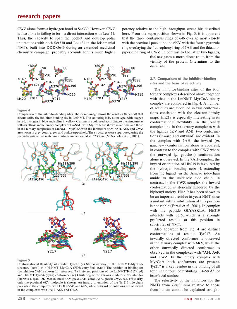
Figure 4Comparison of the inhibitor-binding sites. The stereo image shows the residues (labelled) thatcircumscribe the inhibitor-binding site in LmNMT. The colouring is by atom type, with oxygenin red, nitrogen in blue and sulfur in yellow. C atoms are coloured according to the structure asfollows. Those in the binary complex of LmNMTwith MyrCoA are shown in ice blue and thosein the ternary complexes of LmNMT–MyrCoA with the inhibitors 6KV, 7AH, A6K and CWZare shown in grey, coral, green and pink, respectively. The structures were superposed using thesecondary-structure matching routines implemented in CCP4mg (McNicholas et al., 2011).
Figure 5Conformational flexibility of residue Tyr217. (a) Stereo overlay of the LmNMT–MyrCoAstructure (coral) with HsNMT–MyrCoA (PDB entry 3iu1, cyan). The position of binding forthe inhibitor 7AH is shown for reference. (b) Preferred positions of the LmNMT Tyr217 (red)and HsNMT Tyr296 (cyan) conformers. (c) Clustering of the various inhibitors. No inhibitor(HsNMT), cyan; DDD85646, blue; 6KV, grey; 7AH, coral; A6K, green; CWZ, red. For clarity,only the proximal 6KV molecule is shown. An inward orientation of the Tyr217 side chainprevails in the complexes with DDD85646 and 6KV, while outward orientations are observedin the complexes with 7AH, A6K and CWZ.
forwardly by mapping the residues contacting the inhibitors in
the structures onto the aligned sequences of the proteins. On
the contrary, all of the amino-acid side chains that make
significant interactions with the inhibitors in LdNMT/
LmNMT, defined either by a sizable contribution to the ligand
interface or their participation in polar interactions with the
latter (Table 3), are conserved in human NMT.
To explore the origins of selectivity in more detail, we
compared the structure of the LmNMT complex with
MyrCoA with that of the binary complex of HsNMT with
unpublished work). This comparison confirms the similarity of
the inhibitor-binding pockets in the two enzymes and the close
superposition of corresponding residues (Fig. 5a). The most
obvious differences occur in the Ab loop, which is ‘more
closed’ in the human NMT. The comparison suggests Tyr217
(Tyr296 in HsNMT) as a potential selectivity-conferring
residue. In the binary complexes of LmNMT and HsNMT, the
tyrosine appears as two conformers with inward and outward
side-chain orientations. For HsNMT the inwardly oriented
conformation appears to be preferred, whereas for LmNMT
the outward orientation predominates (Fig. 5b). This may be
significant because the LmNMT inhibitors are juxtaposed
differently with respect to this tyrosine residue. The ligands
with the higher selectivity towards the Leishmania NMTs
(compounds 7AH, A6K and CWZ) bind to the enzyme so as
to occlude the inwardly oriented conformation of the side
chain of the tyrosine residue (Fig. 5c) and would clash with the
preferred conformer of HsNMT Tyr296. In contrast, the less
selective ligands 646 and 6KV allow the inward orientation of
this aromatic side chain. This would introduce selectivity if the
�G value for the inward to outward transition in the Tyr
conformer is more positive for Tyr296 in HsNMT than it is for
Tyr217 in the Leishmania NMTs. The potential role of the
conformation of the corresponding tyrosine residue (211) in
determining the selectivity of benzofuran-based inhibitors for
P. falciparum NMT over HsNMT has been discussed
previously (Yu et al., 2012).
4. Summary and perspectives
The structures presented here reveal the mode of binding of a
set of four Leishmania NMT inhibitors emerging from a high-
throughput screen. The three compounds with higher ligand
efficiency each interact with the C-terminal carboxylate of
the enzyme through a basic centre. Each inhibitor develops
significant interactions with the aromatic side chains of Phe90,
Tyr217 and Tyr345 as well as a set of interactions characteristic
of each ligand. The next step is to use medicinal chemistry
approaches to develop these hits into leads which inhibit in the
nanomolar range. The higher ligand efficiency of the 6KV,
7AH and A6K compounds suggest that the prospects for
success in developing higher affinity are good. The potent
inhibitor 646 makes a polar interaction with Ser330 and such
an interaction could be engineered into compounds from
these three series, most notably 7AH which reaches up
towards this residue. On the other hand, the CWZ molecule,
which does form a polar contact with Ser330, could be adapted
to form a direct interaction with the �-carboxylate. The
ligands approach the Leu421 carboxylate from different
directions and moreover they use different basic/hydrogen-
bond donor groups to form interactions with it, suggesting that
there is scope for the energetic contribution of this interaction
to be augmented.
The structures of the bound inhibitors, which explore
different aspects of the peptide-binding pocket in NMT,
indicate that chimaeric molecules may be an effective route to
explore possibilities for increased binding. For example, the
close superposition of the second (chloro)phenyl ring of
compound A6K and the six-membered ring of the indole of
7AH in their complexes with LmNMT suggests that hybrid
molecules could be developed combining the higher affinity
binding determinants of each molecule. Another possibility
would be to exploit the close overlap of the core elements of
646 and 6KV by grafting the pyrazole element of the latter
onto the former. A third possibility arises from the binding of
two molecules of 6KV to adjacent sites in the inhibitor pocket,
which suggests that a higher affinity covalently linked dimer of
this molecule could be developed. Finally, we note that the
open character of the active site gives scope for finding
additional binding compounds through further screening.
Acknowledgements
This work was supported by the Wellcome Trust (grant No.
087792). We wish to thank Raymond Hui (SGC, Toronto) for
plasmid LmNMT_SGC:B1, David Robinson (Dundee) for
advice on crystal soaking, Nishant Varshney (Pune) for
LdNMT crystallizations, Johan Turkenburg and Sam Hart
(York) for help with X-ray data collection and Diamond Light
Source (Harwell, England) for synchrotron facilities.
References
Alvar, J., Aparicio, P., Aseffa, A., Den Boer, M., Canavate, C., Dedet,J. P., Gradoni, L., Ter Horst, R., Lopez-Velez, R. & Moreno, J.(2008). Clin. Microbiol. Rev. 21, 334–359.
Alvar, J., Velez, I. D., Bern, C., Herrero, M., Desjeux, P., Cano, J.,Jannin, J. & den Boer, M. (2012). PLoS One, 7, e35671.
Bell, A. S., Mills, J. E., Williams, G. P., Brannigan, J. A., Wilkinson,A. J., Parkinson, T., Leatherbarrow, R. J., Tate, E. W., Holder, A. A.& Smith, D. F. (2012). PLoS Negl. Trop. Dis. 6, e1625.
Bhatnagar, R. S., Futterer, K., Farazi, T. A., Korolev, S., Murray, C. L.,Jackson-Machelski, E., Gokel, G. W., Gordon, J. I. & Waksman, G.(1998). Nature Struct. Biol. 5, 1091–1097.
Bhatnagar, R. S., Futterer, K., Waksman, G. & Gordon, J. I. (1999).Biochim. Biophys. Acta, 1441, 162–172.
Bhatnagar, R. S., Jackson-Machelski, E., McWherter, C. A. &Gordon, J. I. (1994). J. Biol. Chem. 269, 11045–11053.
Bowyer, P. W., Gunaratne, R. S., Grainger, M., Withers-Martinez, C.,Wickramsinghe, S. R., Tate, E. W., Leatherbarrow, R. J., Brown, K.A., Holder, A. A. & Smith, D. F. (2007). Biochem. J. 408, 173–180.
Bowyer, P. W., Tate, E. W., Leatherbarrow, R. J., Holder, A. A., Smith,D. F. & Brown, K. A. (2008). ChemMedChem, 3, 402–408.
Brannigan, J. A., Smith, B. A., Yu, Z., Brzozowski, A. M.,Hodgkinson, M. R., Maroof, A., Price, H. P., Meier, F.,Leatherbarrow, R. J., Tate, E. W., Smith, D. F. & Wilkinson, A. J.(2010). J. Mol. Biol. 396, 985–999.
Ducker, C. E., Upson, J. J., French, K. J. & Smith, C. D. (2005). Mol.Cancer Res. 3, 463–476.
research papers
IUCrJ (2014). 1, 250–260 James A. Brannigan et al. � N-Myristoyltransferase 259
Duronio, R. J., Towler, D. A., Heuckeroth, R. O. & Gordon, J. I.(1989). Science, 243, 796–800.
Emsley, P., Lohkamp, B., Scott, W. G. & Cowtan, K. (2010). ActaCryst. D66, 486–501.
Evans, P. (2006). Acta Cryst. D62, 72–82.Farazi, T. A., Waksman, G. & Gordon, J. I. (2001). Biochemistry, 40,
6335–6343.Frearson, J. A. et al. (2010). Nature (London), 464, 728–732.Goncalves, V., Brannigan, J. A., Thinon, E., Olaleye, T. O., Serwa, R.,
Lanzarone, S., Wilkinson, A. J., Tate, E. W. & Leatherbarrow, R. J.(2012). Anal. Biochem. 421, 342–344.
Goncalves, V., Brannigan, J. A., Whalley, D., Ansell, K. H., Saxty, B.,Holder, A. A., Wilkinson, A. J., Tate, E. W. & Leatherbarrow, R. J.(2012). J. Med. Chem. 55, 3578–3582.
Hopkins, A. L., Keseru, G. M., Leeson, P. D., Rees, D. C. & Reynolds,C. H. (2014). Nature Rev. Drug Discov. 13, 105–121.
Johnson, D. R., Bhatnagar, R. S., Knoll, L. J. & Gordon, J. I. (1994).Annu. Rev. Biochem. 63, 869–914.
Kabsch, W. (2010). Acta Cryst. D66, 125–132.Krissinel, E. & Henrick, K. (2007). J. Mol. Biol. 372, 774–797.Lodge, J. K., Jackson-Machelski, E., Devadas, B., Zupec, M. E.,
Getman, D. P., Kishore, N., Freeman, S. K., McWherter, C. A.,Sikorski, J. A. & Gordon, J. I. (1997). Microbiology, 143, 357–366.
Lodge, J. K., Jackson-Machelski, E., Higgins, M., McWherter, C. A.,Sikorski, J. A., Devadas, B. & Gordon, J. I. (1998). J. Biol. Chem.273, 12482–12491.
Lodge, J. K., Johnson, R. L., Weinberg, R. A. & Gordon, J. I. (1994). J.Biol. Chem. 269, 2996–3009.
Masubuchi, M., Ebiike, H., Kawasaki, K., Sogabe, S., Morikami, K.,Shiratori, Y., Tsujii, S., Fujii, T., Sakata, K., Hayase, M., Shindoh,H., Aoki, Y., Ohtsuka, T. & Shimma, N. (2003). Bioorg. Med. Chem.11, 4463–4478.
Masubuchi, M., Kawasaki, K., Ebiike, H., Ikeda, Y., Tsujii, S., Sogabe,S., Fujii, T., Sakata, K., Shiratori, Y., Aoki, Y., Ohtsuka, T. &Shimma, N. (2001). Bioorg. Med. Chem. Lett. 11, 1833–1837.
McNicholas, S., Potterton, E., Wilson, K. S. & Noble, M. E. M. (2011).Acta Cryst. D67, 386–394.
Murshudov, G. N., Skubak, P., Lebedev, A. A., Pannu, N. S., Steiner,R. A., Nicholls, R. A., Winn, M. D., Long, F. & Vagin, A. A. (2011).Acta Cryst. D67, 355–367.
Panethymitaki, C., Bowyer, P. W., Price, H. P., Leatherbarrow, R. J.,Brown, K. A. & Smith, D. F. (2006). Biochem. J. 396, 277–285.
Price, H. P., Guther, M. L., Ferguson, M. A. & Smith, D. F. (2010).Mol. Biochem. Parasitol. 169, 55–58.
Price, H. P., Menon, M. R., Panethymitaki, C., Goulding, D., McKean,P. G. & Smith, D. F. (2003). J. Biol. Chem. 278, 7206–7214.
Rackham, M. D., Brannigan, J. A., Moss, D. K., Yu, Z., Wilkinson,A. J., Holder, A. A., Tate, E. W. & Leatherbarrow, R. J. (2013). J.Med. Chem. 56, 371–375.
Rackham, M. D., Brannigan, J. A., Rangachari, K., Meister, S.,Wilkinson, A. J., Holder, A. A., Leatherbarrow, R. J. & Tate, E. W.(2014). J. Med. Chem. 57, 2773–2788.
Resh, M. D. (2012). Trends Mol. Med. 18, 206–214.Rudnick, D. A., McWherter, C. A., Rocque, W. J., Lennon, P. J.,
Getman, D. P. & Gordon, J. I. (1991). J. Biol. Chem. 266, 9732–9739.
Sirimulla, S., Bailey, J. B., Vegesna, R. & Narayan, M. (2013). J. Chem.Inf. Model. 53, 2781–2791.
Sogabe, S., Masubuchi, M., Sakata, K., Fukami, T. A., Morikami, K.,Shiratori, Y., Ebiike, H., Kawasaki, K., Aoki, Y., Shimma, N.,D’Arcy, A., Winkler, F. K., Banner, D. W. & Ohtsuka, T. (2002).Chem. Biol. 9, 1119–1128.
Tate, E. W., Bell, A. S., Rackham, M. D. & Wright, M. H. (2014).Parasitology, 141, 37–49.
Weston, S. A., Camble, R., Colls, J., Rosenbrock, G., Taylor, I.,Egerton, M., Tucker, A. D., Tunnicliffe, A., Mistry, A., Mancia, F.,de La Fortelle, E., Irwin, J., Bricogne, G. & Pauptit, R. A. (1998).Nature Struct. Biol. 5, 213–221.
Wiley, R. A. & Rich, D. H. (1993). Med. Res. Rev. 13, 327–384.Winter, G. (2010). J. Appl. Cryst. 43, 186–190.Wright, M. H. et al. (2014). Nature Chem. 6, 112–121.Yu, Z., Brannigan, J. A., Moss, D. K., Brzozowski, A. M., Wilkinson,
A. J., Holder, A. A., Tate, E. W. & Leatherbarrow, R. J. (2012). J.Med. Chem. 55, 8879–8890.
research papers
260 James A. Brannigan et al. � N-Myristoyltransferase IUCrJ (2014). 1, 250–260