An Alzheimer’s Disease-Derived BiomarkerSignature Identifies Parkinson’s DiseasePatients with DementiaYosef Berlyand1, Daniel Weintraub2, Sharon X. Xie3, Ian A. Mellis1, Jimit Doshi4,Jacqueline Rick1, Jennifer McBride5,6, Christos Davatzikos4, Leslie M. Shaw5,Howard Hurtig1, John Q. Trojanowski5,6, Alice S. Chen-Plotkin1*
1 Department of Neurology, Perelman School of Medicine at the University of Pennsylvania, Philadelphia,Pennsylvania, United States of America, 2 Department of Psychiatry, Perelman School of Medicine at theUniversity of Pennsylvania, Philadelphia, Pennsylvania, United States of America, 3 Department ofBiostatistics and Epidemiology, Perelman School of Medicine at the University of Pennsylvania, Philadelphia,Pennsylvania, United States of America, 4 Department of Radiology, Perelman School of Medicine at theUniversity of Pennsylvania, Philadelphia, Pennsylvania, United States of America, 5 Department ofPathology and Laboratory Medicine, Perelman School of Medicine at the University of Pennsylvania,Philadelphia, Pennsylvania, United States of America, 6 Center for Neurodegenerative Disease Research,Perelman School of Medicine at the University of Pennsylvania, Philadelphia, Pennsylvania, United States ofAmerica
AbstractBiomarkers from multiple modalities have been shown to correlate with cognition in Parkin-
son’s disease (PD) and in Alzheimer’s disease (AD). However, the relationships of these
markers with each other, and the use of multiple markers in concert to predict an outcome of
interest, are areas that are much less explored. Our objectives in this study were (1) to eval-
uate relationships among 17 biomarkers previously reported to associate with cognition in
PD or AD and (2) to test performance of a five-biomarker classifier trained to recognize AD
in identifying PD with dementia (PDD). To do this, we evaluated a cross-sectional cohort of
PD patients (n = 75) across a spectrum of cognitive abilities. All PD participants had 17
baseline biomarkers from clinical, genetic, biochemical, and imaging modalities measured,
and correlations among biomarkers were assessed by Spearman’s rho and by hierarchical
clustering. We found that internal correlation among all 17 candidate biomarkers was mod-
est, showing a maximum pairwise correlation coefficient of 0.51. However, a five-marker
subset panel derived from AD (CSF total tau, CSF phosphorylated tau, CSF amyloid beta
42, APOE genotype, and SPARE-AD imaging score) discriminated cognitively normal PD
patients vs. PDD patients with 80% accuracy, when employed in a classifier originally
trained to recognize AD. Thus, an AD-derived biomarker signature may identify PDD
patients with moderately high accuracy, suggesting mechanisms shared with AD in some
PDD patients. Based on five measures readily obtained during life, this AD-derived signa-
ture may prove useful in identifying PDD patients most likely to respond to AD-based cross-
over therapies.
PLOS ONE | DOI:10.1371/journal.pone.0147319 January 26, 2016 1 / 17
OPEN ACCESS
Citation: Berlyand Y, Weintraub D, Xie SX, Mellis IA,Doshi J, Rick J, et al. (2016) An Alzheimer’s Disease-Derived Biomarker Signature Identifies Parkinson’sDisease Patients with Dementia. PLoS ONE 11(1):e0147319. doi:10.1371/journal.pone.0147319
Editor: Pradeep Garg, Biomedical ResearchFoundation, UNITED STATES
Data Availability Statement: Data from the UPennUdall cohort is included as S1 Dataset. The de-identified dataset will also be available for downloadfrom the Parkinson's Disease Biomarker ProgramData Management Resource (https://pdbp.ninds.nih.gov). The ADNI dataset is already publicly availablevia the ADNI website (http://www.adni-info.org), butfor transparency of methodology, we also include theextracted data used in this manuscript as S2 Dataset.
Funding: The funding for this actual study was fromthe NIH only (P50 NS053488, U01 NS082134).
IntroductionParkinson disease (PD) is the second most common adult-onset neurodegenerative disease,affecting an estimated 1 million people in the United States alone [1,2]. PD is pathologicallycharacterized by the loss of dopaminergic neurons in the substantia nigra of the brain and thepresence of Lewy body inclusions containing alpha-synuclein [3].
In addition to the hallmark motor symptoms of bradykinesia, tremor, and rigidity, most PDpatients develop cognitive impairment (CI), with up to 83% progressing to dementia [4–7]. PDpatients who progress to dementia (PDD) have reduced quality of life and independence [6,8],which adds to the cost of care [9], and results in additional stress for families and caregivers[9].
The need to develop and to understand the role of various biomarkers for PD, and for endo-phenotypes within PD, is increasingly recognized [10–13]. Defined by the National Institutesof Health Biomarkers Definitions Working Group, a biomarker is “a characteristic that isobjectively measured and evaluated as an indicator of normal biological processes, pathogenicprocesses, or pharmacologic responses to a therapeutic intervention” [14]. While common usesof the term often refer to biochemical or imaging-based measures, in the most generic form,biomarkers might originate from multiple types of data. For the purposes of this paper, we con-sidered clinical, genetic, biochemical, and imaging-based biomarkers.
Although the field is still young, and far from consensus, various candidate biomarkersfrom these disparate modalities have been reported to associate with cognitive performance inPD (S1 Table). Clinical correlates of poorer cognition in PD include older age [4], greatermotor severity [15], male sex [16], a motor phenotype characterized by postural instability andgait disorder (PIGD) [4], increased disease duration [17], and the presence of hallucinations[18] or depression [15]. From the genetics literature, variation in APOE [19],MAPT [20],COMT [20], and GBA [21] have been reported to moderate risk for CI in PD. Previouslyreported biochemical correlates of cognitive decline in PD include lower levels of cerebrospinalfluid (CSF) amyloid beta, specifically the disease-implicated form of amyloid beta known asAβ42 [22,23], and lower levels of plasma epidermal growth factor (EGF) [24,25], while higherlevels of CSF total tau (t-tau) and phosphorylated tau (p-tau) associate with poorer cognitionin Alzheimer disease (AD) [23]. Finally, many imaging-based measures have been reported toassociate with cognitive performance in PD [26]; as one example, a global pattern of brain atro-phy known as the Spatial Pattern of Atrophy for Recognition of AD (SPARE-AD) has beenreported to associate with cognitive decline in both AD [27] and PD [28].
It is notable that many of the biochemical and imaging-based biomarkers for cognitive per-formance in PD are “crossover” biomarkers from AD. No doubt this reflects in part the relativepaucity of available candidate PD biomarkers [29]. In addition, however, neuropathologicaldata suggest that CI in PD and in AD may in fact share some mechanistic underpinnings. Spe-cifically, postmortem examinations demonstrate that 29–50% of PDD patients have Aβ plaquesand neurofibrillary tangles characteristic of AD [30–34] concurrent with PD-defining alpha-synuclein pathology.
While there is an emerging literature on individual PD biomarkers, how biomarkers fromvarious modalities correlate with each other, and whether they represent the same vs. differentunderlying biological processes, is not well understood. In the present study, we cross-section-ally evaluated a panel of 17 biomarkers spanning multiple modalities in a densely-characterizedcohort of 75 PD patients across a spectrum of cognitive abilities. We sought to understand therelationship of these 17 biomarkers to each other. Additionally, because 5/17 markers in ourpanel have been strongly implicated in AD, we tested the hypothesis that an AD-derived bio-marker signature might identify PDD as well.
An AD-Derived Biomarker Signature Identifies PDD Patients
PLOS ONE | DOI:10.1371/journal.pone.0147319 January 26, 2016 2 / 17
Competing Interests: Dr. Trojanowski may accruerevenue in the future on patents submitted by theUniversity of Pennsylvania wherein he is co-Inventorand he received revenue from the sale of Avid to EliLilly as co-inventor on imaging related patentssubmitted by the University of Pennsylvania. Hereceives research support from the NIH, GSK,Janssen, Biogen and several non-profits. Dr. Chen-Plotkin is the inventor of a patent pending from theUniversity of Pennsylvania on biomarkers forParkinson’s disease diagnosis; the biomarkerscovered by this patent do not concern cognition. Dr.Weintraub has received research funding or supportfrom Michael J. Fox Foundation for Parkinson’sResearch, NIH, Novartis Pharmaceuticals,Department of Veterans Affairs, and AvidRadiopharmaceuticals; honoraria from AbbVie, Biotie,Teva Pharmaceuticals, Otsuka, UCB, Clintrex LLC,and the CHDI Foundation; license fee payments fromUPenn for the QUIP and QUIP-RS; royalties fromWolters Kluweland; and fees for legal consultation forlawsuit related to antipsychotic prescribing in a PDpatient.
MethodsPlease see S1 Methods for additional details.
ParticipantsUPenn Udall Cohort. The University of Pennsylvania Institutional Review Board approved
the protocols and consent procedures. Written informed consent was obtained from all partici-pants in the study. In the case of individuals with limited capacity to consent, consent was obtainedfrom an authorized representative of the patient. Additional details may be found in S1Methods.
75 patients were prospectively enrolled into the present study from the UPenn subspecialtymovement disorders clinic, provided that they met the UK Brain Bank diagnostic criteria for PD[35]. These individuals represent the first 75 subjects prospectively enrolled for the Intensive Assess-ment Cohort of the UPenn Udall Center, which has a planned enrollment of 150 PD patients. Allpatients first presented to clinical attention for PD symptoms; however, a subset developed demen-tia over the course of disease, and these individuals already met criteria for PDD by the time ofenrollment into this study. For each patient, 17 candidate biomarkers (Table 1, S1 Table) were cap-tured within one year, representing the baseline visit for each patient’s enrollment into the study.
Candidate biomarkers were nominated from the existing literature on cognitive biomarkersin PD or AD, and they spanned multiple types of data: clinical, genetic, biochemical, and imag-ing [36,37].
ADNI Cohort. Background information on the Alzheimer’s Disease Neuroimaging Initia-tive (ADNI) cohort was recently reviewed [36], and appears in the ADNI database (adni.loni.usc.edu) and the S1 Methods.
For the current study, CSF Aβ42, CSF t-tau, CSF p-tau, SPARE-AD score, and APOE geno-type were downloaded from the ADNI website for all cognitively normal (n = 109) and AD(n = 101) patients with complete information available for all five biomarkers.
Neuropsychological AssessmentTheMattis Dementia Rating Scale-2 (DRS) was used to assess cognitive performance [38]. Age-adjusted scores were used for all analyses. The 15-item Geriatric Depression Scale was used toassess presence of depressive symptoms as previously described [39]. The presence and severity ofhallucinations was ascertained by the Thought Disorder item of the Unified PD Rating Scale(UPDRS) Part I [40]. The Hopkins Verbal Learning Test-Revised (HVLT-R) [41,42] was used as asecond test of memory domain function, as previously described [43]. HVLT-R scores for totalimmediate free recall and recognition discrimination [43], standardized from published norms[42], were analyzed. For those patients for whom severity of dementia prevented completion of theHVLT-R, a floor score of 20 (T-score of 20 = three standard deviations below the mean) was used.
Classification of PD patients as PD-CN, PD-MCI, or PDDCognitive status of our PD cohort, i.e. cognitively normal (PD-CN), mild cognitive impairment(PD-MCI), or dementia (PDD) was determined by expert clinical consensus at the UPennUdall Center as previously described [44]. In brief, data considered for assignment of cognitivecategory included treating clinician impression, clinical chart data, psychometric test data, andmeasures of function in activities of daily living. Additional details are provided in S1 Methods.
Motor AssessmentMotor severity of PD symptoms was assessed by the UPDRS Part III (UPDRS-III) score [40]and by the Modified Hoehn and Yahr (MODHY) score [45,46].
An AD-Derived Biomarker Signature Identifies PDD Patients
PLOS ONE | DOI:10.1371/journal.pone.0147319 January 26, 2016 3 / 17
Genetic TestingPeripheral blood DNAwas genotyped forMAPT, COMT, and APOE variants using real-time alle-lic discrimination with Applied Biosystem (ABI) TaqMan probes as previously described [19].
GBAmutation analysis was performed as previously described [47] using long-range PCRfollowed by sequencing of all 11 GBA exons and intron-exon boundaries.
Additional details are provided in S1 Methods.
Biochemical TestingCSF Aβ42, t-tau, and p-tau were measured as previously described, using the Innogenetics(INNOBIA AlzBio3) reagent on the xMAP Luminex platform [23,48].
Plasma EGF was measured as previously described, using a commercially available enzyme-linked immunosorbent assay (R&D Systems) [25].
Table 1. Comparison of biomarker data among PD-CN, PD-MCI, and PDD patients.
Biomarker Modality Features PD-CN PD-MCI PDD P-value
Clinical Sex 0.347a
Male (%) 35 (74%) 15 (75%) 8 (100%)
Female (%) 12 (26%) 5 (25%) 0 (0%)
Age at Plasma Median in yrs (IQR) 66 (61.5–71.0) 66 (63.0–70.3) 77.5 (71.8–79.0) 0.003b
Disease Duration Median in yrs (IQR) 6.7 (4.7–11.3) 6.9 (3.9–10.9) 10.5 (5.4–14.0) 0.696b
UPDRS III Median (IQR) 19 (13.3–25.0) 23 (18.0–32.5) 34.5 (30.0–41.5) <0.001b
MODHY Median (IQR) 2.0 (2.0–2.5) 2.5 (2.0–3.0) 2.75 (2.0–3.0) 0.009b
GDS Median (IQR) 2.0 (1.0–4.0) 3.0 (2.0–6.0) 3.0 (1.75–3.0) 0.179b
Tremor:PIGD Ratio Median (IQR) 0.8 (0.0–1.5) 0.5 (0.1–1.7) 0 (0–1.4) 0.676b
UPDRS Thought Disorder Median (IQR) 0.0 (0.0–1.0) 1.0 (0.0–1.0) 0.0 (0.0–1.0) 0.51b
Genetic APOE E4 Allele Count 0.534a
0 (%) 33 (70%) 14 (70%) 4 (50%)
1 (%) 14 (30%) 6 (30%) 4 (50%)
2 (%) 0 (0%) 0 (0%) 0 (0%)
MAPT H1/H1 0.592a
No (%) 15 (32%) 5 (25%) 1 (13%)
Yes (%) 32 (68%) 15 (75%) 7 (88%)
GBA Mutant 1a
No (%) 45 (96%) 19 (95%) 8 (100%)
Yes (%) 2 (4%) 1 (5%) 0 (0%)
COMT Met Allele Count 0.774a
0 (%) 14 (30%) 6 (30%) 2 (25%)
1 (%) 21 (45%) 11 (55%) 3 (38%)
2 (%) 12 (26%) 3 (15%) 3 (38%)
Biochemical CSF Aβ42 Median in pg/mL (IQR) 278.0 (232.0–303.7) 253.5 (210.5–281.0) 169.8 (149.4–217.3) 0.014b
CSF t-tau Median in pg/mL (IQR) 40 (33.5–50.0) 44 (35.5–64.3) 57.2 (20.2–97.5) 0.637b
CSF p-tau Median in pg/mL (IQR) 20 (14.0–25) 18 (15.75–28.5) 22.4 (14.8–24.6) 0.718b
Plasma EGF Median in pg/mL (IQR) 17.7 (10.6–52.1) 30 (13.4–76.74) 91 (62.6–196.4) 0.013b
An AD-Derived Biomarker Signature Identifies PDD Patients
PLOS ONE | DOI:10.1371/journal.pone.0147319 January 26, 2016 4 / 17
Additional details of sample collection, measurement, and quality control are provided inS1 Methods.
ImagingA SPARE-AD score was assigned for each participant as previously described [28]. In brief, theSPARE-AD score reflects the overall similarity between pattern of atrophy seen in a particularindividual and a generic pattern reflective of AD.
Statistical AnalysisAll statistical analyses were performed in R (http://www.r-project.org). Additional details areprovided in the S1 Methods, and scripts are available in the S1 Scripts.
Multiple imputation. A panel of 17 biomarkers was assessed in all patients for a 98%complete dataset (1251/1275 data points available). The 24 missing data points were multiplyimputed using the “mi” package in R [49].
Bivariate analyses. Bivariate comparisons of PD-CN vs. PDD for candidate biomarkerswere evaluated by the non-parametric Mann-Whitney U-test, with Bonferroni correction.
Spearman correlation, hierarchical clustering, and logistic regression classifier. Pair-wise Spearman correlation coefficients were calculated for assessment of internal correlationsamong continuous markers and markers with at least 5 categories, comprising 12 candidatemarkers. Partial pairwise Spearman correlation coefficients, adjusted for cognition (age-adjusted DRS score), were similarly calculated.
Patients and biomarkers were hierarchically clustered by Euclidean distance using averagelinkage, and heatmaps generated for visualization. Prior to clustering, distributions for bio-markers were tested for normality (Shapiro-Wilk test); biomarkers that were non-normally dis-tributed were log-transformed (CSF t-tau, age, disease duration, plasma EGF), thenstandardized by setting the mean of each variable to zero with a standard deviation of one.
For classification of AD vs. normal control samples, a five-marker logistic regression classi-fier was trained on ADNI data and ten-fold cross-validated. The classifier was then evaluatedfor performance in identifying PDD patients in the UPenn Udall cohort.
Results
Patient characteristicsSeventy-five PD patients were prospectively enrolled at the UPenn Udall Center. Of these 75patients, 47 (63%) were classified as PD-CN, 20 (27%) as PD-MCI, and 8 (11%) as PDD(Table 1, Fig 1A). Objective assessment with the DRS corroborated a range of cognitive perfor-mance (Fig 1B).
Bivariate cross-sectional analysis of candidate biomarkersdemonstrates an association between cognition and two candidatebiomarkersWe first sought to replicate previously-reported findings [15,23,25,28] of differences betweenPD-CN and PDD patients in our current cohort. Of 17 candidate biomarkers, eight(UPDRS-III, MODHY, depression, CSF Aβ42, CSF t-tau, CSF p-tau, plasma EGF, SPARE-AD)had been previously shown to associate cross-sectionally (vs. longitudinally) with baseline cog-nition in either PD or AD [15,23,25,28].
After Bonferroni correction, significant associations were detected between PDD and twocandidate biomarkers. Specifically, PDD patients had greater motor severity as measured by
An AD-Derived Biomarker Signature Identifies PDD Patients
PLOS ONE | DOI:10.1371/journal.pone.0147319 January 26, 2016 5 / 17
the UPDRS-III (corrected p = 0.002) and exhibited a global brain atrophy pattern similar toAD as captured by the SPARE-AD score [28] (corrected p = 0.031). In addition, PDD patientstrended towards lower levels of CSF Aβ42 (corrected p = 0.062). PD-MCI patients had valuesintermediate to PD-CN and PDD for these three biomarkers (Table 1, Fig 1C–1F).
Fig 1. Cohort characteristics and bivariate cross-sectional analyses. (A) By consensus clinical determination, 47/75 patients were classified as PD-CN,20/75 PD-MCI, and 8/75 as PDD. (B) Histogram and kernel density plot of age-adjusted DRS scores for the study cohort. Dashed vertical lines representseparations between PDD vs. PD-MCI vs. PD-CN ranges for DRS performance. (C) A bivariate analysis of eight candidate markers previously reported toassociate cross-sectionally with cognition confirms associations between PDD and two candidate markers in the present study: Unified Parkinson’s DiseaseRating Scale Motor score (UPDRS III, corrected p = 0.002) and Spatial Pattern of Atrophy for Recognition of AD score (SPARE-AD, corrected p = 0.031). Anadditional marker–CSFmeasures of Aβ42 –trended towards association with PDD (corrected p = 0.062). Vertical line indicates corrected p<0.05.EGF = Epidermal Growth Factor. MODHY = Modified Hoehn and Yahr. The other nine candidate markers assessed in this study have previously beenreported to associate with longitudinal decline in cognition. (D)–(F) Boxplots of the distribution of UPDRS-III score, CSF Aβ42 levels, and SPARE-AD scoreamong the three cognitive classes. Median and interquartile range are shown.
doi:10.1371/journal.pone.0147319.g001
An AD-Derived Biomarker Signature Identifies PDD Patients
PLOS ONE | DOI:10.1371/journal.pone.0147319 January 26, 2016 6 / 17
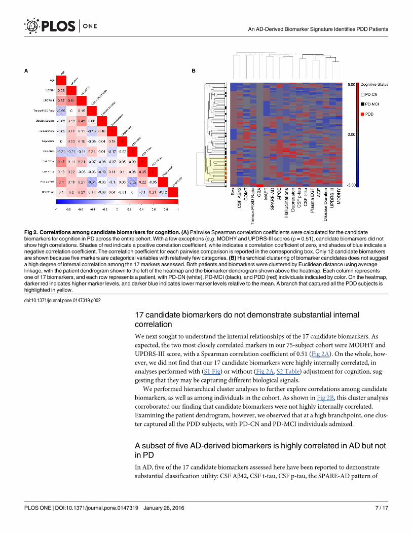
17 candidate biomarkers do not demonstrate substantial internalcorrelationWe next sought to understand the internal relationships of the 17 candidate biomarkers. Asexpected, the two most closely correlated markers in our 75-subject cohort were MODHY andUPDRS-III score, with a Spearman correlation coefficient of 0.51 (Fig 2A). On the whole, how-ever, we did not find that our 17 candidate biomarkers were highly internally correlated, inanalyses performed with (S1 Fig) or without (Fig 2A, S2 Table) adjustment for cognition, sug-gesting that they may be capturing different biological signals.
We performed hierarchical cluster analyses to further explore correlations among candidatebiomarkers, as well as among individuals in the cohort. As shown in Fig 2B, this cluster analysiscorroborated our finding that candidate biomarkers were not highly internally correlated.Examining the patient dendrogram, however, we observed that at a high branchpoint, one clus-ter captured all the PDD subjects, with PD-CN and PD-MCI individuals admixed.
A subset of five AD-derived biomarkers is highly correlated in AD but notin PDIn AD, five of the 17 candidate biomarkers assessed here have been reported to demonstratesubstantial classification utility: CSF Aβ42, CSF t-tau, CSF p-tau, the SPARE-AD pattern of
Fig 2. Correlations among candidate biomarkers for cognition. (A) Pairwise Spearman correlation coefficients were calculated for the candidatebiomarkers for cognition in PD across the entire cohort. With a few exceptions (e.g. MODHY and UPDRS-III scores (ρ = 0.51), candidate biomarkers did notshow high correlations. Shades of red indicate a positive correlation coefficient, white indicates a correlation coefficient of zero, and shades of blue indicate anegative correlation coefficient. The correlation coefficient for each pairwise comparison is reported in the corresponding box. Only 12 candidate biomarkersare shown because five markers are categorical variables with relatively few categories. (B) Hierarchical clustering of biomarker candidates does not suggesta high degree of internal correlation among the 17 markers assessed. Both patients and biomarkers were clustered by Euclidean distance using averagelinkage, with the patient dendrogram shown to the left of the heatmap and the biomarker dendrogram shown above the heatmap. Each column representsone of 17 biomarkers, and each row represents a patient, with PD-CN (white), PD-MCI (black), and PDD (red) individuals indicated by color. On the heatmap,darker red indicates higher marker levels, and darker blue indicates lower marker levels relative to the mean. A branch that captured all the PDD subjects ishighlighted in yellow.
doi:10.1371/journal.pone.0147319.g002
An AD-Derived Biomarker Signature Identifies PDD Patients
PLOS ONE | DOI:10.1371/journal.pone.0147319 January 26, 2016 7 / 17
global brain atrophy, and genotype at the APOE locus [19,22,23,28]. We therefore investigatedthese five AD-derived markers in 109 cognitively normal and 101 AD subjects from the ADNIcohort.
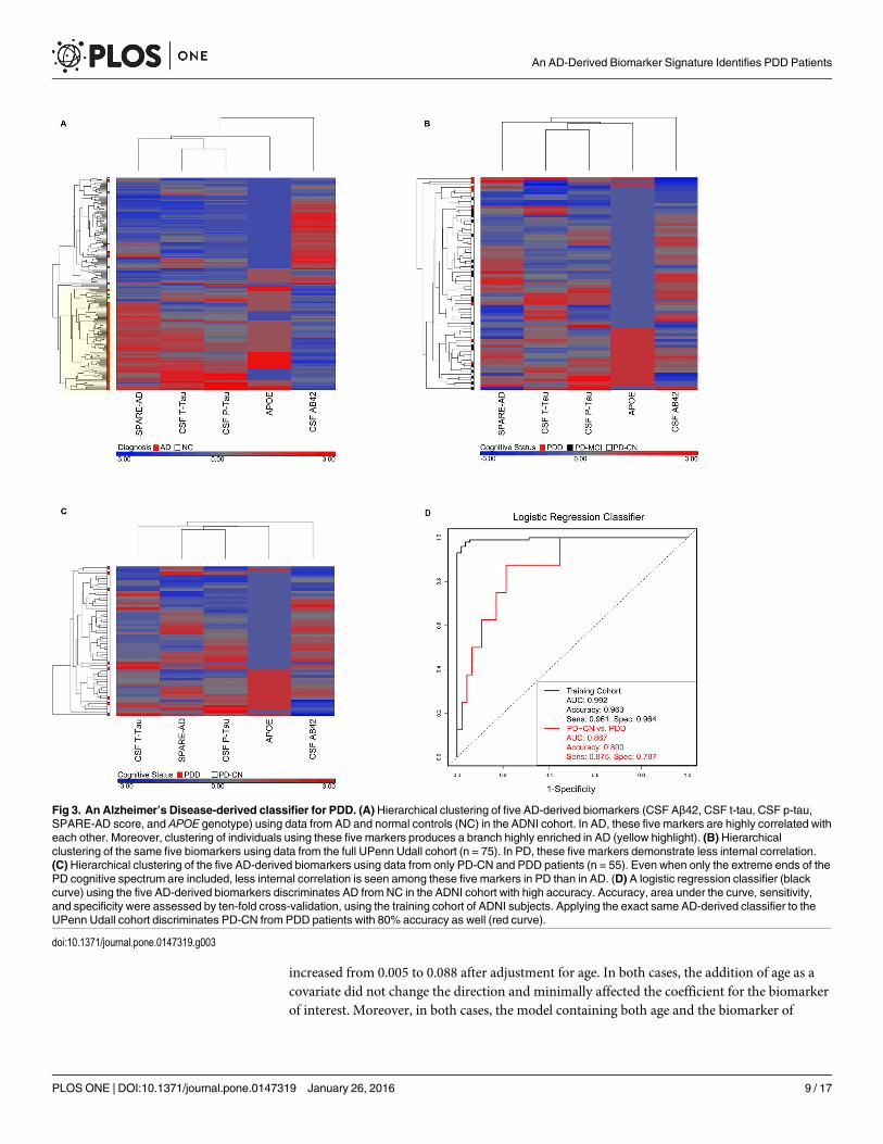
CSF Aβ42, CSF t-tau, CSF p-tau, SPARE-AD score, and APOE genotype were strongly cor-related in ADNI subjects (Fig 3A). Moreover, evaluating subjects by hierarchical cluster analy-sis based on these five markers in the ADNI cohort resulted in a large cluster highly enrichedin AD patients.
When we performed a similar analysis using the five AD biomarkers in the UPenn Udallcohort, however, we found that they were less internally correlated in PD patients (Fig 3B).Omitting the PD-MCI group, in order to focus on the extremes of PDD and PD-CN, did notchange this result (Fig 3C).
An AD-derived classifier identifies PDD subjects with 80% accuracyAlthough the five-marker panel of CSF p-tau, CSF t-tau, SPARE-AD score, APOE genotype,and CSF Aβ42 shows less internal correlation in the PD cohort than in ADNI, these five mark-ers might nevertheless prove useful in classifying PD-CN vs. PDD patients. Indeed, some haveargued that in at least some PD patients, dementia results from an “Alzheimer”-ization of thebrain concomitant with the development of Lewy body inclusions containing alpha-synuclein[50].
To evaluate this hypothesis, we first trained a logistic regression classifier using these fivemarkers to classify cognitively normal vs. AD subjects from ADNI.
As expected based on the cluster analyses, a classifier using CSF p-tau, CSF t-tau, SPA-RE-AD score, APOE genotype and CSF Aβ42 measures performed well in discriminating ADfrom cognitively normal ADNI subjects. Indeed, in ten-fold cross-validation within the ADNIcohort, our logistic regression classifier separated AD vs. normal subjects with an accuracy of>96% (95% CI 0.93–0.98, area under the receiver operating curve (AUC) 0.99, sensitivity 0.96,specificity 0.96, Fig 3D).
The logistic regression classifier was trained in the ADNI cohort to identify AD; we nextasked if this exact classifier could discriminate PD-CN vs. PDD patients from the UPenn Udallcohort. Indeed, the five-marker AD-derived classifier distinguished PD-CN from PDD subjectswith 80% accuracy (95% CI 0.67–0.90, AUC 0.87, sensitivity 0.88, specificity 0.79, Fig 3D). Re-running this analysis using the 74 PD individuals with no imputed data (one SPARE-AD scorefor one PD patient was imputed) did not change the results (accuracy 0.80, AUC 0.86).
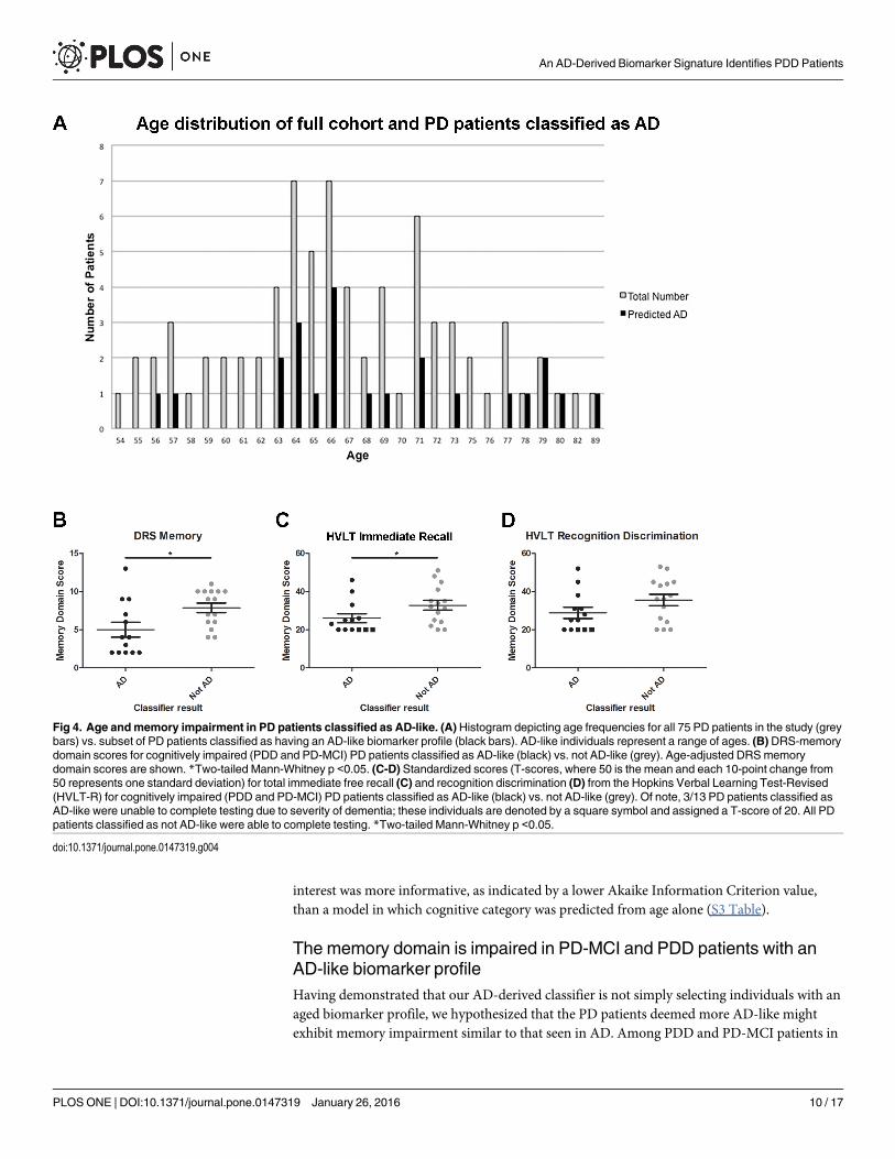
Because our cohort was prospectively enrolled, PD patients in different cognitive categorieswere not age-matched, reflecting, instead, the innate distribution of ages found in PD patientsacross a spectrum of cognitive performance. As a consequence, PDD patients were significantlyolder than individuals in other cognitive categories. We therefore asked whether the older ageof the PDD individuals could be driving the ability of our AD-derived classifier to identifythem. As shown in Fig 4A, however, PD patients classified as AD-like by this five-marker clas-sifier represent a range of ages, with two-thirds falling within the interquartile range of ages forthe PD-CN group.
We further asked whether each of the markers individually associated with cognitive cate-gory and whether this association persisted after adjustment for age. Of the five markers com-prising our AD-derived classifier, only two associated significantly with cognitive category inbivariate logistic regressions–SPARE-AD score and CSF Aβ42. Notably, in the five-markerAD-derived classifier, SPARE-AD score contributed most to classification, with the largestcoefficient. SPARE-AD score remained significantly associated with cognitive category afteradjustment for age, while the p-value for association of CSF Aβ42 levels with cognitive category
An AD-Derived Biomarker Signature Identifies PDD Patients
PLOS ONE | DOI:10.1371/journal.pone.0147319 January 26, 2016 8 / 17
increased from 0.005 to 0.088 after adjustment for age. In both cases, the addition of age as acovariate did not change the direction and minimally affected the coefficient for the biomarkerof interest. Moreover, in both cases, the model containing both age and the biomarker of
Fig 3. An Alzheimer’s Disease-derived classifier for PDD. (A) Hierarchical clustering of five AD-derived biomarkers (CSF Aβ42, CSF t-tau, CSF p-tau,SPARE-AD score, and APOE genotype) using data from AD and normal controls (NC) in the ADNI cohort. In AD, these five markers are highly correlated witheach other. Moreover, clustering of individuals using these five markers produces a branch highly enriched in AD (yellow highlight). (B) Hierarchicalclustering of the same five biomarkers using data from the full UPenn Udall cohort (n = 75). In PD, these five markers demonstrate less internal correlation.(C) Hierarchical clustering of the five AD-derived biomarkers using data from only PD-CN and PDD patients (n = 55). Even when only the extreme ends of thePD cognitive spectrum are included, less internal correlation is seen among these five markers in PD than in AD. (D) A logistic regression classifier (blackcurve) using the five AD-derived biomarkers discriminates AD from NC in the ADNI cohort with high accuracy. Accuracy, area under the curve, sensitivity,and specificity were assessed by ten-fold cross-validation, using the training cohort of ADNI subjects. Applying the exact same AD-derived classifier to theUPenn Udall cohort discriminates PD-CN from PDD patients with 80% accuracy as well (red curve).
doi:10.1371/journal.pone.0147319.g003
An AD-Derived Biomarker Signature Identifies PDD Patients
PLOS ONE | DOI:10.1371/journal.pone.0147319 January 26, 2016 9 / 17
interest was more informative, as indicated by a lower Akaike Information Criterion value,than a model in which cognitive category was predicted from age alone (S3 Table).
The memory domain is impaired in PD-MCI and PDD patients with anAD-like biomarker profileHaving demonstrated that our AD-derived classifier is not simply selecting individuals with anaged biomarker profile, we hypothesized that the PD patients deemed more AD-like mightexhibit memory impairment similar to that seen in AD. Among PDD and PD-MCI patients in
Fig 4. Age andmemory impairment in PD patients classified as AD-like. (A) Histogram depicting age frequencies for all 75 PD patients in the study (greybars) vs. subset of PD patients classified as having an AD-like biomarker profile (black bars). AD-like individuals represent a range of ages. (B) DRS-memorydomain scores for cognitively impaired (PDD and PD-MCI) PD patients classified as AD-like (black) vs. not AD-like (grey). Age-adjusted DRSmemorydomain scores are shown. *Two-tailed Mann-Whitney p <0.05. (C-D) Standardized scores (T-scores, where 50 is the mean and each 10-point change from50 represents one standard deviation) for total immediate free recall (C) and recognition discrimination (D) from the Hopkins Verbal Learning Test-Revised(HVLT-R) for cognitively impaired (PDD and PD-MCI) PD patients classified as AD-like (black) vs. not AD-like (grey). Of note, 3/13 PD patients classified asAD-like were unable to complete testing due to severity of dementia; these individuals are denoted by a square symbol and assigned a T-score of 20. All PDpatients classified as not AD-like were able to complete testing. *Two-tailed Mann-Whitney p <0.05.
doi:10.1371/journal.pone.0147319.g004
An AD-Derived Biomarker Signature Identifies PDD Patients
PLOS ONE | DOI:10.1371/journal.pone.0147319 January 26, 2016 10 / 17
our cohort, 13 were classified by the five-marker panel as AD-like, whereas 15 were not. Thesetwo groups were compared for performance on memory-specific tests. We note that PD-MCIindividuals were also included in this analysis because nearly all the PDD patients were classi-fied as AD-like, with the consequence that an analysis confined to PDD individuals would notallow comparison between balanced groups with vs. without an AD-like biomarker profile.
On the memory domain portion of the DRS, the 13 PD patients identified as AD-like dem-onstrated significantly poorer performance, compared to the 15 PD individuals deemed notAD-like by our logistic regression classifier (two-tailed Mann-Whitney p-value = 0.012, Fig4B). Similar results were observed for a second test of memory, the Hopkins Verbal LearningTest-Revised (HVLT-R) [41,42], for which average T-scores (standardized from publishednorms [42]) on immediate free recall (Fig 4C) and recognition discrimination (Fig 4D) werelower in AD-like PD individuals, compared to PD individuals classified as not AD-like. Whiledifferences in total immediate recall were significant between groups (two-tailed Mann-Whit-ney p-value = 0.050), differences in recognition discrimination showed only a non-significanttrend (two-tailed Mann-Whitney p-value = 0.129).
DiscussionBiomarker studies in neurodegenerative diseases are becoming increasingly common. How-ever, in most cases, a small number of candidate markers, usually from the same modality, areused to predict an outcome of interest. As a consequence, we gain an increasing number ofassociations between individual markers and various clinical outcomes, but the relationshipsamong these individual markers remain unclear.
In PD, the clinical outcome of CI and dementia has been studied in detail, with the adventof multiple candidate biomarkers reported to correlate with current cognitive state and futurecognitive decline [4,15–21,25,51]. The focus on CI and dementia in PD reflects the fact thatthis aspect of PD occurs in the vast majority of patients [4–7], exacting a significant personaland financial cost [9]. Moreover, while there are currently no neuroprotective therapies andonly marginally effective symptomatic therapies for PD-MCI and PDD, at least two broadmechanisms for explaining this PD phenotype exist.
Specifically, Braak and Braak have hypothesized that dementia may develop in PD as alpha-synuclein pathology spreads from brainstem-limited patterns to involvement of the neocortex[52,53]. An alternative, but not mutually exclusive, mechanistic hypothesis for the develop-ment of dementia in PD has emphasized the role of the concomitant AD-defining senile pla-ques and neurofibrillary tangles found in a large proportion of PD patients; put simply,proponents of this theory posit that an AD-like process may drive the development of PDD inat least some patients [34,50,54–57].
Here, we first sought to understand relationships among multiple individual markers thathave been linked to CI and dementia in PD. To this end, we evaluated a deeply characterizedcohort of PD patients, ascertaining their characteristics across 17 candidate biomarkers span-ning multiple modalities. These markers have not previously been studied together in a cohortof patients.
Somewhat surprisingly, these 17 markers did not show a high degree of internal correlation,with the two most highly-correlated markers, MODHY and UPDRS-III score, demonstrating acorrelation coefficient of just 0.51 across the 75 PD patients in our cohort.
Our second objective in this study was to evaluate the hypothesis that an AD-like processmay drive the development of PDD. To address this question, we evaluated a subset of fivemarkers strongly linked to AD–CSF p-tau, CSF t-tau, SPARE-AD score, APOE genotype, andCSF Aβ42 –in our PD patients. Intriguingly, a logistic regression classifier using these five
An AD-Derived Biomarker Signature Identifies PDD Patients
PLOS ONE | DOI:10.1371/journal.pone.0147319 January 26, 2016 11 / 17
markers trained to recognize AD could also discriminate PD-CN from PDD subjects. Indeed,classification accuracy was high, at 80%, with balanced sensitivity and specificity, despite therelative lack of internal correlation among these markers in PD subjects compared to AD sub-jects. Our finding is thus consistent with the hypothesis that the development of dementia inPD and AD may share biological mechanisms, manifesting as a shared biomarker signature.Supporting this hypothesis is our clinical finding that cognitively impaired PD patients deemedAD-like have poorer performance on both the memory subsection of the Mattis DRS and onthe HVLT-R recall than cognitively impaired PD patients who are classified as not AD-like.We note here that AD-like PD patients had lower scores for both immediate free recall and rec-ognition on the HVLT-R, but only the former difference was statistically significant. It is possi-ble that this reflects small sample numbers and that a larger sample size would showdifferences between groups on both aspects of the HVLT-R. However, some PD patients havebeen reported to show impairment of free recall with relative sparing of recognition memorycompared to AD patients [43,58,59], raising the possibility that even in cognitively impairedPD patients with an AD-like biomarker profile, the specific nature of the memory deficit maystill differ from AD.
Our in vivo biomarker findings corroborate multiple lines of evidence suggesting somedegree of shared pathophysiology in AD and PDD. First, postmortem neuropathological exam-ination reveals that up to 50% of PDD patients have concomitant AD pathology [30–34]. Sec-ond, biochemical investigations suggest that the individual proteins aggregating in PD and inAD–and in particular the alpha-synuclein protein characteristic of PD, and tau protein charac-teristic of AD–may each enhance the formation of pathological inclusions of the other protein[60–63].
Several limitations of our study should be acknowledged. First, we studied a relatively smallsample of PD patients. Because of this limitation, we chose not to develop and cross-validateclassifiers within the PD group itself at the present time, as our numbers within this groupmight be insufficient to develop robust classifiers. In addition, further validation in largercohorts of the predictive ability of our five-marker AD-derived classifier to correctly identifyPDD patients would be a valuable addition to the current report. It is important to note, how-ever, that the strength of our study lies not in the number of subjects enrolled, but in the depthof their characterization across multiple modalities, resulting in a dense and highly completedataset collected from a prospectively enrolled cohort, which will be made publicly available.Indeed, it is precisely the depth of characterization in our cohort that allows us to demonstrate,for the first time, the relative lack of correlation among these disparate cognitive biomarkers.
Second, our cohort has moderate PD, with a median disease duration of 6.9 years. Thus, itremains to be seen whether our findings, and in particular the ability of an AD-derived classi-fier to correctly identify PDD subjects, would generalize to other stages of disease.
Third, as is frequently encountered in the clinic, the PDD patients in our study are olderthan the PD-MCI or PD-CN patients. We guarded against the possibility that our AD-derivedclassifier may simply be detecting an aged biomarker profile by evaluating the age distributionof individuals deemed AD-like and by performing further analyses adjusting for age. However,it is possible that at least some of the signal detected by our classifier could still be due toincreased age. In this regard, given the younger age of the PD-MCI individuals in our cohort, itwill be interesting to see if those classified as AD-like now will be more likely to developdementia in the future.
In summary, we show that it is possible to use a small panel of biomarkers to discriminatePD patients with and without dementia, and, further, that the discriminating biomarkers mayreflect an underlying process shared with AD. We note that the concept of shared pathophysi-ology between AD and PD/PDD has important ramifications. In AD, therapies targeting the
An AD-Derived Biomarker Signature Identifies PDD Patients
PLOS ONE | DOI:10.1371/journal.pone.0147319 January 26, 2016 12 / 17
generation and deposition of Aβ are in clinical trials now [64–68]. Should any of these agentsshow promise in large human AD trials, evidence for shared pathophysiology in these two dis-eases might suggest the evaluation of these therapeutics to prevent dementia in PD-CN subjectsat greatest risk of developing dementia, or to treat manifest dementia in PDD patients. More-over, the development of classifiers selecting the most AD-like of PD patients–and thus the PDpatients most likely to respond to “crossover” therapies–may be of substantial practical use.
Supporting InformationS1 Dataset. Biomarker data for Parkinson’s Disease patients.(CSV)
S2 Dataset. Biomarker data for ADNI subjects.(CSV)
S1 Fig. Partial correlations among candidate biomarkers, adjusted for cognitive perfor-mance. Pairwise partial Spearman correlation coefficients were calculated for the candidate bio-markers for cognition in PD across the entire cohort, using the age-adjusted DRS score to controlfor cognitive performance. Candidate biomarkers did not show high correlations. Shades of redindicate a positive correlation coefficient, white indicates a correlation coefficient of zero, andshades of blue indicate a negative correlation coefficient. The correlation coefficient for each pair-wise comparison is reported in the corresponding box. Only 12 candidate biomarkers are shownbecause five markers are categorical variables with relatively few categories.(DOCX)
S1 Methods.(DOCX)
S1 Scripts.(DOCX)
S1 Table. Previously-reported associations between candidate biomarkers and cognition.16 markers previously reported in the literature are summarized, but 17 were evaluated in thepresent study, since motor severity was assessed by both UPDRS-III and MODHY. Full refer-ences are provided in the main text.(DOCX)
S2 Table. R2 values for correlation of categorical markers.We assessed the degree of internalcorrelation among our candidate markers by calculating R2 values for logistic and linear regres-sions involving one or more categorical markers. For categorical-categorical biomarker com-parisons (shaded in grey), logistic regressions were performed and the McFadden R2 value isreported. For categorical-continous biomarker comparisons, linear regressions were performedafter inspection of data for normality, and log-transformation of non-normal biomarkers. Thedegree of correlation observed among markers was low.(DOCX)
S3 Table. Effect of age on classifier. Logistic regression models predicting cognitive category(PDD vs. PD-CN) based on indicated variables. Five models are shown. In each case, the addi-tion of the biochemical or imaging biomarker improves the model compared to age alone, asreflected by a lower Akaike Information Criterion (AIC) measure. In each case, the addition ofage as a covariate does not change the direction and minimally changes the magnitude of effectfor the biochemical or imaging biomarker, as reflected by the coefficient.(DOCX)
An AD-Derived Biomarker Signature Identifies PDD Patients
PLOS ONE | DOI:10.1371/journal.pone.0147319 January 26, 2016 13 / 17
AcknowledgmentsData used in preparation of this article were obtained from the ADNI database (adni.loni.usc.edu). As such, the investigators within the ADNI contributed to the design and implementa-tion of ADNI and/or provided data but did not participate in analysis or writing of this report.A complete listing of ADNI investigators can be found at: http://adni.loni.usc.edu/wpcontent/uploads/how_to_apply/ADNI_Acknowledgment_List.pdf
We thank the many patients who contributed samples for this study. We thank TravisUnger, Vivianna Van Deerlin, and Eunran Suh for technical support and for providing geneticdata.
Author ContributionsConceived and designed the experiments: ACP. Performed the experiments: JD LMS YB. Ana-lyzed the data: IAM SXX YB ACP. Contributed reagents/materials/analysis tools: DW JR JMHH ACP CD JQT. Wrote the paper: YB ACP. Edited the manuscript: YB ACP DW JR JM HHIAM SXX LMS JQT JD CD.
References1. Tanzi RE, Bertram L. Twenty years of the Alzheimer’s disease amyloid hypothesis: a genetic perspec-
tive. Cell. 2005; 120: 545–555. PMID: 15734686
2. Wright Willis A, Evanoff BA, Lian M, Criswell SR, Racette BA. Geographic and ethnic variation in Par-kinson disease: a population-based study of US Medicare beneficiaries. Neuroepidemiology. 2010; 34:143–151. doi: 10.1159/000275491 PMID: 20090375
3. Fearnley JM, Lees AJ. Ageing and Parkinson's disease: substantia nigra regional selectivity. Brain.1991; 114 (Pt 5): 2283–2301. PMID: 1933245
5. Hely MA, Reid WG, Adena MA, Halliday GM, Morris JG. The Sydney multicenter study of Parkinson'sdisease: the inevitability of dementia at 20 years. 2008;23: 837–844.
6. Buter TC, van den Hout A, Matthews FE, Larsen JP, Brayne C, Aarsland D. Dementia and survival inParkinson disease: a 12-year population study. Neurology. 2008; 70: 1017–1022. doi: 10.1212/01.wnl.0000306632.43729.24 PMID: 18362281
7. Mayeux R, Denaro J, Hemenegildo N, Marder K, Tang M, Cote LJ, et al. A population-based investiga-tion of Parkinson's disease with and without dementia: relationship to age and gender. Arch Neurol.1992; 49: 492–497. PMID: 1580811
8. Rosenthal E, Brennan L, Xie S, Hurtig H, Milber J, Weintraub D, et al. Association between cognitionand function in patients with Parkinson disease with and without dementia. 2010;25: 1170–1176.
9. Pressley JC, Louis ED, Tang MX, Cote L, Cohen PD, Glied S, et al. The impact of comorbid diseaseand injuries on resource use and expenditures in parkinsonism. Neurology. 2003; 60: 87–93. PMID:12525724
10. Marek K, Jennings D, Lasch S, Siderowf A, Tanner C, Simuni T, et al. The parkinson progressionmarker initiative (PPMI). Prog Neurobiol. 2011; 95: 629–635. doi: 10.1016/j.pneurobio.2011.09.005PMID: 21930184
11. Marek K, Jennings D, Tamagnan G, Seibyl J. Biomarkers for Parkison's disease: tools to assess Par-kinson's disease onset and progression. Ann Neurol. 2008; 64: S111–S121. doi: 10.1002/ana.21602PMID: 19127587
13. Parnetti L, Chiasserini D, Bellomo G, Giannandrea D, De Carlo C, Qureshi MM, et al. Cerebrospinalfluid Tau/α‐synuclein ratio in Parkinson's disease and degenerative dementias. 2011;26: 1428–1435.
14. Strimbu K, Tavel JA. What are biomarkers? Curr Opin HIV AIDS. 2010; 5: 463–466. doi: 10.1097/COH.0b013e32833ed177 PMID: 20978388
15. Marder K, Tang M, Cote L, Stern Y, Mayeux R. The frequency and associated risk factors for dementiain patients with Parkinson's disease. Arch Neurol. 1995; 52: 695–701. PMID: 7619026
An AD-Derived Biomarker Signature Identifies PDD Patients
PLOS ONE | DOI:10.1371/journal.pone.0147319 January 26, 2016 14 / 17
16. Uc EY, McDermott MP, Marder KS, Anderson SW, Litvan I, Como PG, et al. Incidence of and risk fac-tors for cognitive impairment in an early Parkinson disease clinical trial cohort. Neurology. 2009; 73:1469–1477. doi: 10.1212/WNL.0b013e3181bf992f PMID: 19884574
17. Hughes TA, Ross HF, Musa S, Bhattacherjee S, Nathan RN, Mindham RH, et al. A 10-year study of theincidence of and factors predicting dementia in Parkinson's disease. Neurology. 2000; 54: 1596–1602.PMID: 10762499
18. Aarsland D, Andersen K, Larsen JP, Lolk A, Nielsen H, Kragh-Sorensen P. Risk of dementia in Parkin-son's disease: a community-based, prospective study. Neurology. 2001; 56: 730–736. PMID:11274306
19. Morley JF, Xie SX, Hurtig HI, Stern MB, Colcher A, Horn S, et al. Genetic influences on cognitivedecline in Parkinson's disease. 2012;27: 512–518.
20. Williams-Gray CH, Evans JR, Goris A, Foltynie T, Ban M, Robbins TW, et al. The distinct cognitive syn-dromes of Parkinson's disease: 5 year follow-up of the CamPaIGN cohort. Brain. 2009; 132: 2958–2969. doi: 10.1093/brain/awp245 PMID: 19812213
21. Neumann J, Bras J, Deas E, O'Sullivan SS, Parkkinen L, Lachmann RH, et al. Glucocerebrosidasemutations in clinical and pathologically proven Parkinson's disease. Brain. 2009; 132: 1783–1794. doi:10.1093/brain/awp044 PMID: 19286695
22. Siderowf A, Xie SX, Hurtig H, Weintraub D, Duda J, Chen-Plotkin A, et al. CSF amyloid {beta} 1–42 pre-dicts cognitive decline in Parkinson disease. Neurology. 2010; 75: 1055–1061. doi: 10.1212/WNL.0b013e3181f39a78 PMID: 20720189
23. Shaw LM, Vanderstichele H, Knapik‐Czajka M, Clark CM, Aisen PS, Petersen RC, et al. Cerebrospinalfluid biomarker signature in Alzheimer's disease neuroimaging initiative subjects. Ann Neurol. 2009;65: 403–413. doi: 10.1002/ana.21610 PMID: 19296504
24. Pellecchia MT, Santangelo G, Picillo M, Pivonello R, Longo K, Pivonello C, et al. Serum epidermalgrowth factor predicts cognitive functions in early, drug-naive Parkinson’s disease patients. J Neurol.2013; 260: 438–444. doi: 10.1007/s00415-012-6648-6 PMID: 22911513
25. Chen‐Plotkin AS, HuWT, Siderowf A, Weintraub D, Goldmann Gross R, Hurtig HI, et al. Plasma epider-mal growth factor levels predict cognitive decline in Parkinson disease. Ann Neurol. 2011; 69: 655–663. doi: 10.1002/ana.22271 PMID: 21520231
26. Mollenhauer B, Rochester L, Chen‐Plotkin A, Brooks D. What can biomarkers tell us about cognition inParkinson's disease?. 2014;29: 622–633.
27. Davatzikos C, Xu F, An Y, Fan Y, Resnick SM. Longitudinal progression of Alzheimer's-like patterns ofatrophy in normal older adults: the SPARE-AD index. Brain. 2009; 132: 2026–2035. doi: 10.1093/brain/awp091 PMID: 19416949
28. Weintraub D, Dietz N, Duda JE, Wolk DA, Doshi J, Xie SX, et al. Alzheimer's disease pattern of brainatrophy predicts cognitive decline in Parkinson's disease. Brain. 2012; 135: 170–180. doi: 10.1093/brain/awr277 PMID: 22108576
29. Chen-Plotkin AS. Unbiased Approaches to Biomarker Discovery in Neurodegenerative Diseases. Neu-ron. 2014; 84: 594–607. doi: 10.1016/j.neuron.2014.10.031 PMID: 25442938
30. Mattila P, Rinne J, Helenius H, Dickson D, Röyttä M. Alpha-synuclein-immunoreactive cortical Lewybodies are associated with cognitive impairment in Parkinson’s disease. Acta Neuropathol. 2000; 100:285–290. PMID: 10965798
31. Hughes AJ, Daniel SE, Blankson S, Lees AJ. A clinicopathologic study of 100 cases of Parkinson's dis-ease. Arch Neurol. 1993; 50: 140–148. PMID: 8431132
32. Sabbagh MN, Adler CH, Lahti TJ, Connor DJ, Vedders L, Peterson LK, et al. Parkinson disease withdementia: comparing patients with and without Alzheimer pathology. Alzheimer Dis Assoc Disord.2009; 23: 295–297. doi: 10.1097/WAD.0b013e31819c5ef4 PMID: 19812474
33. Irwin DJ, White MT, Toledo JB, Xie SX, Robinson JL, Van Deerlin V, et al. Neuropathologic substratesof Parkinson disease dementia. Ann Neurol. 2012; 72: 587–598. doi: 10.1002/ana.23659 PMID:23037886
34. Jellinger KA, Attems J. Prevalence and impact of vascular and Alzheimer pathologies in Lewy body dis-ease. Acta Neuropathol. 2008; 115: 427–436. doi: 10.1007/s00401-008-0347-5 PMID: 18273624
35. GibbWR, Lees AJ. The relevance of the Lewy body to the pathogenesis of idiopathic Parkinson's dis-ease. J Neurol Neurosurg Psychiatry. 1988; 51: 745–752. PMID: 2841426
36. Weiner MW, Veitch DP, Aisen PS, Beckett LA, Cairns NJ, Cedarbaum J, et al. 2014 Update of the Alz-heimer's Disease Neuroimaging Initiative: A review of papers published since its inception. AlzheimersDement. 2015; 11: e1–e120.
37. Mollenhauer B, Rochester L, Chen‐Plotkin A, Brooks D. What can biomarkers tell us about cognition inParkinson's disease?. 2014;29: 622–633.
An AD-Derived Biomarker Signature Identifies PDD Patients
PLOS ONE | DOI:10.1371/journal.pone.0147319 January 26, 2016 15 / 17
38. Lucas JA, Ivnik RJ, Smith GE, Bohac DL, Tangalos EG, Kokmen E, et al. Normative data for the Mattisdementia rating scale. 1998;20: 536–547.
39. Weintraub D, Oehlberg KA, Katz IR, Stern MB. Test characteristics of the 15-item geriatric depressionscale and Hamilton depression rating scale in Parkinson disease. 2006;14: 169–175.
40. Fahn S, Elton R. Unified rating scale for Parkinson’s disease. 1987: 153–163.
41. Shapiro AM, Benedict RH, Schretlen D, Brandt J. Construct and concurrent validity of the Hopkins Ver-bal Learning Test-revised. Clin Neuropsychol. 1999; 13: 348–358. PMID: 10726605
43. Weintraub D, Moberg PJ, Culbertson WC, Duda JE, Stern MB. Evidence for impaired encoding andretrieval memory profiles in Parkinson disease. Cogn Behav Neurol. 2004; 17: 195–200. PMID:15622014
44. Chahine LM, Qiang J, Ashbridge E, Minger J, Yearout D, Horn S, et al. Clinical and biochemical differ-ences in patients having Parkinson disease with vs without GBAmutations. 2013;70: 852–858.
45. Hoehn M. Yahr. 1967: V7.
46. Jankovic J, McDermott M, Carter J, Gauthier S, Goetz C, Golbe L, et al. Variable expression of Parkin-son's disease: a base-line analysis of the DATATOP cohort. The Parkinson Study Group. Neurology.1990; 40: 1529–1534. PMID: 2215943
47. Tsuang D, Leverenz JB, Lopez OL, Hamilton RL, Bennett DA, Schneider JA, et al. GBAmutationsincrease risk for Lewy body disease with and without Alzheimer disease pathology. Neurology. 2012;79: 1944–1950. doi: 10.1212/WNL.0b013e3182735e9a PMID: 23035075
48. Olsson A, Vanderstichele H, Andreasen N, De Meyer G, Wallin A, Holmberg B, et al. Simultaneousmeasurement of beta-amyloid(1–42), total tau, and phosphorylated tau (Thr181) in cerebrospinal fluidby the xMAP technology. Clin Chem. 2005; 51: 336–345. PMID: 15563479
49. Su Y, Yajima M, Gelman AE, Hill J. Multiple imputation with diagnostics (mi) in R: Opening windowsinto the black box. 2011;45: 1–31.
50. Irwin DJ, Lee VM, Trojanowski JQ. Parkinson's disease dementia: convergence of [alpha]-synuclein,tau and amyloid-[beta] pathologies. 2013;14: 626–636.
51. Mollenhauer B, Trenkwalder C, von Ahsen N, Bibl M, Steinacker P, Brechlin P, et al. Beta-amlyoid1–42 and tau-protein in cerebrospinal fluid of patients with Parkinson's disease dementia. Dement Ger-iatr Cogn Disord. 2006; 22: 200–208. PMID: 16899997
52. Braak H, Del Tredici K, Rüb U, de Vos RA, Steur ENJ, Braak E. Staging of brain pathology related tosporadic Parkinson’s disease. Neurobiol Aging. 2003; 24: 197–211. PMID: 12498954
53. Braak H, Müller C, Rüb U, Ackermann H, Bratzke H, De Vos R, et al. Pathology associated with spo-radic Parkinson’s disease—where does it end? In: Anonymous Parkinson’s Disease and Related Dis-orders.: Springer; 2006. pp. 89–97.
55. Compta Y, Parkkinen L, O'Sullivan SS, Vandrovcova J, Holton JL, Collins C, et al. Lewy- and Alzhei-mer-type pathologies in Parkinson's disease dementia: which is more important? Brain. 2011; 134:1493–1505. doi: 10.1093/brain/awr031 PMID: 21596773
56. Jellinger KA. A critical reappraisal of current staging of Lewy-related pathology in human brain. ActaNeuropathol. 2008; 116: 1–16. doi: 10.1007/s00401-008-0406-y PMID: 18592254
57. Jellinger K, Seppi K, Wenning G, PoeweW. Impact of coexistent Alzheimer pathology on the naturalhistory of Parkinson's disease. J Neural Transm. 2002; 109: 329–339. PMID: 11956955
58. Kramer JH, Levin BE, Brandt J, Delis DC. Differentiation of Alzheimer's, Huntington's, and Parkinson'sdisease patients on the basis of verbal learning characteristics. Neuropsychology. 1989; 3: 111–120.
59. Pillon B, Deweer B, Agid Y, Dubois B. Explicit memory in Alzheimer's, Huntington's, and Parkinson'sdiseases. Arch Neurol. 1993; 50: 374–379. PMID: 8460958
60. Clinton LK, Blurton-Jones M, Myczek K, Trojanowski JQ, LaFerla FM. Synergistic Interactions betweenAbeta, tau, and alpha-synuclein: acceleration of neuropathology and cognitive decline. J Neurosci.2010; 30: 7281–7289. doi: 10.1523/JNEUROSCI.0490-10.2010 PMID: 20505094
61. Duda JE, Giasson BI, Mabon ME, Miller DC, Golbe LI, Lee VM, et al. Concurrence of α-synuclein andtau brain pathology in the Contursi kindred. Acta Neuropathol. 2002; 104: 7–11. PMID: 12070658
62. Lee VM, Giasson BI, Trojanowski JQ. More than just two peas in a pod: common amyloidogenic proper-ties of tau and α-synuclein in neurodegenerative diseases. Trends Neurosci. 2004; 27: 129–134.PMID: 15036877
An AD-Derived Biomarker Signature Identifies PDD Patients
PLOS ONE | DOI:10.1371/journal.pone.0147319 January 26, 2016 16 / 17
63. Giasson BI, Forman MS, Higuchi M, Golbe LI, Graves CL, Kotzbauer PT, et al. Initiation and synergisticfibrillization of tau and alpha-synuclein. Science. 2003; 300: 636–640. PMID: 12714745
64. Doody RS, Thomas RG, Farlow M, Iwatsubo T, Vellas B, Joffe S, et al. Phase 3 trials of solanezumabfor mild-to-moderate Alzheimer's disease. N Engl J Med. 2014; 370: 311–321. doi: 10.1056/NEJMoa1312889 PMID: 24450890
65. Salloway S, Sperling R, Fox NC, Blennow K, KlunkW, Raskind M, et al. Two phase 3 trials of bapineu-zumab in mild-to-moderate Alzheimer's disease. N Engl J Med. 2014; 370: 322–333. doi: 10.1056/NEJMoa1304839 PMID: 24450891
66. AstraZeneca. An Efficacy and Safety Study of AZD3293 in Early Alzheimer's Disease (AMARANTH).
67. Eli Lilly and Company. Clinical Trial of Solanezumab for Older Individuals Who May be at Risk for Mem-ory Loss (A4).
68. Eisai Inc. Dose-Finding Study To Evaluate Safety, Tolerability, and Efficacy of E2609 in Subjects WithMild Cognitive Impairment Due to Alzheimer's Disease (Prodromal Alzheimer's Disease) and MildDementia Due to Alzheimer's Disease.
An AD-Derived Biomarker Signature Identifies PDD Patients
PLOS ONE | DOI:10.1371/journal.pone.0147319 January 26, 2016 17 / 17


![Helicobacterpylori inParkinson sDisease ......drugs[4]. H.pylorihas beenassociatedwith avarietyofautoimmunedisorders.AlthoughH. pylori colonizationtakes placemainlyinthe antrum,H.pylori-driven](https://static.documents.pub/doc/80x56/5fbc5630034fd614550b9327/helicobacterpylori-inparkinson-sdisease-drugs4-hpylorihas-beenassociatedwith.jpg)


