The TNFα-Transgenic Rat: HippocampalSynaptic Integrity, Cognition, Function, andPost-Ischemic Cell LossL. Creed Pettigrew1,2,3,4*, Richard J. Kryscio2,5, Christopher M. Norris2,6
1 Paul G. Blazer, Jr. Stroke Research Laboratory, University of Kentucky, Lexington, Kentucky, UnitedStates of America, 2 Sanders-Brown Center on Aging, University of Kentucky, Lexington, Kentucky, UnitedStates of America, 3 Department of Neurology, University of Kentucky, Lexington, Kentucky, United Statesof America, 4 Veterans Administration (VA) Medical Center, Lexington, Kentucky, United States of America,5 Department of Statistics and School of Public Health, University of Kentucky, Lexington, Kentucky, UnitedStates of America, 6 Department of Pharmacology and Nutritional Sciences, University of Kentucky,Lexington, Kentucky, United States of America
CA3-CA1 synaptic strength, recorded in acute brain slices, was not significantly different
between eight-week-old TNFα Tg rats and non-Tg rats. In contrast, slices from TNFα Tg
rats showed significantly greater levels of long-term potentiation (LTP) in response to 100
Hz stimulation, suggesting that synaptic networks may be hyperexcitable in the context of
elevated TNFα. Cognitive and motor deficits (assessed on the Morris Water Maze and
Rotarod task, respectively) were present in TNFα Tg rats in the absence of significant differ-
ences in the loss of cortical and hippocampal neurons. TNF overexpression exacerbated
MCAO-dependent deficits on the rotarod, but ameliorated cortical neuron loss in response
to MCAO.
IntroductionTumor necrosis factor-α (TNFα) is a pleiotropic cytokine suspected to enhance or deter cellu-lar survival through activation of receptor-mediated signal transduction. When present atsupra-physiological levels after injury, it is known to modulate neural cell loss in cerebral ische-mia [1], intracerebral hemorrhage [2], chronic cerebral oligemia [3], and trauma [4]. The levelof TNFα in human brain becomes elevated after cerebral infarction [5] and appears sequen-tially in the infarct core and peri-infarct areas before expression in tissue within the unaffected
PLOSONE | DOI:10.1371/journal.pone.0154721 May 4, 2016 1 / 20
a11111
OPEN ACCESS
Citation: Pettigrew LC, Kryscio RJ, Norris CM (2016)The TNFα-Transgenic Rat: Hippocampal SynapticIntegrity, Cognition, Function, and Post-Ischemic CellLoss. PLoS ONE 11(5): e0154721. doi:10.1371/journal.pone.0154721
Editor: Malú G. Tansey, Emory University, UNITEDSTATES
Data Availability Statement: All relevant data arewithin the paper and its supporting information files.
Funding: NIH/NINDS grant R01 NS047395 and aMcGeorge Neurological and Spinal Cord Grantawarded through the Research & DevelopmentOffice, Lexington VA Medical Center, Lexington,Kentucky (L.C.P.). NIH/NIA grant R01 AG027297 anda Kentucky Spinal Cord and Head Injury ResearchTrust Award 12-10A (C.M.N.).
Competing Interests: The authors have declaredthat no competing interests exist.
hemisphere [6]. Elevated levels of TNFα have been observed consistently in serum [7–9] andin cerebrospinal fluid [8, 9] after acute ischemic stroke. In animal models of cerebral ischemia,high levels of TNFα have been found after global [10, 11] and focal [12] ischemic injury. Sev-eral investigators reported reduction of infarct volume through inhibition of TNFα [13–15],although Nawashiro and colleagues [16] showed that pretreatment of mice by intra-cisternaladministration of TNFα reduced infarct volume paradoxically without an inhibitor. Anti-TNFα antibodies have been shown to be neuroprotective and may reduce infarct volume infocal ischemic models by as much as 85% [13, 17, 18].
Although TNFα is well recognized as an inflammatory mediator that may enhance neuronalloss after cerebral ischemia, recent evidence suggests that it may have an alternate, beneficialrole in augmenting neural recovery. This multi-faceted capacity of the cytokine appears to bedriven by complex interactions between TNFα in its active, soluble form, its less active precur-sor, and its principal receptors on mammalian cells, p55/tumor necrosis factor-receptor 1(TNF-R1) and p75/TNF-R2 [19, 20]. Some of the most intriguing reports have been in regardto TNF receptor-mediated regulation of neurogenesis. Iosif and colleagues [21] demonstratedthat TNF-R1-/- or TNF-R1/R2-/- mice had enhanced expression of mature hippocampal neu-rons in vivo, an effect that was not observed in TNF-R2-/- mice. The same investigators furtherpursued the seemingly unique role of TNF-R1 in the regulation of progenitor cell growth aftercerebral ischemia [22]. They demonstrated that progenitor cell proliferation would beenhanced within the subventricular zone (SVZ) in the brain of TNF-R1-/- mice subjected tomiddle cerebral artery occlusion (MCAO). Furthermore, addition of TNFα to cultured neuro-spheres harvested from the SVZ decreased cellular growth through a TNF-R1-mediated mech-anism, without causing cell death. These findings suggest that TNF-R1 is a negative regulatorof SVZ progenitor growth and indicate that a selective antagonist of this receptor, if adminis-tered effectively in vivo, may augment neural cell replacement after ischemia.
To complement its dualistic roles in cell recovery or death after physiological stress, TNFαis recognized as an important contributor to neurotransmission and synaptic plasticity. GlialTNFα can cause an increase in cell-surface expression of excitatory, neuronal α-amino-3-hydroxy-5-methyl-4-isoxazole propionic acid (AMPA) receptors [23, 24], which wouldimprove synaptic efficacy and/or increase neuronal excitability. This TNFα-induced AMPAreceptor exocytosis has been shown to be mediated by activation of TNF-R1 through a phos-pho-inositol triphosphate (PI3) kinase-dependent pathway [25]. The same action of glialTNFα can also lead to endocytosis of inhibitory gamma-aminobutyric acid (GABA) receptors.TNFα is also known to act as a regulator of synaptic plasticity in the dentate gyrus of the hippo-campus. TNFα knock-out mice show reduced dendritic arborization in the CA1 and CA3regions while at the same time have accelerated development within the dentate gyrus [26],probably by ligand-mediated activation of TNF-R2. Patho-physiological levels of TNFα in theCA1 region and in the dentate gyrus are known to inhibit long-term potentiation (LTP; [27–29], a long-lasting increase in synaptic efficacy that is considered to be an important underlyingmechanism of learning and memory formation [30]. Butler and colleagues [31] found thatimpairment of early-phase (<120 minutes post-tetanic stimulation) LTP by TNFα was depen-dent upon activation of mitogen-activated protein kinase (MAPK) p38. Inhibition of p38MAPK with a selective antagonist blocked the impairment of early-phase LTP by TNFα, buthad no effect on late-phase (>120 minutes post-tetanic stimulation) LTP. However, TNFα-mediated inhibition of LTP is not dependent on the presence of active TNF-R1/2, as observedby Albensi and Mattson [32] who found that LTP could be stimulated in hippocampal slicestaken from TNF-R1/R2-/- mice.
In the present experiments, we sought to determine the effect of constitutive upregulation ofTNFα synthesis on hippocampal synaptic plasticity and on cognitive and functional performance
TNF-Overexpressing Rat
PLOS ONE | DOI:10.1371/journal.pone.0154721 May 4, 2016 2 / 20
after ischemic brain injury. Here, we used TNFα-transgenic (TNFα-Tg) rats that over-expressthe murine TNFα gene from its native promoter. In these animals, levels of biologically activeTNFα in brain tissue are 5-fold higher than in non-Tg littermates [33]. In our previous studies[33], we demonstrated that TNFα-Tg rats have increased caspase 3 activity within the infarctcore and ischemic penumbra and are more susceptible to apoptotic cellular death after 24 hoursof reperfusion, when compared to non-Tg littermates. The overall goal of the current study wasto determine if increased levels of TNFα protein in cerebral tissue, as may be found in humanbrain after ischemic stroke, will exacerbate memory impairment after ischemia and contribute tothe risk of vascular cognitive impairment (VCI). Utilizing the novel resource of the TNFα-Tg rat,we tested the hypothesis that elevated brain levels of TNFα protein will undermine hippocampalsynaptic integrity or contribute to enhanced neuronal loss after cerebral ischemia, thereby wors-ening cognitive or functional impairment.
Methods
Ethics StatementAll experimental methods using animal subjects were approved by the Institutional AnimalCare & Use Committee of the University of Kentucky in Lexington, Kentucky. Every effort wasmade to provide ethical, humane and compassionate care for animals during experimental pro-cedures, in full observance of federal guidelines and of recommendations issued by the Ameri-can Veterinary Medical Association.
Construction and Genotyping Transgenic RatsConstruction of the TNF-Tg rat and its detailed characterization have been described [33]. Allrat pups underwent genotyping from tail-snip tissue at 21 post-natal days, to confirm the pres-ence or absence of the murine TNFα transgene construct [33]. Hemizygous TNFα-Tg ratsexpressed the murine TNFα gene and its native promoter; affected animals had elevated levelsof TNF mRNA and biologically active TNF protein in brain tissue [33].
Electrophysiologic Analysis of Hippocampal Synaptic IntegrityMethods for preparing hippocampal slices and recording synaptic responses in area CA1 of rathippocampus have been reported elsewhere in greater detail [34–37]. All recordings of synapticfunction were performed by an examiner blinded to genotype conditions (C.M.N.).
TNFα-Tg rats and non-Tg littermates (and in some cases, two-to-three week-old non-Tgrats) were euthanized in a CO2 chamber and decapitated. Brains were removed en bloc andstored briefly in ice-cold, oxygenated (95% O2, 5% CO2) artificial cerebrospinal fluid (ACSF)that contained (in mM) 124 NaCl, 2 KCl, 1.25 KH2PO4, 2 MgSO4, 0.5 CaCl2, 26 NaHCO3, and10 dextrose at pH ~ 7.4. Hippocampi were removed and sliced (450 μm sections) parallel to thealvear fibers using a McIlwain tissue chopper. Slices were then transferred to netting in a cus-tom plexiglass holding chamber [38] and bathed in recording medium (oxygenated ACSF con-taining 2 mM CaCl) at an interface with humidified air. Slices equilibrated for at least 1.5 hbefore transfer to a modified RC-22 recording chamber (Warner Instruments, Hamden, CT)secured to the stage of a Nikon E600FN microscope where they were perfused with recordingmedium (32°C) at a rate of 1–2 mL/min.
The recording electrode, consisting of a glass pipette (~8 MO resistance) filled with ACSFand a silver chloride wire, was positioned extracellularly in stratum radiatum of area CA1.Field EPSPs were elicited by diphasic (100 μs) current pulses delivered through a bipolar plati-num/iridium wire positioned in stratum radiatum near the CA3 border. Stimulus intensity was
TNF-Overexpressing Rat
PLOS ONE | DOI:10.1371/journal.pone.0154721 May 4, 2016 3 / 20
controlled by a constant current stimulus isolation unit (World Precision Instruments). At theoutset of each recording session, a full input/output (I/O) curve was constructed using ninestimulus intensity levels (30, 50, 100, 150, 200, 250, 300, 400, and 500 μA), with five field EPSPselicited at each level at a rate of 0.1 Hz. Twin diphasic pulses at each stimulus level, separatedby a 50 ms interpulse interval, were used to assess paired-pulse facilitation (PPF). After the I/Ocurve, stimulus intensity was adjusted to elicit an approximately 1 mV field EPSP and singlestimulus pulses were delivered at a rate of 0.033 Hz. LTP was induced using two 100 Hz stimu-lus trains (1 s duration) separated by 10 s at baseline stimulus intensity. EPSPs were then fol-lowed for at least 60 minutes after LTP-induction.
Field potential parameters and measures of synaptic strength andplasticityFor I/O curves, the presynaptic fiber volley amplitude (mV) was plotted against the stimulationintensity level to determine fiber excitability levels. EPSP slope, calculated as the differencebetween two cursors spaced 1 ms apart on the middle of the descending phase of the EPSP, wasmeasured to determine CA3-CA1 monosynaptic response magnitudes. EPSP slopes were plot-ted against the respective fiber volley amplitudes to determine relative synaptic strength. Pairedpulse facilitation, determined from the delivery of twin stimulus pulses (see above), was indi-cated as the percent change in the EPSP slope following pulse two relative to the EPSP slopefollowing pulse one. For LTP experiments, EPSP slopes were averaged during the last 10 min-utes of the post-100 Hz baseline and expressed as a percentage of the average EPSP slope dur-ing the last 10 minutes of the pre-100 Hz baseline.
Electrophysiologic Data AcquisitionField potentials were amplified 100X, bandpass-filtered between 1 Hz and 1 kHz by a Multi-clamp 700B amplifier (Axon Instruments), converted to digital units, and stored on a computerfor off-line analysis. Stimulus timing and data acquisition were controlled by pClamp Ver.9software (Axon Instruments).
Focal Cerebral IschemiaBecause the murine TNFα gene was expressed within Sprague-Dawley rats, the Zea Longatechnique [39] was selected for suture-occlusion of the MCA. Male transgenic and non-Tg ratsof 275–325 gm body weight were subjected to one hour of suture-occlusion by our modifica-tion [40] of the original Zea Longa method. Each animal was fasted overnight in preparationfor surgery and then anesthetized by IP injection of chloral hydrate (350 mg/kg) and xylazine(4 mg/kg). Rectal and temporalis muscle temperatures were maintained at 36.5–37.5°C byexternal warming. A suture-occluder prepared by the method of Belayev and others [41] wasadvanced retrograde through the external carotid artery and into the internal carotid artery toocclude the MCA. After one hour, the suture-occluder was withdrawn.
Assessment of Reference Memory by Water MazeBoth TNFα-transgenic and non-Tg rats underwent assessment of reference memory perfor-mance using the Morris water maze technique [42–44]. All animals were trained in the watermaze before MCAO and were tested after seven days of post-ischemic recovery. Rats were trainedand assessed in a circular tank (170 cm diameter, 56 cm height) that was painted black, filledwith water maintained at 27°C, and placed in a well-lit room with black walls. A circular goalplatform of clear Plexiglass (13 cm diameter) was placed with the surface lying approximately
TNF-Overexpressing Rat
PLOS ONE | DOI:10.1371/journal.pone.0154721 May 4, 2016 4 / 20
1.5 cm below water level in the tank. Non-toxic black powder paint was added to the water to fur-ther obscure the goal platform. For each series of training blocks, the goal platform was locatedrandomly within one of 4 quadrants in the tank and remained constant relative to visual cuesplaced in the testing room. A video camera mounted directly above the center of the tankrecorded swimming performance. Each record was analyzed by a video motion analyzer (Video-max I, Columbus Instruments, Columbus, OH).
Animals were habituated to the tank by 30 s of free swim. Training consisted of 5 blocks of3 trials per block, with all training completed within one day. Inter-trial intervals were 20 sand inter-block intervals were 15 minutes. Each trial began by manually releasing the rat intothe water from one of four start locations (N, S, E, and W) spaced at equivalent distancesalong the perimeter of the tank. During each trial, the rat was allowed 60 s to find and ascendthe goal platform. If the animal did not escape within 60 s, then it was directed to the plat-form. Acquisition goal latency was defined as the time elapsed between the release of the ani-mal into the tank and its successful ascension of the hidden platform. Rats that were unableto ascend the goal platform were given an goal latency of 60 sby definition. Each animalremained on the platform between trials and was warmed in a heated incubator betweenblocks. Fifteen minutes following the end of training, a free-swim probe trial was adminis-tered in order to test reference memory. Probe trials consisted of placing the animal in thetank for one minute without the platform and recording the time the animal spent in eachquadrant [42].
Assessment of Functional Performance by RotarodTransgenic rats and non-Tg littermates were tested serially for motor function before and for28 days after focal cerebral ischemia using a Rotarod (Economex; Columbus Instruments,Columbus, OH), with slight modfications described by Candelario-Jalil and colleagues [45].The rotating rod was fitted with dividers to accommodate rats. For each animal, training beganon the day of surgical preparation for MCAO. On the first training day, the rat was acclimatedto the Rotarod device by being placed on the stationary rod for 120 s. Animals unable to remainin place on the stationary rod for this duration were repositioned serially until able to do so.After the rat was acclimated, it was removed from the rod for a one-minute rest period. The ratwas then repositioned on the stationary rod for an acceleration trial that began at 2 revolutionsper minute (RPM) and increased linearly to 20 RPM within 300 s. Each animal was required toremain on the moving rod for a minimum fall latency (FL) of 30 s. If the animal was unable tomeet this criterion, then the acceleration trial was repeated up to five times with one-minuterest periods in between. If up to five trials were required for the animal to remain on the mov-ing rod for the 30 s minimum, then the two longest FLs were averaged as the pre-ischemicbaseline. If the rat remained on the rotating rod for the entire 300 s duration, then this timewas assigned as the pre-ischemic FL.
RotaRod sessions were undertaken at 24 and 48 hours after reversal of MCAO (or aftersham surgery), and twice weekly thereafter on weeks 2–4. The twice-weekly examinations wereseparated by two days. The last examination did not exceed 28 days from the date of ischemiaby MCAO. Beginning with the 24-hour post-ischemic examination, each rat was placed on thestationary rod and had to maintain this position for a minimum of 30 s before being advancedto an acceleration trial. If the animal remained in place on the stationary rod for 120 s, thenthis value was defined as the maximum score in the stationary position. Once the animalremained in place on the stationary rod for a minimum of 30 s, it would be rested for one min-ute before beginning an acceleration trial (2–20 RPM within 300 s) to define FL. For each dateof post-ischemic examination, the best two FLs determined from a minimum of three trials
TNF-Overexpressing Rat
PLOS ONE | DOI:10.1371/journal.pone.0154721 May 4, 2016 5 / 20
were averaged for an overall performance score. The animal was allowed one minute of restbetween each attempt.
Stereological Analysis in Hippocampus and CortexAfter completion of the 28-day Rotarod experiment, animals were euthanatized for removal ofbrain en bloc. Quantitative stereological analysis of neuronal preservation in the hippocampusand ischemic cortex was performed as described previously [46–49]. Each whole brain wasimmersed in OCT Embedding Compound for cryostat sectioning and 30-μm frozen coronalsections were collected. Each section contained the ischemic and contralateral, unaffectedhemispheres. The hippocampus was sampled in an anterior to posterior direction (3.3 to 4.3mm posterior to bregma). Every 20th section was mounted on pre-cleaned Fisher Super-frostTMmicroscope slides. The resulting sections were Nissl-stained with 1% cresyl violet,according to a standard histological protocol.
Neuronal counting was performed in the selectively vulnerable CA1 and CA3 regions of thedorsal hippocampus and in the overlying cortex. Pyramidal neurons in CA1 and CA3 of thehippocampus and in the cortex were imaged sequentially using a 40 × objective (Olympus OX-40 microscope). The CA1 region was identified as the layer consisting of small pyramidal neu-rons in the dorsal hippocampus. The CA3 region was identified as the layer of large pyramidalneurons in the ventral hippocampus extending from the dentate gyrus to the transition zoneadjacent to CA1. Pyramidal neurons in layer 5 of the cortex were identified and cell countingwas performed in both hemispheres. Manual counting of the neurons in each subfield was per-formed using Infinity Camera1 and the analysis was done using 5.0.6 software (Lumenera Cor-poration). All cells, regardless of the intensity of staining, were counted within defined samplegrids (100 × 100 μm2 for the hippocampus and 400 × 400 μm2 for the cortex); four grids werecounted in each region. Only histologically normal-appearing neurons with clearly defined cellbodies and nuclei were counted. Neurons that were partially obscured due to the level of sec-tioning were not included. Three adjacent coronal sections were analyzed from each animal.Mean values were calculated for these regions of interest, as previously described in the proce-dure. Each cresyl violet-stained section was analyzed by two independent investigators whowere blinded to the experimental design.
Statistical AnalysisAll electrophysiological data acquired during recordings from hippocampal slices wereexpressed as means ± SEM and were analyzed using Statview Ver. 5. For measurements of syn-aptic function, data were obtained in at least two brain slices per animal. Long-term potentia-tion and PPF values across slices were then averaged to yield a single data point from eachanimal for every measurement, such that sample sizes equaled the number of animals pergroup defined by genotype.
For analysis of reference memory testing in the water maze, Fisher’s exact test was employedto detect differences between AGLs in the first and last blocks of trials and repeated measuresANOVA was performed to test for differences in AGL reduction over time. To evaluate probetrial data, one-sample t-tests were used to compare the percent search time in specific quad-rants of the water maze relative to chance (i.e., 25%).
For statistical analysis of Rotarod data, group mean responses were compared by construct-ing a linear mixed model for a 2 x 2 factorial design (TNFα-Tg or non-Tg groups; MCAO orsham-ischemia). Repeated measurements were obtained from each animal, beginning 24 hoursafter MCAO or sham-ischemia and concluding on post-ischemia Day 28. The dependent vari-able was FL. Since the variability in this measurement was considerable across time, a
TNF-Overexpressing Rat
PLOS ONE | DOI:10.1371/journal.pone.0154721 May 4, 2016 6 / 20
univariate structure was adopted for the correlation matrix of the repeated measurements.Degrees of freedom for the F tests and all comparisons among group means were estimated bySatterthwaite’s procedure.
For statistical analysis of hippocampal and cortical histology at 28 days after focal ischemia,cell counts were pooled by region (CA1, CA3, and cortex layer 5) and by experimental condi-tion (hemispheric laterality and sham v. ischemic) for one-way ANOVA. When appropriateafter confirmation of a statistically significant primary effect by ANOVA, post hoc analysis wasperformed with Tukey’s test for multiple comparisons. (p< 0.05).
Results
TNFα-rats show alterations in hippocampal synaptic plasticityWe first determined if chronic overexpression of TNF alters synaptic function and plasticity inthe hippocampus, a structure that is essential for cognitive function and is highly vulnerable tosecondary injury from MCAO. Field potentials were examined in CA1 stratum radiatum ofhippocampal slices obtained from naïve TNFα-Tg rats and non-Tg littermates (n = 6–8 pergroup) that did not undergo focal cerebral ischemia. All animals were examined at 8 weeks ofpost-natal age, to maintain consistency with other studies performed in rats subjected toMCAO (see below). Measures of fiber volley (FV) amplitude and excitatory post-synapticpotential (EPSP) slope were taken in response to electrical stimulation of Schaffer collateralsusing our standard methods [36–38, 50, 51]. As depicted in Fig 1A, synaptic strength curvesappeared very similar across genotypes, indicating that both fiber excitability and basal synap-tic strength are not appreciably affected by chronically elevated TNFα. In contrast, paired-pulse facilitation (PPF), a transient form of synaptic plasticity, and long-term potentiation(LTP), a long-lasting form of plasticity, were both increased by a similar extent in TNFα Tgrats (Fig 1B–1D). LTP differences reached statistical significance (51 ± 7.3% vs 29.6 ± 1.6%,p< 0.05), while PPF did not (53 ± 10.5% vs 34.6 ± 5.4%, p = 0.15), due to overall greater vari-ability. Elevated PPF and LTP levels at hippocampal synapses have also been reported in neo-natal (< 3 weeks old) relative to adult rats [52, 53]. When we investigated CA3-CA1 synapticfunction in two-three week old Non-Tg rats (n = 5), we observed PPF and LTP levels that werecomparable to adult TNFα Tg rats (PPF 58.9 ± 10.6%, p> 0.05; LTP 49 ± 4.1%, p> 0.05), butmarkedly greater than that shown by adult Non-Tg rats (PPF p = 0.1; LTP p< 0.05). Together,these observations suggest that chronically elevated TNFαmay retard the maturation of hippo-campal circuitry.
TNFα-rats show mild spatial cognition deficitsReference memory performance of TNFα-Tg and non-Tg rats in a spatial discrimination taskis shown in Fig 2 (n = 10–13 per group). All animals were assessed with the Morris water mazebefore (Fig 2A and 2B) and seven days after (Fig 2C and 2D) MCAO. Testing consisted of fivetraining blocks of three trials followed by a “free-swim” probe trial. The time to mount anescape platform, or the acquisition goal latency (CGL) during each training block and the per-cent time spent searching the goal quadrant during the 60 s probe trial were used as indices forspatial learning.
Prior to MCAO, both TNFα-Tg and non-Tg rats exhibited learning across training (Fig2A), as shown by statistically significant reductions in AGLs over the first to the fifth block oftrials (TNFα-Tg rats, p< 0.01; non-Tg rat, p< 0.001). However, by the fifth training block,TNFα-Tg rats showed significantly slower escape times relative to the non-Tg group(p� 0.05). On the probe trial, the time spent searching the goal quadrant for non-Tg rats wassignificantly greater (p< 0.0001, one sample t-test) than what would be expected if a random
TNF-Overexpressing Rat
PLOS ONE | DOI:10.1371/journal.pone.0154721 May 4, 2016 7 / 20
search strategy was used (i.e. 25% time in the goal quadrant). In contrast, the time spent in thegoal quadrant for TNFα-Tg rats was more variable and did not differ significantly from chance.TNFα-Tg rats also spent more time than non-Tg rats searching the “decoy” quadrant, (i.e. thequadrant opposite to the goal quadrant), consistent with poorer spatial learning.
When the same rats were tested on the Morris Water Maze 7 days after MCAO, neithergroup showed significant improvement (i.e. faster AGLs) over training (Fig 2C), suggestingthat MCAO caused learning impairments for both genotype groups. Time spent searchingthe goal quadrant in the probe trial (Fig 2D) also went down for both groups followingMCAO (i.e. compare search times in Fig 2D and 2B). Nonetheless, non-Tg rats continued totarget the goal quadrant above chance levels (vs. 25%, p< 0.05), while TNFα-Tg rats did not.Together these results indicate that the overexpression of TNF is associated with a mild
Fig 1. Measurement of CA3-CA1 synaptic function in hippocampal slices from TNFα-Tg and non-Tg rats. A,Mean ± SEMCA1 excitatory postsynaptic potentials (EPSPs) in slices from adult Non-Tg and TNFα-Tg rats (n = 6-8/group)plotted against the CA3 fiber volley (FV) amplitude generated from nine successively increasing stimulus intensities. Themaximal EPSP slope was slightly, though insignificantly, increased in non-Tg hippocampus. B, Mean ± SEM paired-pulsefacilitation (PPF) of the EPSP is shown in response to twin stimulus pulses administered with a 50 ms inter-pulse interval. Non-Tg neonatal (Neo) rats (2–3 weeks-old, n = 5) are shown for comparison. PPF was greater (albeit, nonsignificantly) in TNFα-Tgrats and Non-Tg neonatal rats relative to Non-Tg adult rats (p > 0.05). C, Time plot showing normalized mean ± SEM EPSPslope amplitudes in slices from adult Non-Tg and TNFα-Tg rats (n = 6-8/group) before and after the delivery of two 100-Hztrains (arrow) to induce LTP. Inset shows representative EPSP waveforms averaged during the last 10 minutes of the pre-LTP(1) and post-LTP (2) baselines (calibration bars, 0.5 mV/5 ms). D, Mean ± SEM LTPmeasured 60 min after the delivery of 100Hz stimulation. Non-Tg neonatal (Neo) rats (2–3 weeks-old, n = 5) are shown for comparison. LTP was significantly elevated inTNFα-Tg rats and Non-Tg neonatal rats relative to Non-Tg adult rats.
doi:10.1371/journal.pone.0154721.g001
TNF-Overexpressing Rat
PLOS ONE | DOI:10.1371/journal.pone.0154721 May 4, 2016 8 / 20
hippocampal-dependent cognitive deficit. However, it does not appear that ischemia-depen-dent cognitive changes are worsened by TNF.
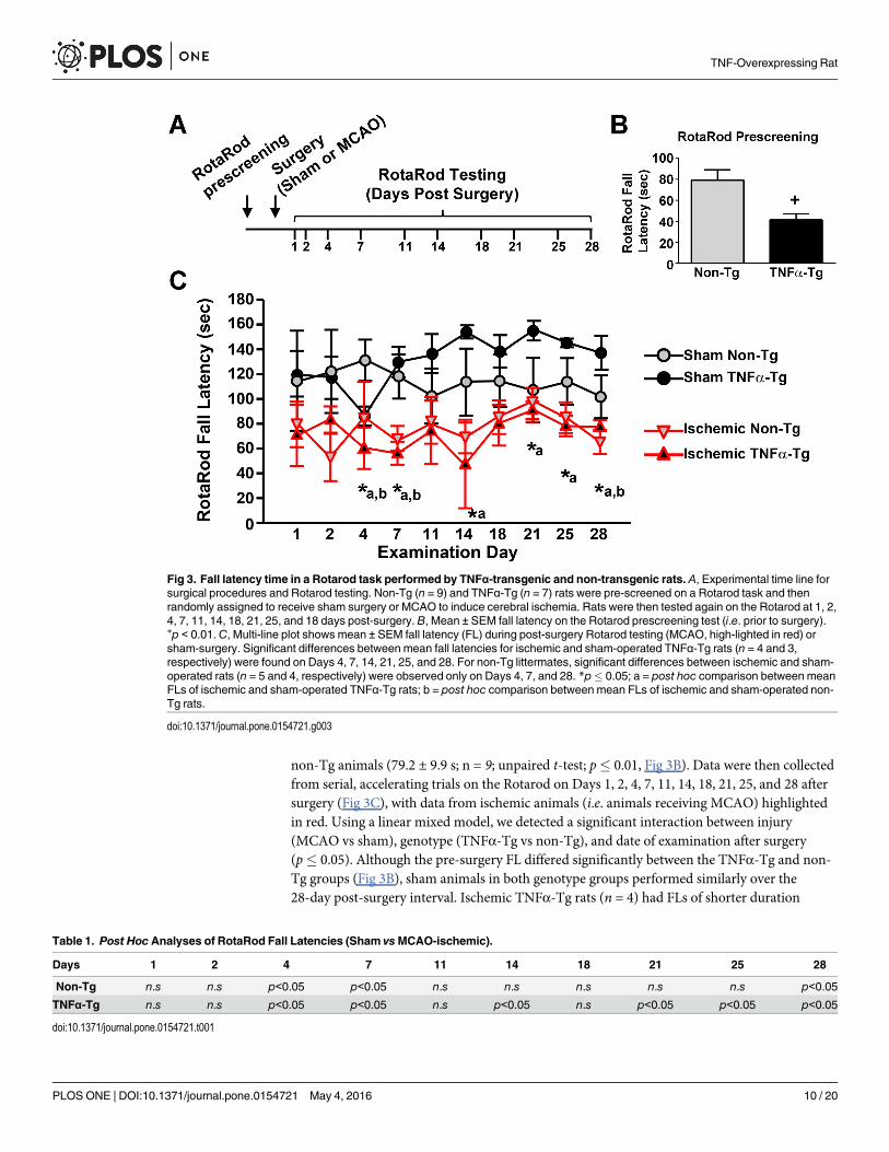
TNFα exacerbates motor deficits following MCAOTo determine the effects of chronically elevated TNFα on motor function, non-Tg and TNFα-Tg rats were tested on the Rotarod then randomly assigned to receive MCAO or sham surgery(Fig 3A). Prior to surgery, each animal was able to maintain its balance on the stationary rodfor 120 s. Fall latency (FL) was defined by averaging the longest two durations achieved by eachanimal after five trials on the accelerating rod. Prior to MCAO or sham surgery, themean ± SEM FL for TNFα-Tg rats (41.3 ± 5.7 s; n = 7) was significantly shorter than that of
Fig 2. Spatial learning in the Morris Water Maze before and after MCAO. Five training blocks (3 trials/block) were given across one day onthe Morris Water Maze (A,C), followed by a free-swim probe trial (B, D). A, Mean acquisition goal latency (AGL) ± SEM in seconds (averagedacross 3 trials within each block) for TNFα-Tg and non-Tg littermates prior to MCAO (n = 10–13 rats per genotype/treatment group).Significant reductions in AGL were observed over training in both groups, with non-Tg mice outperforming TNFα-Tg mice by block 5. B, Meanpercent time ± SEM spent searching the goal quadrant or a decoy quadrant (opposite the goal) during the free-swim probe prior to MCAO. Thetime spent by non-Tg mice in the goal quadrant was significantly above chance levels (i.e. 25%), (***p < 0001). In contrast, the percent timespent by TNFα-Tg mice in the goal quadrant did not differ significantly from chance.C, AGL ± SEM on the Morris Water maze assessed sevendays after MCAO. Neither TNFα-Tg nor non-Tg rats showed significant reductions in AGL across training. D, Mean percent time ± SEM spentsearching the goal and decoy quadrants at seven days post-MCAO. Non Tg rats continued to search above chance levels in the goalquadrant (*p < 0.05). Conversely, goal search times for TNFα-Tg rats did not differ from chance levels.
doi:10.1371/journal.pone.0154721.g002
TNF-Overexpressing Rat
PLOS ONE | DOI:10.1371/journal.pone.0154721 May 4, 2016 9 / 20
non-Tg animals (79.2 ± 9.9 s; n = 9; unpaired t-test; p� 0.01, Fig 3B). Data were then collectedfrom serial, accelerating trials on the Rotarod on Days 1, 2, 4, 7, 11, 14, 18, 21, 25, and 28 aftersurgery (Fig 3C), with data from ischemic animals (i.e. animals receiving MCAO) highlightedin red. Using a linear mixed model, we detected a significant interaction between injury(MCAO vs sham), genotype (TNFα-Tg vs non-Tg), and date of examination after surgery(p� 0.05). Although the pre-surgery FL differed significantly between the TNFα-Tg and non-Tg groups (Fig 3B), sham animals in both genotype groups performed similarly over the28-day post-surgery interval. Ischemic TNFα-Tg rats (n = 4) had FLs of shorter duration
Fig 3. Fall latency time in a Rotarod task performed by TNFα-transgenic and non-transgenic rats. A, Experimental time line forsurgical procedures and Rotarod testing. Non-Tg (n = 9) and TNFα-Tg (n = 7) rats were pre-screened on a Rotarod task and thenrandomly assigned to receive sham surgery or MCAO to induce cerebral ischemia. Rats were then tested again on the Rotarod at 1, 2,4, 7, 11, 14, 18, 21, 25, and 18 days post-surgery. B, Mean ± SEM fall latency on the Rotarod prescreening test (i.e. prior to surgery).+p < 0.01.C, Multi-line plot shows mean ± SEM fall latency (FL) during post-surgery Rotarod testing (MCAO, high-lighted in red) orsham-surgery. Significant differences between mean fall latencies for ischemic and sham-operated TNFα-Tg rats (n = 4 and 3,respectively) were found on Days 4, 7, 14, 21, 25, and 28. For non-Tg littermates, significant differences between ischemic and sham-operated rats (n = 5 and 4, respectively) were observed only on Days 4, 7, and 28. *p� 0.05; a = post hoc comparison between meanFLs of ischemic and sham-operated TNFα-Tg rats; b = post hoc comparison between mean FLs of ischemic and sham-operated non-Tg rats.
doi:10.1371/journal.pone.0154721.g003
Table 1. Post Hoc Analyses of RotaRod Fall Latencies (Sham vsMCAO-ischemic).
PLOS ONE | DOI:10.1371/journal.pone.0154721 May 4, 2016 10 / 20
compared to TNFα-Tg sham-operated rats (n = 3) on Days 4, 7, 14, 21, 25, and 28 (p� 0.05 oneach Day, see Fig 3C and Table 1). In contrast, non-Tg ischemic rats (n = 5) differed signifi-cantly from sham-operated non-Tg sham rats (n = 4) on only three examinations (Days 4, 7,and 28; p� 0.05 each, see Fig 3C and Table 1). There was no significant difference in FLbetween TNFα-Tg and non-Tg ischemic rats over 28 days. Overall, TNFα-Tg rats exposed toischemia had shorter mean FLs, indicating greater impairments in the Rotarod task when com-pared to TNFα-Tg sham-operated rats, for twice as many days as were observed with non-Tgrats.
TNFα alleviates cortical neuron loss following MCAOThe results of quantitative histological analysis conducted on TNFα-Tg rats and non-Tg litter-mates exposed to MCAO or sham surgery are shown in Fig 4. Regional neuron counting wasperformed on CA1 and CA3 and in cortical layer 5 in both the ipsilateral (i.e. the side receivingMCAO) and contralateral hemisphere of ischemic TNFα-Tg rats (n = 6) and ischemic non-Tg
Fig 4. Neuronal loss in hippocampus and cortex of TNFα-transgenic and non-transgenic rats at 28 days aftermiddle cerebral artery occlusion. A bar graph shows Nissl-stained cells per counting grid ± SEM in control (normallyperfused) and ischemic hippocampus (CA1 and CA3) and cortex (neocortical layer 5) in TNFα-transgenic (TNFα-Tg) andnon-transgenic (non-Tg) littermates subjected to middle cerebral artery occlusion (MCAO). Non-Tg rats had significantlyfewer surviving cells in ischemic cortex when compared to the normally perfused hemisphere or when compared to shamoperated animals. ***p� 0.001. ns = 4 Non-Tg Sham, 8 Non-Tg Ischemia, 3 TNFα-Tg Sham, 6 TNFα-Tg Ischemia.
doi:10.1371/journal.pone.0154721.g004
TNF-Overexpressing Rat
PLOS ONE | DOI:10.1371/journal.pone.0154721 May 4, 2016 11 / 20
rats (n = 8). TNFα-Tg and non-Tg sham-operated rats had similar numbers of pyramidal neu-rons in CA1, CA3, and cortical layer 5, regardless of hemisphere, and were therefore combinedinto a single group (n = 7) for statistical comparisons to ischemic rats. A one-way ANOVArevealed statistically significant differences in the number of cresyl violet-stained CA1 pyrami-dal neurons across treatment groups (p< 0.05). However, post hoc comparisons, found no sig-nificant differences between the mean numbers of CA1 neurons sampled from eitherhemisphere in ischemic TNFα-Tg and non-Tg rats, compared to sham controls. No genotypeor ischemia dependent effects were observed for CA3 neuron counts.
The most remarkable differences were observed in pyramidal neuron counts in corticallayer 5, which would be directly affected by ischemic injury. Group variation in layer 5 neuro-nal count was statistically significant by ANOVA (p� 0.001). Post hoc comparisons showedsignificantly fewer cortical layer 5 neurons in the ischemic hemisphere of non-Tg rats (22 ± 11cells per grid; mean ± SEM) than in the unaffected cortex in the same animals (68 ± 3 cells pergrid; Tukey’s test; p� 0.001) or in sham-ischemic rats (57 ± 3 cells per grid; p< 0.01). Therewas no significant difference between the number of surviving cortical layer 5 neurons in theischemic hemisphere of TNFα-Tg rats (36 ± 12 cells per grid) and those in non-Tg rats. Theseresults suggest that TNF overexpression helps to promote cortical neuron survival followingischemic insult.
DiscussionWe examined indices of synaptic integrity in the TNFα-Tg rat, to determine if this animal hasfundamental alterations of hippocampal circuitry that could affect its learning performance.Our data revealed a small nonsignificant decrease in maximal EPSP magnitudes in 8-weekTNFα-Tg rats compared to non-Tg littermates. In contrast, measures of short-term and long-term synaptic potentiation (i.e. PPF and LTP) were enhanced in TNFα-Tg rats (Fig 1B–1D).These changes were qualitatively and quantitatively similar to those observed in the hippocam-pus of immature (< 3-week old) animals, as reported here and previously [52]. In neonatal hip-pocampus, reduced basal synaptic strength and increased PPF correspond to a reduction inneurotransmitter release capacity [53], while augmented neonatal LTP requires differentmolecular mechanisms (for review, see [54]) than LTP in mature animals. Intriguingly, adultTNFα-Tg rats also showed impaired performance on hippocampal-dependent behavioraltasks, as discussed below, that are similar to those observed in neonatal animals [55]. Basedupon these results, we would hypothesize that elevated brain levels of biologically active TNFαhelp maintain hippocampal circuitry in a state that mimics early development. Clearly, furtherstudies will be required to fully evaluate this hypothesis.
Though others have previously shown that TNFα can facilitate LTP in hippocampal slicesunder certain conditions [56], consistent with our findings, several earlier studies reported animpairment in LTP induction in both the dentate and the CA1 regions of the hippocampus fol-lowing exogenous application of pathophysiological quantities of TNFα [27–29]. These appar-ent discrepancies may be attributable to several fundamental methodological differences. Mostimportantly, previous studies investigated acute effects of TNFα on LTP induction (~20 minapplication). Chronically elevated levels of TNFα in our transgenic animal may recruit tran-scriptional mechanisms not explored in earlier studies. Also, the synaptic changes observed inthe present work may reflect compensatory reactions to the many deleterious effects of TNFα.In addition, earlier studies investigated very long-lasting LTP (~2 hours), whereas we investi-gated LTP for only one hour after induction. It is possible that deficits in LTP would emerge inthe TNFα-Tg rat if synaptic changes were monitored for longer time periods. Finally, it’simportant to note that TNF is coupled to divergent signaling pathways via interactions with its
TNF-Overexpressing Rat
PLOS ONE | DOI:10.1371/journal.pone.0154721 May 4, 2016 12 / 20
different receptor subtypes (TNF-R1and TNF-R2), which could, in turn, result in complex neu-rologic and cognitive phenotypes. Strategies for teasing apart the differential contributionsTNF-Rs to the phenotype of TNFα-Tg rats are considered further below.
We tested spatial reference memory in the TNFα-Tg rat to determine if chronic elevation ofTNFα affects cognitive performance before and after focal cerebral ischemia. We found thatboth TNFα-Tg rats and non-Tg littermates demonstrated successive reductions in AGL duringserial training at pre-ischemic baseline, implying progressively more effective learning withrepetition in both groups. Transgenic rats showed a trend for more prolonged AGL in compar-ison to non-Tg littermates, although a statistically significant between-group difference wasfound only at the fifth and final trial block. While the two groups showed similarly impairedcognitive function at 7 days post MCAO, with overall elevations of mean AGL times comparedto pre-ischemic baseline, no significant reduction in AGL over five successive trial blocks, andno between-group difference were detected at any point. However, in probe trial experimentsdesigned to test the accuracy of memory recall, non-Tg rats performed more effectively thanTNFα-Tg animals at pre-ischemic baseline and after seven days of post-ischemic recovery.
The incremental, but significant, differences in pre-ischemic performance on the referencememory task suggest that the TNFα-Tg rat showed less effective learning with repetition whencompared to non-Tg littermates. Although both animal groups demonstrated similarlyimpaired learning after being subjected to the physiological stress of ischemia, the probe trialexperiments showed that non-Tg rats had more accurate memory recall relative to TNFα-Tgrats. These results indicate that the TNFα-Tg rat showed less effective mastery of referencememory tasks when compared to non-Tg littermates, with or without secondary injury fromfocal cerebral ischemia. This observation, implying an underlying impairment of cognitive per-formance in the TNFα-Tg rat, is presaged by an earlier report of impaired learning in two linesof transgenic mice overexpressing TNFα, one of which showed inflammatory changes in thebrain and another that showed no inflammatory changes [57]. Overall, the occurrence of reten-tive memory impairment in our transgenic rat, before and after focal ischemia (and in thetransgenic mice with or without active brain inflammation), suggests that chronic overexpres-sion of TNFα alters the molecular basis of learning, even in the absence of cytokine-mediatedcellular injury.
We examined performance of the TNFα-Tg rat in the Rotarod task to address two ques-tions: 1) is there an overall difference in functional performance after focal cerebral ischemiathat is observed uniquely in the transgenic animal and 2) if present, does this comparative dif-ference persist for an extended duration after ischemia (28 days)? We found that pre-ischemic,mean FL was significantly shorter in TNFα-Tg rats, suggesting that elevated TNF levels precip-itate a behavioral phenotype characterized by impaired balance and/or motor coordination. It’spossible that elevated TNF levels adversely affect the vestibular system and/or the neuromuscu-lature, both of which show high vulnerability to multiple autoimmune disorders [58–62]. If so,then it seems likely that this phenotype is overcome or compensated with increased practice,since Non-Tg and TNFα-Tg rats performed similarly on the rotarod during the 28 day testingperiod following surgery. The effects of elevated TNF on balance and motor coordinationwould therefore appear to be mild, at least in young adult animals. MCAO had a significantimpact on Rotarod performance in both genotype groups, resulting in overall shorter fall laten-cies. However, ischemic TNFα-Tg rats had shorter, mean test-day FLs for almost twice asmany days as non-Tg animals, when compared to sham-ischemic controls. These between-group differences persisted for the 28-day duration of the Rotarod experiment. Again, this sug-gests that mild deficits in balance and motor coordination are important phenotype traits ofthe TNFα-Tg rat.
TNF-Overexpressing Rat
PLOS ONE | DOI:10.1371/journal.pone.0154721 May 4, 2016 13 / 20
We anticipated that TNFα-Tg rats would have relatively smaller numbers of surviving neu-rons in ischemic cortex and/or in the CA1 region of ipsilateral hippocampus, when examined28 days after focal cerebral ischemia as compared to non-Tg littermates. Instead, we found thatthere were no significant differences in the number of surviving neurons in ischemic corticallayer 5 or in the CA1 region of hippocampus, between the 2 animal groups. The severity ofischemic injury was actually more pronounced in the non-Tg group, in that the number of sur-viving cortical layer 5 neurons was significantly smaller than in sham-ischemic rats. We specu-late that this lack of difference between neuronal counts in ischemic TNFα-Tg rats and non-Tganimals is driven by a “survivor effect” that favored the transgenic animal. In our previouswork, we demonstrated that TNFα-Tg rats have significantly greater neural cell apoptosis inischemic cortex at 24–72 hours relative to non-Tg controls [33]. Before the 28-day, post-ische-mic interval, evaluated in the present study, the synergistic interaction between chronically ele-vated regional TNFα and ischemic physiological stress may have already exerted its maximaleffect on cellular apoptosis in ischemic cortex and hippocampus. Consistent with this idea isabundant evidence showing the importance of TNFα in mediating inflammation and apopto-sis, characteristic of most neurodegenerative diseases. In vascular cognitive impairment (VCI)and other disorders resulting from cerebral ischemia, TNFα (expressed by vascular and peri-vascular cells) can promote pro-inflammatory and pro-coagulant effects on the endothelia[63]. In a rat model of VCI, produced by surgical occlusion of both carotid arteries, subcorticalvessels become immuno-reactive for TNFα [64, 65]. Nasal administration of recombinant E-selectin, a glycoprotein adhesion molecule that stimulates the synthesis of regulatory T-cellstargeting activated endothelia, suppresses TNFα vascular immuno-reactivity in this model.Moreover, induction of mucosal tolerance to E-selectin prevents ischemic and hemorrhagicstroke in spontaneously hypertensive stroke-prone rats [66] and reduces infarct volume afterpermanent MCAO [67], suggesting that the deleterious effects of vascular pathology are medi-ated, at least partly, through the activation of TNF signaling mechanisms.
As alluded to above, TNF can mediate complex phenotypic effects through actions on itsdifferent receptor subtypes. Soluble TNFα interacts preferentially with type TNF-R1s, whiletransmembrane TNFα shows greater selectivity for TNF-R2s [68, 69]. In turn, TNF-R subtypesare coupled to divergent downstream signaling pathways: TNF-R1 activation stimulates cas-pase activity and cell death, while TNF-R2 activation stimulates PI3-kinase signaling and pro-motes cell survival [70]. It is widely believed that many of the detrimental pro-inflammatoryactions of TNF are mediated by TNF-R1s, while several of the more beneficial actions of TNFare carried out by TNF-R2s [70]. Presently, it is unclear to what extent TNF-R1 and TNF-R2signaling is engaged in TNFα overexpressing rats, or how these different receptor mechanismsimpact the cellular and behavioral phenotypes investigated in this study. Biologics that prefer-entially target different TNF-Rs have been used in other model systems to tease apart the rela-tive contributions of soluble and transmembrane TNF. For instance, XPro1595 is a solubleTNF mimetic that exhibits dominant-negative inhibition of TNF-R1, but not TNF-R2, signal-ing [71]. This drug has revealed selective deleterious roles of TNF-R1 in animal models of Par-kinson’s disease [72, 73], Alzheimer’s disease [74], and autoimmune encephalomyelitis [75],Previously, we used XPro1595 and a variety of biochemical approaches to show that solubleTNF/TNF-R1 interactions contributes to cognitive decline, neuronal Ca2+ dysregulation, andaltered synaptic plasticity in aged rats [34]. Future use of XPro1595, or similar reagents, couldtherefore be a powerful way to dissect apart the apparently complex phenotype of TNFα over-expressing rats.
Finally, changes in glial activation and inflammatory profiles were not investigated in thepresent study, and as such, it is unclear whether the phenotypic traits reported in TNFα over-expressing rats are attributable to direct effects of TNF on neuronal signaling pathways, or to
TNF-Overexpressing Rat
PLOS ONE | DOI:10.1371/journal.pone.0154721 May 4, 2016 14 / 20
indirect effects on glial cells (both microglia and astrocytes), which can alter neuronal functionthrough a myriad of mechanisms. Indeed, TNF can cause profound glial activation, which hasbeen noted in similar TNFα overexpressing mouse models [76, 77]. Numerous glial-derivedcytokines that are induced or potentiated by exogenously applied TNF can significantly modu-late synaptic function, neuronal morphology, and neuronal survival/death pathways [78–82].In addition, TNF application to both astrocytes and microglia can cause the release of numer-ous reactive oxygen species known to erode synaptic efficacy and/or to precipitate neurodegen-eration [83, 84]. Finally, TNF and related cytokines have been shown to negatively affectnumerous protective properties of astrocytes, including glutamate uptake [85, 86] and gapjunction coupling [87], to name a few. All of these mechanisms could be in play in shaping theneurologic and cognitive phenotype of TNFα overexpressing rats and should be investigated infuture studies.
ConclusionsOur results suggest that elevated brain levels of TNFα, similar to that found in a number ofneurodegenerative disorders, can contribute to impaired cognition and functional perfor-mance. Our LTP studies in non-ischemic TNFα-Tg rats suggest that constitutive upregulationof TNFα synthesis may facilitate maintenance of hippocampal circuitry in a nascent stage.Although such a fundamental alteration could be beneficial in preserving normal hippocampalfunction under the physiological stress of cerebral ischemia, we found that post-ischemicTNFα-Tg rats showed impaired performance on both cognitive and functional tasks atextended time intervals after MCAO. By histological analysis, this impairment was not associ-ated with excessive neuronal loss from the CA1 or CA3 regions of hippocampus or from post-ischemic cortex. We cannot exclude the possibility that constitutive upregulation of TNFα syn-thesis produces other alterations in the transgenic rat that could influence performance on cog-nitive and functional tasks, such as the release of inhibitory or excitatory neurotransmitters orthe modulation of cortico-hippocampal connections. Overall, our results substantiate theimpression that increased brain synthesis of TNFα will precipitate long-term cognitive effectsand contribute to higher risk of dementia after cerebral ischemia.
Supporting InformationS1 File. Raw data for producing figures and for conducting statistical analyses.(XLS)
AcknowledgmentsWe thank Thomas C. Foster, Ph.D., for advice regarding the water maze experiments. Susan D.Craddock, Mary Louise Holtz, Ph.D., and Ravikumar Rangaswamy Rao, Ph.D., made valuedtechnical contributions to this work. Sherry Chandler Williams, ELS, edited the manuscriptand prepared the illustrations.
Author ContributionsConceived and designed the experiments: LCP CMN. Performed the experiments: LCP CMN.Analyzed the data: RJK CMN. Contributed reagents/materials/analysis tools: LCP. Wrote thepaper: LCP CMN.
TNF-Overexpressing Rat
PLOS ONE | DOI:10.1371/journal.pone.0154721 May 4, 2016 15 / 20
References1. Emsley HC, Tyrrell PJ. (2002) Inflammation and infection in clinical stroke. J Cereb Blood FlowMetab.
22: 1399–419. PMID: 12468886
2. Mayne M, Ni W, Yan HJ, Xue M, Johnston JB, Del Bigio MR, et al. (2001) Antisense oligodeoxynucleo-tide inhibition of tumor necrosis factor-alpha expression is neuroprotective after intracerebral hemor-rhage. Stroke. 32: 240–8. PMID: 11136943
3. Tomimoto H, Ihara M, Wakita H, Ohtani R, Lin JX, Akiguchi I, et al. (2003) Chronic cerebral hypoperfu-sion induces white matter lesions and loss of oligodendroglia with DNA fragmentation in the rat. ActaNeuropathol. 106: 527–34. PMID: 13680276
4. Lotocki G, Alonso OF, Dietrich WD, Keane RW. (2004)Tumor necrosis factor receptor 1 and its signal-ing intermediates are recruited to lipid rafts in the traumatized brain. J Neurosci. 24: 11010–6. PMID:15590916.
5. Tomimoto H, Akiguchi I, Wakita H, Kinoshita A, Ikemoto A, Nakamura S, et al. (1996) Glial expressionof cytokines in the brains of cerebrovascular disease patients. Acta Neuropathol. 92: 281–7. PMID:8870830.
6. Sairanen T, Carpén O, Karjalainen-Lindsberg ML, Paetau A, Turpeinen U, Kaste M, et al. (2001) Evolu-tion of cerebral tumor necrosis factor-alpha production during human ischemic stroke. Stroke. 32:1750–8. PMID: 11486101
7. Carlstedt F, Lind L, Lindahl B. (1997) Proinflammatory cytokines, measured in a mixed population onarrival in the emergency department, are related to mortality and severity of disease. J Intern Med. 242:361–5. PMID: 9408063
8. Vila N, Castillo J, Davalos A, Chamorro A. (2000) Proinflammatory cytokines and early neurologicalworsening in ischemic stroke. Stroke. 31: 2325–9. PMID: 11022058
9. Zaremba J, Skrobanski P, Losy J. (2001) Tumor necrosis factor-alpha is increased in the cerebrospinalfluid and serum of ischaemic stroke patients and correlates with the volume of evolving brain infarct.Biomed Pharmacother. 55: 258–63. PMID: 11428551
10. Sairanen TR, Lindsberg PJ, Brenner M, Carpén O, Sirén AL. (2001) Differential cellular expression oftumor necrosis factor-α and Type I tumor necrosis factor receptor after transient global forebrain ische-mia. J Neurol Sci. 186: 87–99. PMID: 11412877
11. Saito K, Suyama K, Nishida K, Sei Y, Basile AS. (1996) Early increases in TNF-α, IL-6, and IL-1β levelsfollowing transient cerebral ischemia in gerbil brain. Neurosci Lett. 206: 149–52. PMID: 8710173
12. Liu Y, Jacobowitz DM, Barone F, McCarron R, Spatz M, Feuerstein G, et al. (1994) Quantitation of peri-vascular monocytes and macrophages around cerebral blood vessels of hypertensive and aged rats. JCereb Blood Flow Metab. 14: 348–52. PMID: 8113330
13. Barone FC, Arvin B, White RF, Miller A, Webb CL, Willette RN, et al. (1997) Tumor necrosis factor-alpha: a mediator of focal ischemic brain injury. Stroke. 28: 1233–44. PMID: 9183357
14. Dawson DA, Martin D, Hallenbeck JM. (1996) Inhibition of tumor necrosis factor-alpha reduces focalcerebral ischemic injury in the spontaneously hypertensive rat. Neurosci Lett. 218: 41–4. PMID:8939476
15. Nawashiro H, Martin D, Hallenbeck JM. (1997) Inhibition of tumor necrosis factor and amelioration ofbrain infarction in mice. J Cereb Blood Flow Metab. 17: 229–32. PMID: 9040503
16. Nawashiro H, Tasaki K, Ruetzler CA, Hallenbeck JM. (1997) TNF-alpha pretreatment induces protec-tive effects against focal cerebral ischemia in mice. J Cereb Blood Flow Metab. 17: 483–90. PMID:9183285
17. Lavine SD, Hofman FM, Zlokovic BV. (1998) Circulating antibody against tumor necrosis factor-alphaprotects rat brain from reperfusion injury. J Cereb Blood Flow Metab. 18: 52–8. PMID: 9428305
18. Yang GY, Gong C, Qin Z, YeW, Mao Y, Bertz AL. (1998) Inhibition of TNF-α attenuates infarct volumeand ICAM-1 expression in ischemic mouse brain. Neuroreport. 9: 2131–4. PMID: 9674607
19. Marchetti L, Klein M, Schlett K, Pfizenmaier K, Eisel UL. (2004) Tumor necrosis factor (TNF)-mediatedneuroprotection against glutamate-induced excitotoxicity is enhanced by N-methyl-D-aspartate recep-tor activation. Essential role of a TNF receptor 2-mediated phosphatidylinositol 3-kinase-dependentNF-kappa B pathway. J Biol Chem. 279: 32869–81. PMID: 15155767
20. Wajant H, Pfizenmaier K, Scheurich P. (2003) Tumor necrosis factor signaling. Cell Death Differ. 10:45–65. PMID: 12655295
21. Iosif RE, Ekdahl CT, Ahlenius H, Pronk CJ, Bonde S, Kokaia Z, et al. (2006) Tumor necrosis factorreceptor 1 is a negative regulator of progenitor proliferation in adult hippocampal neurogenesis. J Neu-rosci. 26: 9703–12. PMID: 16988041
TNF-Overexpressing Rat
PLOS ONE | DOI:10.1371/journal.pone.0154721 May 4, 2016 16 / 20
25. Stellwagen D, Beattie EC, Seo JY, Malenka RC. (2005) Differential regulation of AMPA receptor andGABA receptor trafficking by tumor necrosis factor-alpha. J Neurosci. 25: 3219–28. PMID: 15788779
26. Golan H, Levav T, Mendelsohn A, Huleihel M. (2004) Involvement of tumor necrosis factor alpha in hip-pocampal development and function. Cereb Cortex. 14: 97–105. PMID: 14654461
27. Tancredi V, D'Arcangelo G, Grassi F, Tarroni P, Palmieri G, Santoni G, et al. (1992) Tumor necrosisfactor alters synaptic transmission in rat hippocampal slices. Neurosci Lett. 146: 176–8. PMID:1337194
28. Cunningham AJ, Murray CA, O'Neill LA, Lynch MA, O'Conner JJ. (1996) Interleukin-1 beta (IL-1 beta)and tumour necrosis factor (TNF) inhibit long-term potentiation in the rat dentate gyrus in vitro. NeurosciLett. 203: 17–20. PMID: 8742036
29. Butler MP, O'Connor JJ, Moynagh PN. (2004) Dissection of tumor-necrosis factor-alpha inhibition oflong-term potentiation (LTP) reveals a p38 mitogen-activated protein kinase-dependent mechanismwhich maps to early-but not late-phase LTP. Neuroscience. 124: 319–26. PMID: 14980382
30. Bliss TV, Collingridge GL. (1993) A synaptic model of memory: long-term potentiation in the hippocam-pus. Nature. 361: 31–9. PMID: 8421494
31. Butler MP, Moynagh PN, O'Connor JJ. (2002) Methods of detection of the transcription factor NF-kappa B in rat hippocampal slices. J Neurosci Methods. 119: 185–90. PMID: 12323422
32. Albensi BC, Mattson MP. (2000) Evidence for the involvement of TNF and NF-κB in hippocampal syn-aptic plasticity. Synapse. 35: 151–9. PMID: 10611641
33. Pettigrew LC, Kindy MS, Scheff S, Springer JE, Kryscio RJ, Li Y, et al. (2008) Focal cerebral ischemiain the TNFalpha-transgenic rat. J Neuroinflammation. 5: 47. doi: 10.1186/1742-2094-5-47 PMID:18947406
34. Sama DM, Mohmmad Abdul H, Furman JL, Artiushin IA, Szymkowski DE, Scheff SW, et al. (2012) Inhi-bition of soluble tumor necrosis factor ameliorates synaptic alterations and Ca2+ dysregulation in agedrats. PLoS One. 7(5):e38170. doi: 10.1371/journal.pone.0038170 PMID: 22666474
35. Furman JL, Sama DM, Gant JC, Beckett TL, Murphy MP, Bachstetter AD, et al. (2012) Targeting astro-cytes ameliorates neurologic changes in a mouse model of Alzheimer's disease. J Neurosci. 32:16129–40. PMID: 23152597 doi: 10.1523/JNEUROSCI.2323-12.2012
36. Furman JL, Sompol P, Kraner SD, Pleiss MM, Putman EJ, Dunkerson J, et al. (2016) Blockade of Astro-cytic Calcineurin/NFAT Signaling Helps to Normalize Hippocampal Synaptic Function and Plasticity ina Rat Model of Traumatic Brain Injury. J Neurosci. 36: 1502–15. PMID: 26843634 doi: 10.1523/JNEUROSCI.1930-15.2016
37. Norris CM, Sompol P, Roberts KN, Ansari M, Scheff SW. (2016) Pycnogenol protects CA3-CA1 synap-tic function in a rat model of traumatic brain injury. Exp Neurol. 276: 5–12. PMID: 26607913 doi: 10.1016/j.expneurol.2015.11.006
38. Mathis DM, Furman JL, Norris CM. (2011) Preparation of acute hippocampal slices from rats and trans-genic mice for the study of synaptic alterations during aging and amyloid pathology. J Vis Exp. 2011(49). doi: 10.3791/2330 PMID: 21490565
39. Zea Longa E, Weinstein PR, Carlson S, Cummins R. (1989) Reversible middle cerebral artery occlu-sion without craniotomy in rats. Stroke. 20:84–91. PMID: 2643202
41. Belayev L, Alonso OF, Busto R, ZhaoW, Ginsberg MD. (1996) Middle cerebral artery occlusion in therat by intraluminal suture. Neurological and pathological evaluation of an improved model. Stroke. 27:1616–23. PMID: 8784138
42. Foster TC, Sharrow KM, Kumar A, Masse J. (2003) Interaction of age and chronic estradiol replace-ment on memory and markers of brain aging. Neurobiol Aging. 24: 839–52. PMID: 12927766
43. Foster TC, Sharrow KM, Masse JR, Norris CM, Kumar A. (2001) Calcineurin links Ca2+ dysregulationwith brain aging. J Neurosci. 21:4066–73. PMID: 11356894
44. Norris CM, Foster TC. (1999) MK-801 improves retention in aged rats: implications for altered neuralplasticity in age-related memory deficits. Neurobiol Learn Mem. 71: 194–206. PMID: 10082639
TNF-Overexpressing Rat
PLOS ONE | DOI:10.1371/journal.pone.0154721 May 4, 2016 17 / 20
45. Candelario-Jalil E, Mhadu NH, Gonzalez-Falcon A, Garcia-Cabrera M, Munoz E, Leon OS, et al. (2005)Effects of the cyclooxygenase-2 inhibitor nimesulide on cerebral infarction and neurological deficitsinduced by permanent middle cerebral artery occlusion in the rat. J Neuroinflammation. 2(1): 3. PMID:15656909
46. Yulug B, Kilic U, Kilic E, Bahr M. (2004) Rifampicin attenuates brain damage in focal ischemia. BrainRes. 996: 76–80. PMID: 14670633
48. Tejadilla D, Cerbon M, Morales T. (2010) Prolactin reduces the damaging effects of excitotoxicity in thedorsal hippocampus of the female rat independently of ovarian hormones. Neuroscience. 169: 1178–85. PMID: 20570717 doi: 10.1016/j.neuroscience.2010.05.074
49. de la Tremblaye PB, Plamondon H. (2011) Impaired conditioned emotional response and object recog-nition are concomitant to neuronal damage in the amygdala and perirhinal cortex in middle-aged ische-mic rats. Behav Brain Res. 219: 227–33. PMID: 21238489 doi: 10.1016/j.bbr.2011.01.009
50. Norris CM, Scheff SW. (2009) Recovery of afferent function and synaptic strength in hippocampal CA1following traumatic brain injury. J Neurotrauma. 26: 2269–78. PMID: 19604098 doi: 10.1089/neu.2009.1029
51. Furman JL, Sama DM, Gant JC, Beckett TL, Murphy MP, Bachstetter AD, et al. (2012) Targeting astro-cytes ameliorates neurologic changes in a mouse model of Alzheimer's disease. J Neurosci. 32:16129–40. PMID: 23152597 doi: 10.1523/JNEUROSCI.2323-12.2012
52. Dumas TC. (2012) Postnatal alterations in induction threshold and expression magnitude of long-termpotentiation and long-term depression at hippocampal synapses. Hippocampus. 22: 188–99. PMID:21069779 doi: 10.1002/hipo.20881
53. Dumas TC, Foster TC. (1995) Developmental increase in CA3-CA1 presynaptic function in the hippo-campal slice. J Neurophysiol. 73: 1821–8. PMID: 7623083
54. Dumas TC. (2005) Late postnatal maturation of excitatory synaptic transmission permits adult-likeexpression of hippocampal-dependent behaviors. Hippocampus. 15: 562–78. PMID: 15884034
55. Rudy JW, Stadler-Morris S, Albert P. (1987) Ontogeny of spatial navigation behaviors in the rat: dissoci-ation of "proximal"- and "distal"-cue-based behaviors. Behav Neurosci. 101: 62–73. PMID: 3828056
56. Wall AM, Mukandala G, Greig NH, O'Connor JJ. (2015) Tumor necrosis factor-alpha potentiates long-term potentiation in the rat dentate gyrus after acute hypoxia. J Neurosci Res. 93: 815–29. PMID:25641742 doi: 10.1002/jnr.23540
57. Aloe L, Properzi F, Probert L, Akassoglou K, Kassiotis G, Micera A, et al. (1999) Learning abilities, NGFand BDNF brain levels in two lines of TNF-α transgenic mice, one characterized by neurological disor-ders, the other phenotypically normal. Brain Res. 840: 125–37 PMID: 10517960
58. Greco A, De Virgilio A, Gallo A, Fusconi M, Ruoppolo G, Turchetta R, et al. (2014) Idiopathic bilateralvestibulopathy: an autoimmune disease? Autoimmun Rev. 13: 1042–7. PMID: 25173622 doi: 10.1016/j.autrev.2014.08.035
59. Arbusow V, Strupp M, Dieterich M, Stocker W, Naumann A, Schulz P, et al. (1998) Serum antibodiesagainst membranous labyrinth in patients with "idiopathic" bilateral vestibulopathy. J Neurol. 245: 132–6. PMID: 9553841
60. Merrill JE, Graves MC, Mulder DG. (1992) Autoimmune disease and the nervous system. Biochemical,molecular, and clinical update. West J Med. 156: 639–46. PMID: 1319627
61. Solomon AJ, Spain RI, Kruer MC, Bourdette D. (2011) Inflammatory neurological disease in patientstreated with tumor necrosis factor alpha inhibitors. Mult Scler. 17:1472–87. PMID: 21816758 doi: 10.1177/1352458511412996
62. De Paepe B, Creus KK, De Bleecker JL. (2012) The tumor necrosis factor superfamily of cytokines inthe inflammatory myopathies: potential targets for therapy. Clin Dev Immunol. 2012:369432. doi: 10.1155/2012/369432 PMID: 22110532
63. Hallenbeck JM. (2002) The many faces of tumor necrosis factor in stroke. Nat Med. 8: 1363–8. PMID:12457181
64. Ruetzler CA, Furuya K, Takeda H, Hallenbeck JM. (2001) Brain vessels normally undergo cyclic activa-tion and inactivation: evidence from tumor necrosis factor-alpha, heme oxygenase-1, and manganesesuperoxide dismutase immunostaining of vessels and perivascular brain cells. J Cereb Blood FlowMetab. 21: 244–52. PMID: 11295879
65. Wakita H, Ruetzler C, Illoh KO, Chen Y, Takanohashi A, Spatz M, et al. (2008) Mucosal tolerization toE-selectin protects against memory dysfunction and white matter damage in a vascular cognitiveimpairment model. J Cereb Blood FlowMetab. 28: 341–53. PMID: 17637705
TNF-Overexpressing Rat
PLOS ONE | DOI:10.1371/journal.pone.0154721 May 4, 2016 18 / 20
66. Takeda H, Spatz M, Ruetzler C, McCarron R, Becker K, Hallenbeck J. (2002) Induction of mucosal tol-erance to E-selectin prevents ischemic and hemorrhagic stroke in spontaneously hypertensive geneti-cally stroke-prone rats. Stroke. 33: 2156–63. PMID: 12215580
67. Chen Y, Ruetzler C, Pandipati S, Spatz M, McCarron RM, Becker K, et al. (2003) Mucosal tolerance toE-selectin provides cell-mediated protection against ischemic brain injury. Proc Natl Acad Sci U S A.100:15107–12. PMID: 14645708
68. Grell M, Douni E, Wajant H, Lohden M, Clauss M, Maxeiner B, et al. (1995) The transmembrane form oftumor necrosis factor is the prime activating ligand of the 80 kDa tumor necrosis factor receptor. Cell.83: 793–802. PMID: 8521496
69. Grell M, Wajant H, Zimmermann G, Scheurich P. (1998) The type 1 receptor (CD120a) is the high-affin-ity receptor for soluble tumor necrosis factor. Proc Natl Acad Sci U S A. 95: 570–5. PMID: 9435233
70. McCoy MK, Tansey MG. (2008) TNF signaling inhibition in the CNS: implications for normal brain func-tion and neurodegenerative disease. J Neuroinflammation. 5:45. doi: 10.1186/1742-2094-5-45 PMID:18925972
71. Steed PM, Tansey MG, Zalevsky J, Zhukovsky EA, Desjarlais JR, Szymkowski DE, et al. (2003) Inacti-vation of TNF signaling by rationally designed dominant-negative TNF variants. Science. 301: 1895–8.PMID: 14512626
72. McCoy MK, Martinez TN, Ruhn KA, Szymkowski DE, Smith CG, Botterman BR, et al. (2006) Blockingsoluble tumor necrosis factor signaling with dominant-negative tumor necrosis factor inhibitor attenu-ates loss of dopaminergic neurons in models of Parkinson's disease. J Neurosci. 26: 9365–75. PMID:16971520
73. Barnum CJ, Chen X, Chung J, Chang J, Williams M, Grigoryan N, et al. (2014) Peripheral administra-tion of the selective inhibitor of soluble tumor necrosis factor (TNF) XPro(R)1595 attenuates nigral cellloss and glial activation in 6-OHDA hemiparkinsonian rats. J Parkinsons Dis. 4: 349–60. PMID:25061061 doi: 10.3233/JPD-140410
74. McAlpine FE, Lee JK, Harms AS, Ruhn KA, Blurton-Jones M, Hong J, et al. (2009) Inhibition of solubleTNF signaling in a mouse model of Alzheimer's disease prevents pre-plaque amyloid-associated neu-ropathology. Neurobiol Dis. 34: 163–77. PMID: 19320056
75. Brambilla R, Ashbaugh JJ, Magliozzi R, Dellarole A, Karmally S, Szymkowski DE, et al. (2011) Inhibi-tion of soluble tumour necrosis factor is therapeutic in experimental autoimmune encephalomyelitis andpromotes axon preservation and remyelination. Brain. 134: 2736–54. PMID: 21908877 doi: 10.1093/brain/awr199
76. Probert L, Akassoglou K, Pasparakis M, Kontogeorgos G, Kollias G. (1995) Spontaneous inflammatorydemyelinating disease in transgenic mice showing central nervous system-specific expression of tumornecrosis factor alpha. Proc Natl Acad Sci U S A. 92: 11294–8. PMID: 7479982
77. Probert L, Akassoglou K, Alexopoulou L, Douni E, Haralambous S, Hill S, et al. (1996) Dissection of thepathologies induced by transmembrane and wild-type tumor necrosis factor in transgenic mice. J Leu-koc Biol. 59: 518–25. PMID: 8613699
78. Katsuki H, Nakai S, Hirai Y, Akaji K, Kiso Y, Satoh M. (1990) Interleukin-1 beta inhibits long-term poten-tiation in the CA3 region of mouse hippocampal slices. Eur J Pharmacol. 181: 323–6. PMID: 2166677
79. Bellinger FP, Madamba S, Siggins GR. (1993) Interleukin 1 beta inhibits synaptic strength and long-term potentiation in the rat CA1 hippocampus. Brain Res. 628: 227–34. PMID: 8313151
80. D'Arcangelo G, Grassi F, Ragozzino D, Santoni A, Tancredi V, Eusebi F. (1991) Interferon inhibits syn-aptic potentiation in rat hippocampus. Brain Res. 564: 245–8. PMID: 1725767
81. Bellinger FP, Madamba SG, Campbell IL, Siggins GR. (1995) Reduced long-term potentiation in thedentate gyrus of transgenic mice with cerebral overexpression of interleukin-6. Neurosci Lett. 198: 95–8. PMID: 8592650
82. Sama DM, Norris CM. (2013) Calcium dysregulation and neuroinflammation: discrete and integratedmechanisms for age-related synaptic dysfunction. Ageing Res Rev. 12: 982–95. PMID: 23751484 doi:10.1016/j.arr.2013.05.008
83. Knapp LT, Klann E. (2002) Role of reactive oxygen species in hippocampal long-term potentiation: con-tributory or inhibitory? J Neurosci Res. 70: 1–7. PMID: 12237859
84. Serrano F, Klann E. (2004) Reactive oxygen species and synaptic plasticity in the aging hippocampus.Ageing Res Rev. 3: 431–43. PMID: 15541710
85. SamaMA, Mathis DM, Furman JL, Abdul HM, Artiushin IA, Kraner SD, et al. (2008) Interleukin-1beta-dependent signaling between astrocytes and neurons depends critically on astrocytic calcineurin/NFAT activity. J Biol Chem. 283: 21953–64. PMID: 18541537 doi: 10.1074/jbc.M800148200
TNF-Overexpressing Rat
PLOS ONE | DOI:10.1371/journal.pone.0154721 May 4, 2016 19 / 20
86. Szymocha R, Akaoka H, Dutuit M, Malcus C, Didier-Bazes M, Belin MF, et al. (2000) Human T-cell lym-photropic virus type 1-infected T lymphocytes impair catabolism and uptake of glutamate by astrocytesvia Tax-1 and tumor necrosis factor alpha. J Virol. 74: 6433–41. PMID: 10864655
87. Kielian T. (2008) Glial connexins and gap junctions in CNS inflammation and disease. J Neurochem.106: 1000–16. PMID: 18410504 doi: 10.1111/j.1471-4159.2008.05405.x
TNF-Overexpressing Rat
PLOS ONE | DOI:10.1371/journal.pone.0154721 May 4, 2016 20 / 20