[ 237 ] RESPIRATION IN THE DESERT LOCUST II. THE CONTROL OF THE SPIRACLES BY P. L. MILLER Department of Zoology, University of Cambridge* (Received 14 September 1959) INTRODUCTION The observations of a number of authors have shown that the spiracles of various insects are capable of graded opening. In the absence of ventilation this is termed diffusion control. Hazelhoff (1927) demonstrated diffusion control in Periplaneta: in 1 % carbon dioxide the spiracles are slightly open, 2 % causes further opening and in 3 % they are wide open. It has been observed in the flea (Wigglesworth, 1935), in houseflies (Case, 1957) and in the tsetse fry (Bursell, 1957), and its occurrence has been inferred in the pupae of Hyalophora (Buck, 1958), whose spiracles are very slightly open during the 'interburst'. Graded opening has not previously been demonstrated in an insect whose spiracles are normally synchronized with abdominal ventilation, and in this paper it will be shown that the spiracles of the locust can individually modify their behaviour within the overall pattern of synchronization, and suggestions will be made as to how this is controlled. The first four pairs of spiracles of the locust open during the inspiratory phase of abdominal ventilation and the remainder during expiration (McArthur, 1929). That these synchronized movements in fact produce an anterior to posterior flow of air through the insect has been demonstrated by Fraenkel (1932). More recent measurements of the volume of air pumped through the insect in flight (Weis-Fogh, i960) have shown that hyperventilation does not greatly increase the flow, so that probably a modification of spiracular behaviour takes place. MATERIAL, METHODS AND NOMENCLATURE Material. Adult Schistocerca gregaria were obtained and kept as described else- where (Miller, 1960a). The first three pairs of spiracles have been studied in detail, and observations made on the remainder suggest that they are similar to the third. Methods. To record the behaviour of several spiracles simultaneously, very small mirrors were attached to the spiracle valves with a resin and wax mixture. The mirrors were made from silvered fragments of drawn out coverslips and each weighed approximately 0-05 mg.—about the same weight as one valve of spiracle 2. A weight twenty times heavier did not hamper the movements of the valves. A beam of light from a Baker microscope lamp was reflected by the mirrors through • Present address: Makerere College, Kampala, Uganda.
Transcript
[ 237 ]
RESPIRATION IN THE DESERT LOCUST
II. THE CONTROL OF THE SPIRACLES
BY P. L. MILLER
Department of Zoology, University of Cambridge*
(Received 14 September 1959)
INTRODUCTION
The observations of a number of authors have shown that the spiracles of variousinsects are capable of graded opening. In the absence of ventilation this is termeddiffusion control. Hazelhoff (1927) demonstrated diffusion control in Periplaneta:in 1 % carbon dioxide the spiracles are slightly open, 2 % causes further openingand in 3 % they are wide open. It has been observed in the flea (Wigglesworth,1935), in houseflies (Case, 1957) and in the tsetse fry (Bursell, 1957), and itsoccurrence has been inferred in the pupae of Hyalophora (Buck, 1958), whosespiracles are very slightly open during the 'interburst'.
Graded opening has not previously been demonstrated in an insect whosespiracles are normally synchronized with abdominal ventilation, and in this paper itwill be shown that the spiracles of the locust can individually modify their behaviourwithin the overall pattern of synchronization, and suggestions will be made as tohow this is controlled.
The first four pairs of spiracles of the locust open during the inspiratory phaseof abdominal ventilation and the remainder during expiration (McArthur, 1929).That these synchronized movements in fact produce an anterior to posterior flowof air through the insect has been demonstrated by Fraenkel (1932). More recentmeasurements of the volume of air pumped through the insect in flight (Weis-Fogh,i960) have shown that hyperventilation does not greatly increase the flow, so thatprobably a modification of spiracular behaviour takes place.
MATERIAL, METHODS AND NOMENCLATURE
Material. Adult Schistocerca gregaria were obtained and kept as described else-where (Miller, 1960a). The first three pairs of spiracles have been studied in detail,and observations made on the remainder suggest that they are similar to the third.
Methods. To record the behaviour of several spiracles simultaneously, very smallmirrors were attached to the spiracle valves with a resin and wax mixture. Themirrors were made from silvered fragments of drawn out coverslips and eachweighed approximately 0-05 mg.—about the same weight as one valve of spiracle 2.A weight twenty times heavier did not hamper the movements of the valves.A beam of light from a Baker microscope lamp was reflected by the mirrors through
concentrating lenses on to a moving roll of photographic recording paper (KodakR.P. 20), in a simple electrically driven camera. Movements of the spot on thefilm which resulted from ventilation were clearly distinguishable from thosecaused by opening and shutting of the valves. Where only the extreme positionsof valve movement were required, the beam was focused onto a ground-glass screenand the positions marked by pencil. A slight but negligible error arose from notusing a cylindrical screen.
Before a mirror can be attached to spiracle 1, part of the overlying pronotummust be cut away and the prothorax rigidly waxed to the mesothorax. Spiracles3 and 10 are sunk in pits, and the mirror must be supported on a very small glassrod waxed to the valve. With this technique it was possible to record from threespiracles simultaneously.
Nerve impulses in the spiracular nerves were recorded with two hooked platinumand 10% iridium wire electrodes, 0-03 mm. in diameter, and insulated down tothe hooks with 'Araldite'. The nerve was lifted into a drop of paraffin oil containedin a polythene ring (diameter 1 mm.), which was glued to one electrode. Frequentlythis failed to contain the oil, but it was still possible to record impulses when morewas added. Provided that oil did not spread into the cut ends of tracheae, the nerveremained alive for at least an hour. The impulses were amplified, displayed andphotographed as described elsewhere (Miller, 1960 a). Impulses were recordedfrom the spiracle nerves close to the ganglia and close to the spiracles but nodifferences were detected.
Spiracle movements and other events were recorded on the oscilloscope by the'buzzer' method (Miller, 1960a).
Nerves were stimulated with two coupled, single channel, square-wave stimu-lators, using pulses of 1 msec, duration at various frequencies. Most stimulationwas carried out under oil.
Sections of the spiracles and their muscles were cut after fixation in Carnoy orBaker's formaldehyde-calcium and double embedding by Peterfi's Celloidinparaffin technique (Pantin, 1948). The course of the spiracle nerves was traced infresh material after supravital staining with methylene blue.
Gassing techniques are described elsewhere (Miller, 1960 a).Nomenclature. Nerves are named according to Ewer's (1953) scheme for Acan-
thacris ruficornis. The muscle numbering is that used by Snodgrass (1929, 1935).The spiracles are numbered 1-10. It should be remembered that spiracle 3,situated on the first abdominal segment, supplies tracheae principally to thethorax, while spiracle 4 does so exclusively.
GENERAL OBSERVATIONS ON THE SPIRACLE RHYTHM
Mirror recordings of the activity of spiracles 1—4 and 10, and direct observationson the remainder, have confirmed the conclusions of earlier authors concerningtheir synchronization with ventilation. Spiracle 10 has been reported as inspiratoryat rest but expiratory in flight (Hamilton, 1937). All the present records have
Respiration in the desert locust. II 239
shown that spiracle 10 opens towards the end of expiration, although when ventila-tion is weak it may in addition open during inspiration.
At rest, spiracles 3 and 5-9 remain closed, so that air probably enters by spiracles1, 2 and 4, and leaves by spiracle 10. During greater activity, and normally inmature females, spiracles 5-9 are brought into use, the more posterior first. Insome locusts spiracle 3 may open only during and for a short period after flight.
Sf>2a 4
JiHUHHiMiM
10 sec.
Fig. 1. Mirror recordings of the synchronized movements of pairs of spiracles of the locust. A,spiracles I and 2 opening with inspiration. B, spiracles 10 and 2. C, spiracles 10 and 2 in 1 %carbon dioxide. D, spiracles 10 and 2 in 3 % carbon dioxide. E, spiracles 10 and 2 in 5 %carbon dioxide. SP, spiracle; Cl, closed; O, open.
The durations of the open and closed phases of the spiracles are very variable(Hoyle, 1959), but in a resting locust ventilating at 30/min., the open phase ofthe inspiratory spiracles seldom lasts more than 20%, and that of the expiratory
240 P. L. MILLER
spiracles 5-10% of the whole ventilatory cycle (Fig. 1). Opening of the inspiratoryspiracles always follows that of the expiratory spiracles immediately: there is thena pause, which includes the compression phase (McCutcheon, 1940), before theexpiratory spiracles re-open. When ventilation is accelerated, the duration of theclosed phase is reduced while that of the open phase stays approximately the same,and the compression phase disappears.
In an atmosphere of 5 % carbon dioxide, the inspiratory and expiratory spiraclesmay each be open for as much as 80-90 % of the whole cycle: the considerableoverlap allows air to be inspired and expired to some extent through all spiracles.Indirect measurements of the intratracheal pressure of various insects (Watts, 1951)show an increase with moderate ventilation from 1 to 7-10 mm. Hg, whereas inhyperventilation induced by carbon dioxide the pressure falls back to 1-2 mm.These figures are probably explained by changes in spiracular behaviour, similarto those described here.
SPIRACLE 1
Morphology. Adequate general descriptions of the spiracles of Acrididae havebeen given by Snodgrass (1929, 1935), Jannone (1940), Karandikar (1939) andAlbrecht (1953, 1956). Only a brief description and a number of details notmentioned by these authors will be given here.
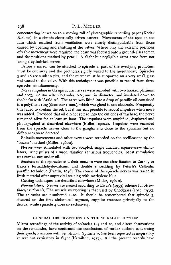
Spiracle 1 is situated on the soft membrane between the pro- and mesothorax,underneath the posterior expansion of the pronotum, which is kept clear of thespiracle by a small knob. Unlike the other spiracles the atrium leads into twoseparate tracheal trunks: the dorsal one supplies the head and prothorax and thesmaller ventral one the first pair of legs and the flight muscles. The openings ofthese trunks into the atrium will be termed the dorsal and ventral orifices. Thespiracle is contained on a small peritreme to which the anterior grooved valve isfixed and the posterior valve hinged. The ventral part of the posterior valvecomprises well sclerotized cuticle and is saucer-shaped; the dorsal part is of softcuticle with the typical' thorns' of intersegmental membrane (Fig. 2). A sclerotizedrod runs down the anterior margin of the valve; it passes between the orifices andthen inwards and round the ventral orifice. Half way round it bears a large processwhich curves anteriorly and upwards. The closer muscle (79), arising from anapodeme on the peritreme, runs dorsally to this process. One end of the opener (80)shares the insertion on the peritreme with the closer, and the other is attached tothe posterior margin of the hinged valve near its ventral end.
The closer muscle is short (0-5 xo-3 mm.) with outer fibres 20—30/i in diameterand an inter-Z distance of 3-5^. The central fibres are more slender, 10-15^. indiameter with an inter-Z distance of 5-8 /x. The opener is longer and thinner(0-15 x 07 mm.) with fibres 10-20 y. in diameter and an inter-Z distance of3-5 /*•
The spiracle is innervated by the median nerve of the prothoracic ganglion(Fig. 3). The course of the nerve is very similar to that of Acanthacris ruficorms(Ewer, 1954a). Near the spiracle it divides and two axons enter each muscle.
Respiration in the desert locust. II 241
As in the cockroach (Case, 1957) and the dragonfly nymph (Zawarzin, 1924),the motor axons themselves divide at the branching of the median nerve into twotransverse nerves. This has been shown in two ways; by simultaneously recordingfrom the two transverse nerves close to the spiracles, when identical patterns ofimpulses are obtained from each (Fig. 4), and secondly, after cutting the mediannerve close to the spiracle, by electrically stimulating one transverse nerve whenidentical movements are seen in both spiracles. Similar tests have shown this tobe true for spiracles 2 and 3 as well.
Posterior
Dorsal
Opener
D CloserProceis
Fig. 2. Spiracle i, inner view.
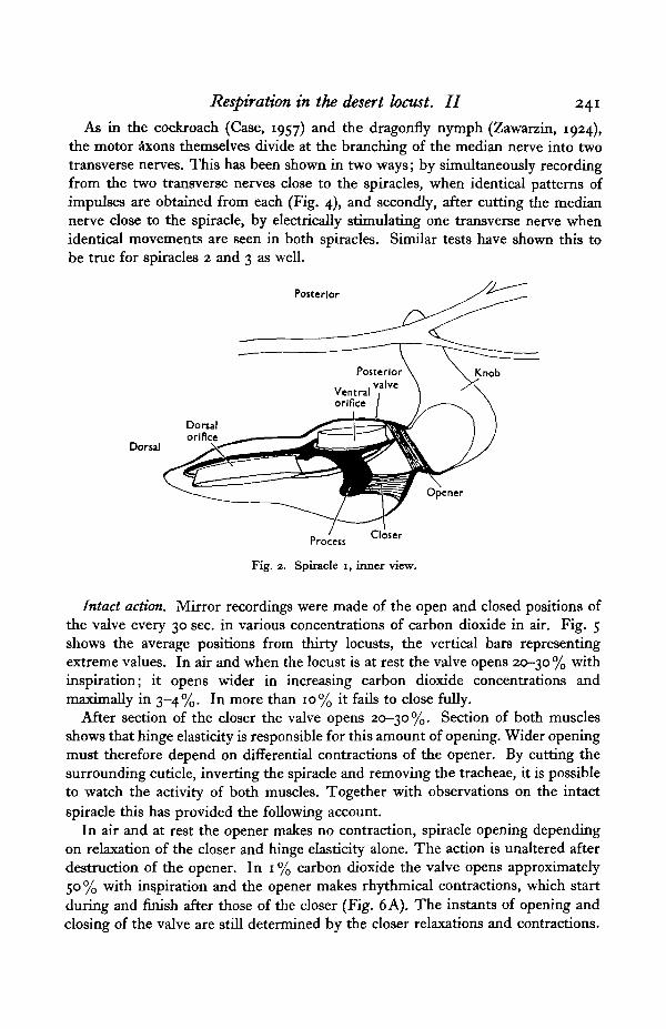
Intact action. Mirror recordings were made of the open and closed positions ofthe valve every 30 sec. in various concentrations of carbon dioxide in air. Fig. 5shows the average positions from thirty locusts, the vertical bars representingextreme values. In air and when the locust is at rest the valve opens 20-30 % withinspiration; it opens wider in increasing carbon dioxide concentrations andmaximally in 3-4%. In more than 10% it fails to close fully.
After section of the closer the valve opens 20-30%. Section of both musclesshows that hinge elasticity is responsible for this amount of opening. Wider openingmust therefore depend on differential contractions of the opener. By cutting thesurrounding cuticle, inverting the spiracle and removing the tracheae, it is possibleto watch the activity of both muscles. Together with observations on the intactspiracle this has provided the following account.
In air and at rest the opener makes no contraction, spiracle opening dependingon relaxation of the closer and hinge elasticity alone. The action is unaltered afterdestruction of the opener. In 1 % carbon dioxide the valve opens approximately50% with inspiration and the opener makes rhythmical contractions, which startduring and finish after those of the closer (Fig. 6A). The instants of opening andclosing of the valve are still determined by the closer relaxations and contractions.
242 P. L. MILLER
The slower contraction of the opener draws the posterior margin of the movingvalve inwards and thereby tenses a cuticular spring. The more the valve is distorted,the more energy is stored by the spring and the wider the valve opens when thecloser relaxes. Before the closer contracts again, the opener relaxes and the valvecloses from half to.about a quarter open. In 2% carbon dioxide the phasing
11
Fig. 3. Dorsal view of the innervation of spiracles 1, 2 and 3. GI, Gil, GUI, pro-, meso- andmetathoracic ganglia; 79, closer and 80, opener of spiracle 1; m , closer of spiracle a; 148,closer, 147, opener of spiracle 3; MN, median nerve; PSM, pleuro-subalar muscle; SPM,spino-pleural muscle; LOIM, lateral oblique intersegmental muBcle; SD salivary duct; TN,tympanal nerve.
changes (Fig. 6B); the opener now contracts while the closer is relaxed so that thespiracle opens wider towards the end of inspiration, from 25 % to about 50-70 %open. A further change takes place in 3-4% carbon dioxide, when both relaxationand contraction of the opener take place while the spiracle is closed and the closercontracted (Fig. 6C). Throughout inspiration the opener is therefore contracted
Respiration in the desert locust. II 243
y U ^\\y
Fig. 4. Oscilloscope records from the transverse nerves of the locust. A, synchronous impulses inthe nerves to right and left spiracles 3. B, Simultaneous records from the nerves to spiracles 1(top) and 2 (bottom). C, simultaneous records from the nerves to spiracles 1 (top) and 3(bottom).
Horizontal scale for A and B = 1 sec.; for C = 100 msec.
iihfsi iihft; Q g
• III Illl100—1
10 12 1Percentage COj in air
16 18 20
Fig. 5. The percentage opening of spiracle 1 in various carbon dioxide concentrations duringinspiration ('open positions') and expiration ('closed positions'). The vertical bars representextreme values. Above the graph the activity of the opener is represented.
244 P. L. MILLER
and the spiracle 70% or more open. Finally, in more than 4-5% carbon dioxidethe relaxations of the opener get smaller and disappear, so that the opener remainscontinuously contracted and the closer shuts the valve against the fully tensedspring (Fig. 6C).
Open
Spiracle valve •
Closed
Spiraclemuscles
Contracted
\/
/J
\ A.f \
, • \
Posteriorvalve
Opener
Process attachedto posterior
valve /
Closer
Fig. 6. A-C, the relation of the activity of the closer (continuous line) and the opener (broken line)to spiracle movements. A, in 1 % carbon dioxide. B, in 2 % carbon dioxide. C, in 4 % carbondioxide; three phases of opener activity are shown. D, transverse section near the ventralend of spiracle 1 illustrating the amount of spiracle opening (broken line) when the opener isrelaxed and, E, when the opener a contracted.
Since both muscles make contractions while the spiracle is closed, it is notsurprising that both have been termed closers by some authors (Lee, 1925), andsome other Orthoptera have been described with two closers in spiracle 1 (Maki,I938)-
A volley of large impulses (up to 80/sec.), recorded from the transverse nerve,corresponds to the contraction of the closer, while a volley of smaller impulses(smaller by a factor of five or six and usually at frequencies lower than 20/sec.)corresponds to the contraction of the opener (Fig. 7 G). In air when the opener staysrelaxed no small impulses appear, and in 5% carbon dioxide when the openerremains contracted the small impulses are continual. No change in the pattern ofimpulses has been detected after squashing the nerve peripherally, but after itssection near the ganglion they cease. There is no evidence for sensory nerves in thetransverse nerve, which are associated with the spiracle. Sensory impulses havebeen recorded from nerve IAOL, which probably innervates cuticular sense organsnear the spiracle, but they remain unaltered in carbon dioxide.
Inspection of the records reveals that the large (closer) impulses often occur inpairs and are of two sizes; moreover, one usually occurs at a frequency slightly
Respiration in the desert locust. II 245
different from that of the other. This means that they come in and out of phase,that is, they beat (Fig. 7 A, B). The same is usually apparent in the small (opener)impulses. When they coincide, the impulses are superimposed and an extra-largeimpulse is recorded. Similar patterns have been observed in recordings fromspiracles 2 and 3.
* " H » M
• • " ' - " " " 1
Fig. 7. Oscilloscope records from the transverse nerve to spiracle i of an intact locust. A, duringa temporary pause in ventilation, closer impulses are maintained and the spiracle remainsclosed. B and C, same at faster film speed Arrows indicate extra large impulses resulting fromthe coincidence of two normal impulses. D, a silent period between two volleys of closerimpulses, during which the spiracle opens 20 %. E, small opener impulses appear during andfor a short time after volleys of closer impulses, cf. Fig. 6 A. F and G, in 5 % carbon dioxidethe opener impulses are maintained throughout, cf. Fig. 6C. H and J, simultaneous recordsfrom two pairs of electrodes 3 mm. apart on the transverse nerve of spiracle 1. H, closer andopener impulses. J, opener impulses alone. Time markers: A—G, 50 eye./sec. (trace) andi-o sec. (dots); H. and J, 20 msec. pips.
Hoyle (1959) describes spiracle 2 of the locust as being innervated by twoaxons, a 'slow' and a 'fast'. They often fire together but sometimes the slowdischarge is absent, while the fast is always there. He has observed pairs ofimpulses, but states that both members occur in the same axon. However, inspiracle 1, since one member of the pair is of a slightly but constantly different
16 Exp. Biol. 37, 2
246 P. L. MILLER
size from the other, and since the second sometimes follows the first by 1 msec,or less (presumably within the absolute refractory period of the first) and mayeven be superimposed on it, it seems more likely that each axon provides onemember of the pair, and that both axons are therefore involved in the normaloperation of the spiracle. Hoyle draws attention to paired impulses in recordsfrom the cockroach (Case, 1957); in these, too, extra large impulses occurinfrequently.
Measurements of the speed of conduction in the four axons of the spiracle 1transverse nerve have been made by recording under oil from two pairs of electrodesseparated by 3 mm. (Fig. 7H). Impulses in the two axons to the closer travel atapproximately 1-09 and 0-97 m./sec.; in the two axons to the opener they travelat approximately 0-43 and 0-36 m./sec. These speeds are slow compared with6-7 m./sec. in the giant fibres and 2-3 m./sec. in the cereal nerves of the cockroach(Roeder, 1948).
Stimulation of the transverse nerve of spiracle 1 with single shocks, after de-struction of the opener, produces twitches in the closer, and these summate to givea smooth tetanus at frequencies greater than 15/sec. The opener, however, doesnot respond to single shocks; at 3/sec. a very slow and weak contraction occurs;at 6/sec it contracts more strongly still taking 2-3 sec. to do so, and at higherfrequencies the contractions become stronger and faster. The artificial and naturalfrequencies of impulses produce about the same speed and amount of contractionin each muscle, and they show that the closer is a fast (phasic) muscle and theopener a slow and at times a tonic muscle.
The site of spiracle 1 regulation. The foregoing experiments have shown thatcarbon dioxide affects the activity of the opener. To determine whether this isinitiated centrally or peripherally, mirrors were waxed to the valves of both spiracles1 and a very gentle stream of carbon dioxide directed into one. After a delay ofseveral seconds increased opening occurred in both spiracles simultaneously. Bycomparison with results from spiracle 2, and since each spiracle receives the samemotor input, the reaction probably takes place through the ganglion, rather thanby means of a local system. The possibility remains that an extra-ganglionic systemmay control both spiracles simultaneously. However, after section of the mediannerve close to the ganglion the limited amount of spontaneous activity which thenappears is not related in the two spiracles (see p. 256).
Modifications in the patterns of nerve impulses in the transverse nerve, whichtook place as a result of carbon dioxide treatment, were unaltered after crushingthe nerve peripherally and after completely denervating the prothoracic ganglion,except for the median nerve and the anterior and posterior connectives. Conse-quently, carbon dioxide reception must take place within the ganglion or in anothersegment.
After section of the nerve cord between pro- and mesothoracic ganglia (orbetween meso- and metathoracic ganglia) all trace of synchronized action disappearsfrom spiracle 1. The valve usually remains 10 or 20% open with continual flutteringmovements due to the closer. Prodding the insect, or struggling, causes immediate
Respiration in the desert locust. II 247
full closing, and afterwards the valve opens more widely for a few seconds. In 1-2 %carbon dioxide it opens wide and in higher concentrations all fluttering ceases:opening results from a maintained contraction of the opener and relaxation of thecloser.
Fig. 8. Oscilloscope records from the transverse nerve-to spiracle i after section of the nerve cordbetween the pro- and mesothoracic ganglia. A, in air. B, in i % carbon dioxide. C, in 2 %.D, in 4%. E, touching the antenna ('buzzer') during treatment with 2% carbon dioxide.F, after section of the nerve cord in the neck. Time marker: 50 cyc./sec. (trace) and 0-5 sec.(dots).
Records from the transverse nerve show a constant stream of pairs of largecloser impulses (7-12 pairs/sec.)—too slow apparently to maintain a tetanus in thecloser (Fig. 8). The slight difference in the frequencies in the axons is more constantthan in the intact insect, and the number of extra-large impulses (beats) gives anindication of the frequency difference in the firing of the two axons. With strugglingthe frequency in both axons increases and the same phasing is maintained. In 1 %carbon dioxide a few small (opener) impulses appear but the large impulses areunchanged (Fig. 8B); in 2%, the small impulses increase in frequency and thelarge decrease; in 3-4% the large disappear altogether (Fig. 8D).
Since section of the nerve cord between the pro- and mesothoracic gangliaabolishes the rhythmic opening and closing movements in spiracle 1, but not itssensitivity to carbon dioxide, the latter must depend on the head or prothoracicganglion.
Head-perfusion experiments were undertaken to demonstrate a carbon-dioxidereceptor which controlled the activity of the opener. (The technique is describedelsewhere, Miller, 1960a.) 1-2% carbon dioxide was injected into the mandibularair-sac and it produced almost immediate contraction of the opener. After section
16-2
248 P. L. MILLER
of the nerve cord in the neck the injections had no effect, although 4-5 % carbondioxide directed at the ganglion then produced a weak contraction.
It seems that the head contains sensitive carbon-dioxide receptors whose stimula-tion increases abdominal ventilation, induces neck and prothoracic ventilation andcauses contraction of the opener of spiracle 1. It is possible that one receptorcontrols all the reactions. In addition the prothoracic ganglion has less sensitivereceptors affecting abdominal ventilation and the spiracle 1 opener. Stimulationof the head receptors produces neck and prothoracic ventilation only when themetathoracic ganglion is intact: for opener contractions, however, no more thanthe prothoracic ganglion need be intact. Since the dorsal trunk leads directly fromspiracle 1 to the head, it is not surprising that the head should have an overridinginfluence on the activity of the spiracle 1 opener. It has no comparable influenceover other spiracles.
The ventral orifice. The opener is inserted on the posterior margin of the movingvalve behind the ventral orifice, and the closer in front on the upturned process(Fig. 2). When both muscles contract strongly and simultaneously, the long narroworifice is completely constricted by the scissor-like action of the rod bearing theprocess and the posterior margin. In more than 4—5 % carbon dioxide, when theopener remains contracted, the orifice opens slightly only during inspiration as thecloser relaxes. On the other hand, when the opener remains relaxed the orifice isopen all the time. The significance of this surprising consequence of opener con-tractions (a misnomer in the context) is discussed elsewhere (Miller, 19606).
SPIRACLE 2Anatomy. A detailed account of the structure of spiracle 2, together with a
description of the histology of the closer muscle and its innervation, has been givenby Hoyle (1959). Only a small number of additional points will be mentionedhere.
Spiracle 2 possesses a single closer muscle (111) and the valves are opened by theelasticity of the cuticular surround. Hoyle points out the occurrence of two typesof muscle fibre in the closer: a central core of thin fibres with an inter-Z distanceof 6-7/n, and surrounding thicker fibres with an inter-Z distance of 3-6 /x. Innerfibres with an inter-Z distance as great as 10/i have been observed during thepresent investigation. The thin fibres are more numerous than in the closer ofspiracle i, and the inter-Z distance of many is greater. Hoyle suggests that theymight be normal fibres whose development has been arrested, but neither Tiegs(1955), Wigglesworth (1956) nor Smith (1958) mentions a stage in fibre developmentwhen the striations appear more widely separated than in the mature muscle.
Secretory tissue occurs in the thickness of the anterior valve, and yellow orcolourless droplets have been observed hanging inside the valve after roughhandling of the insect. Spiracle 2 of Romalea nricroptera froths a black noxiousliquid when the insect is irritated, and the secretions in Schistocerca may be similarto this.
Respiration in the desert locust. II 249
After section of the closer muscle the valves open and are separated maximallyby o-1-0-2 mm. Observations made during flight, however, show the valves thento be separated by o^-o^ mm. This wide-opening is achieved by a cuticulardevice.
Bridge
Bridge '
Ligament
Closer
Fig. 9. The wide-opening mechanism in spiracle 2. A, external view showing position of the ligament.B, transverse section of the suture immediately dorsal to the spiracle. C, diagrammatic trans-verse section of the spiracle. Arrow indicates the movement of the mesothorax. Explanationin text.
The suture between the meso- and metathoracic lateral wall, dorsal to spiracle 2,comprises on the anterior margin of the metepisternum a well-formed apodeme towhich the metacoxal muscle (125) is attached. The apodeme is joined to thethickened posterior margin of the mesepimeron by a bridge of cuticle with specificstaining properties (Fig. 9 B). The bridge, lying under the mesepimeron and barelyvisible from the exterior, extends about three-quarters of the way from the spiracleto the wings. It is continuous with the cuticle surrounding the spiracle valves but,while the latter acts hke a metal spring, the bridge is rubber-like and has many ofthe properties of the elastic ligaments of the wings (Weis-Fogh, 1958). The rubberyquality is not lost after fixation in alcohol or after drying and re-wetting. In sectionsfixed in Carnoy or Baker's fixatives and stained with Hansen's haematoxylin orchlorazol black, the bridge stains more intensely than normal cuticle: with Mallory'striple stain it is red.
The bridge allows the mesothorax to be pulled very slightly anteriorly and up-wards from the metathorax; on release it snaps back into position. By pulling bothends of a thin horizontal section of the lateral wall under a microscope, the actionof the elastic part can be watched. A thin superficial layer of sclerotized cuticleprevents the movement from exceeding about 0-05 mm.
In the nymphs of Schistocerca this movement pulls the base of the spiracle valvesapart and tends to close the spiracle. In the adult, however, a small and sometimesdarkened ligament runs in the cuticle from a knob on the dorsal end of the anterior
250 P. L. MILLER
valve to the apodeme of the metepisternum (Fig. <)A). It is attached very closebut just medial to the vertical axis of rotation of the valve, so that a small anteriormovement of the mesothorax pivots the valve wide open on the ligament, and thisin turn cants open the posterior valve (Fig. 9C). After section of the ligament nowide-opening occurs, although the normal movements are unaffected.
Wide-opening has been observed momentarily during struggling and always inflight. It can be artificially induced by gentle pressure on the cuticle just anteriorto the spiracle. After section of both pleural apophyses through their respectivecoxal cavities, it no longer occurs in flight: section of the metathoracic apophysisalone is nearly as effective. Electrical stimulation at 100/sec. of nerve IVBbi,which innervates the metathoracic sternopleural muscle (115), causes a slowcontraction of that muscle but wide-opening does not occur. Locusts were flownafter section of this nerve and of the homologous nerve in the mesothorax, butwide-opening was unaffected. Apparently the rigidity provided by the sterno-pleural apophysis is essential for wide-opening, but it is not dependent on contrac-tion of the sterno-pleural muscles. Section of the coxal muscles which are insertedon the pleural and intersegmental sutures does not prevent wide-opening in flight,although these muscles are probably responsible for its occurrence during struggling.The basalar and subalar muscles in the mesothorax (98, 99) and in the metathorax(128, 129) assist in the downstroke of the wings (Pringle, 1957), and after theirunilateral section, the wing movements of that side are much reduced and wide-opening almost disappears. Comparison with the intact side, where wide-openingcontinues, suggests that contraction of the basalars and subalars causes it. Electricalstimulation of the mesothoracic basalar and subalar muscles is more effective inproducing wide-opening than stimulation of the metathoracic muscles. Weis-Fogh(unpublished) has suggested the presence of tonic components in these muscles,which may be responsible for wide-opening.
Innervation. The spiracle is innervated by the median nerve of the mesothoracicganglion (Fig. 3). The course of the nerve is similar to that of Acanthacris ruficorms(Ewer, 1954a). Near the salivary duct the transverse nerve is joined by a branchfrom nerve 1 of the metathoracic ganglion. Before and after crossing the duct itsupplies a number of branches (meso IF) to the vestige of the nymphal spino-pleural muscle. Close to the spiracle muscle the transverse nerve divides. Onebranch, meso IG (meso IE of Ewer), supplies the vestige of the mesothoracicpleuro-aubalar muscle: as it passes dorsal to the spiracle, very slender branchesleave it and appear to end on the surface of air-sacs. At the bases of these branchesthere are swellings in the nerve, which contain large cells. The other branch,meso IE, sends two axons into the spiracle muscle.
Intact action. Mirror tests on the open and closed positions of the valves havebeen made in various carbon-dioxide concentrations (Fig. 10). The valves neveropen more than 30-40% with inspiration; that is, there is no wide-opening incarbon dioxide. In more than 7-8% carbon dioxide they usually fail to closecompletely, but this value is variable and in some locusts the valves may make onlythe weakest closing movements in 5 %, whereas in others more or less full closing
Respiration in the desert locust. II 251
continues in 10-12% carbon dioxide. A comparable variation in the sensitivity ofthe dragonfly spiracles to carbon dioxide has been shown to depend on the waterbalance of the insect (Miller, unpublished), and the same may be true for thelocust. To conclude, spiracle 2 in the non-flying locust shows much less of a gradedresponse than spiracle 1.
100-1
c 75H2LoV
fa Open position
6 8 10 12Percentage COj in air
14I
16 20
Fig. 10. The percentage opening of spiracle a in different carbon dioxide concentrations duringinspiration (' open positions') and expiration (' closed positions ')• Vertical bars represent extremevalues.
Unilateral gassing with 10 % carbon dioxide has demonstrated that one spiraclemay remain open while its partner continues to make full closing movements: thevolleys of impulses remain unchanged in both transverse nerves. The direct actionof carbon dioxide on the neuromuscular junction, reducing the electrical responsesand the tension developed, has recently been demonstrated (Hoyle, i960), andis no doubt responsible for the independent action of one spiracle.
The volleys of impulses giving rise to spiracle closing nearly always comprisepairs of impulses of slightly different sizes. For the reasons already given forspiracle 1, both axons appear to be involved in the normal operation of the closer(Fig. 11).
After section of the nerve cord between the meso- and metathoracic ganglia, thebehaviour of spiracle 2 is similar to that of spiracle 1. The volleys of impulses inthe transverse nerve are replaced by continual pairs of impulses, usually between6 and 12 pairs/sec. (Fig. 11D). In some locusts the spiracle remains closed, butin many it is 20 % open and continually fluttering: with struggling it closes fully.The sensitivity to carbon dioxide is much increased and it will open slightly in 1—2 %and completely in 2-3%, when all fluttering ceases. As in the intact insect, theimpulses in the transverse nerve remain unaffected during opening. The sensitivityof the response remains undiminished for several weeks.
After the spiracle has been uncoupled from ventilation, the differing frequenciesin the two axons become more regular. By watching the valves and listeningthrough earphones to the impulses recorded close to the spiracle, the apparent
up- P. L. MILLER
drop in frequency as the impulses nearly and then completely coincide can be seento correspond to a larger twitch of the valve. Periods of coincidence and largertwitches occur about once a second, while there may be 8-10 small twitches persecond. Hoyle (1959) describes a rhythm persisting in spiracle 2 after sectionbetween meso- and metathoracic ganglia, often seen as a regular rise and fall in the
Fig. 11. Oscilloscope records from the transverse nerve to spiracle 2. A, a volley of impulsescoinciding with spiracle closing. B, the same at faster film speed. Arrows indicate extra largeimpulses resulting from the coincidence of two normal impulses. C, during weak ventilationthe spiracle opens less than 10 % with inspiration and impulses continue at a reduced frequency(horizontal line = inspiration). D, records after section of the nerve cord between meso-and metathoracic ganglia. E, record from an intact locust at low film speed, showing the regularoccurrence of extra large impulses. Time markers: A, ioo msec. dots. B, 50 msec. dots.C-E, 50 cyc/sec. and i-o sec. dots.
frequency of impulses. The asynchronous firing of the two axons gives the im-pression of a rise and fall of frequency. This rhythm is clearly of a different naturefrom that which occurs in the intact insect involving a rhythmic cessation of allimpulses. Hoyle states that a pair of impulses separated by 3-4 msec, gives riseto a considerably greater tension than would be evoked by either alone; his accountrefers to impulses in the same fast axon, whereas the pairs under considerationhere, which give rise to larger twitches, occur in different axons.
It is not surprising that the apparent sensitivity of the peripheral carbon-dioxidereaction should be greater after the spiracle is uncoupled from ventilation, sincethe frequency of motor impulses reaching the spiracle is then less. In an intactbut immobilized locust the spiracle may fail to close fully in 7% carbon dioxide,whereas after release full closing may occur in higher concentrations. Variation ofthe impulse frequency provides a method of altering the sensitivity of the spiraclereaction to carbon dioxide. To what extent it is used, perhaps in relation to thewater balance, is unknown.
Anterior
Respiration in the desert locust. II 253
To conclude, gradations of the open position of spiracle 2 are affected primarilythrough the wide-opening mechanism and appear only in flight, although areduced amount of opening (10-20%) is infrequently seen in resting locusts.Gradations of the closed position do not normally take place in less than 5-7 %carbon dioxide, and then depend on the local action of carbon dioxide on the neuro-muscular junction (Hoyle, i960).
SPIRACLE 3
Anatomy. Spiracle 3, lying just anterior to the auditory capsule and belongingto the first abdominal segment, supplies large tracheae to the thoracic air-sacs andflight muscles, and also to the gut. The inner endof the atrium is closed by the hinged valve which isproduced into a manubrium for the attachment of thedorsal closer and ventral opener muscles (Fig. 12).The short closer, 148 (0-32 x 0-096 mm.), is insertedon a rigid apodeme immediately above the spiracle,and the long narrow opener, 147 (2-6 x 0-064 t 0
0-032 mm.), is inserted on the soft ventral interseg-mental membrane, beside the tensor of the tympanum(146). For over half its length the opener runs parallelto muscle 146 and is bound to it by connective tissue.The opener comprises fibres normal in appearancewith an inter-Z distance of 3-5 /A. The structure ofthe closer is similar to that in spiracle 1. Section ofboth muscles shows the hinge to possess limitedelasticity sufficient to open the spiracle valve 20%.
Iimervation. The innervation is again similar tothat of Acanthacris ruficornis (Ewer, 1953, Fig. 3).The anterior median nerve divides into transversenerves shortly after leaving dorsally from the meta-thoracic ganglion. Each transverse nerve (meta IC)sends a branch (meta iF) to muscle 117 and one tonerve ABDi. It then crosses the ventral end ofcoxal muscles 119 and 120, sending several branchesinto the former. Shortly before reaching the spiracle a further branch leaves tosupply the vestige of the nymphal longitudinal oblique intersegmental muscle ofthe metathorax (140). The transverse nerve joins the opener half way along itslength and after sending several twigs into that muscle, runs parallel and into thecloser.
Intact action. Three patterns of behaviour are commonly seen in spiracle 3 ofthe non-flying locust: in the first it remains closed, in the second it opens slightlyduring inspiration and in the third it opens fully.
The first pattern usually appears when the locust is at rest or moderately activeand ventilation is not greatly stimulated. At such a time records from the transverse
Fig. iz. Spiracle 3, inner view.148, closer, 147, opener; 146,tensor of the tympanum.
254 P- L. MILLER
nerve show a constant stream of impulses (Fig. 13 A). The second occurs whenventilation is rapid (c. 60/min.) but shallow, and the valve opens about 10% or lessin phase with the other inspiratory spiracles: sometimes it opens twice per cycle.Opening results from partial relaxation of the closer and from hinge elasticity; theaction is not disturbed by section of the opener. Records from the transverse nerveshow alternate volleys of impulses and silent periods (Fig. 13B).
B
Fig. 13. Oscilloscope records from the transverse nerve to spiracle 3 of an intact locust. A, restinglocust; the spiracle remains closed. B, spiracle opens less than 10% with inspiration. C, inz % carbon dioxide the spiracle opens c. 50 %, and small impulses appear during opening.D and E, slow ventilation of full amplitude, the spiracle opens fully with inspiration whilecloser and opener impulses alternate. Horizontal line = spiracle opening. Time marker,50 eye/sec (trace) and i-o sec. (dots).
With slow deep ventilation (30/min.), the third pattern appears when the spiracleopens 50—100% during inspiration. The amount of opening is dependent ondifferential contractions of the opener, although unlike spiracle 1 there is nostoring of energy in an elastic system and the contractions of opener and closeralternate regularly. This is reflected in the patterns of nerve impulses from thetransverse nerve (Fig. 13C-E), which show a regular alternation of large (closer)and small (opener) impulses—smaller by a factor of 4-5. The small impulses neveroverlap the large and occur at comparable frequencies (6o-7o/sec.) when thespiracle opens 100%, and at lower frequencies (10-20/sec.) when the spiracleopens less.
Mirror tests on the open and closed positions of the valve in different carbondioxide concentrations show that the behaviour of the spiracle is comparable to
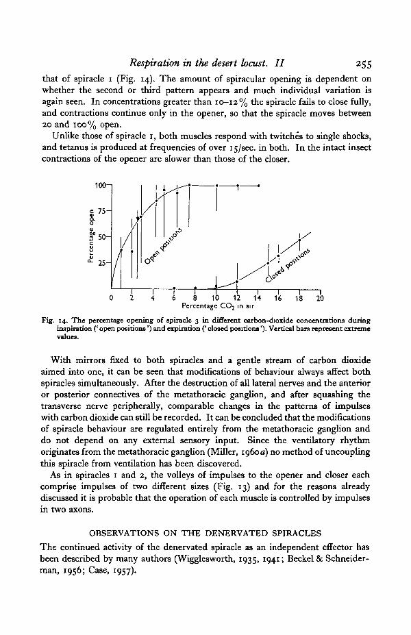
Respiration in the desert locust. II 255
that of spiracle 1 (Fig. 14). The amount of spiracular opening is dependent onwhether the second or third pattern appears and much individual variation isagain seen. In concentrations greater than 10-12% the spiracle fails to close fully,and contractions continue only in the opener, so that the spiracle moves between20 and 100% open.
Unlike those of spiracle 1, both muscles respond with twitches to single shocks,and tetanus is produced at frequencies of over 15/sec. in both. In the intact insectcontractions of the opener are slower than those of the closer.
6 8 10 12 14Percentage CO2 in air
Fig. 14. The percentage opening of spiracle 3 in different carbon-dioxide concentrations duringinspiration (' open positions') and expiration (' closed positions'). Vertical bars represent extremevalues.
With mirrors fixed to both spiracles and a gentle stream of carbon dioxideaimed into one, it can be seen that modifications of behaviour always affect bothspiracles simultaneously. After the destruction of all lateral nerves and the anterioror posterior connectives of the metathoracic ganglion, and after squashing thetransverse nerve peripherally, comparable changes in the patterns of impulseswith carbon dioxide can still be recorded. It can be concluded that the modificationsof spiracle behaviour are regulated entirely from the metathoracic ganglion anddo not depend on any external sensory input. Since the ventilatory rhythmoriginates from the metathoracic ganglion (Miller, 1960 a) no method of uncouplingthis spiracle from ventilation has been discovered.
As in spiracles 1 and 2, the volleys of impulses to the opener and closer eachcomprise impulses of two different sizes (Fig. 13) and for the reasons alreadydiscussed it is probable that the operation of each muscle is controlled by impulsesin two axons.
OBSERVATIONS ON THE DENERVATED SPIRACLESThe continued activity of the denervated spiracle as an independent effector hasbeen described by many authors (Wigglesworth, 1935, 1941; Beckel & Schneider-man, 1956; Case, 1957).
256 P. L. MILLER
Spiracles 1 and 3. After section of the transverse nerve of spiracle 1 or 3, theopener and closer muscles show no activity. One or two hours later the closercontracts and the spiracle is closed. Subsequently, it relaxes in more than 5%carbon dioxide, but the opener makes no contractions.
Spiracle 2. The left transverse nerve of sixty locusts was cut close to the spiracle,but medial to its junction with meso IG. In the majority the closer showed nomore activity for 1 or 2 days. Later it contracted and the spiracle closed. Thelocusts were kept in a cage under normal conditions (see Part I) and were inspectedthree times a day. At each inspection the locusts were pinned on one side to a corkand observed for 5 or 10 min. During closing, five stages can be recognized spreadover about 24 hr.:
(i) Valves are open and motionless, but close momentarily after one is pulledout and released.
(ii) Small spontaneous movements appear.(iii) Valves are half closed and make continual small movements.(iv) Valves are almost fully closed but still moving.(v) Valves are fully closed.
Most spiracles close after 2 or 3 days—some take as long as 5.In a further batch of twelve locusts the transverse nerve was cut close to the
ganglion, but the subsequent behaviour was unchanged.Fig. 15 shows the times of closing: eight spiracles closed immediately after
nerve section. When the locusts were kept for 3 hr. in the dark or were pinneddown and kept at 40 C , then in about 70% spiracle closing followed nerve sectionimmediately. If the locusts were kept immobilized the spiracles remained closed,but shortly after their release they opened. This suggests that the denervatedspiracle is normally kept open for the first day or two after nerve section by thelevel of metabolic end-products in the insect, and that only after 3 hr. immobiliza-tion do these fall sufficiently low for the spiracle to close. Herber & Slifer (1928)concluded that the ventilation frequency of Melanoplus femur-rvbrum did not reacha true 'resting rate' for at least 1 hr. after the insect was pinned down and thenonly in the complete absence of struggling. It would follow from the hypothesisthat spontaneous closing is normally inhibited in the intact locust by the level ofmetabolic end-products, so that the spiracle is able to open when the motor im-pulses cease; but this seems improbable and there is no evidence to suggest thatthe spontaneous closing mechanism is operative in the innervated muscle.
The subsequent closing of spiracle 2 after 2 or 3 days may result from a decreasein sensitivity to metabolic products or an increase in the excitability of the muscle.That it was not a result of water loss through the open spiracle was shown bykeeping locusts in moist and in dry air following nerve section, and observing thatspiracle closing occurred after approximately the same interval in each.
The closed denervated spiracle is very sensitive to carbon dioxide, opening fullyin 1-2 % and reclosing shortly after return to air. It opens a few seconds after thestart of struggling or flight. It does not react to oxygen lack, and remains closed
Respiration in the desert locust. II 257
in 100% nitrogen until nearly all movement of the locust has ceased. The move-ments are always slow and smooth—very different from the fluttering seen in thespiracle uncoupled from ventilation. This behaviour is quite unlike that of thedenervated cockroach spiracle 2 (Case, 1957) which closes within 24 hr. of denerva-tion and then will not open in less than 15 % carbon dioxide. Closing is followedafter 4-6 days by ' fasciculation', which Case associates with the degeneration of theperipheral nerve stump.
MesolG/ /pnCloier
60
1 2 3 4 5Days after cutting transverse nerve
Fig. 15. The time taken by the denervated spiracles a of sixty locusts to close. Black area, spiraclesat stage 5; hatched area, spiracles at stages 2-3. Eight spiracles close immediately after theoperation. Explanation in text.
In the locust the sensitivity remains for 2-3 months; occasionally, when thesensitivity was much reduced, the tracheae supplying the closer muscle were foundto be partially filled with liquid. Hoyle (i960) comments on the need for the airpassages into the muscle to be unblocked for carbon dioxide to affect the muscle,and this observation suggests that the same mechanism is responsible for musclerelaxation in the denervated spiracle. Nerve regeneration, indicated by the re-sumption of synchronized movements, occurred in two locusts only (3 %). Case(1957) reported regeneration in 36% of operated cockroaches.
It seemed that the independent behaviour of the spiracles could be explained ifmotor nerve cells were present in the muscle. To investigate this possibility,
258 P. L. MILLER
6/x sections of the muscle were cut and stained for cholinesterases with myristoylcholine and light green (Denz, 1953; Bowden & Lowy, 1955). Dark areas appearedin the sections but they gave no clear indication of the presence of nerve cells.
The effect of nicotine on insect nervous tissue is well known (Roeder, 1939).After initial excitation, it causes the complete block of ganglia. To test for thepresence of nerve cells in the closer, the spiracles were cut out and floated on asolution of o-ooi % nicotine sulphate in Ringer. They closed immediately andremained closed for several hours, but would open in 5% carbon dioxide. Instronger nicotine solutions (o-oi %) they would not open in less than 20% carbondioxide. Nicotine excites the closer to contract and its subsequent failure todestroy the autonomous action suggests that contraction of the closer is notdependent on continual excitation from a motor neurone in the muscle, but is aproperty of the muscle fibres themselves.
Hoyle (1959) pointed out that the thinner muscle fibres with a long inter-Zdistance might be expected to perform slow tonic contractions. An attempt wasmade to destroy most of the large outer fibres with a small scalpel and a tungstenhook. In other spiracles the thin inner fibres, together with the large fibres of oneside, were destroyed. The operations were performed on twenty denervatedspiracles, and after observation the muscles were fixed in Carnoy and stained withacid fuchsin, so that an estimate of the remaining fibres could be made. Providedthat the thin central fibres were not damaged, the spiracle remained closed andcontinued to react to carbon dioxide. If, however, they were destroyed, but morethan half of the large fibres were left intact, the spiracle opened and showed nofurther activity.
Although not conclusive, the results suggest that the maintained contractionof the denervated spiracle and the subsequent relaxations in low carbon-dioxideconcentrations depend on the small central fibres. The observation that the closersof spiracles 1 and 3, possess a few thin fibres and make maintained contractionsaccords with this suggestion.
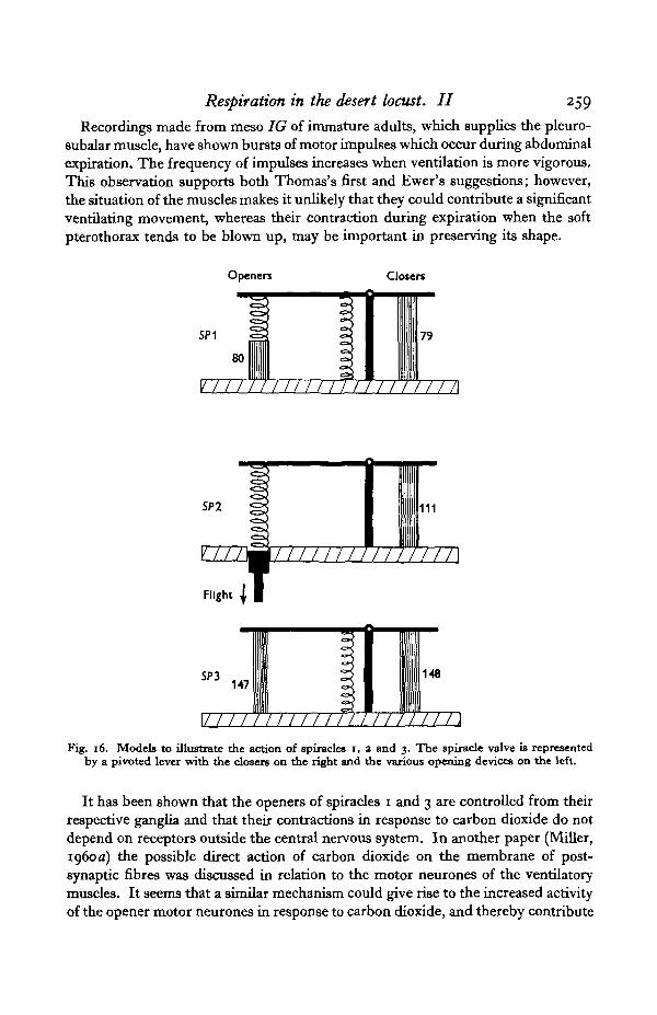
DISCUSSION
The operation of the first three pairs of spiracles can be compared when theirvalves are represented by levers moved by springs and muscles (Fig. 16). Thisemphasizes the similarity of the closing and differences in the opening mechanisms,the latter being effected in spiracle 1 by hinge elasticity and a muscle in series witha spring; in spiracle 2 by a spring with variable tension (the piston), and in spiracle 3and the remaining spiracles by a muscle which probably includes a variable elasticelement and at times works like a rubber band (Miller, 19606).
It is noteworthy that the median nerves in each segment, in addition to innervatingthe spiracles, supply branches to some of the nymphal muscles which degeneratein the adult (Ewer, 19546). Thomas (1954) suggested that these muscles may makenymphal respiratory movements or possibly that they help to rupture the cuticleat ecdysis. Ewer (19546) has proposed that they may be important in preservingthe shape of the pterothorax immediately after moulting.
Respiration in the desert locust. II 259
Recordings made from meso IG of immature adults, which supplies the pleuro-subalar muscle, have shown bursts of motor impulses which occur during abdominalexpiration. The frequency of impulses increases when ventilation is more vigorous.This observation supports both Thomas's first and Ewer's suggestions; however,the situation of the muscles makes it unlikely that they could contribute a significantventilating movement, whereas their contraction during expiration when the softpterothorax tends to be blown up, may be important in preserving its shape.
Openers Closers
SP1
80
v/////////m nrr / / / /i
SP2
V ///Imi/ /
Flight I I
IITJITTT
111
/TT\
SP3147
148
\i 1111111111111 mn n\Fig. 16. Models to illustrate the action of spiracles i, 3 and 3. The spiracle valve is represented
by a pivoted lever with the closers on the right and the various opening devices on the left.
It has been shown that the openers of spiracles 1 and 3 are controlled from theirrespective ganglia and that their contractions in response to carbon dioxide do notdepend on receptors outside the central nervous system. In another paper (Miller,1960a) the possible direct action of carbon dioxide on the membrane of post-synaptic fibres was discussed in relation to the motor neurones of the ventilatorymuscles. It seems that a similar mechanism could give rise to the increased activityof the opener motor neurones in response to carbon dioxide, and thereby contribute
260 P. L. MILLER
further to the economy of interconnexions in the central nervous system, as wassuggested elsewhere (Miller, 1960a).
Spiracle 2 is an 'odd-man-out' and it is difficult to explain why this spiraclealone in the locust should have one muscle and a peripheral control mechanism.Observations on the one-muscle spiracles of Diptera and Odonata have shown thatthey have a similar peripheral control, and this may be a property of all suchspiracles. No insect is known to have an opener muscle in spiracle 2 (Maki, 1938),and openers are not found in spiracle 1 outside the Orthoptera. It may be thatthe peripheral mechanism has some particular significance for the locomotorysegments, although what part it plays in the adult locust remains uncertain.Alternatively, the peripheral mechanism may represent a more primitive systemwhich is overridden in the locust in the interests of synchronization with ventilation.There is plenty of evidence (to be discussed in a later paper), however, that inOdonata and Diptera it takes part in the normal control of the spiracle. In thefirst instar locust spiracle 2 is rarely synchronized with ventilation; more often itappears to act independently and most probably under the influence of the peri-pheral mechanism.
Both the ventilatory and the spiracular rhythms are autonomous although modifiedby sensory input (Hoyle, 1959). The presence of carbon dioxide in the ganglionmay be necessary for their initiation, but each cycle is not dependent on the build-upand release of the gas. A comparison may be made with the cockroach whereventilation and synchronized movements appear only in more than 10% carbondioxide (Hazelhoff, 1927); below this the spiracles are regulated perhaps bya peripheral mechanism, similar to that of the locust. The locust, possessingthe same mechanisms, is much more strongly biased in favour of ventilationsynchronization.
Attention has been drawn to the firing of two motor axons at slightly differentfrequencies during the normal operation of the spiracle. This has suggested anhypothetical mechanism to account for the rhythmical disappearance of motorimpulses in the nerves to the spiracle muscles during ventilation, as follows.Two internuncial neurones each firing spontaneously and continually, but atslightly different frequencies, relay onto the motor neurones of the spiracle nerves.One internuncial is inhibitory and the other excitatory. The excitatory impulse isinhibited at the synapse if, say, the inhibitory impulse precedes it by less than200 msec. For peripheral a-inhibition in Crustacea the inhibitory impulse mustprecede the excitatory by only a few msec, to be effective (Katz, 1949); however,at least in vertebrates, a central inhibitory volley can still be effective after 100 msec.(Sherrington, 1947). If, for example, the excitatory internuncial in the locust firesat 10/sec, and the inhibitory at 11/sec, two excitatory impulses will be inhibitedat the synapse every second. If two controlling cells are now postulated, oneinfluencing the frequency in both internuncials by the same amount, and the otheraltering the frequency in one only, both changes in frequency and variations inthe duration of periods of firing and of silence can be achieved (Fig. 17). To obtainsome combinations it may be necessary to postulate a second inhibitory internuncial.
Respiration in the desert locust. II 261
By some such plan two continually firing cells can produce a much slower rhythmin a post-synaptic fibre without any sensory feed-back. Such a system could beresponsible for the rhythmic firing of the motor cells of the ventilatory muscles aswell as those of the spiracle muscles.
SSpiracle motor neurone - -
. A - . I I I » M i l — H + l — - :
I I I I I I I I L.I I I I I I I I I I I /"^
Excitatory Intemuncial
Fig. 17. Diagram to illustrate the theory of the means of producing the ventilatory rhythm.Explanation in text.
The same principle is used in beat-frequency electronic oscillators, where twooscillators produce a third frequency much lower than that of either alone.
After section of the nerve cord between the meso- and metathoracic ganglia,the rhythm disappears from spiracles 1 and 2, and is replaced by continual firingin the motor nerves. This can be explained if the inhibitory intemuncial is in themetathoracic and the excitatory in the mesothoracic ganglion. During strongflight, motor impulses in the nerve to spiracle 2 cease entirely (Miller 1960Z1);this is explicable if at such times the excitatory and inhibitory internuncials firein phase.
It should be emphasized that this scheme is entirely hypothetical, and recalledthat the patterns of impulses in the transverse nerves involve probably two motorfibres, so that the scheme must perhaps be duplicated.
SUMMARY
1. Small mirrors are used to record the movements of the spiracle valves ofSchistocerca gregaria, and some general observations are made on the synchronizedmovements of the spiracles with ventilation.
2. Spiracles 1-3 are shown to alter their positions of opening and closing indifferent carbon dioxide concentrations, within the pattern of synchronizedmovements.
3. Modifications of the amount of opening of spiracles 1 and 3 take place as aresult of differential contractions of the openers. In spiracle 1 the energy is storedin an elastic system, and the opener does not necessarily make contractions everycycle. Oscilloscope recordings show these reactions, resulting from carbon-dioxidestimulation, are controlled entirely from within the central nervous system anddo not depend on any sensory input.
4. The movements of spiracle 2 are controlled peripherally through the directaction of carbon dioxide on the muscle membrane (Hoyle, i960) and also througha cuticular wide-opening device.
J 7 Exp. B10L 37, 2
262 P. L. MILLER
5. The maintained sensitivity of spiracle 1 and the increased sensitivity ofspiracle 2 after they are uncoupled from ventilation are discussed.
6. Some remarks are made on the activity of the denervated spiracle 2, and thesuggestion is made that it depends on a central core of thin fibres.
7. A central nervous mechanism is postulated which could account for therhythmic cessation of impulses in spiracle and ventilatory nerves.
I would like to thank Prof. V. B. Wigglesworth for encouragement and super-vision of this work. I am most grateful to Prof. T. Weis-Fogh for much usefuldiscussion and help. My thanks are due to Dr F. S. J. Hollick for the supply ofsmall mirrors, to Dr G. Hoyle and Dr D. S. Smith for permission to quote theirunpublished results, and to the Agricultural Research Council for financial support.
REFERENCESALBRECHT, F. O. (1953). The Anatomy of the migratory locust. London: Athlone Press.ALBRECHT, F. O. (1956). Anatomy of the red locust (Nomadacris septemfasciata). Anti-Locust Bull. 23.BECKEL, W. E. & SCHNEIDERMAN, H. A. (1956). Spiracle of the Cecropia moth as an independent
effector. Anat. Rec. ia$, no. 3. 559.BoWDEN, J. & LoWY, J. (1955). The lamellibranch muscle innervation. Nature, Lond., 176, 346.BUCK, J.B. (1958). A theory of the mechanism of cyclic carbon dioxide release in insects. BioL Bull.,
Wood's Hole, 1x4, 118-40.BuBftKT.T., E. (1957). Spiracular control of water loss in the tsetse fly. Proc. R. ent. Soc. Lond. A,
3a, ai-a9.CASE, J. F. (1957). Median nerves and cockroach apiracular function. J. ins. Physiol. 1, 85-94.DBNZ, F. A. (1953). On the histochemistry of the myoneural junction. Brit. J. exp. Path. 34,
329-39.EwBR, D. W. (1953). The anatomy of the nervous system of the tree locust, Acanthacris ruficornis.
I. The adult metathorax. Ann. Natal Mus. ia, 367-81.EWER, D. W. (1954 a). The anatomy of the nervous system of the tree locust, Acanthacris ruficornis
II. The adult mesothorax. J. ent. Soc. S. Afr. 17, 27-37.EWER, D. W. (19546). On the nymphal musculature of the pterothorax of certain Acrididae (Ortho-
ptera). Aim. Natal Mus. 13, 79-89.FRABNKEL, G. (193a). Untersuchungen Uber die Koordination von Reflexen und automatisch-
nervosen Rhythmen bei Insekten. III. Daa Problem des gerichteten Atemstromes in denTracheen der Insekten. Z. vergl. Physiol. 16, 418-43.
HAMILTON, A. G. (1937). The mechanism of respiration of locusts and its bearing on the problemof the inhalation of poisonous dusts. Bull. ent. Res. 38, 53-68.
HAZELHOFF, E. H. (1927). Die Regulierung der Atmung bei Insekten und Spinnen. Z. vergl.Physiol. 5, I79-9O-
HERBER, E. C. SC SLJFER, E. H. (1928). The regularity of respiratory movements of Locustidae.Physiol. Zool. 1, 593-602.
HOYLE, G. (1959). The neuromuscular mechanism of an insect spiracular muscle. J. ins. Physiol.3. 378-94-
HOYLE, G. (i960). Action of carbon dioxide on an insect spiracular muscle. J. ins. Physiol. (in thePress).
JANNONE, G. (1040). Studio morfologico, anatomico e istologico del Dodostaurus maroccanus.Boll. Lab. Ent. agr. Portici, 4, 1-433.
KARANDIKAR, J. R. (1939). External structure of the desert locust. J. Univ. Bombay. (A) 9, 3-57.KATZ, B. (1949). Neuromuscular transmission in invertebrates. Biol. Rev. 24, 1-20.LEE, M. 0 (1925). On the mechanism of respiration in certain Orthoptera. J. exp. Zool. 41, 125-54.MCARTHUR, J. M. (1929). Functions of spiracles in Orthoptera. J. exp. Zool. 53, 117-28.MCCUTCHEON, F. H. (1940). The respiratory mechanism in the grasshopper. Ann. ent. Soc. Arner.
33. 35-55-MAKI, T. (1938). Studies on the thoracic musculature of insects. Mem. Fac. Sci. Agric. Tdhoku,Xi,
J-343-
Respiration in the desert locust. II 263MILLER, P. L. (1960a). Respiration in the desert locust. I. The control of ventilation. J. exp. Biol.
(in the Press).MILLER, P. L. (19606). Respiration in the desert locust. III. Ventilation and the spiracles during
flight. J. exp. Biol. (in the Press).PANTIN, C. F. A. (1948). Notes on Microscopical Technique for Zoologists. Cambridge.PBTNOLE, J. W. S. (1957). Insect Flight. Cambridge.ROEDER, K. D. (1939). The action of certain drugs on the insect central nervous system. BioL Bull,
Wood's Hole, 76, 183-9.ROEDER, K. D. ( I 948). Organization of the ascending giant fibre system in the cockroach (Periplancta
amencana). J. exp. Zool. 108, 243-62.SHERRINGTON, C. (1947). The integrative Action of the Nervous System. 2nd edition. London:
Cambridge University Press.SMITH, D. S. (1958). Flight muscles of beetles. Unpublished thesis, Cambridge.SNODGRASS, R. E. (1929). The thoracic mechanism of a grasshopper and its antecedents. Smithsonian
Misc. Coll. 82, no. a, 1-11.SNODGRASS, R. E. (1935). Abdominal mechanisms of a grasshopper. Smithsonian Misc. Coll. 94,
no. 6.THOMAS, J. G. (1954). 0 ° the post-embryonic development of the pterothoracic skeleton and certain
muscles of Locusta migratoria migratorioides R. and F. Proc. zool. Soc. Lond. 124, 229-38.TIEGS, O. W. (1955). Flight muscles of insects—their anatomy and histology. Phil. Trans. B, 238,
221-348.WATTS, D. T. (1951). Intratracheal pressure in insect respiration. Ann. ent. Soc. Amer. 44, 527-38.WEIS-FOGH, T. (1958). Elasticity in arthropod locomotion: a neglected subject, illustrated by the
wing system. XVth Int. Zool. Congr. London.WEIS-FOGH, T. (i960). (In preparation.)WIOGLESWORTH, V. B. (1935). The regulation of respiration in the flea, Xenopsylla cheopis Roths.
(Pulicidae). Proc. roy. Soc. B, 118, 397-419.WICKJLESWORTH, V. B. (1941). Effect ofpyrethrum on the spiracular mechanism of insects. Proc. R.
ent. Soc. Lond. A, 16, n-14.WIGGLESWORTH, V. B. (1956). Formation and involution of striated muscle fibres during the growth
and moulting cycles of Rhodnius prolixus (Hemiptera). Quart. J. micr. Sci. 97, 465-80.ZAWARZIN, A. (1924). Histologie studien Uber Insekten. V. Ober die histologische Beschaffenheit
des unpaaren Ventralnervs der Insekten. Z. miss. Zool. 132, 97-115. VI. Das Bauchmark derInsekten. Z. vrist. Zool. 122, 523-4.