In Practice ● A PRIL 2008 200 LOWER RESPIRATORY TRACT DISEASE Acute bacterial and viral pneumonia can affect any age of animal, but most commonly occurs in lambs aged three to 12 months after acquired maternal antibody titres have waned. Cases of chronic pneumonia usually present with illthrift and varying degrees of respiratory compromise, with respiratory distress usually initially evident during exercise. The infectious agents described below have been identified in cases of acute pneumonia, although sub- acute and chronic disease can also occur. BACTERIAL PNEUMONIA Mannheimia haemolytica Mannheimia haemolytica is a major cause of bacterial pneumonia, either as a primary or secondary infection. In the majority of combined infections it is presumed that the other respiratory pathogens involved disrupt the lung’s protective mechanisms and predispose the animals to a secondary M haemolytica pneumonia. The source of infection is usually carrier adults, although the organism can survive in the environment, on grass or bedding, and in water. Outbreaks can occur at any time of the year and in any age group, but most cases affect lambs less than 12 months old, although ewes at lambing are also sus- ceptible. M haemolytica infections are generally most prevalent during the spring and summer, particularly between May and July, and more specifically in store and fattening lambs in the autumn and winter. FARM ANIMAL PRACTICE RESPIRATORY disease is commonly encountered in sheep flocks, affecting groups or individuals. It often involves a combination of infectious causes as well as predisposing management factors, potentially leading to significant losses. However, cases of respiratory disease represent only 5·6 per cent of sheep submissions received by the Veterinary Laboratories Agency – a figure that is likely to be lower than the actual prevalence. In many instances, a presumptive diagnosis will be made following clinical and on-farm postmortem examinations. This article discusses the causes of respiratory disease in sheep and describes the diagnostic procedures that can be performed, with a checklist of potential pathogens for veterinary surgeons in the field. An article in the next issue will describe methods for its control and treatment. Respiratory disease in sheep 1. Differential diagnosis and epidemiology SUZANNA BELL In Practice (2008) 30, 200-207 Factors predisposing lambs to outbreaks of M haemo- lytica pneumonia include: ■ Housing. Cases often occur three to six weeks after animals are housed, and can last for two weeks; ■ Moving lambs from poor pasture to richer aftermath pasture in late summer. A move back to poorer pasture often resolves the outbreak; ■ Extreme weather conditions or changes; ■ Concurrent infections involving other respiratory pathogens and tickborne fever; ■ Stress due to handling. Clinical signs vary. Mild cases may involve a cough and oculonasal discharge, whereas in more severe out- breaks some animals may be found dead and those that are affected and still alive often have a high temperature in the region of 40·4 to 42°C, hyperpnoea/dyspnoea, loud respira- tory sounds, serous nasal and ocular discharges, and froth emanating from the mouth. The clinical course is usually 12 hours to death without treatment, but some animals can survive for as long as three days. Chronic cases can occur following initial recovery, resulting in persistent ill health and poor growth rates. Lambs up to 12 weeks of age will often have a septicaemia with lung involvement. In outbreaks involving lambs, the morbidity can be up to 40 per cent, with a mortality between 2 and 20 per cent. Mycoplasma species Mycoplasmas have a worldwide distribution. Infection principally occurs in housed or densely stocked store lambs, particularly following two months of hous- ing. Outbreaks can take place following the mixing of Suzanna Bell graduated from Glasgow in 1999. She worked in small animal and mixed practice in Shropshire before joining the Veterinary Laboratories Agency in 2003, where she was a member of the Small Ruminant Advisory Group, focusing on respiratory diseases in sheep. She is currently studying for an MSc in tropical veterinary diseases through the University of Pretoria in South Africa and is working on a project on bluetongue. If left undiagnosed, respiratory disease in sheep can lead to significant losses. Picture, Hamish Newton
Transcript
In Practice ● A P R I L 20 0 8200
LOWER RESPIRATORY TRACT DISEASE
Acute bacterial and viral pneumonia can affect any age of animal, but most commonly occurs in lambs aged three to 12 months after acquired maternal antibody titres have waned. Cases of chronic pneumonia usually present with illthrift and varying degrees of respiratory compromise, with respiratory distress usually initially evident during exercise.
The infectious agents described below have been identified in cases of acute pneumonia, although sub-acute and chronic disease can also occur.
BACTERIAL PNEUMONIAMannheimia haemolyticaMannheimia haemolytica is a major cause of bacterial pneumonia, either as a primary or secondary infection. In the majority of combined infections it is presumed that the other respiratory pathogens involved disrupt the lung’s protective mechanisms and predispose the animals to a secondary M haemolytica pneumonia. The source of infection is usually carrier adults, although the organism can survive in the environment, on grass or bedding, and in water.
Outbreaks can occur at any time of the year and in any age group, but most cases affect lambs less than 12 months old, although ewes at lambing are also sus-ceptible. M haemolytica infections are generally most prevalent during the spring and summer, particularly between May and July, and more specifically in store and fattening lambs in the autumn and winter.
FA
RM
AN
IMA
L P
RA
CT
ICE
RESPIRATORY disease is commonly encountered in sheep flocks, affecting groups or individuals. It often involves a combination of infectious causes as well as predisposing management factors, potentially leading to significant losses. However, cases of respiratory disease represent only 5·6 per cent of sheep submissions received by the Veterinary Laboratories Agency – a figure that is likely to be lower than the actual prevalence. In many instances, a presumptive diagnosis will be made following clinical and on-farm postmortem examinations. This article discusses the causes of respiratory disease in sheep and describes the diagnostic procedures that can be performed, with a checklist of potential pathogens for veterinary surgeons in the field. An article in the next issue will describe methods for its control and treatment.
Respiratory disease in sheep
1. Differential diagnosis and
epidemiology SUZANNA BELL
In Practice (2008) 30, 200-207
Factors predisposing lambs to outbreaks of M haemo-lytica pneumonia include:■ Housing. Cases often occur three to six weeks after animals are housed, and can last for two weeks;■ Moving lambs from poor pasture to richer aftermath pasture in late summer. A move back to poorer pasture often resolves the outbreak;■ Extreme weather conditions or changes;■ Concurrent infections involving other respiratory pathogens and tickborne fever;■ Stress due to handling.
Clinical signs vary. Mild cases may involve a cough and oculonasal discharge, whereas in more severe out-breaks some animals may be found dead and those that are affected and still alive often have a high temperature in the region of 40·4 to 42°C, hyperpnoea/dyspnoea, loud respira-tory sounds, serous nasal and ocular discharges, and froth emanating from the mouth. The clinical course is usually 12 hours to death without treatment, but some animals can survive for as long as three days. Chronic cases can occur following initial recovery, resulting in persistent ill health and poor growth rates. Lambs up to 12 weeks of age will often have a septicaemia with lung involvement.
In outbreaks involving lambs, the morbidity can be up to 40 per cent, with a mortality between 2 and 20 per cent.
Mycoplasma speciesMycoplasmas have a worldwide distribution. Infection principally occurs in housed or densely stocked store lambs, particularly following two months of hous-ing. Outbreaks can take place following the mixing of
Suzanna Bell graduated from Glasgow in 1999. She worked in small animal and mixed practice in Shropshire before joining the Veterinary Laboratories Agency in 2003, where she was a member of the Small Ruminant Advisory Group, focusing on respiratory diseases in sheep. She is currently studying for an MSc in tropical veterinary diseases through the University of Pretoria in South Africa and is working on a project on bluetongue.
If left undiagnosed, respiratory disease in sheep can lead to significant losses. Picture, Hamish Newton
In Practice ● A P R I L 20 0 8 201
market-bought lambs, particularly in the autumn and winter, and also during periods of high temperatures during the summer. Although acute disease has been seen in young lambs, a chronic infection is often found in older lambs and adults.
Mycoplasma pneumonia in sheep is usually caused by Mycoplasma ovipneumoniae, which results in an interstitial bronchopneumonia that is often subclinical. However, when clinical signs do occur they include a chronic, persistent, soft cough (which can lead to rectal prolapse), an ocular discharge and a mucopurulent nasal discharge, with signs potentially lasting for six months or more. Cases are usually afebrile, although acute cases can be febrile, anorexic and listless with respiratory distress; in many animals, more severe clinical signs are due to a secondary M haemolytica infection.
Clinical signs can be seen in lambs from five weeks of age. Although variable numbers of animals will ini-tially show signs, most of the group may be affected after several weeks. In general, the disease frequently goes undiagnosed and is rarely fatal; however, growth rates may be slowed.
M ovipneumoniae is the most frequently isolated organism from cases of chronic enzootic pneumonia complex in a mixed infection with other respiratory patho-gens such as parainfluenza virus type 3 (PI3) and adeno-virus. It is thought that the organism may predispose the lower respiratory tract to other infections. It has also been isolated from cases of mastitis and keratoconjunctivitis, although its role in these diseases is unknown.
Mycoplasma argininii may contribute to disease when in mixed infections, but is not necessarily a pri-mary pathogen. Mycoplasma bovis has also occasionally been isolated from sheep respiratory tracts.
Exotic mycoplasmas should always be considered in sheep with respiratory problems, as respiratory dis-ease can feature in outbreaks of contagious agalactia (a notifiable disease), together with keratoconjunctivitis, polyarthritis and mastitis.
Bibersteinia trehalosi Bibersteinia (formerly Pasteurella) trehalosi is usually associated with systemic pasteurellosis; however, sero-types T3 and T4 have been isolated from pneumonic lungs in ewes and lambs, although serotype T3 appears to be frequently associated with pneumonic lesions.
Pasteurella multocidaPasteurella multocida can cause pneumonia in sheep, with outbreaks occurring in lambs, sometimes concur-rently with septicaemia.
Bordetella parapertussisBordetella parapertussis infection in lambs can produce a mild pyrexia, mild bronchopneumonia and tracheo-bronchitis, with rapid clearance of the organism from
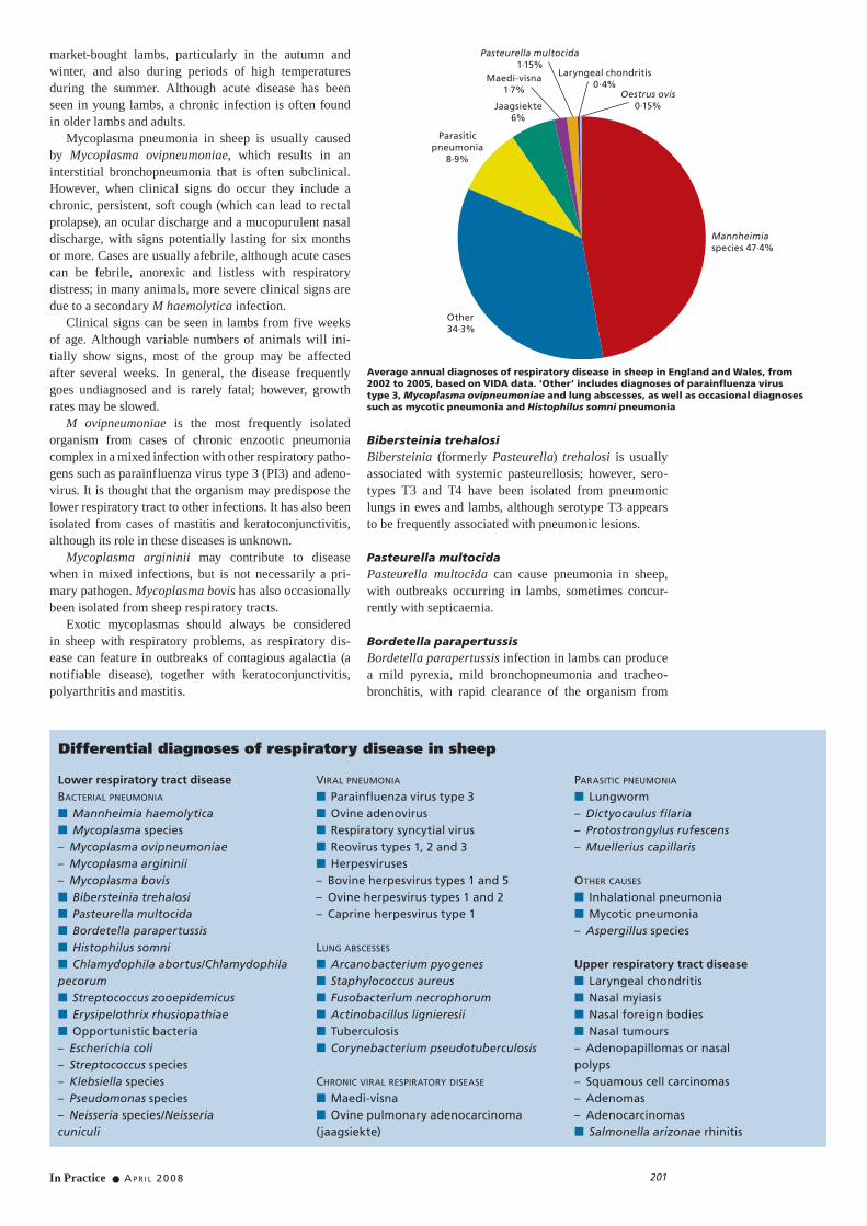
Average annual diagnoses of respiratory disease in sheep in England and Wales, from 2002 to 2005, based on VIDA data. ‘Other’ includes diagnoses of parainfluenza virus type 3, Mycoplasma ovipneumoniae and lung abscesses, as well as occasional diagnoses such as mycotic pneumonia and Histophilus somni pneumonia
Laryngeal chondritis 0·4%
Mannheimia species 47·4%
Other 34·3%
Parasitic pneumonia
8·9%
Jaagsiekte 6%
Maedi-visna 1·7%
Pasteurella multocida1·15%
Oestrus ovis 0·15%
Differential diagnoses of respiratory disease in sheep
Baermann apparatus set up to examine faecal samples for lungworm larvae. A sieve and some gauze are positioned in the funnel, onto which the faeces and some water are placed and left for 24 to 48 hours. The larvae migrate through the gauze and settle in the neck of the funnel. The water is then drained out of the tube at the bottom and the sediment examined microscopically for the presence of first-stage larvae
■ Clinical history■ Clinical signs■ Postmortem examination – gross pathology, lung bacterial culture, fluorescent antibody test (FAT) on lung tissue or tissue or bronchiole swabs, histopathology of lung tissue■ Bronchoalveolar lavage – bacterial culture, FAT, jaagsiekte PCR■ Single serology – maedi-visna, jaagsiekte PCR■ Paired serology – parainfluenza virus type 3 and Mycoplasma ovipneumoniae■ Faecal examination – isolation of lungworm larvae (using the Baermann technique)
Bronchoalveolar lavage technique
■ Restrain the sheep in a standing posi-tion with its neck slightly extended. Clip a 50 x 70 mm area 50 mm distal to the larynx. Disinfect the area with 4 per cent chlor hexidine gluconate followed by 70 per cent alcohol■ Infiltrate the area with lidocaine using a 21 gauge needle■ Identify an area between two tracheal rings by palpation and insert a 12 gauge, 52 x 2·7 mm intravenous catheter at an angle of 45° to the skin. On entering the
tracheal lumen, retract the needle and advance the catheter■ Determine the required length of a 1·7 (5 French gauge) x 570 mm polyethyl-ene extension male to female Luer lock suction catheter by measuring from the entry point on the trachea to a point behind the shoulder. On average, 40 to 45 cm of tubing is required for an adult sheep■ Introduce the tube through the cath-eter and advance it down the trachea until
resistance is encountered. At this point, the animal usually coughs■ Steadily infuse 30 ml of sterile isoton-ic saline through the tube using a 60 ml syringe■ Withdraw the fluid after five seconds, by gently moving the intratracheal tube backwards and forwards to aid recovery. Remove the tube in one movement. On average, 10 ml of fluid is recovered
From Sheehan and others (2005)
METHODS OF DIAGNOSIS
Method Comments
Viral pneumoniaParainfluenza virus type 3
FAT on BAL fluid or lung tissue during the first six days of infectionELISAImmunohistocytochemistry on fixed lung
Paired serology confirms recent infection
Ovine adenovirus AGIDT Seroconversion takes four to five weeks. Test not currently available
Respiratory syncytial virus
FAT on lung tissue or BAL fluidImmunohistochemistry on fixed lung tissue
Maedi-visna AGIDT or ELISAPostmortem examination and lung histopathology Lungs are two to four times the normal weight of 300 to 500 g
Ovine pulmonary adenocarcinoma
‘Wheelbarrow’ testPostmortem examination and lung histopathologyJaagsiekte retrovirus PCR
Lifting the hindlimbs produces profuse fluid from the nostrils
Detects virus in peripheral blood leucocytes in infected sheep. Sensitivity is highest in sheep with actual lesions. Also detects virus in tissues and respiratory tract exudate
Bacterial pneumoniaPasteurella species Bacterial culture of lung tissue
Histopathology Lesions contain oat-shaped cells
Mycoplasma species Mycoplasma culture of lung tissuePCR/denaturing gradient gel electrophoresis on tissue or tissue swabELISA
Needs transport medium
Antibody titres peak 41 days postinfection and last for 16 weeks. Paired serology indicates recent exposure
Bordetella species Bacterial culture Sampling from the airways is necessary for reliable isolation
Other bacteria Bacterial culture of lung tissue or BAL fluid
Tuberculosis Positive tuberculin testIsolation from lesions or secretions
ParasitesLungworm Presumptive mild respiratory disease in lambs
during late summer and early autumn Baermann techniquePostmortem examination
Identification of first-stage larvaeIdentification of adult worms in the trachea/bronchioles. Muellerius capillaris can be seen by pressing the lung nodule between two slides
Nasal myiasis Frenzied behaviour of sheepChronic nasal dischargePostmortem examination Larvae can be seen in the sinuses
FAT Fluorescent antibody test, BAL Bronchoalveolar lavage, AGIDT Agar gel immunodiffusion test
In Practice ● A P R I L 20 0 8 203
the lower respiratory tract. The bacterium may poten-tially initiate more severe respiratory disease by damag-ing the tracheal epithelium, thus predisposing lambs to a secondary M haemolytica infection.
Histophilus somniHaemophilus somnus, Histophilus ovis and Haemo-philus agni have now been reclassified as a single fam-ily, Histophilus somni, the ovine strain of which is also able to infect cattle and goats. However, the bovine strain appears unable to infect sheep.
There are infrequent reports of the involvement of H somni in incidents of respiratory disease in sheep, but it has been associated with outbreaks of septicaemia and lung lesions in lambs. H somni (H ovis) was confirmed as the cause of pneumonia, as well as epididymo-orchitis, meningoencephalitis, septicaemia, mastitis and metritis in sheep in an Australian study (Philbey and others 1991). The organism has also been isolated from sheep with fibrinous pleuritis, bronchopneumonia, necrotising vas-culitis and grossly visible lung abscesses (Rahaley 1978, Poonacha and Donahue 1984).
VIRAL PNEUMONIA Parainfluenza virus type 3PI3 occurs worldwide and is considered to be a common virus based on serological prevalence studies carried out in various countries. Ovine PI3 is distinct from both bovine and human PI3.
Most clinical cases occur in lambs when protective maternal antibodies wane, although adults can some-times be affected. PI3 infection causes a mild, undiffer-entiated interstitial pneumonia. Clinical signs are usually mild or inapparent and most cases are afebrile, although occasionally animals may have pyrexia (40 to 41°C). Some animals may have a copious serous nasal discharge and an ocular discharge. Severe clinical disease is seen following high viral loads in experimental infections or when there is a concurrent infection, particularly with M haemolytica. Lung lesions usually persist for two months following infection.
Ovine adenovirusSheep seroprevalence studies have revealed that ovine adenovirus is common in many countries. The virus has, in most cases, been isolated from apparently healthy lambs, but has also been isolated from animals with severe pneumonia and enteritis. Clinical signs seen fol-lowing the experimental infection of lambs include pyrexia, anorexia, nasal discharge, sneezing and pneu-monia, with the most severe lesions found in the ter-minal airways. Central nervous signs, hepatic necrosis, lymphangitis and necrotising enteritis have also all been seen with adenovirus infections. In most cases, the virus can be isolated from the nasal secretions for up to 10 days after infection.
The virus can potentially be transmitted between cattle, goats and sheep.
Respiratory syncytial virusRespiratory syncytial virus (RSV) has been detected in sheep with rhinitis, and experimental virus inoculation has produced mild pneumonia. A seroprevalence of 84·5 per cent has been reported in the USA (Lehmkuhl and others 1985), and seropositive ewes have been identified in Northern Ireland (Adair and others 1984). There are
two ovine RSV isolates, which are antigenically distinct from bovine and caprine RSV isolates.
Bovine RSV infection in sheep produces severe pathological changes, but usually without apparent associated clinical signs.
PARASITIC PNEUMONIA LungwormPotentially pathogenic lungworms that can be found in sheep include:■ Dictyocaulus filaria;■ Protostrongylus rufescens;■ Muellerius capillaris.
When seen, clinical signs are usually due to D filaria infections and occur most commonly in young sheep during the autumn and winter, although cases can be seen between June and November – a warm, wet summer enhances the survival of the larvae.
The spring hatching of first-stage larvae from ewe and yearling faeces or overwintering third-stage larvae on the pasture provide a source of infective Dictyocaulus species larvae. During the grazing season, at least three generations of parasites can be present, with adult worms developing four weeks after infection. Most lambs are infected in their first year, resulting in a light, prolonged infection. Strong immunity develops following exposure so there are usually few worms in older sheep, unless they are suffering from a concurrent immuno suppressing condition.
Adult Dictyocaulus filaria worms (lungworms) found in a lamb’s trachea during postmortem examination. Picture, Sian Mitchell
Perc
enta
ge
7
6
5
4
3
2
1
0 Jan-Mar Apr-Jun Jul-Sep Oct-Dec
Mannheimia species pneumoniaOther pneumoniaParasitic pneumonia
Seasonal trends in respiratory disease diagnoses in sheep, from 2002 to 2005, based on diagnosable submissions to the Veterinary Laboratories Agency
Maedi-visnaOvine pulmonary adenocarcinoma
In Practice ● A P R I L 20 0 8204
and weight loss. Deaths are uncommon unless there is a secondary bacterial infection.
The incidence of Protostrongylus and Muellerius species depends on the distribution of their mollusc intermediate hosts. Large numbers of P rufescens worms can result in respiratory distress and weight loss, and M capillaris can occasionally cause mild respiratory signs. Mixed worm infections can also occur.
The more frequent routine usage of anthelmintics in sheep may contribute to the apparent reduced clini-cal significance of lungworm infections when compared with cattle.
Hydatid cystsThe intermediate stages of the tapeworm Echinococcus granulosus occur as ‘hydatid cysts’, which are fluid-filled cysts of up to 20 cm in diameter that often contain numerous worm scolices. In sheep, approximately 70 per cent of hydatid cysts are found in the lungs; these lesions are then partially embedded in the lung tissue, although usually with very little local reaction and no associated clinical signs.
OTHER POTENTIAL PATHOGENSThe following pathogens have all been found in cases of acute respiratory disease in sheep, but their prevalence in natural infections is uncertain: ■ Reovirus types 1, 2 and 3;■ Bovine herpesvirus types 1 and 5;■ Ovine herpesvirus types 1 and 2;■ Caprine herpesvirus type 1;■ Chlamydophila abortus/Chlamydophila pecorum;■ Streptococcus zooepidemicus;■ Erysipelothrix rhusiopathiae;■ Opportunistic bacteria following primary airway or lung damage from other pathogens: Escherichia coli, Streptococcus species, Klebsiella species, Pseudomonas species and Neisseria (formerly Branhamella) species/Neisseria cuniculi; ■ Fungi: Aspergillus species, Mortierella wolfii.
Herpesviruses and Chlamydophila abortus have produced a mild interstitial pneumonia experimen-tally (Whetstone and Evermann 1988, Scott and others 1990, Navarro and others 2004, Li and others 2005). Streptococcus zooepidemicus was found in 10-month-old lambs with a fibrinous pleuritis, pericarditis and pneu-monia; co-grazing carrier ponies were suspected to be the source of infection (Stevenson 1974). Erysipelothrix rhusiopathiae has been isolated from adult sheep with a severe necrotising alveolitis (Griffiths and others 1991).
Mycotic pneumonia, which is often due to Aspergillus fumigatus, is rarely encountered, but can occur as either a primary infection or secondary to disseminated sys-temic infections, particularly following mammary infec-tions (Pérez and others 1999). It produces lung oedema and greyish-white pyogranulomatous nodules. M wolfii has also produced lesions in experimentally infected sheep (Cordes and others 1972).
Immunosuppression may predispose cases to mycotic infection and spoilt feed is often thought to be the likely source.
LUNG ABSCESSESLung abscesses are often caused by secondary oppor-tunistic bacteria such as Arcanobacterium pyogenes and Staphylo coccus aureus following previous lung damage,
for example, after an acute pneumonic incident or sec-ondary to ovine pulmonary adenocarcinoma (jaagsiekte). They may also follow embolic spread from another focus of infection. S aureus lung abscesses can occur with tick pyaemia, and Fusobacterium necrophorum can produce abscesses in cases of inhalational pneumonia or second-ary to pharyngeal abscessation. Similarly, Actinobacillus lignieresii may cause lung lesions secondary to oral abscesses. Pyaemia can develop following lung absces-sation, sometimes presenting as sudden death.
Tuberculosis (TB) and caseous lymphadenitis (CLA) are both contagious conditions that potentially lead to lung abscesses. Tuberculosis is a rare condition in sheep but has been found in animals aged two years and older that have had close contact with TB-infected cattle. It results in either caseous lung lesions or abscesses containing liquid pus. CLA is caused by the bacterium Corynebacterium pseudo-tuberculosis, which produces abscesses in the superficial lymph nodes or skin, although lung abscesses can also occur. Spread occurs when purulent material discharges from ruptured abscesses and contaminates the environ-ment and fomites (eg, shearing equipment). Lung lesions usually occur secondary to a disseminated haematogenous spread, although they may follow aerosol spread.
CHRONIC RESPIRATORY VIRUSESMaedi-visnaMaedi-visna virus is a lentivirus – a non-oncogenic retro-virus – causing primarily an interstitial pneumonia, as well as indurative mastitis, arthritis and potentially neurological signs. The first cases in the UK were iden-tified in 1979, but a low annual diagnostic rate suggests the disease is not widespread in the UK, although it is hard to detect infection due to the insidious nature of the virus.
The virus can be transmitted via milk, respiratory secretions and faeces, and survival for up to two weeks outside the body has been suggested. A low level of intrauterine transmission is believed to occur (Cutlip and others 1981), and the virus has also been detected in semen from rams with a concurrent orchitis.
Clinical disease is usually not evident in a flock until at least 50 per cent of ewes are seropositive. Clinical signs are seen in sheep at least two years of age, but most are four to five years old.
There is a protracted period of lesion development, con-sisting of a slow, replicating, lymphoid hyperplasia, with infected animals never developing a protective immunity to the disease. Many sheep remain asymptomatic carriers, with 25 to 30 per cent developing clinical signs.
The production of antibodies can occur several months following infection, while some infected animals never develop an antibody response. The mortality in uncomplicated cases is low, with deaths generally occur-ring due to a secondary bacterial infection, pregnancy toxaemia or harsh climatic conditions. Right ventricular hypertrophy can develop due to the lung lesions.
Cases present as bright and alert, with loss of condi-tion and slow-onset afebrile respiratory distress, which is usually only evident on exertion. Coughing and a nasal discharge are occasionally seen. In advanced cases, the ewes flare their nostrils, extend their necks and breathe through the mouth. Signs progress over a three- to six-month period.
The economic impact in a flock is from earlier cull-ing of ewes and a decrease in lamb weights at weaning.
In Practice ● A P R I L 20 0 8 205
In extreme conditions, losses of 10 to 20 per cent gross margin per ewe have been reported.
Ovine pulmonary adenocarcinomaOvine pulmonary adenocarcinoma is caused by the jaagsiekte retrovirus, which induces bronchioloalveolar adenocarcinoma lung tumours that account for 70 per cent of all sheep tumours.
Horizontal transmission of the virus occurs via aero-sol, and contaminated food and water troughs. The pos-sibility of intrauterine infection has been suggested but is thought to occur at a low level, if at all. The disease incubation period is long and clinical signs are usually not seen until sheep are two to four years old. In excep-tional cases, tumours have been seen in lambs as young as two months and in sheep as old as 11 years. The inci-dence of clinical cases in endemic flocks ranges between 2 and 10 per cent annually, with management practices, such as length of housing period and stocking densities, influencing infection rates.
Clinical signs consist of weight loss and a progres-sive, afebrile respiratory distress, particularly follow-ing exertion. Auscultation reveals high-pitched, moist lung sounds due to the accumulation of large amounts of fluid in the lungs that can exude from the nostrils, which is a characteristic finding of the disease. Death usually occurs due to a secondary bacterial pneumonia, although in some cases right ventricular hypertrophy can lead to heart failure.
MISCELLANEOUS DISEASESInhalational pneumoniaInhalational pneumonia frequently affects an individ-ual or a small number of animals. It occurs most often in bottle-reared lambs or following incorrect drench-ing or dipping, or after the inhalation of a foreign body or inhalation of rumen contents in laterally recumbent animals.
UPPER RESPIRATORY TRACT DISEASE
Upper respiratory tract infections usually present with a unilateral or bilateral serous or mucopurulent nasal discharge, coughing and sneezing. The viral causes of pneumonia discussed earlier can present with primarily upper respiratory tract signs.
The following diseases specifically affect the upper respiratory tract and are encountered sporadically in sheep flocks.
LARYNGEAL CHONDRITISLaryngeal chondritis is an acute obstructive upper res-piratory condition resulting from chronic suppurative lesions in the arytenoid cartilages of the larynx. The cause is uncertain but there appears to be a genetic pre-disposition in short-necked breeds such as Texels and Southdowns, although any breed can be affected. Other suggested causes include drenching injuries, rough hand-ling, dry, dusty feed or grass awns. There is a higher incidence in rams than ewes.
Bacteria isolated from outbreaks include A pyogenes and mixed environmental bacteria, including E coli and F necrophorum.
Cases present with severe dyspnoea and laryngeal stridor, which can result in inhalational pneumonia. The condition is fatal if not treated promptly.
NASAL MYIASISNasal myiasis, caused by Oestrus ovis larvae, produces a chronic rhinitis and sinusitis due to mucosal irritation by larvae located in the frontal, maxillary and nasal sinuses. First-stage larvae are deposited by adult flies on sheep nostrils between June and October, and the larvae then live and develop in the sinuses for a two- to 10-month period. First-stage larvae overwinter in the
Postmortem examination is necessary for the diagnosis of ovine pulmonary adenocarcinoma. These lungs from an adult ewe show extensive neoplastic changes in the caudal lung lobes due to jaagsiekte retrovirus infection. The neoplastic tissue has a characteristic grey-purple appearance. Picture, Tim Jones
Profuse quantities of fluid are produced by the neoplastic tissue in sheep affected by ovine pulmonary adenocarcinoma. Fluid draining from the nose, particularly when the hindlimbs are lifted (the ‘wheelbarrow’ test), allows a presumptive diagnosis. Picture, Hamish Newton Laryngeal chondritis in a Texel shearling ram that had died soon after developing
acute-onset respiratory distress
In Practice ● A P R I L 20 0 8206
sinuses and complete their development in May and June of the following spring.
Up to 80 per cent of a flock may be affected and the main associated problem is sheep annoyance. The animals tend to stamp their feet, sneeze and press their noses to the ground. Breathing can be difficult due to blocked and inflamed nostrils and a mucopurulent nasal discharge. If seen, clinical signs are usually observed in older ewes and often in late summer. Occasionally, the aberrant loca-
Oestrus ovis larvae, an incidental finding in the sinuses of an adult ewe during postmortem examination
tion of larvae or secondary bacterial infections can cause an inflammatory reaction that interrupts feeding. This can lead to illthrift or, in some cases, a cerebral bacterial infection, pneumonia and occasionally death.
NASAL FOREIGN BODIESProblems due to foreign bodies frequently involve inhaled plant material, such as grass seeds, resulting in a unilateral nasal discharge, which may become mucopu-rulent and even malodorous due to secondary bacterial infection.
NASAL TUMOURSNasal tumours, such as adenopapillomas or nasal polyps, squamous cell carcinomas, adenomas or adeno carcinomas occur sporadically in sheep in the UK. Clinical signs include a seromucous nasal discharge, which may be uni-lateral and can become muco purulent due to secondary bacterial infection. Increased respiratory noise and distor-tion of the maxillary bones may also occur. Contagious nasal adenocarcinomas or enzootic nasal tumours are caused by a virus related to the jaagsiekte retrovirus. This disease occurs in other countries but has not yet been identified in the UK.
SALMONELLA ARIZONAE RHINITISA mild proliferative rhinitis was diagnosed in seven-month-old lambs, with Salmonella arizonae confirmed as the causal pathogen (Brogden and others 1994).
In Practice ● A P R I L 20 0 8 207
AcknowledgementsThe author thanks Graham David, Ian Davies, Michael Millar and Jackie Willmington, of the Veterinary Laboratories Agency (VLA), and Nick Bell, for their comments on and assistance with this article. The author also thanks the VLA for the VIDA data.
ReferencesADAIR, B. M., McFERRAN, J. B., McKILLOP, E. R. & McCULLOUGH, S. J. (1984) Survey for antibodies to respiratory viruses in two groups of sheep in Northern Ireland. Veterinary Record 115, 403-406BROGDEN, K. A., MEEHAN, J. T. & LEHMKUHL, H. D. (1994) Salmonella arizonae infection and colonisation of the upper respiratory tract of sheep. Veterinary Record 135, 410-411CORDES, D. O., DI MENNA, M. E. & CARTER, M. E. (1972) Mycotic pneumonia and placentitis caused by Mortierella wolfii. I. Experimental infections in cattle and sheep. Veterinary Pathology 9, 131-141CUTLIP, R. C., LEHMKUHL, H. D. & JACKSON, T. A. (1981) Intrauterine transmission of ovine progressive pneumonia virus. American Journal of Veterinary Research 42, 1795-1797GRIFFITHS, I. B., DONE, S. H. & READMAN, S. (1991) Erysipelothrix pneumonia in sheep. Veterinary Record 128, 382-383 LEHMKUHL, H. D., CUTLIP, R. C., BOLIN, S. R. & BROGDEN, K. A. (1985) Seroepidemiologic survey for antibodies to selected viruses in the respiratory tract of lambs. American Journal of Veterinary Research 46, 2601-2604LI, H., O’TOOLE, D., KIM, O., OAKS, J. L. & CRAWFORD, T. B. (2005) Malignant catarrhal fever-like disease in sheep after intranasal inoculation with ovine herpesvirus-2. Journal of Veterinary Diagnostic Investigation 17, 171-175NAVARRO, J. A., GARCÍA DE LA FUENTE, J. N., SÁNCHEZ, J., MARTÍNEZ, C. M., BUENDÍA, A. J., GUTIÉRREZ-MARTÍN, C. B., RODRIGUEZ-FERRI, E. F., ORTEGA, N. & SALINAS, J. (2004) Kinetics of infection and effects on the placenta of Chlamydophila abortus in experimentally infected pregnant ewes. Veterinary Pathology 41, 498-505 PÉREZ, V. P., CORPA, J. M., GARCÍA, J. F., MARÍN, A., ADÚRIZ, J. J. & JENSEN, H. E. (1999) Generalized aspergillosis in dairy sheep. Zentralblatt für Veterinärmedizin B 46, 613-621PHILBEY, A. W., GLASTONBURY, J. R., ROTHWELL, J. T., LINKS, I. J.
& SEARSON, J. E. (1991) Meningoencephalitis and other conditions associated with Histophilus ovis infection in sheep. Australian Veterinary Journal 68, 387-390POONACHA, K. B. & DONAHUE, J. M. (1984) Haemophilus ovis infection in lambs. Veterinary Medicine 79, 541–542RAHALEY, R. S. (1978) Pathology of experimental Histophilus ovis infection in sheep. I. Lambs. Veterinary Pathology 15, 631-637SCOTT, F. M., ANGUS, K. W. & DEWAR, P. (1990) Pneumonia of lambs following inoculation of isolates of sheep herpesvirus (caprine herpesvirus 1) of different DNA genotypes. Journal of Comparative Pathology 102, 111-117SHEEHAN, M., MARKEY, B., CASSIDY, J., BALL, H., DUANE, M. & DOHERTY, M. L. (2005) New transtracheal bronchoalveolar lavage technique for the diagnosis of respiratory disease in sheep. Veterinary Record 157, 309-313STEVENSON, R. G. (1974) Streptococcus zooepidemicus infection in sheep. Canadian Journal of Comparative Medicine 38, 243-250WHETSTONE, C. A. & EVERMANN, J. F. (1988) Characterization of bovine herpesviruses isolated from six sheep and four goats by restriction endonuclease analysis and radioimmunoprecipitation. American Journal of Veterinary Research 49, 781-785
Further readingHINDSON, J. C. & WINTER, A. C. (2002) Respiratory disease. In Manual of Sheep Diseases, 2nd edn. Oxford, Blackwell Science. pp 196-209LI, H., O’TOOLE, D., KIM, O., OAKS, J. L. & CRAWFORD, T. B. (2005) Malignant catarrhal fever-like disease in sheep after intranasal inoculation with ovine herpesvirus-2. Journal of Veterinary Diagnostic Investigations 17, 171-175MARTIN, W. B. & AITKEN, I. D. (Eds) (2000) Diseases of the respiratory system. In Diseases of Sheep, 3rd edn. Oxford, Blackwell Science. pp 177-204RADOSTITS, O. M., GAY, C. C., BLOOD, D. C. & HINCHCLIFF, K. W. (2000a) Diseases caused by bacteria. In Veterinary Medicine: A Textbook of the Diseases of Cattle, Sheep, Pigs, Goats and Horses, 9th edn. London, W. B. Saunders. pp 852-855 RADOSTITS, O. M., GAY, C. C., BLOOD, D. C. & HINCHCLIFF, K. W. (2000b) Diseases caused by Mycoplasma spp. In Veterinary Medicine: A Textbook of the Diseases of Cattle, Sheep, Pigs, Goats and Horses, 9th edn. London, W. B. Saunders. p 998 RADOSTITS, O. M., GAY, C. C., BLOOD, D. C. & HINCHCLIFF, K. W. (2000c) Diseases caused by viruses and chlamydia. In Veterinary Medicine: A Textbook of the Diseases of Cattle, Sheep, Pigs, Goats and Horses, 9th edn. London, W. B. Saunders. pp 1184-1191 SWIFT, B. L., KIMBERLING, C. V. & JENSEN, R. (1987) Diseases of the respiratory system. In Jenson and Swift’s Diseases of Sheep, 3rd edn. Philadelphia, Lea & Febiger. pp 173-181, 265-279WARD, A. C., WEISER, G. C., ANDERSON, B. C., CUMMINGS, P. J., ARNOLD, K. F. & CORBEIL, L. B. (2006) Haemophilus somnus (Histophilus somni) in bighorn sheep. Canadian Journal of Veterinary Research 70, 34-42


![Edinburgh Research Explorer...sheep with bacterial respiratory infections, followed by absence of residual bronchial catarrh in the same sheep during recovery [13]. Authors in more](https://static.documents.pub/doc/80x56/611f532bee560775465ee0f9/edinburgh-research-sheep-with-bacterial-respiratory-infections-followed-by.jpg)


