63
RESPIRATORY SYSTEM DISORDERS By DR. Areefa Albahri (Alkasseh) Assistance Prof. of MCH Islamic University of Gaza ١٤٤٣/٠٨/٢٣ DR. Areefa Albahri
| Date post: | 19-Dec-2015 |
| Category: |
Documents |
| Upload: | kathryn-stephanie-summers |
| View: | 216 times |
| Download: | 1 times |

RESPIRATORY SYSTEM DISORDERS
By
DR. Areefa Albahri (Alkasseh)
Assistance Prof. of MCH
Islamic University of Gaza
/ /١٤٤٤ ٠٩ ٢٨
DR. Areefa Albahri

• Functions of Respiratory System:• Taking in O2 and breathing out the CO2• Maintenance of acid-base balance • Regulation of H2O and heat balance• Production of speech• Facilitate the sense of smell
DR. Areefa Albahri

• Respiratory system during childhood years:• Infants are obligatory nasal breathers and they use their
abdominal muscle for breathing • Use more accessory muscles for breathing• At 2yrs the Rt bronchus become shorter, wider & more
vertical•Walls of the airways are small in size and have less
cartilage thus lung collapse after expiration is easier.
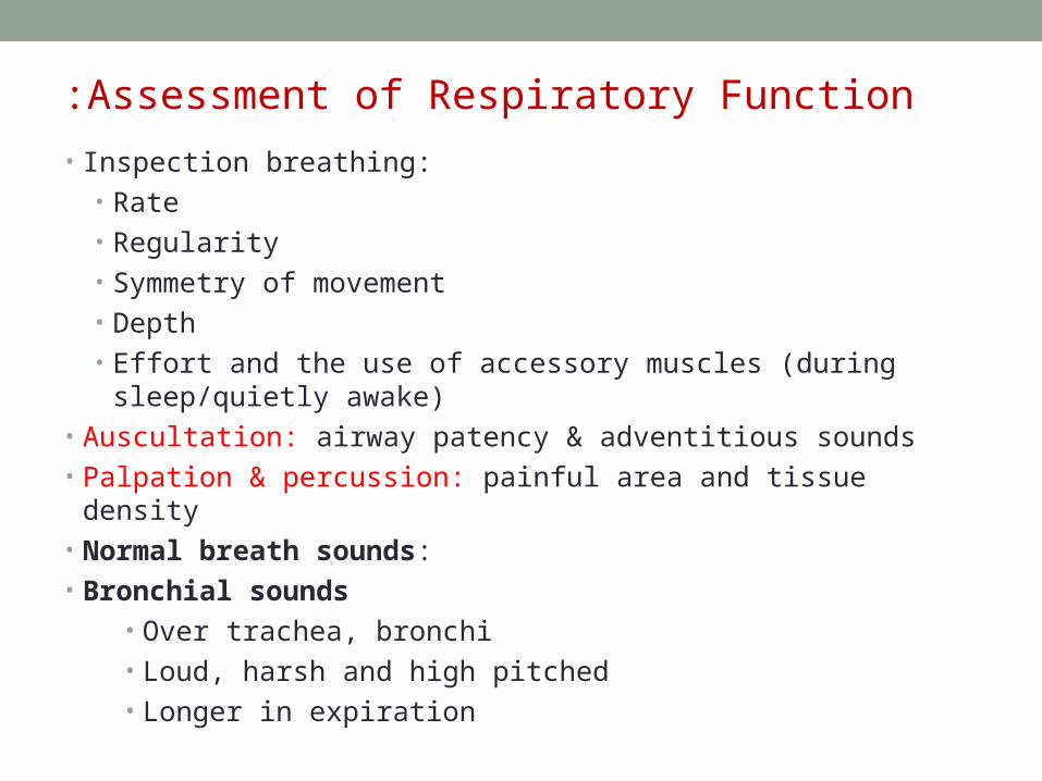
Assessment of Respiratory Function:
• Inspection breathing: • Rate• Regularity• Symmetry of movement• Depth• Effort and the use of accessory muscles (during sleep/quietly
awake)• Auscultation: airway patency & adventitious sounds• Palpation & percussion: painful area and tissue density• Normal breath sounds:• Bronchial sounds
• Over trachea, bronchi• Loud, harsh and high pitched• Longer in expiration
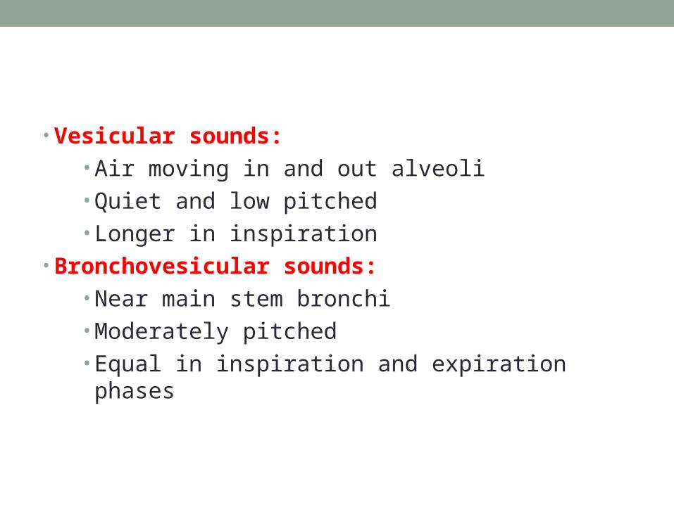
• Vesicular sounds:• Air moving in and out alveoli• Quiet and low pitched• Longer in inspiration
• Bronchovesicular sounds:• Near main stem bronchi• Moderately pitched• Equal in inspiration and expiration phases

Adventitious sounds:
• Crackles (Rales)• Result of air passing through fluid in small airways • Simulated by rubbing a few strands of hair between
fingers next to the ear• Most common during inspiratory phase• Associated with COPD and pulmonary edema
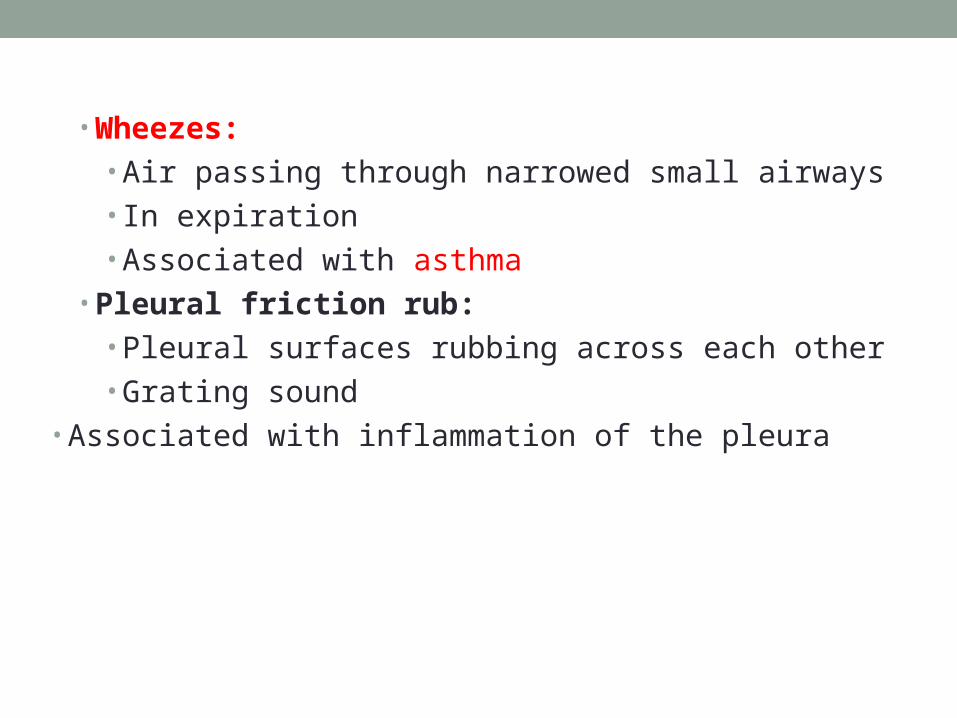
• Wheezes:• Air passing through narrowed small airways• In expiration• Associated with asthma
• Pleural friction rub:• Pleural surfaces rubbing across each other• Grating sound
• Associated with inflammation of the pleura

• Noisy Breathing: •
•Snoring: Indicates partial obstruction of the upper airway that causes vibration of the air as it passes the nasopharynx and oropharynx. May cause sleep apnea.•
•Stridor: A harsh, continuous crowing sound. Mostly occurs during inspiration. Croup (Laryngotracheobronchitis).•
•Wheezing: Whistling/musical sound. Indicates a narrowing of the airways. Mainly heard during expiration. Accompanied by tightness of the chest and labored breathing. Mostly caused by asthma.
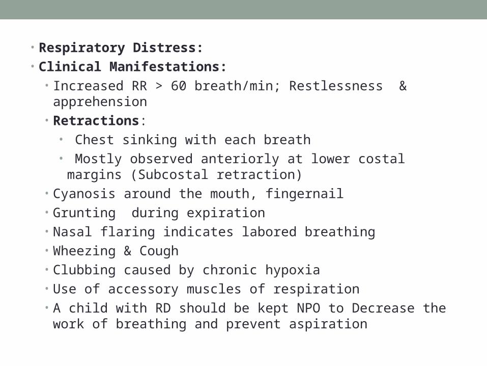
• Respiratory Distress:• Clinical Manifestations:
• Increased RR > 60 breath/min; Restlessness & apprehension • Retractions:
• Chest sinking with each breath• Mostly observed anteriorly at lower costal margins (Subcostal
retraction)• Cyanosis around the mouth, fingernail• Grunting during expiration• Nasal flaring indicates labored breathing• Wheezing & Cough• Clubbing caused by chronic hypoxia• Use of accessory muscles of respiration• A child with RD should be kept NPO to Decrease the work of
breathing and prevent aspiration

• Neonatal Respiratory Distress Syndrome (RDS)• RDS is caused by immature lungs anatomy & physiology
or genetic problem with lungs
• Mainly due to Insufficient surfactant (production of surfactant starts around 26 weeks of gestation)
• Deficient surface area for gas exchange• Pulmonary capillary bed is deficient
• Neonatal RDS mostly seen in neonates born before 28 wks of gestation
• Sometimes called Hyaline membrane disease
Clinical manifestations:
• Tachypnea > 60 breaths/minute• Grunting on expiration• Retractions of the chest wall• Auscultation reveals decreased breath sounds and audible rales
(crackles)• Pallor/cyanosis at room air• Increase in respiratory effort (flaring and dyspnea)• Flaccidity• Apnea• Hypotension • Prolonged capillary refill
Diagnostic Procedures for assessing pulmonary functioning:
• Pulmonary function tests (forced vital capacity, tidal volume, functional residual volume
• Tracheal aspiration• Bronchoscopy• Blood gas (pulse oximetry, ABG)• Sputum Studies• Lung biopsy

• Therapeutic Techniques:• Chest tubes• Tracheostomy• O2 Therapy• Nebulizer• Postural drainage• Chest Physiotherapy (CPT)• Suctioning of Airways• Thoracentesis• Mechanical Ventilation
• Tracheostomy:• It is a surgical opening in the trachea (2-4 tracheal rings)• Temporary tracheostomy (epiglottitis, croup, foreign body
aspiration) • Long term tracheostomy for ventilation Care after the surgery
• Hemorrhage, edema, aspiration
• Regular care:• Airway patency (remove secretions)• Humidification• Cleansing

• O2 Therapy: • To treat hypoxemia• Delivered by mask, nasal cannula, tent, hood, face tent or ventilator• Should be ordered by physician • Should be humidified before administered to the patient• Therapeutic Techniques:
• Aerosol /nebulizer therapy: used to administer medication and avoid systemic side effect
• Observe patient during aerosol therapy b/c it may cause bronchospasm.• Chest physiotherapy: (CPT)• When the children get a chest infection, the inability to take good deep
breaths and cough forcefully may make the clearance of excess thick mucus more difficult. This is when chest physiotherapy is useful
• CPT helps to clear the lungs and get the phlegm into the mouth where it can be spat out, or to the back of the throat where it can be swallowed
• CPT uses of postural drainage in adjunctive techniques including percussion, vibration, deep breathing & coughing exercise
•

Pharmacology: Respiratory System Disorders:Bronchodilators:
• Reverse bronchoconstriction thus opening the air passages in the lungs by acting:
• Stimulating beta-adrenergic sympathetic nervous system receptors • Short acting beta agonists (ventolin); A quick relief
of acute exacerbations and for the prevention of exercise-induced airway constriction
• Long acting (Foradil) directly relaxing bronchial smooth muscles ( Aminophylline, Theophylline)
• Major Side Effect:• Dizziness (decrease in BP), CNS stimulation,
Palpitation, GI irritation
• Corticosteroids:• Anti-inflammatory effect & decrease edema• Control Asthma and improve pulmonary function• Inhaled preparation (Fluticasone) oral (Prednisone),
IV (hydrocortisone)• Major side effect• Inhaled: oropharyngeal candiadiasis, growth
retardation and osteoporosis• Oral: with long-term use immunosuppression,
increase Wt, osteoporosis, gastric ulcer,
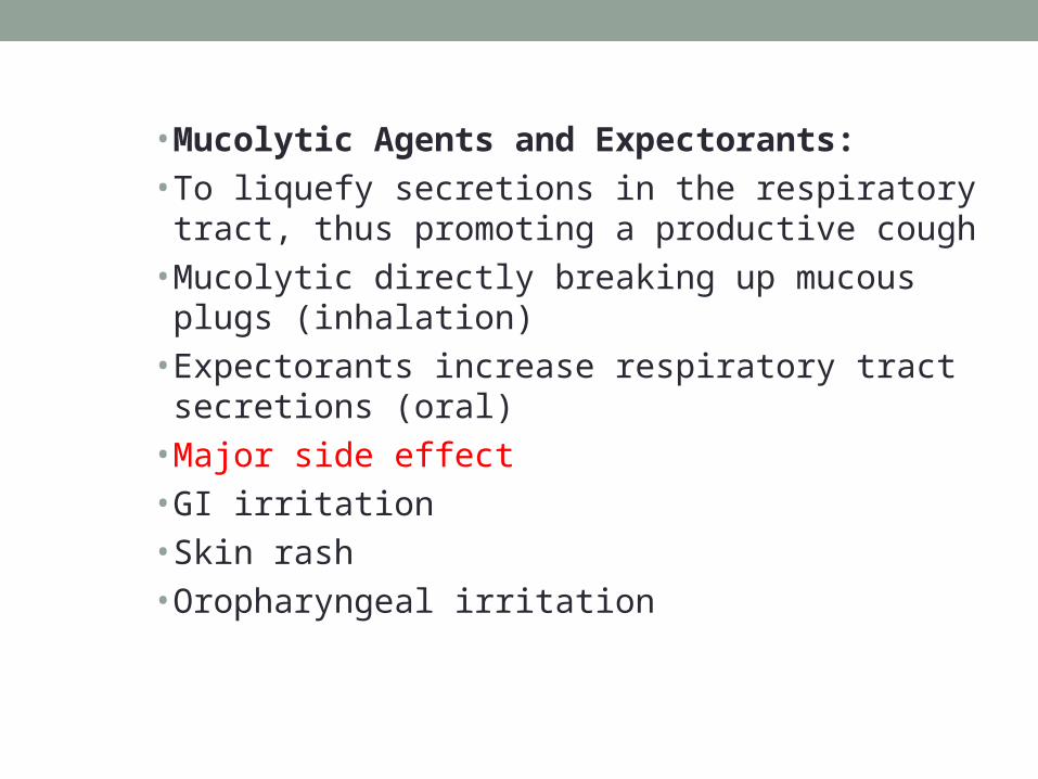
• Mucolytic Agents and Expectorants:• To liquefy secretions in the respiratory tract, thus
promoting a productive cough• Mucolytic directly breaking up mucous plugs
(inhalation)• Expectorants increase respiratory tract secretions
(oral)• Major side effect• GI irritation• Skin rash• Oropharyngeal irritation

• Antitussives: • To suppress the cough reflex• Preparation with Narcotic (Codeine) without Narcotic (Tessalon)• Major side effect• Drowsiness• Nausea• Dry mouth (anticholingeric effect of antihistamine in combination products)• Antihistamines:• To relieve symptoms of common cold and allergies• Act by blocking the action of histamine at receptor sites, also exert antiemetic,
anticholinergic and CNS depressant effect• Major side effect• Drowsiness, Dizziness• GI irritation• Dry mouth (anticholingeric effect of antihistamine in combination products)

Upper respiratory tract infections ( Resp. system) emergency
• Otitis media• Croup (laryngotracheobronchitis)• Epiglottitis
• Respiratory Dysfunction: • 1. Otitis Media (OM)
• Acute infection of the middle ear • Prevalent between 6 months to 2 years
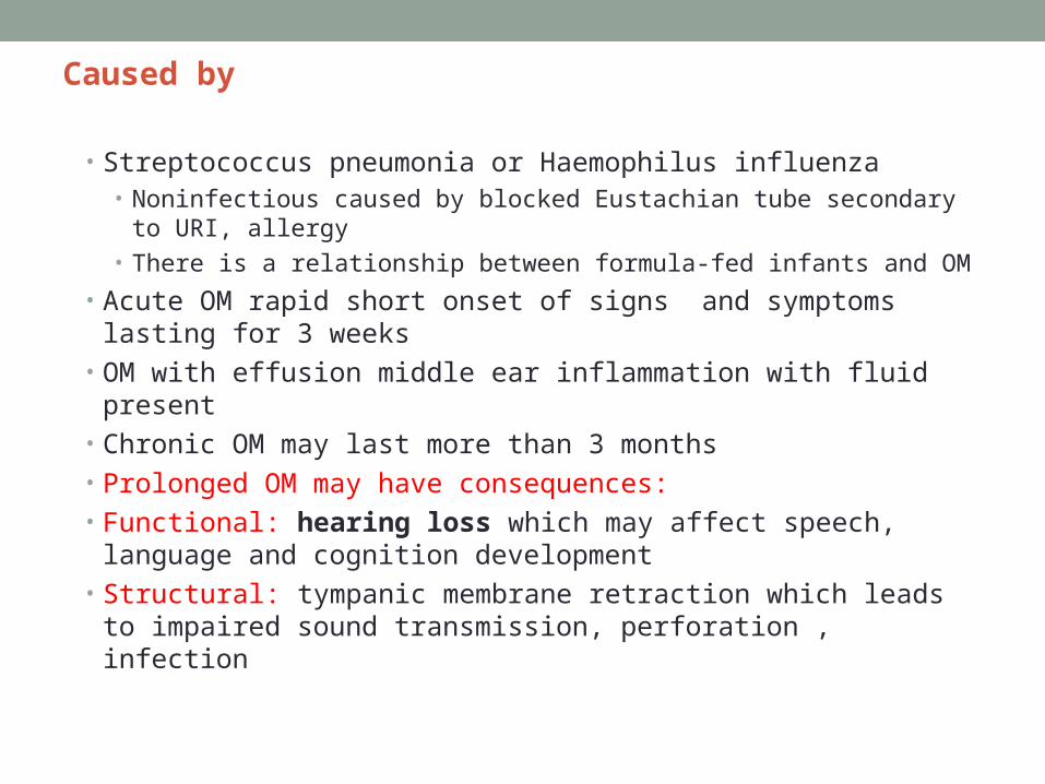
Caused by
• Streptococcus pneumonia or Haemophilus influenza• Noninfectious caused by blocked Eustachian tube secondary to URI,
allergy• There is a relationship between formula-fed infants and OM
• Acute OM rapid short onset of signs and symptoms lasting for 3 weeks
• OM with effusion middle ear inflammation with fluid present• Chronic OM may last more than 3 months• Prolonged OM may have consequences:• Functional: hearing loss which may affect speech, language and
cognition development• Structural: tympanic membrane retraction which leads to impaired
sound transmission, perforation , infection

Clinical Manifestations:
• Acute OM:• Pain: infant rubs ear or rolls head from side to side
• Temp. 40, Vomiting & diarrhea• Loss of appetite• Tympanic membrane is bright red and bulging, no light reflex
• Discharge from the external auditory canal
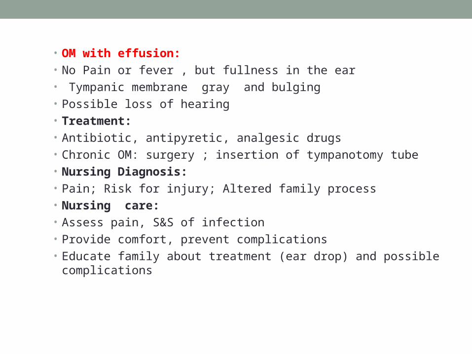
• OM with effusion:• No Pain or fever , but fullness in the ear• Tympanic membrane gray and bulging• Possible loss of hearing • Treatment:• Antibiotic, antipyretic, analgesic drugs• Chronic OM: surgery ; insertion of tympanotomy tube• Nursing Diagnosis:• Pain; Risk for injury; Altered family process• Nursing care:• Assess pain, S&S of infection• Provide comfort, prevent complications• Educate family about treatment (ear drop) and possible complications
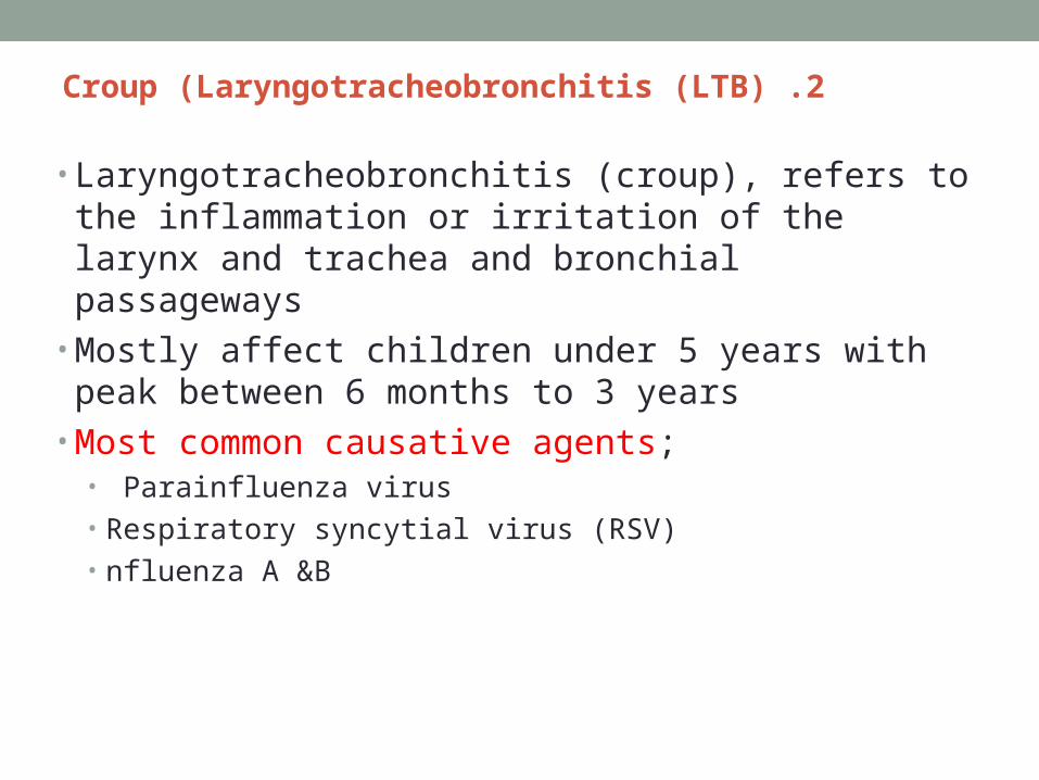
2 .Croup (Laryngotracheobronchitis (LTB)
• Laryngotracheobronchitis (croup), refers to the inflammation or irritation of the larynx and trachea and bronchial passageways
• Mostly affect children under 5 years with peak between 6 months to 3 years
• Most common causative agents;• Parainfluenza virus • Respiratory syncytial virus (RSV)• nfluenza A &B

Croup: Signs/Symptoms
• “Cold” progressing to hoarseness, cough• Low grade fever• Night-time increase in edema with:
• Stridor• “Seal bark” cough• Respiratory distress• Cyanosis• Hypoxia• Resp. acidosis

• Treatment:• Mild Croup
• Reassurance• Moist, cool air• Maintain patent airway• Ibuprofen
• Severe Croup• Humidified high concentration oxygen• NPO if there is respiratory distress and start IV fluid• For sever cases Nebulized epinephrine or dexamethasone• Possible intubation

• Nursing Care• Observation and assessment of respiratory status• Prepare for possible intubation if the patient develops signs of
airway obstructions including: • Increased HR, RR, • Retractions,• Flaring nares• Increased restlessness• Provide comfort, avoid eliciting gag reflex (Laryngo-spasm)
•

Respiratory Dysfunction: 3. Epiglottitis:
• Acute obstructive inflammatory process of epiglottis• Mostly occurs in children 2-6 years• Most often the causative agent is Hemophilus influenza B • Abrupt symptoms • CM:
• Sore throat and pain on swallowing• Fever, • Muffled voice & stridor, • Tripod sit• Drooling saliva, • Irritable and restless & possible retraction• Epiglottis red, inflamed, large, cherry red and edematous• Airway obstruction leads to hypoxia and acidosis
Complete Airway Obstruction
•Respiratory distress+ Sore throat+Drooling =
•Epiglottitis
• Treatment:• IV fluid until the patient can swallow• Antibiotic • Corticosteroids to reduce the edema• Tracheal intubation in severe cases• Nursing Care• Reduce the anxiety• Comfortable position• Avoid using tongue depressor to inspect epiglottius• Monitor respiratory status•

• Lower respiratory tract infections• Acute bronchitis• Bronchiolitis/respiratory syncytial virus (RSV)• Pneumonia

• Acute bronchitis• Inflammation of trachea , bronchi & bronchioles • Common in children older than 6 yrs• Acute bronchitis usually occurs in association with viral
respiratory tract infection.• Causative agent of acute bronchitis is Mycoplasma
pneumonia. Other causes include chemical agent• CM:• Productive cough • Sometimes retrosternal pain during deep breathing or
coughing• It is a self-limited disease (5-10 days)

• Treatment:• Rest, use of antipyretics, adequate hydration • Symptomatic treatment

• Bronchiolitis/respiratory syncytial virus (RSV)• Infection of the lower respiratory tract• Rarely occurs in children over 2 years old (peak 2-5 months)• Primarily occurs in winter & spring• 50% of cases caused by RSV, bacteria also cause bronchiolitis• The bronchi and bronchioles are inflamed that leads to obstruction
of the airway• Narrowing of the airways during expiration causes overinflation
(emphyasema)
• Starts with URT infection then spreads to lower tract
• CM:• Earlier S&S; poor feeding and irritability• Initial S &S; Rhinorrhea, low-grade fever, pharyngitis and possible OM,
conjunctivitis, cough• Progressive sign increased cough, air hunger, tachypnea, retractions &
cyanosis• Severe S&S, RR >70, listless, apneic spells• Symptomatically treatment• Antiviral medication may be used• Humidity• O2• Fluid & rest• If the patient is tachypnea NPO
•

Pneumonia• Is an inflammation of pulmonary parenchyma• Types of pneumonia based on the way the child gets the
infection or the germ;• Aspiration pneumonia; occurs when food or drink accidently gets
into lungs• Community Acquired pneumonia
• Most common type• Caused by viruses, bacteria or chemical irritants• Mostly occurs in winter and spring• Fast breathing is a sign of pneumonia;• 1 wk-2months 60 B/M or more• 2mon- 12 mon 50 B/M or more• 1 2months – 5 yrs 40 B/M or more
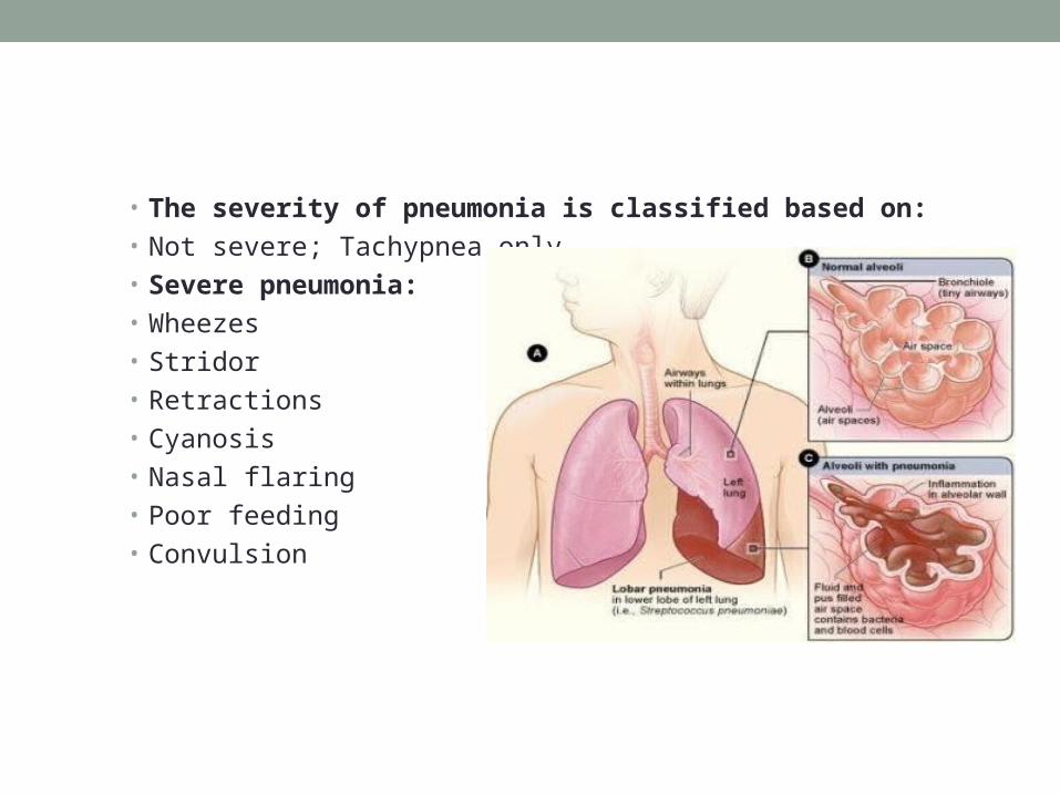
• The severity of pneumonia is classified based on: • Not severe; Tachypnea only • Severe pneumonia:• Wheezes• Stridor• Retractions • Cyanosis • Nasal flaring• Poor feeding• Convulsion


Viral Pneumonia:
• mostly caused by RSV in children under 5 yrs• Gradual onset• Viral infection make the pt susceptible to bacterial pneumonia• Treatment is symptomatically
• O2• Comfort• Fluid• CPT• Postural drainage
Bacterial Pneumonia:
• Streptococcus pneumonia is the most causative bacterium• Others causative agent; group B streptococcus, hemophilus
influenza type b, group A streptococcus• Abrupt onset• CM:
• Productive cough, tachypnea, fever, ronchi or fine crackles, chest pain, retraction, nasal flaring, cyanosis, lethargy
• Chest X-ray shows patchy infiltration• Irritable • Anorexia, vomiting, diarrhea and abdominal pain

Treatment:
• Penicillin G ( for allergic pt erythromycin, chloramphenical, cephalosporin)
• Antipyretic, antitussive (cough)• Rest• Increase fluid intake• O2 may be required for RD children
• Nursing diagnosis: Ineffective breathing pattern R/T inflammatory effects of pneumonia
• Nursing Care:• Thorough respiration assessment (signs of RD)• Provide comfort and O2, Cool humidification• Encourage cough and deep breathing • Increase fluid intake & Monitor I &O• Provide rest & Maintain semi-fowler’s position• Standard precautions & use of air-borne and droplet precautions
•

• Long-term respiratory dysfunction• Asthma• Cystic fibrosis

• Asthma:• Chronic inflammatory disorder
of the airway• Asthma causes recurrent
episodes of wheezing, breathlessness, chest tightness & cough particularly at night or in the early morning
• Associated with reversible airflow limitation or obstruction
• Asthma causes bronchial hyper-responsiveness to stimuli
Bronchospasm
Bronchial Edema
Increased Mucus
Production
Asthma
Factors aggravate asthmatic exacerbation:
• Allergens (air pollution, dust), Irritants (odor spray, smoking)• Changes in weather temperature, Cold air• Environmental changes ( new home)• Infections• Animals• Strong emotions• Food additives, nuts, dairy product• Other factors ( menses, pregnancy).

The mechanisms responsible for the obstructive symptoms in asthma include:
• Inflammation and edema of the mucus membrane.• Accumulation of tenacious secretions from mucus glands.• Spasm of the smooth muscle of the bronchi and bronchioles, which
decreases the diameter of bronchioles.• Pathophysiology
ALL THAT WHEEZES IS NOT ASTHMA
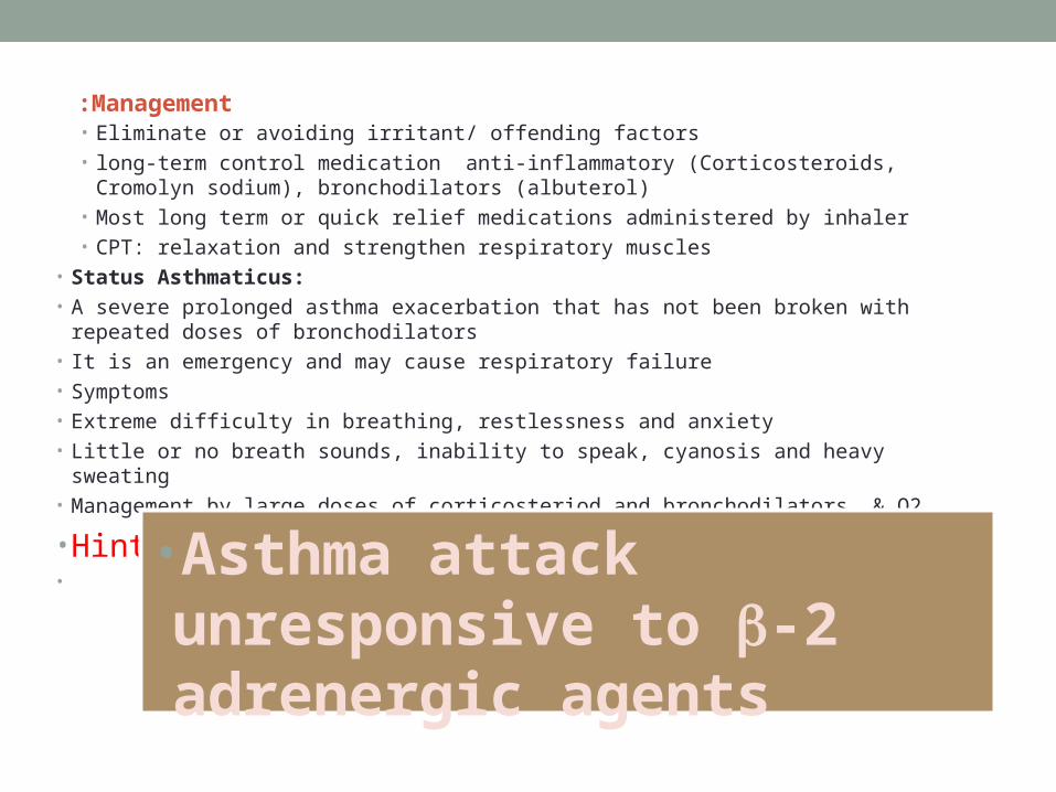
Management: • Eliminate or avoiding irritant/ offending factors• long-term control medication anti-inflammatory (Corticosteroids, Cromolyn sodium),
bronchodilators (albuterol)• Most long term or quick relief medications administered by inhaler • CPT: relaxation and strengthen respiratory muscles
• Status Asthmaticus:• A severe prolonged asthma exacerbation that has not been broken with repeated doses of
bronchodilators• It is an emergency and may cause respiratory failure• Symptoms• Extreme difficulty in breathing, restlessness and anxiety• Little or no breath sounds, inability to speak, cyanosis and heavy sweating• Management by large doses of corticosteriod and bronchodilators, & O2
• Hint•
•Asthma attack unresponsive to -2 adrenergic agents

Cystic Fibrosis (CF)
Sweat gland: high Na & Cl (3-5 times higher than normal)sweat chloride test above 60 mEq/L are diagnostic


• Pancreas: Becomes fibrotic, decrease production of pancreatic enzymes• Decrease in Lipase cause steatorrhea (fatty, foul, bulky stool)• Decrease in Trypsin increase nitrogen in stool• Decrease Amylase cause inability to break down polysacharides
• DM is late complication• Rectal prolapse:• Liver; possible cirrhosis from biliary obstruction• • Early manifestations:
• Meconium ileus in newborn infants• Baby tastes salty• Failure to regain normal 10% weight loss at birth• Presence of cough or wheezing during first 6 months of age• Sweat chloride test > 60mEq/L• Stool fat
•Chest X-ray shows patchy atelactesis
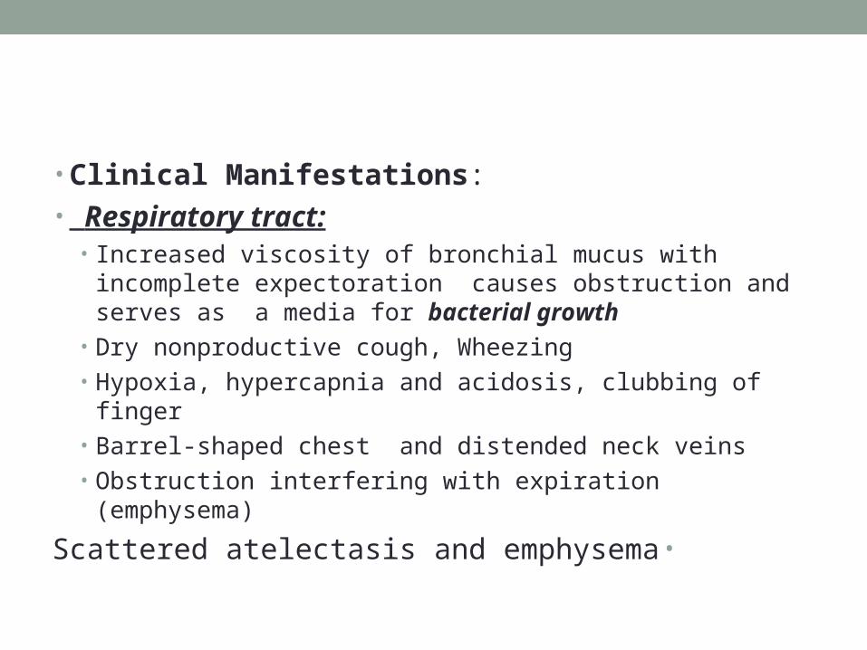
• Clinical Manifestations:• Respiratory tract:
• Increased viscosity of bronchial mucus with incomplete expectoration causes obstruction and serves as a media for bacterial growth
• Dry nonproductive cough, Wheezing• Hypoxia, hypercapnia and acidosis, clubbing of finger• Barrel-shaped chest and distended neck veins• Obstruction interfering with expiration (emphysema)
•Scattered atelectasis and emphysema

• GI tract:• High thick secretion blocked the ducts in pancreas leading fibrosis• Marked impairment of pancreatic enzymes which affects digestion
of fats and protein thus affecting normal growth• Increased bulk of feces ( undigested and unabsorbed fat and protein)• Wt loss & FTT
• High incidence of DM in children with CF

• Cardiovascular; Cardiac enlargement• Reproductive: • Delayed puberty in females, mostly males are sterile
• Integumentary: salty taste, risk of hypochloremic and hyponatremic

• Cystic Fibrosis (CF): Treatment• Pulmonary problems:
• Chest physiotherapy• Bronchodilators• Antibiotic therapy as indicators• Gastrointestinal problems:• Pancreatic enzyme supplements• Balanced nutritional intake
• • Cystic Fibrosis (CF): Nursing Diagnosis• Ineffective airway clearance R/T secretion of thick tenacious mucus• Ineffective breathing pattern R/T mechanical tracheobronchial obstruction• Altered family process R/T situational crisis• Altered nutrition R/T inability to digest nutrients•

• Respiratory Dysfunction• Physical defects of respiratory tract
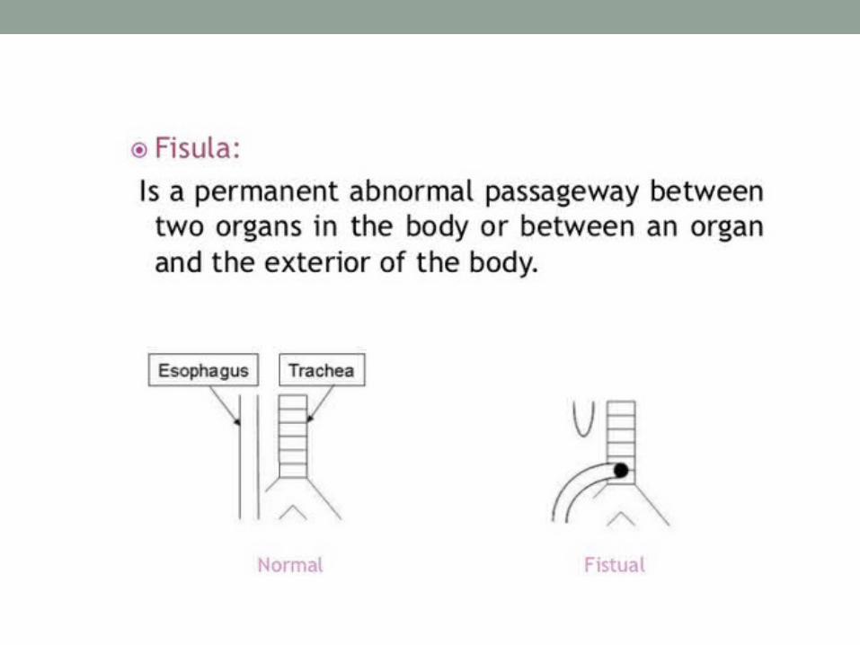
• Esophageal atresia• Tracheoesophageal fistula• Diaphragmatic hernia
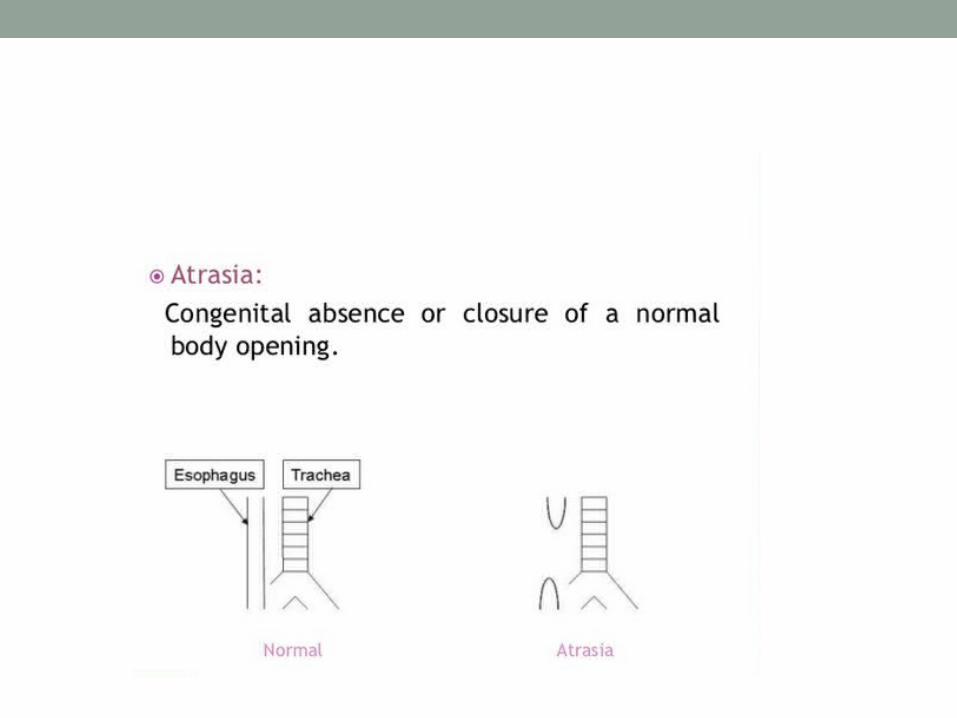
• Esophageal Atresia & Tracheoesophageal Fistula• Most common type proximal esophagus ends in a blind
pouch and distal esophagus is connected to the trachea• Usually occurs in low birth weight• May be associated with other abnormalities• Occurrence is 1 in 800 to 5000 births• Indications:
• Excessive salivation (drooling) in newborn• chocking, coughing, cyanosis (3 Cs) sneezing & when newborn fed
the fluid returns through the nose and the mouth
• Complication: aspiration and RD• Treatment: surgical repair


Congenital diaphragmatic hernia (CHD) Absent of the diaphragm on
one side or both
Mostly accompanied with other fetus malformations
CM: difficulty in breathing, tachycardia, cyanosis, concave abd. Abnormal chest development
Tx: Surgical repair

• Foreign Body Aspiration• Risk for old infant and 1-3 years• Most common foreign body: food (peanuts), balloon, coins• CM:
• Chocking, gagging, cough• Laryngotracheal obstruction: dyspnea, cough, stridor, hoarseness,
possible cyanosis• Bronchial obstruction: paroxysmal cough (sudden severe attack),
wheezing, asymmetric breathing sound, dyspnea• Progressive obstruction: discoloration of the face , no voice ,
unconscious and asphyxiation
• Management: For infants• 5 times Chest thrust 5 times Back blow


