64
Respiratory System Dr. zahiri In the name of God
| Date post: | 29-Jan-2016 |
| Category: |
Documents |
| Upload: | christine-lynch |
| View: | 225 times |
| Download: | 0 times |

Respiratory System
Dr. zahiri
In the name of God

Respiratory systemcomprising the lungs and a sequence of airways leading to the external environment
This system providing Oxygen and eliminating Carbon dioxide
is subdivided into:
conducting portions
respiratory portions

Conducting portion
Parts:nasal cavity, nasopharynx, larynx, trachea, primary bronchi,
secondary (lobar) bronchi, tertiary (segmental) bronchi, and terminal bronchioles
functions :clean, warm and moisten air prior to reaching respiratory portion


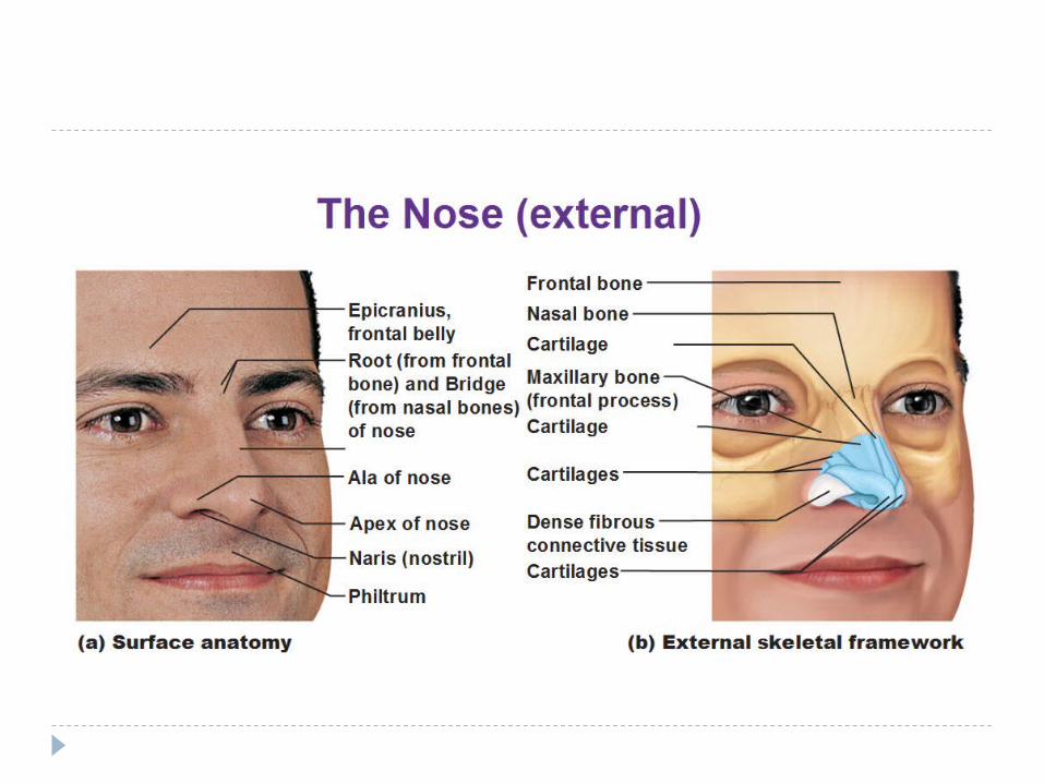
Nasal cavity
Nasal cavity composed of three regions( vestibule, respiratory and olfactory regions)

Vestibule
Anterior portion of nasal cavity near of the nares dilated and known as the vestibule
lined with skin and has short, stiff hairs named vibrissae that prevent large dust particles to enter
dermis houses numerous sebaceous glands


Posterior aspect of Nasal cavity
lined by pseudostratified ciliated columnar epithelium (respiratory epithelium)
respiratory epithelium
Ciliated columnar cells: most common, each cell has about 300 ciliaGoblet cells: secret mucous
Brush cells: have short microvilli, nerve fibers, sensory function
Basal cells: are rounded stem cells that located near basal lamina and show mitotic figuresSmall granule cells (kulchitsky cells= DNEs)


Subepithelial CT (lamina propria)
is richly vascularized, containing large arterial plexuses and venous sinuses, many seromucous glands and lymphoid elements

Olfactory region the olfactory epithelium
lamina propria (serous secreting Bowman’s glands, a rich vascular plexus and many axons arising from olfactory cells of the olfactory epithelium)

olfactory epithelium comprises three types of cells:
Olfactory cells olfactory cells are bipolar neurons whose apical aspect (dendrite) is modified to form a bulb known as olfactory vesicle
Sustentacular cells • has a striated border composed of microvilli, and secretory granules • they provide physical support, nourishment
Basal cells • are short basophilic cells• their apical aspects do not reach the epithelial surface
they proliferate and replace both two other cells
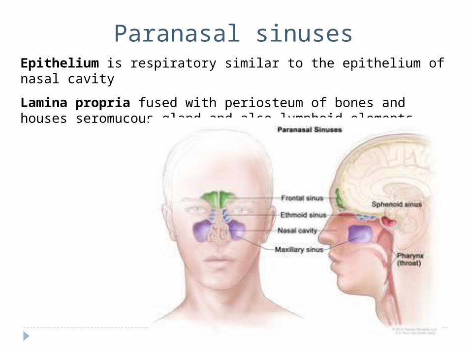
Paranasal sinusesEpithelium is respiratory similar to the epithelium of nasal cavity
Lamina propria fused with periosteum of bones and houses seromucous gland and also lymphoid elements

Sinusitis also known as a sinus infection or rhinosinusitis,
is inflammation of the sinuses
Primary ciliary dyskinesia (PCD), also immotile ciliary syndrome or Kartagener syndrome, is a rare genetic disorder that causes defects in the action of cilia lining the respiratory tract (lower and upper, sinuses, Eustachian tube, middle ear) and fallopian tube
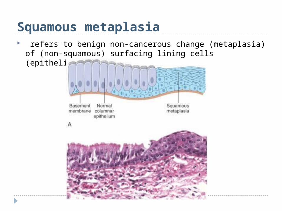
Squamous metaplasia refers to benign non-cancerous change (metaplasia) of
(non-squamous) surfacing lining cells (epithelium) to a squamous morphology

Metaplasia: an initial change from normal cells to a different cell type (such as chronic irritation of cigarette smoke causing ciliated pseudostratified epithelium to be replaced by squamous epithelium more able to withstand the insult).

Dysplasia: an increasing degree of disordered growth or maturation of the tissue .
Dysplasia is still a reversible process. However, once the transformation to neoplasia has been
made, the process is not reversible.
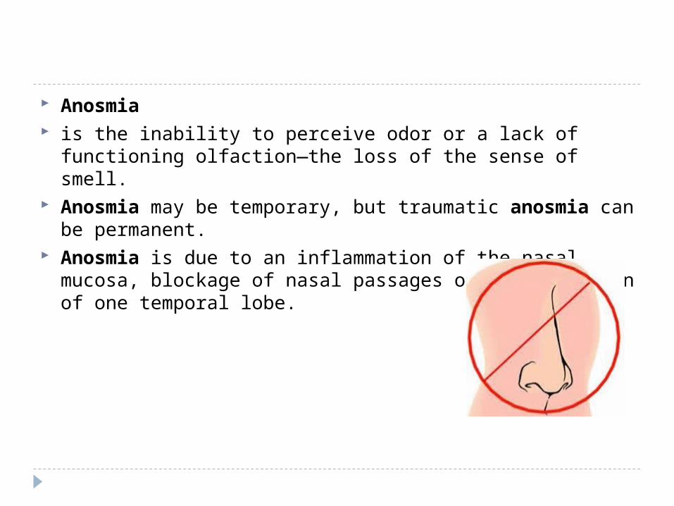
Anosmia is the inability to perceive odor or a lack of functioning
olfaction—the loss of the sense of smell. Anosmia may be temporary, but traumatic anosmia can
be permanent. Anosmia is due to an inflammation of the nasal mucosa,
blockage of nasal passages or a destruction of one temporal lobe.

nasopharynx Epithelium is respiratory similar to the epithelium of nasal
cavity
Lamina propria : pharyngeal tonsil

Larynx
connects pharynx to trachea is lined by respiratory epithelium Laryngeal cartilages(hyaline and elastic) are located in lamina propria The cartilages connected to each other by ligaments and move with respect to
one another by some striated muscles
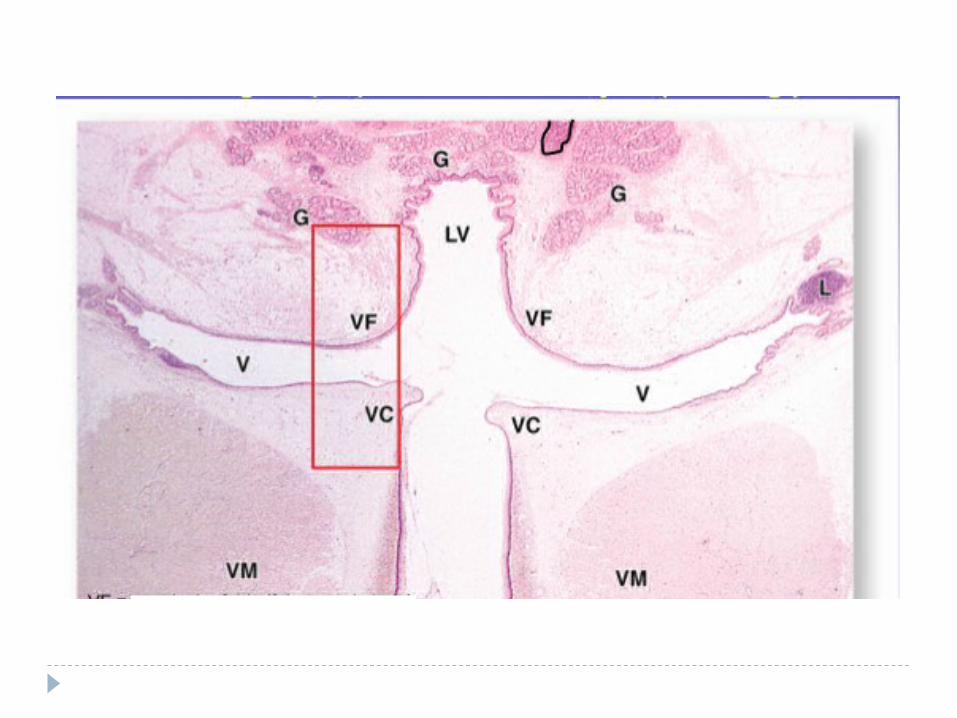
Larynx has two folds consist of superior and inferior Superior vestibular folds lined by respiratory epithelium
Inferior vocal folds lined by stratified squamous nonkeratinized epithelium

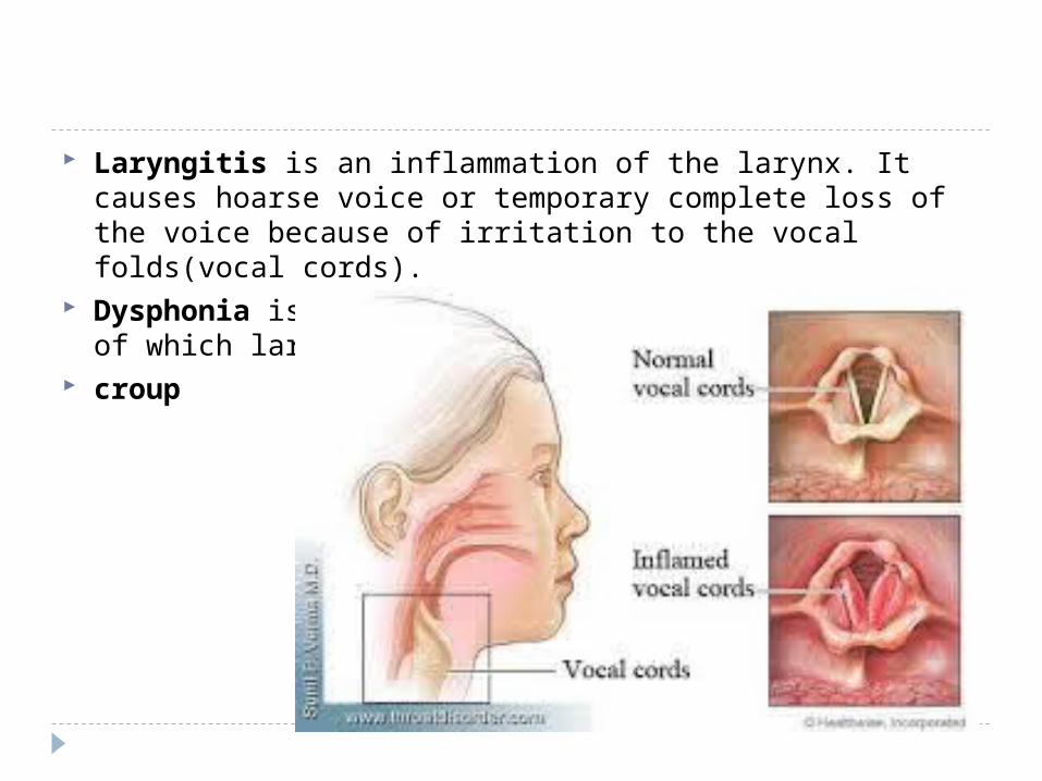
Laryngitis is an inflammation of the larynx. It causes hoarse voice or temporary complete loss of the voice because of irritation to the vocal folds(vocal cords).
Dysphonia is the medical term for a vocal disorder, of which laryngitis is one cause.
croup

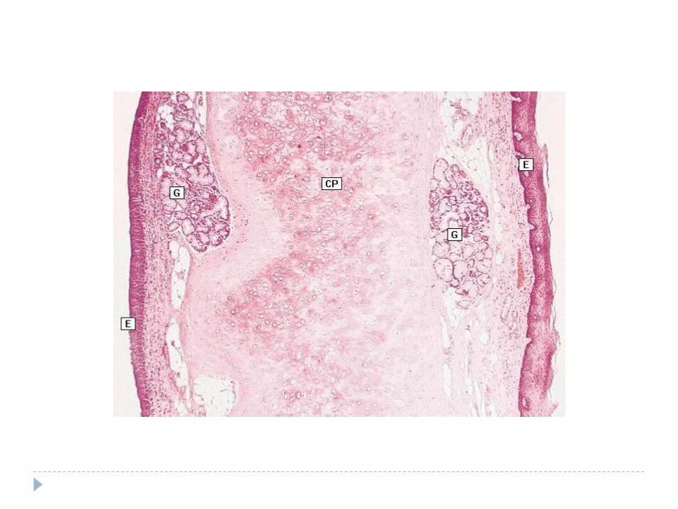
is an elastic cartilage of larynx It is lined by stratified squamous epithelium on lingual surfacePseudostratified ciliated columnar epithelium lined the laryngeal sideSerous and mucous glands located in lamina propria
Epiglottis
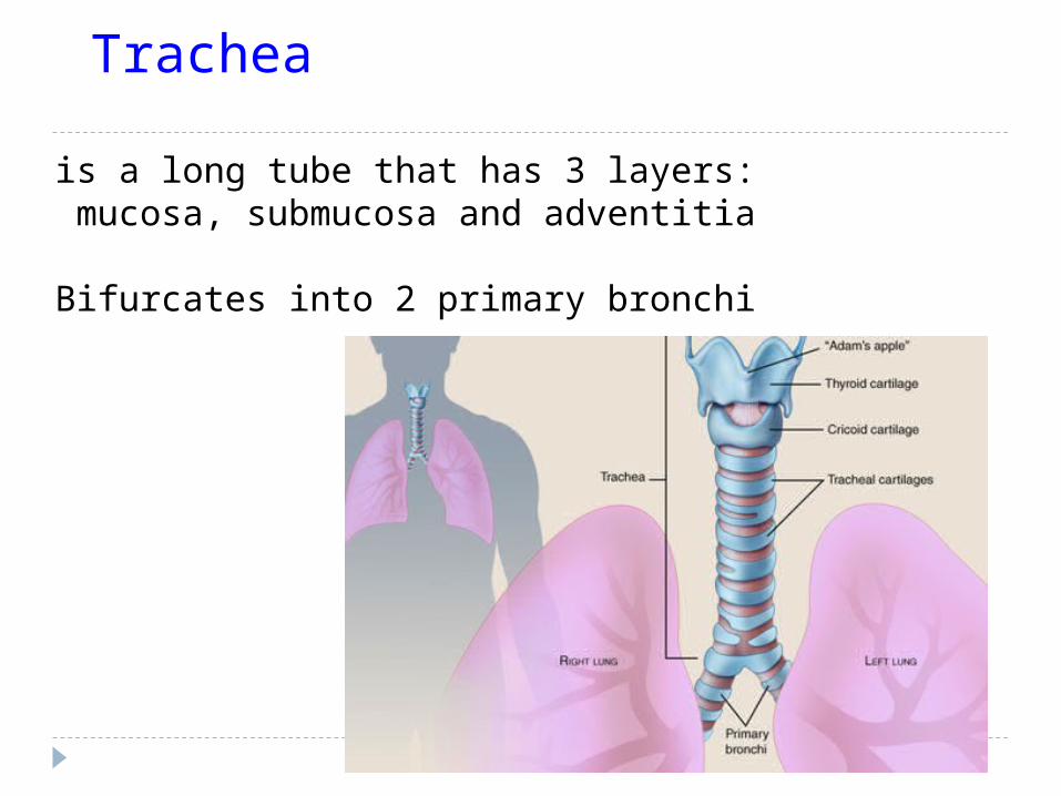
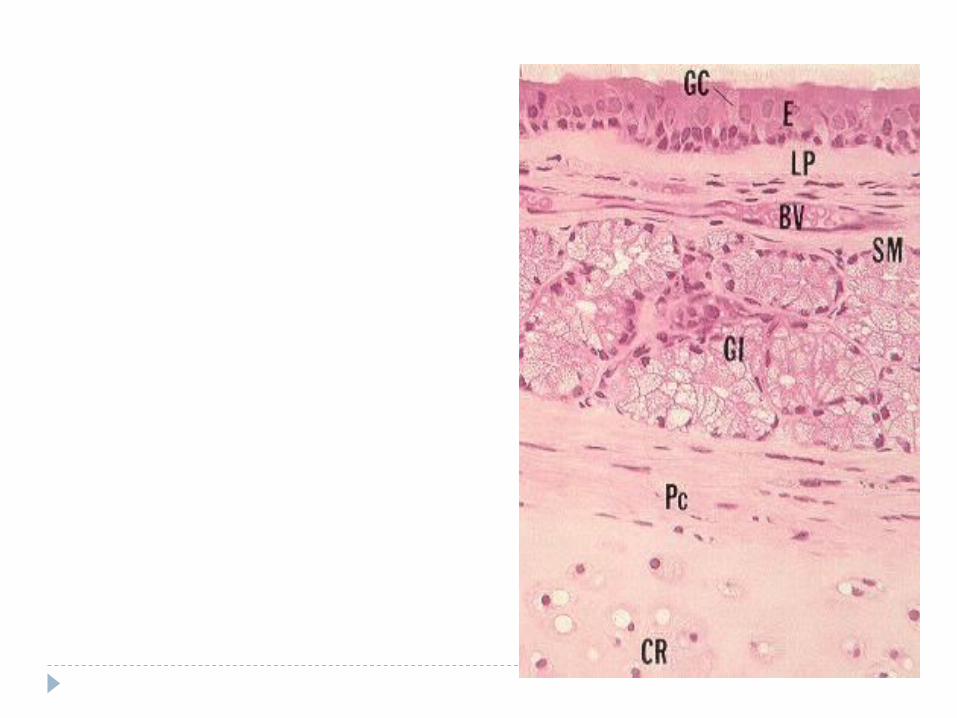
Trachea
is a long tube that has 3 layers: mucosa, submucosa and adventitia
Bifurcates into 2 primary bronchi
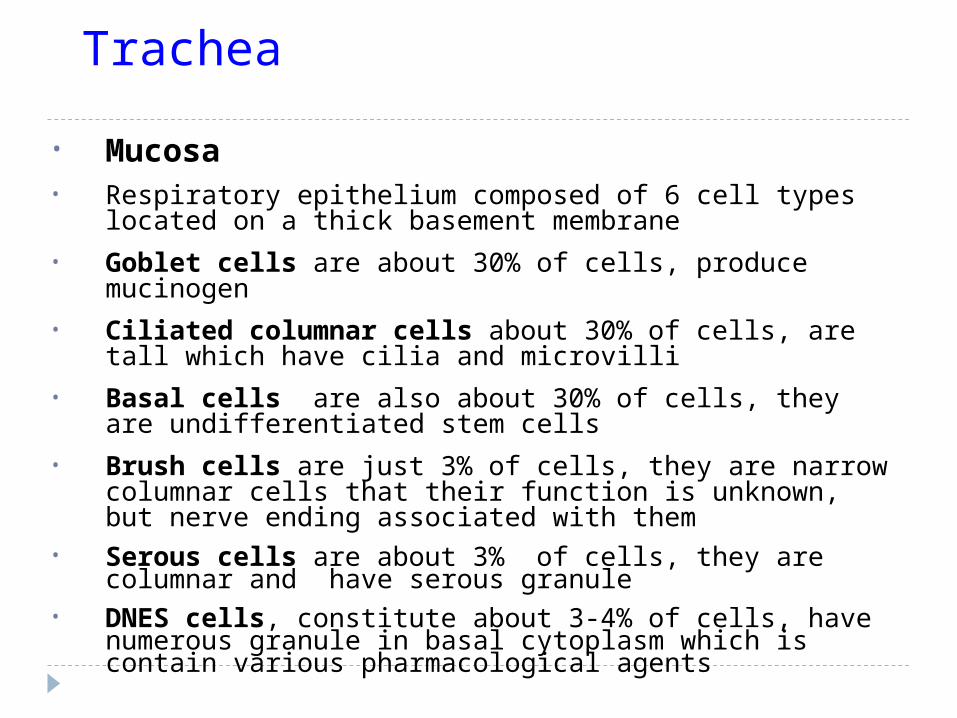
Trachea
• Mucosa• Respiratory epithelium composed of 6 cell types located on a thick
basement membrane
• Goblet cells are about 30% of cells, produce mucinogen
• Ciliated columnar cells about 30% of cells, are tall which have cilia and microvilli
• Basal cells are also about 30% of cells, they are undifferentiated stem cells
• Brush cells are just 3% of cells, they are narrow columnar cells that their function is unknown, but nerve ending associated with them
• Serous cells are about 3% of cells, they are columnar and have serous granule
• DNES cells, constitute about 3-4% of cells, have numerous granule in basal cytoplasm which is contain various pharmacological agents

Lamina propria
composed of loose fibroelastic CT, contain seromucous glands and lyphoid elements, elastic lamina separate this layer from submucosa
Submucosa Subnucosa is composed of dense irregular fibroelastic CT that houses mucous and
seromucous glands, rich in blood and lymph supply
Adventitia Adventitia is a fibroelastic CT that houses C-shaped hyaline cartilage, at
posterior aspect of cartilage, there is a dense band of smooth muscle cells known as trachealis muscle

Bronchial Tree
is composed of: 2 primary bronchus that enter lungs 3 lobar ( secondry) bronchus on right and 2 on the left Segmental (tertiary) bronchus bronchioles
Terminal bronchioles
Respiratory bronchioles
Progressively airways decreased in size and cartilage, glands, goblet cells, and the height of epithelial cells
But increase smooth muscle cells and elastic tissue

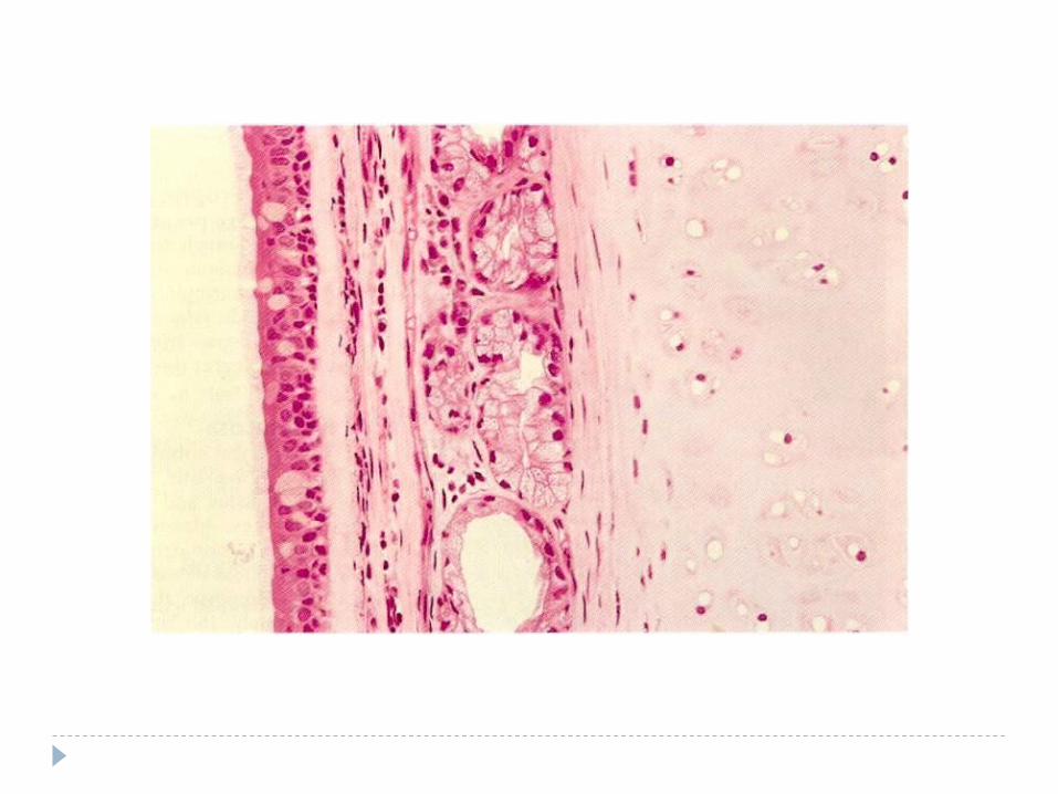
Primary Bronchi (Extrapulmonary) Primary bronchi is identical to
trachea, but have smaller diameter and thinner wall
Cartilage is in form of irregular plates
Smooth muscle located between lamina propria and submucosa as 2 distinct layers


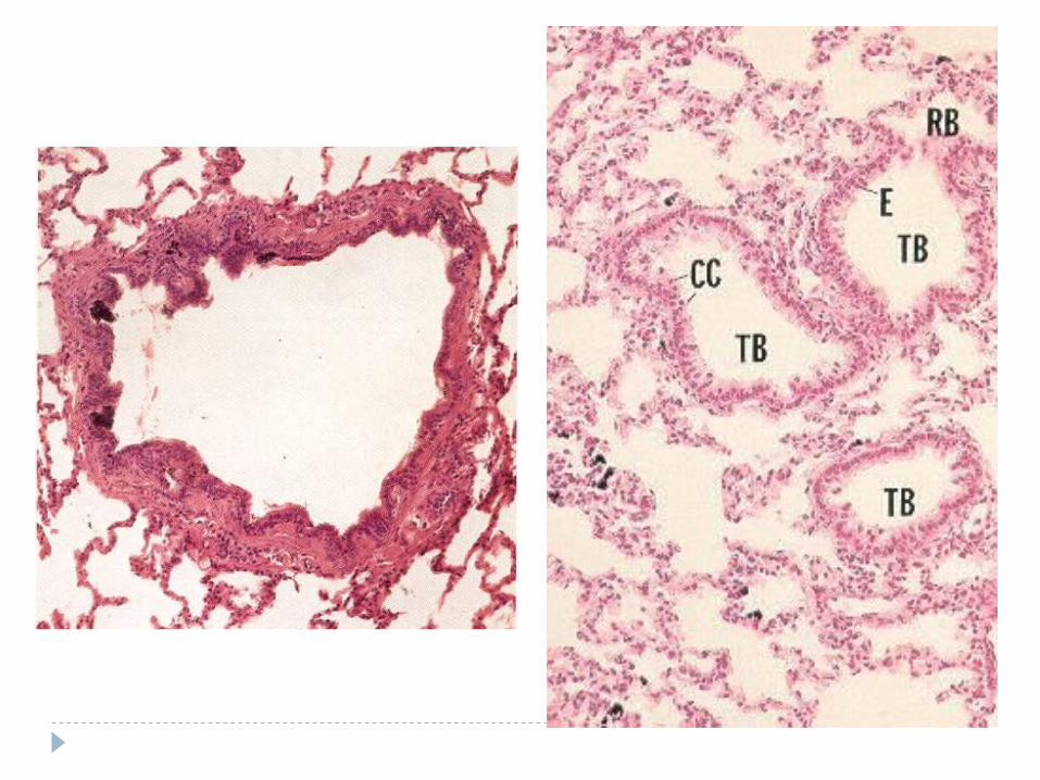
Bronchioles
have not any cartilage or glands but have few goblet cells In larger bronchioles epithelium is simple columnar ciliated, with
occasional goblet cells In smaller bronchioles epithelium change to simple cuboidal, with
no goblet cells Bronchioles have a smooth muscle coats surrounded by fibroelastic
connective tissue

Bronchioles
Terminal bronchioles are terminus of conducting portion they are lined by cuboidal cells(some with cilia) and Clara cells which have
domed apical surface
Lamina propria is a fibroelastic CT, 1-2 layer of smooth muscle cells separate it from adventitia
Clara cells ( exocrine bronchiolar cells)• Clara cells are columnar with dome-shaped apex• secretory granules• RER, which secret glycoproteins and surfactant-like materials• degrade toxins(SER)• divide to replace other cells• antimicrobial peptide

bronchiolitis Bronchiolitis is inflammation of the bronchioles, the
smallest air passages of the lungs. Obliterative bronchiolitis
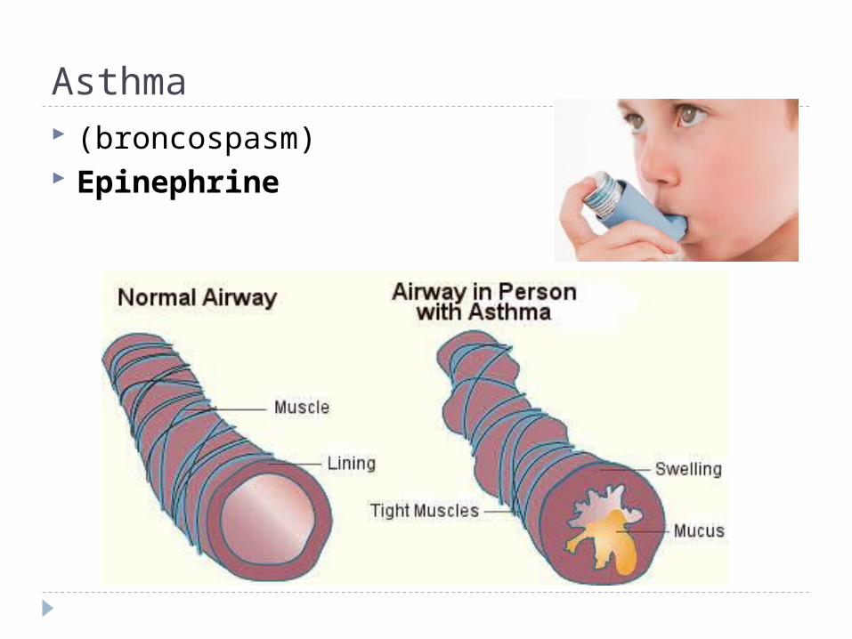
Asthma (broncospasm) Epinephrine

Respiratory Bronchioles
are a transitional zone between conducting and respiratory tissues
Alveoli branching from their walls
are lined by ciliated cuboidal epithelium with Clara cells that change to type I alveolar cells
Smooth muscle cells and elastic fibers underlie epithelium


Atelectasis is defined as the collapse or closure of the lung resulting in
reduced or absent gas exchange. It may affect part or all of one lung.

Alveolar Ducts do not have wall of their own They are only a linear arrangements of alveoli they end as a blind out pouching known as alveolar sac Opening of alveolus to AD controlled by a single smooth muscle cell
embedded

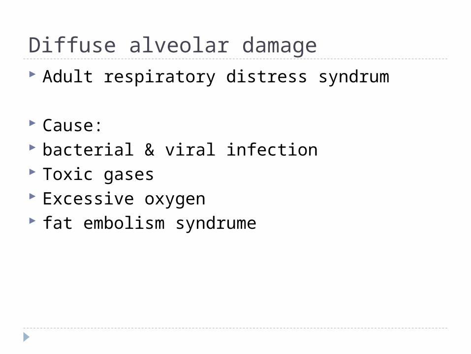
Diffuse alveolar damage Adult respiratory distress syndrum
Cause: bacterial & viral infection Toxic gases Excessive oxygen fat embolism syndrume
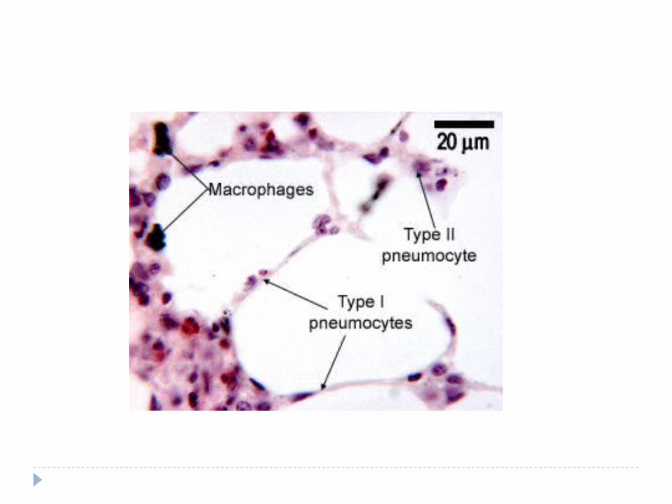
Alveolus
•Alveolus has 200 micrometer in diameter and is the functional unit of respiratory system
•Open as out pouching from RB, AD
•Composed of attenuated type I and type II pneumocytes
•Connective tissue between them are very scant
•Air space of two adjacent alveoli communicate through an alveolar pore
•Interalveolar septum is between alveoli have an extensive capillary bed
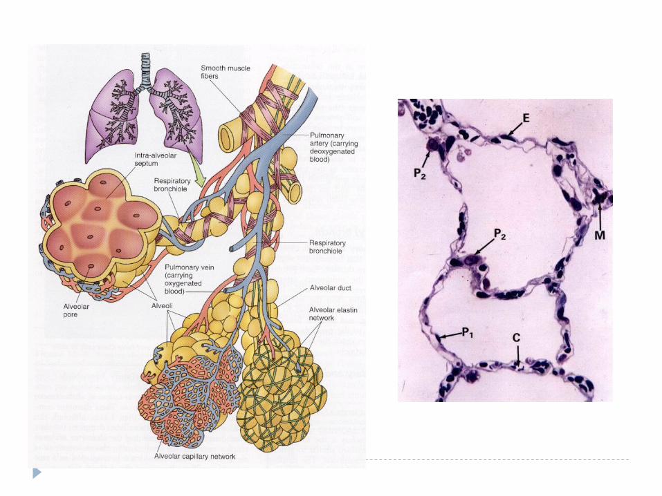
Cells of the Alveolar Septa Endothelial cells are nonfenestrated with a thin dark nucleus, and pinocytotic
vesicles Type I squamous cells that cover most of alveolar surface area, they have
pinocytotic vesicles Type II (greater alveolar) cells are cuboidal, located on alveolar surface
where septa intersect, they have foamy cytoplasm, surfactant granules (reduces surface tension to keep alveoli open during expiration)
Alveolar macrophages that are known as dust cells Interstitial cells consist of fibroblasts and mast cells Elastic & reticular fibers


•95% of the alveolar surface is composed of the simple squamous cells which are known as type I pneumocytes
•occluding junction attaches to other cells
•have basal lamina,
•alveolar pore formed by fusion of two adjacent type I cells
Type I pneumocytes (Squamus alveolar cells)
•They are more numerous than type I
•cover just 5% of the alveolar surface
• located among type I cells, cuboidal with dome-shaped apical
•Located where adjacent alveoli separated by septum
•They have an abundance of RER, developed golgi complex, their lamellar bodies contain pulmonary surfactant
Type II pneumocytes (Septal cells)

Alveolar Macrophages (Dust cells)•Known as type III pneumocytes
•Originate from monocytes that migrate to pulmonary interstitium
•Migrate between type I cells and enter alveolar lumen
•Maintain a sterile environment
•Assist type II to uptake surfactant
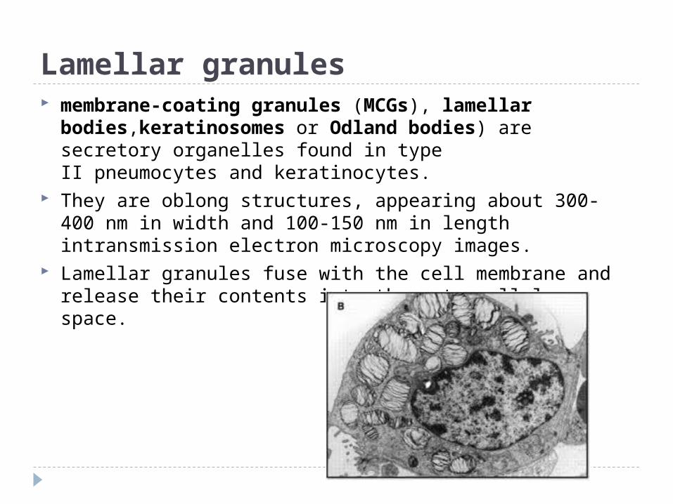
Lamellar granules membrane-coating granules (MCGs), lamellar
bodies,keratinosomes or Odland bodies) are secretory organelles found in type II pneumocytes and keratinocytes.
They are oblong structures, appearing about 300-400 nm in width and 100-150 nm in length intransmission electron microscopy images.
Lamellar granules fuse with the cell membrane and release their contents into the extracellular space.
Blood Gas Barrier•BGB is the thinnest regions of the interalveolar septum where gases can be exchanged
This barrier is composed of:
•Type I alveolar cells
• surfactant that covers them
•Common basal lamina of the type I and endothelial cells
•Endothelial cells of capillary network

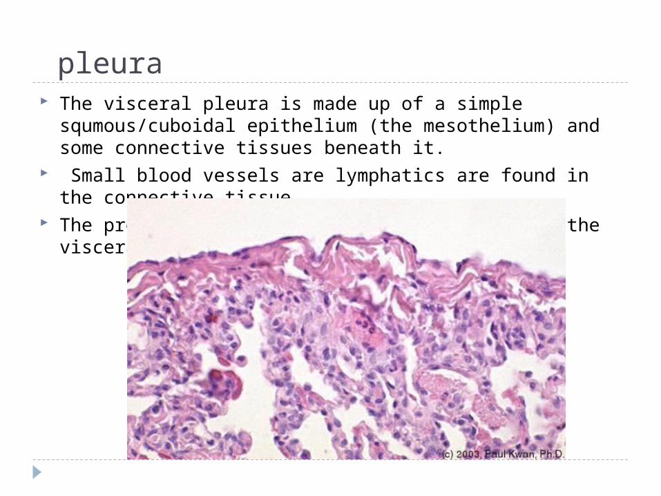
pleura The visceral pleura is made up of a simple
squmous/cuboidal epithelium (the mesothelium) and some connective tissues beneath it.
Small blood vessels are lymphatics are found in the connective tissue.
The presence of alveoli indicate that this is the visceral and not the parietal pleura.

pneumothorax is an abnormal collection of air or gas in the pleural
space that causes an uncoupling of the lung from the chest wall.
Like pleural effusion (liquid buildup in that space), pneumothorax may interfere with normal breathing.
It is often called collapsed lung, although that term may also refer to atelectasis
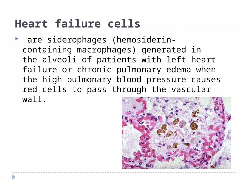
Heart failure cells are siderophages (hemosiderin-
containing macrophages) generated in the alveoli of patients with left heart failure or chronic pulmonary edema when the high pulmonary blood pressure causes red cells to pass through the vascular wall.

Chronic obstructive pulmonary disease (COPD), also known as chronic obstructive lung disease (COLD), and chronic obstructive airway disease (COAD), among others, is a type of obstructive lung disease characterized by chronically poor airflow.
Long-term exposure to the irritants causes an inflammatory response in the lungs resulting in narrowing of the small airways and breakdown of lung tissue, known as emphysema