Respiratory System Objectives - Describe the components and functions of the conducting and respiratory portions of the respiratory system. - Describe the embryologic steps in the development of the respiratory system. - Describe and compare the characteristic microscopic features and functions of the different regions and airways of the respiratory system, including lining epithelium, glands, cartilage, smooth muscle and elastic fibers.
Transcript
Respiratory System Objectives
- Describe the components and functions of the conducting and
respiratory portions of the respiratory system.
- Describe the embryologic steps in the development of the
respiratory system.
- Describe and compare the characteristic microscopic features and
functions of the different regions and airways of the respiratory
system, including lining epithelium, glands, cartilage, smooth
muscle and elastic fibers.
Respiratory System
* Introduction
* Conducting portion
* Respiratory portion
* Pleura
Respiratory System
• Introduction
– Anatomy
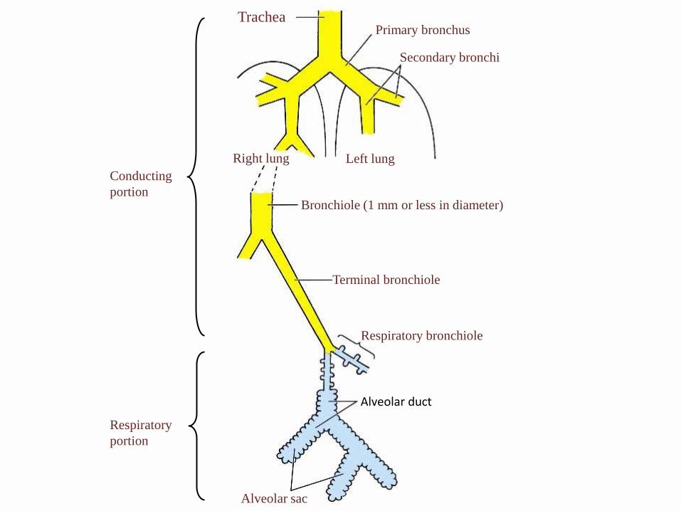
Anatomy of the
respiratory system
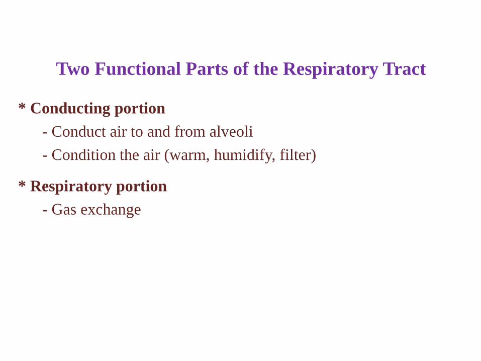
Two Functional Parts of the Respiratory Tract
* Conducting portion
- Conduct air to and from alveoli
- Condition the air (warm, humidify, filter)
* Respiratory portion
- Gas exchange
Trachea
Right lung Left lung
Primary bronchus
Secondary bronchi
Bronchiole (1 mm or less in diameter)
Terminal bronchiole
Respiratory bronchiole
Alveolar duct
Alveolar sac
Conducting
portion
Respiratory
portion
Respiratory System
* Embryology
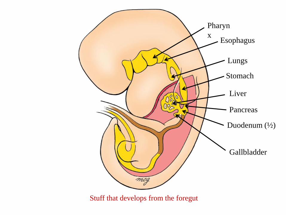
Pharyn
x
Lungs
Esophagus
Gallbladder
Stomach
Liver
Stuff that develops from the foregut
Duodenum (½)
Pancreas
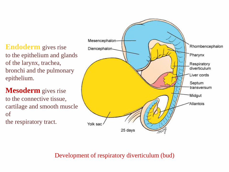
Development of respiratory diverticulum (bud)
Endoderm gives rise
to the epithelium and glands
of the larynx, trachea,
bronchi and the pulmonary
epithelium.
Mesoderm gives rise
to the connective tissue,
cartilage and smooth muscle
of
the respiratory tract.
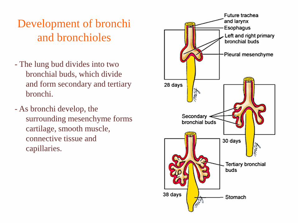
- The lung bud divides into two
bronchial buds, which divide
and form secondary and tertiary
bronchi.
- As bronchi develop, the
surrounding mesenchyme forms
cartilage, smooth muscle,
connective tissue and
capillaries.
Development of bronchi
and bronchioles
- By week 24, respiratory bronchioles are present and lungs are vascularized.
Respiration is possible – but chances of survival outside placenta are slim.
- Weeks 24-26: terminal alveolar sacs develop and type II pneumocytes secrete
surfactant (decreases surface tension)
- By week 26-28: there are enough alveolar sacs and surfactant to allow a
prematurely born infant to survive without medical intervention.
When can the baby breathe?
- 95% of mature alveoli develop after birth!
- The newborn infant has only 1/6 – 1/8 the number of alveoli as adults.
- Most alveolar growth is done by age 8.
- At birth, the lungs are half-filled with amniotic fluid.
- This fluid is cleared through the mouth and nose by pressure on thorax during
delivery.
- It also is reabsorbed into pulmonary blood vessels and lymphatics.
Lungs at Birth
Respiratory System
* Introduction
* Conducting portion
- Nasal cavity
- Larynx
- Trachea
- Bronchi
- Bronchioles
* The walls of the conducting system change in thickness and composition from
region to region.
* Components include:
- Epithelium (“respiratory epithelium”)
- Lamina propria
- Mucous and serous glands
- Cartilage
- Smooth muscle
- Adventitia
Walls of the Conducting System
Respiratory System
* Introduction
* Conducting portion
- Nasal cavity
* Respiratory epithelium everywhere except at the top (which has
specialized olfactory epithelium).
* Serous and mucous glands and numerous blood vessels in lamina
propria.
* Nasal septum: midline structure consisting of bone and hyaline
cartilage.
* Nasal fossa: chambers on each side of septum.
Nasal Cavity
* NASAL CAVITIES- The nasal cavities are paired chambers separated by a bony and
cartilaginous septum.
• They are elongated spaces with a wide base and a narrow apex .
• The skeletal framework is formed by bones and cartilages.
• Each cavity communicates anteriorly with the external environment
through the anterior nares (nostrils);
- posteriorly with the nasopharynx through the choanae; and
laterally with the paranasal sinuses and nasolacrimal duct.
- The chambers are divided into three regions:
• Nasal vestibule, is lined by skin
• Respiratory region, (inferior two-thirds) of the nasal cavities and is
lined by respiratory mucosa
• Olfactory region, at the apex (upper one-third) of each nasal cavity
and is lined by specialized olfactory mucosa
* Respiratory Region of the Nasal Cavity- It is lined by the respiratory mucosa that contains a ciliated,
pseudostratified columnar epithelium.
- lamina propria is firmly attached to the periosteum and perichondrium .
- The medial wall (the nasal septum) , is smooth, but the lateral walls are
thrown into folds by the presence of three shelf-like, bony projections
called conchae or turbinates.
• The conchae play a dual role:
- They increase surface area and
- cause turbulence in airflow to allow conditioning of inspired air.
- The ciliated, pseudostratified columnar epithelium is composed of five
cell types:
• Ciliated cells, tall columnar cells with cilia.
• Goblet cells that synthesize and secrete mucus
• Brush cells, bear short, blunt microvilli.
• Small granule cells (Kulchitsky cells) that contain secretory
granules (diffuse neuroendocrine system (DNES)
• Basal cells, stem cells from which the other cell types arise
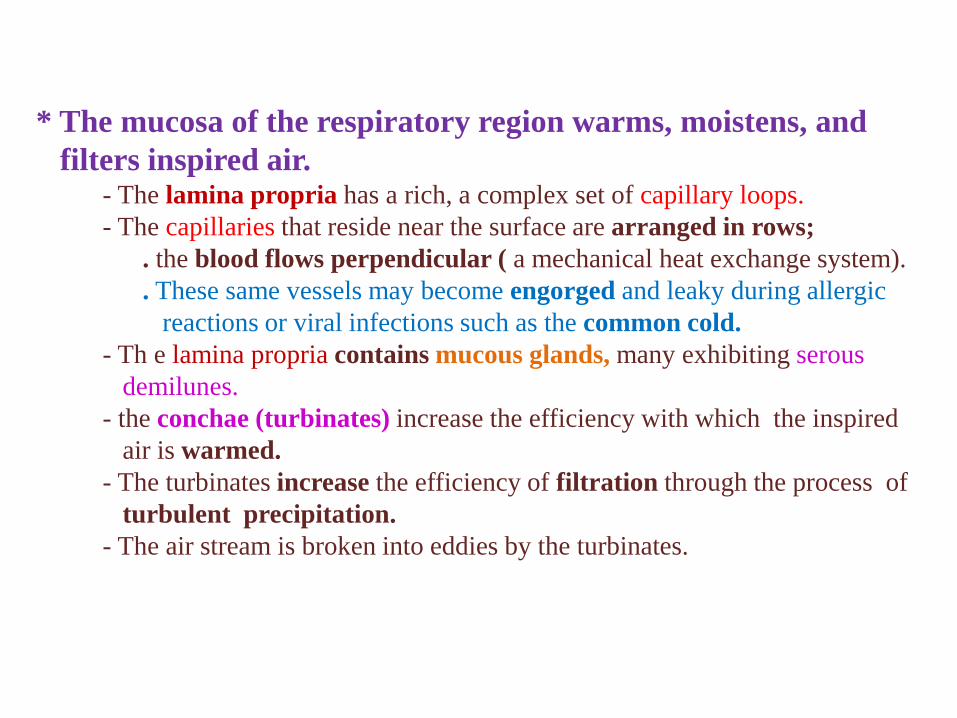
* The mucosa of the respiratory region warms, moistens, and
filters inspired air.- The lamina propria has a rich, a complex set of capillary loops.
- The capillaries that reside near the surface are arranged in rows;
. the blood flows perpendicular ( a mechanical heat exchange system).
. These same vessels may become engorged and leaky during allergic
reactions or viral infections such as the common cold.
- Th e lamina propria contains mucous glands, many exhibiting serous
demilunes.
- the conchae (turbinates) increase the efficiency with which the inspired
air is warmed.
- The turbinates increase the efficiency of filtration through the process of
turbulent precipitation.
- The air stream is broken into eddies by the turbinates.
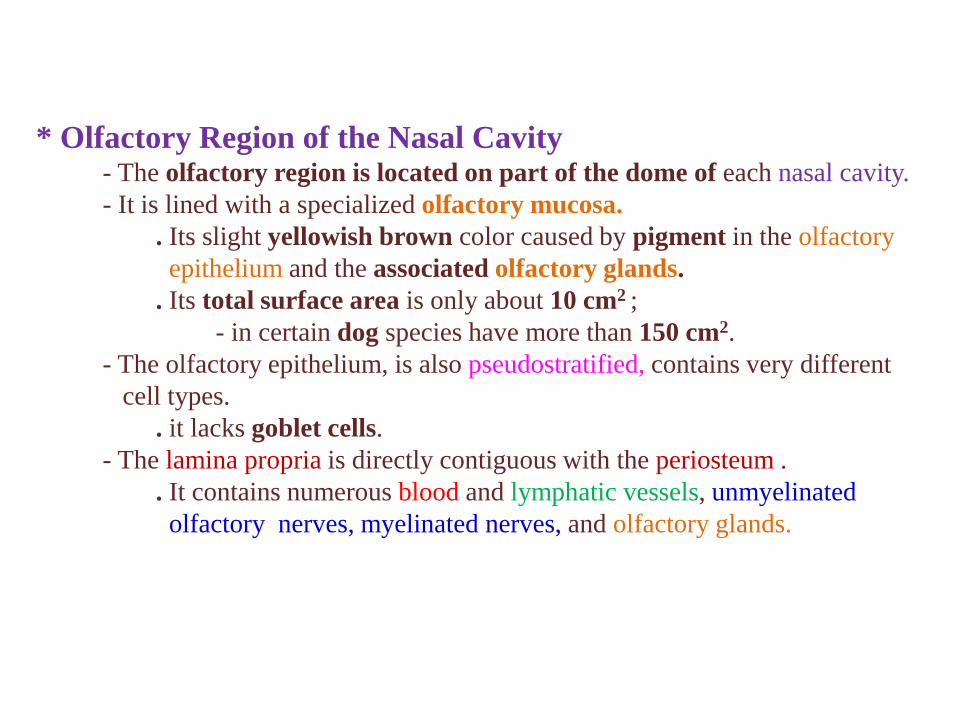
* Olfactory Region of the Nasal Cavity- The olfactory region is located on part of the dome of each nasal cavity.
- It is lined with a specialized olfactory mucosa.
. Its slight yellowish brown color caused by pigment in the olfactory
epithelium and the associated olfactory glands.
. Its total surface area is only about 10 cm2 ;
- in certain dog species have more than 150 cm2.
- The olfactory epithelium, is also pseudostratified, contains very different
cell types.
. it lacks goblet cells.
- The lamina propria is directly contiguous with the periosteum .
. It contains numerous blood and lymphatic vessels, unmyelinated
olfactory nerves, myelinated nerves, and olfactory glands.
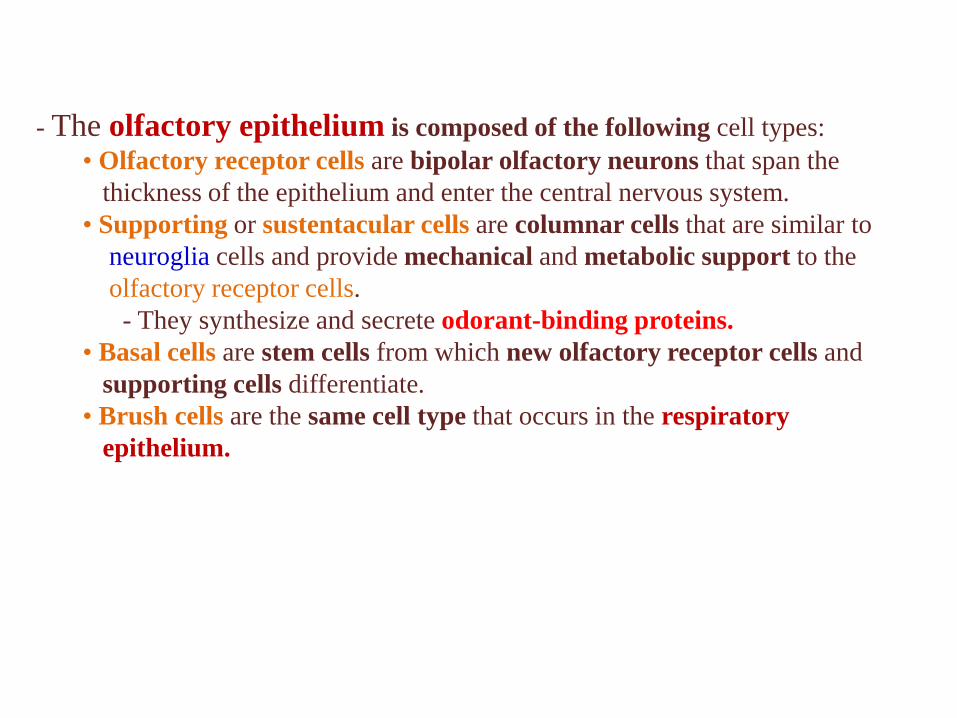
- The olfactory epithelium is composed of the following cell types:
• Olfactory receptor cells are bipolar olfactory neurons that span the
thickness of the epithelium and enter the central nervous system.
• Supporting or sustentacular cells are columnar cells that are similar to
neuroglia cells and provide mechanical and metabolic support to the
olfactory receptor cells.
- They synthesize and secrete odorant-binding proteins.
• Basal cells are stem cells from which new olfactory receptor cells and
supporting cells differentiate.
• Brush cells are the same cell type that occurs in the respiratory
epithelium.
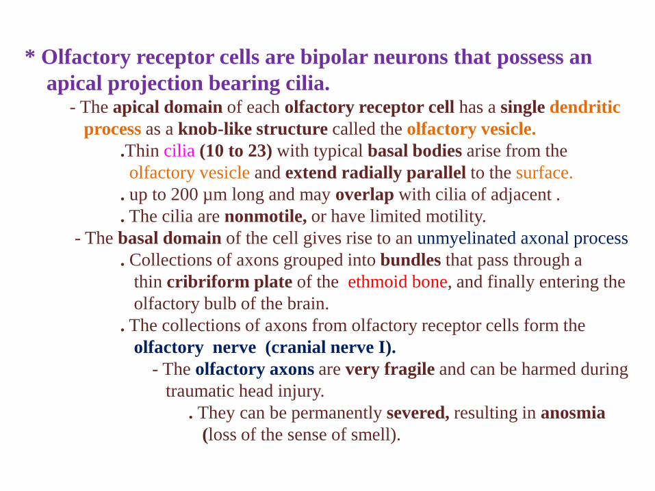
* Olfactory receptor cells are bipolar neurons that possess an
apical projection bearing cilia.- The apical domain of each olfactory receptor cell has a single dendritic
process as a knob-like structure called the olfactory vesicle.
.Thin cilia (10 to 23) with typical basal bodies arise from the
olfactory vesicle and extend radially parallel to the surface.
. up to 200 µm long and may overlap with cilia of adjacent .
. The cilia are nonmotile, or have limited motility.
- The basal domain of the cell gives rise to an unmyelinated axonal process
. Collections of axons grouped into bundles that pass through a
thin cribriform plate of the ethmoid bone, and finally entering the
olfactory bulb of the brain.
. The collections of axons from olfactory receptor cells form the
olfactory nerve (cranial nerve I).
- The olfactory axons are very fragile and can be harmed during
traumatic head injury.
. They can be permanently severed, resulting in anosmia
(loss of the sense of smell).
- The olfactory receptor cells have a life span of about 1 month.
. If injured, they are quickly replaced.
- Olfactory receptor cells (and some neurons of the enteric division ) are
readily replaced during postnatal life.
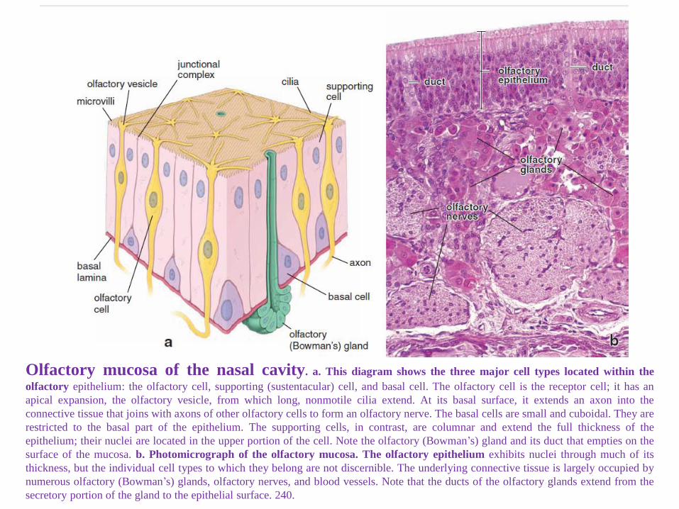
Olfactory mucosa of the nasal cavity. a. This diagram shows the three major cell types located within the
olfactory epithelium: the olfactory cell, supporting (sustentacular) cell, and basal cell. The olfactory cell is the receptor cell; it has an
apical expansion, the olfactory vesicle, from which long, nonmotile cilia extend. At its basal surface, it extends an axon into the
connective tissue that joins with axons of other olfactory cells to form an olfactory nerve. The basal cells are small and cuboidal. They are
restricted to the basal part of the epithelium. The supporting cells, in contrast, are columnar and extend the full thickness of the
epithelium; their nuclei are located in the upper portion of the cell. Note the olfactory (Bowman’s) gland and its duct that empties on the
surface of the mucosa. b. Photomicrograph of the olfactory mucosa. The olfactory epithelium exhibits nuclei through much of its
thickness, but the individual cell types to which they belong are not discernible. The underlying connective tissue is largely occupied by
numerous olfactory (Bowman’s) glands, olfactory nerves, and blood vessels. Note that the ducts of the olfactory glands extend from the
secretory portion of the gland to the epithelial surface. 240.
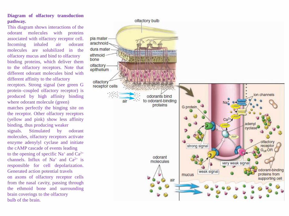
Diagram of olfactory transduction
pathway.
This diagram shows interactions of the
odorant molecules with proteins
associated with olfactory receptor cell.
Incoming inhaled air odorant
molecules are solubilized in the
olfactory mucus and bind to olfactory
binding proteins, which deliver them
to the olfactory receptors. Note that
different odorant molecules bind with
different affinity to the olfactory
receptors. Strong signal (see green G
protein–coupled olfactory receptor) is
produced by high affinity binding
where odorant molecule (green)
matches perfectly the binging site on
the receptor. Other olfactory receptors
(yellow and pink) show less affinity
binding, thus producing weaker
signals. Stimulated by odorant
molecules, olfactory receptors activate
enzyme adenylyl cyclase and initiate
the cAMP cascade of events leading
to the opening of specific Na+ and Ca2+
channels. Influx of Na+ and Ca2+ is
responsible for cell depolarization.
Generated action potential travels
on axons of olfactory receptor cells
from the nasal cavity, passing through
the ethmoid bone and surrounding
brain coverings to the olfactory
bulb of the brain.
* Paranasal Sinuses Paranasal sinuses are air-filled spaces in the
bones of the walls of the nasal cavity.- The paranasal sinuses are extensions of the respiratory region of the
nasal cavity and are lined by respiratory epithelium.
- The sinuses are named for the bone in which they are found (i.e., the
ethmoid, frontal, sphenoid, and maxillary bones).
- The sinuses communicate with the nasal cavities via narrow openings
onto the respiratory mucosa.
- The mucosal surface of the sinuses is a thin, ciliated, pseudostratified
columnar epithelium with numerous goblet cells.
- Mucus produced in the sinuses is swept into the nasal cavities by
coordinated ciliary movements.
- The sinuses are often subject to acute infection after viral infection of the
upper respiratory tract.
Respiratory System
* Introduction
* Conducting portion
- Nasal cavity
- Larynx
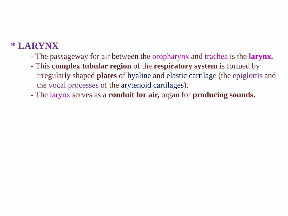
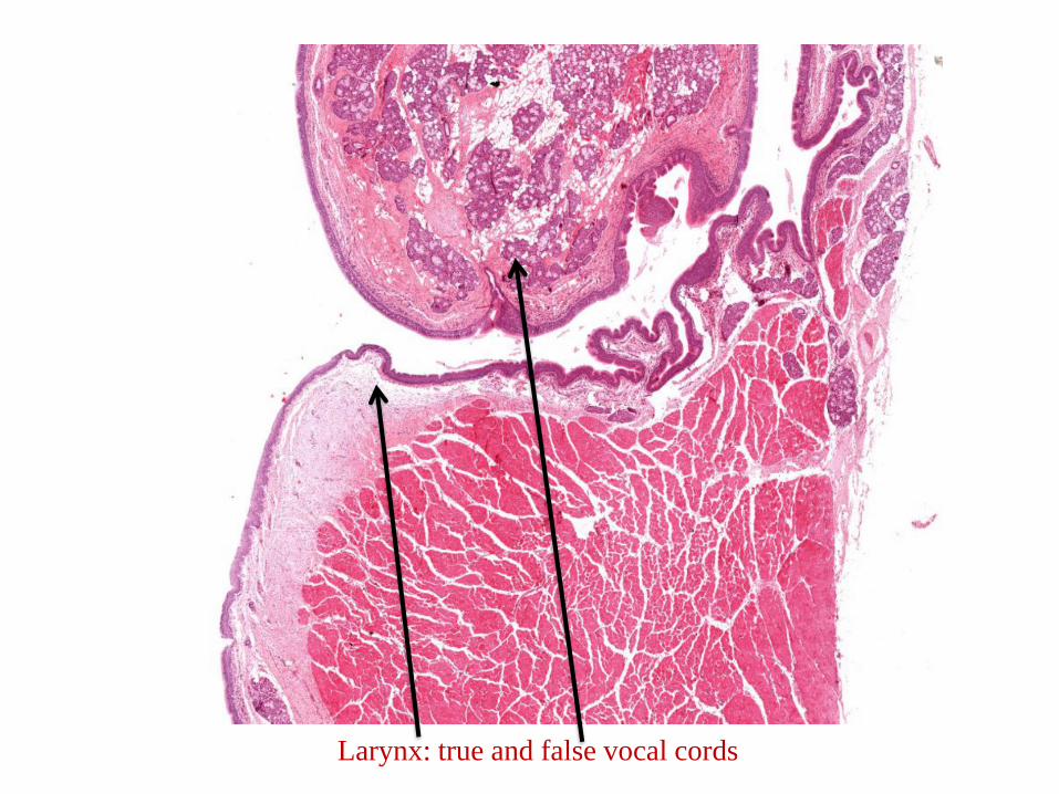
* LARYNX- The passageway for air between the oropharynx and trachea is the larynx.
- This complex tubular region of the respiratory system is formed by
irregularly shaped plates of hyaline and elastic cartilage (the epiglottis and
the vocal processes of the arytenoid cartilages).
- The larynx serves as a conduit for air, organ for producing sounds.
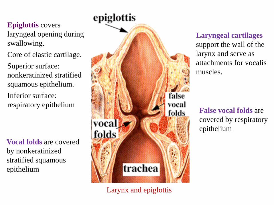
Epiglottis covers
laryngeal opening during
swallowing.
Core of elastic cartilage.
Superior surface:
nonkeratinized stratified
squamous epithelium.
Inferior surface:
respiratory epithelium
Laryngeal cartilages
support the wall of the
larynx and serve as
attachments for vocalis
muscles.
Vocal folds are covered
by nonkeratinized
stratified squamous
epithelium
False vocal folds are
covered by respiratory
epithelium
Larynx and epiglottis
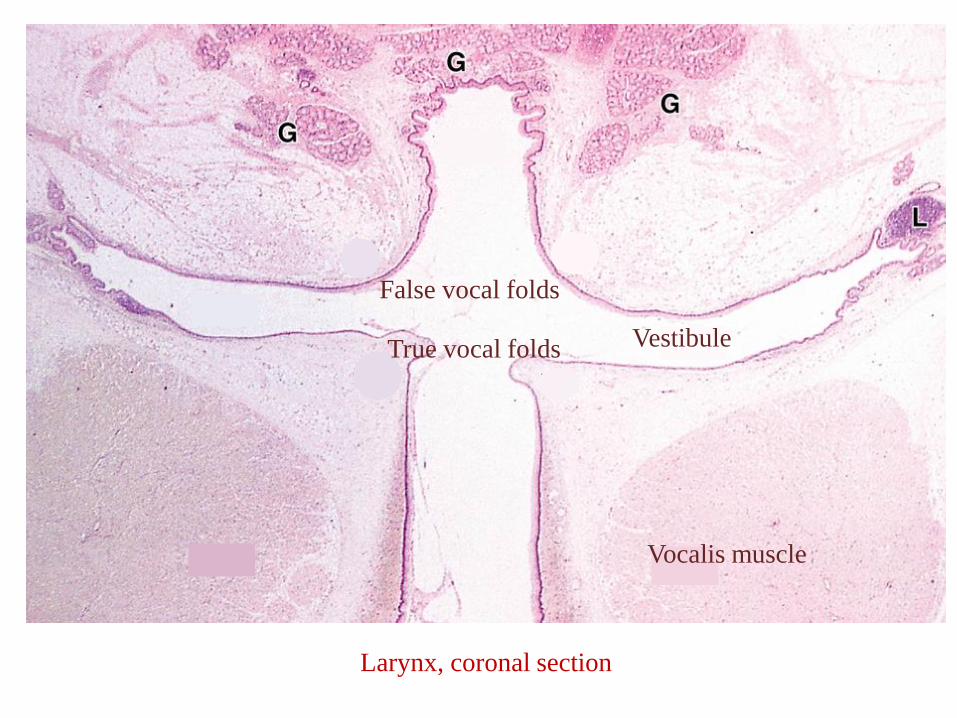
False vocal folds
True vocal folds
Vocalis muscle
Vestibule
Larynx, coronal section
Larynx: true and false vocal cords
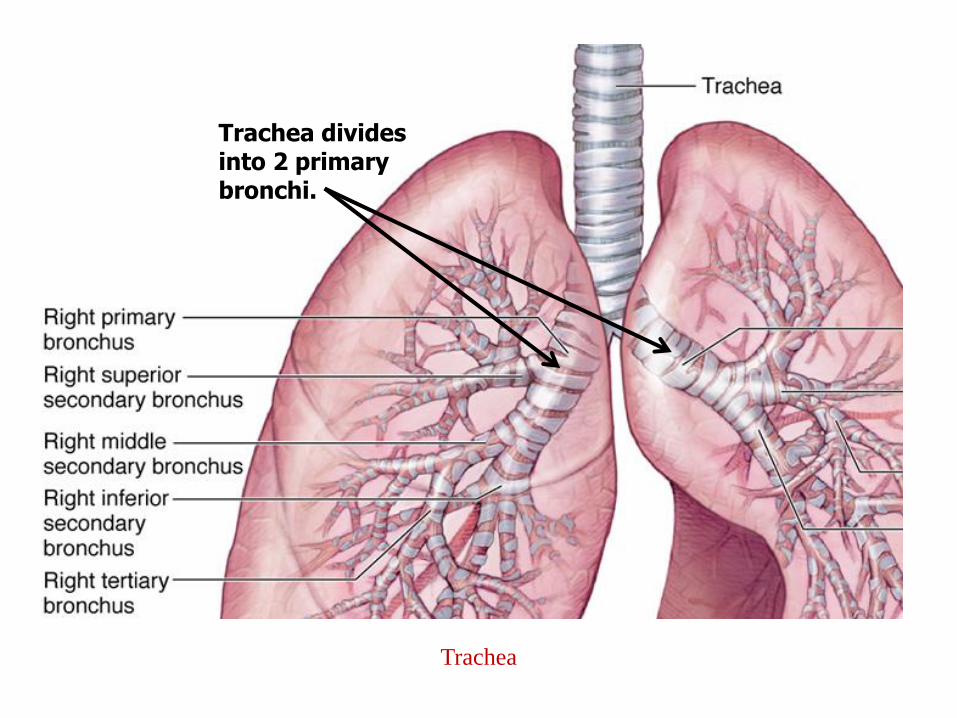
* The trachea is a short, flexible, air tube about 2.5 cm in diameter
and about 10 cm long.
* Extends from larynx and divides into two primary bronchi.
* Contains 16-20 C-shaped hyaline cartilage rings with the dorsal
opening bridged by smooth muscle (trachealis muscle).
* Lined by respiratory epithelium.
* Seromucous glands in lamina propria and submucosa.
Trachea
Trachea
Trachea divides into 2 primary bronchi.
- Ciliated columnar cells: most abundant cell type. Cilia beat in unison and move
mucus and trapped particles to oropharynx, where it is swallowed or
expectorated.
- Goblet cells: produce mucus.
- Basal cells: stem cells that replenish epithelium. Hard to see.
- Brush cells: Columnar cells. No cilia but have apical microvilli. Hard to see.
- Neuroendocrine cells: Small granule cells (Kulchitsky cells) epithelial cells
containing hormones. Hard to see.
Respiratory Epithelium Cell Types
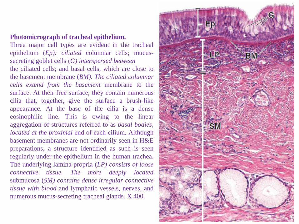
Photomicrograph of tracheal epithelium.
Three major cell types are evident in the tracheal
epithelium (Ep): ciliated columnar cells; mucus-
secreting goblet cells (G) interspersed between
the ciliated cells; and basal cells, which are close to
the basement membrane (BM). The ciliated columnar
cells extend from the basement membrane to the
surface. At their free surface, they contain numerous
cilia that, together, give the surface a brush-like
appearance. At the base of the cilia is a dense
eosinophilic line. This is owing to the linear
aggregation of structures referred to as basal bodies,
located at the proximal end of each cilium. Although
basement membranes are not ordinarily seen in H&E
preparations, a structure identified as such is seen
regularly under the epithelium in the human trachea.
The underlying lamina propria (LP) consists of loose
tissue with blood and lymphatic vessels, nerves, and
numerous mucus-secreting tracheal glands. X 400.
Trachea: respiratory epithelium
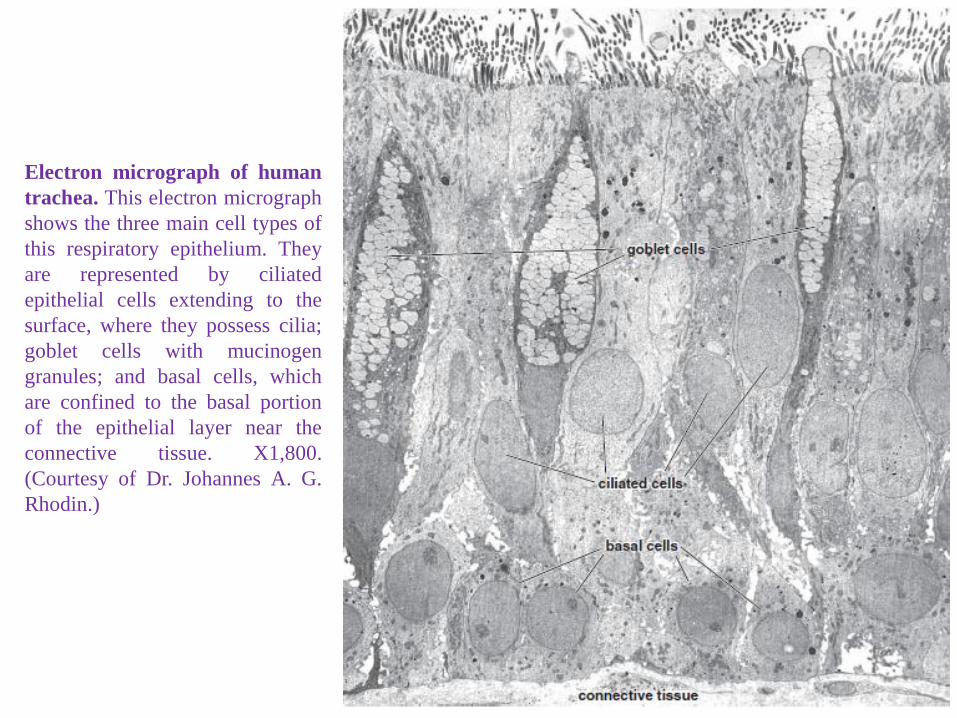
Electron micrograph of human
trachea. This electron micrograph
shows the three main cell types of
this respiratory epithelium. They
are represented by ciliated
epithelial cells extending to the
surface, where they possess cilia;
goblet cells with mucinogen
granules; and basal cells, which
are confined to the basal portion
of the epithelial layer near the
connective tissue. X1,800.
(Courtesy of Dr. Johannes A. G.
Rhodin.)
* BRONCHI- The trachea divides into two main (primary) bronchi.
- Anatomically, these divisions are more frequently described
as simply the right and left main bronchi.
. The right bronchus is wider and significantly shorter
than the left.
- On entering the hilum of the lung, each main bronchus
divides into the lobar bronchi (secondary bronchi).
- The right bronchus divides into three lobar bronchial
branches and the left into two lobar bronchial branches, with
each branch supplying one lobe.
- The lobar bronchi give rise to 10 segmental bronchi (
tertiary bronchi);
- The lobar bronchi of the left lung give rise to 8 segmental
bronchi.
* As branching progresses:
- Connective tissue decreases in thickness
- Relative amount of smooth muscle and elastic tissue increases
- Cartilage disappears (gone by bronchioles)
Morphologic Changes as Bronchi Branch
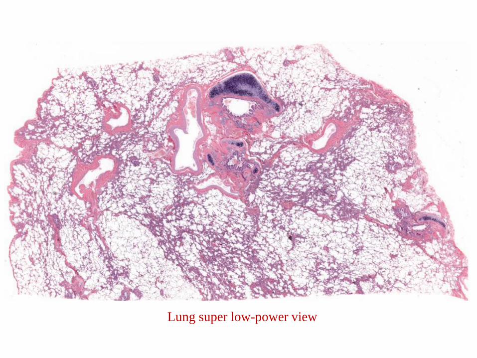
Lung super low-power view
Bronchus
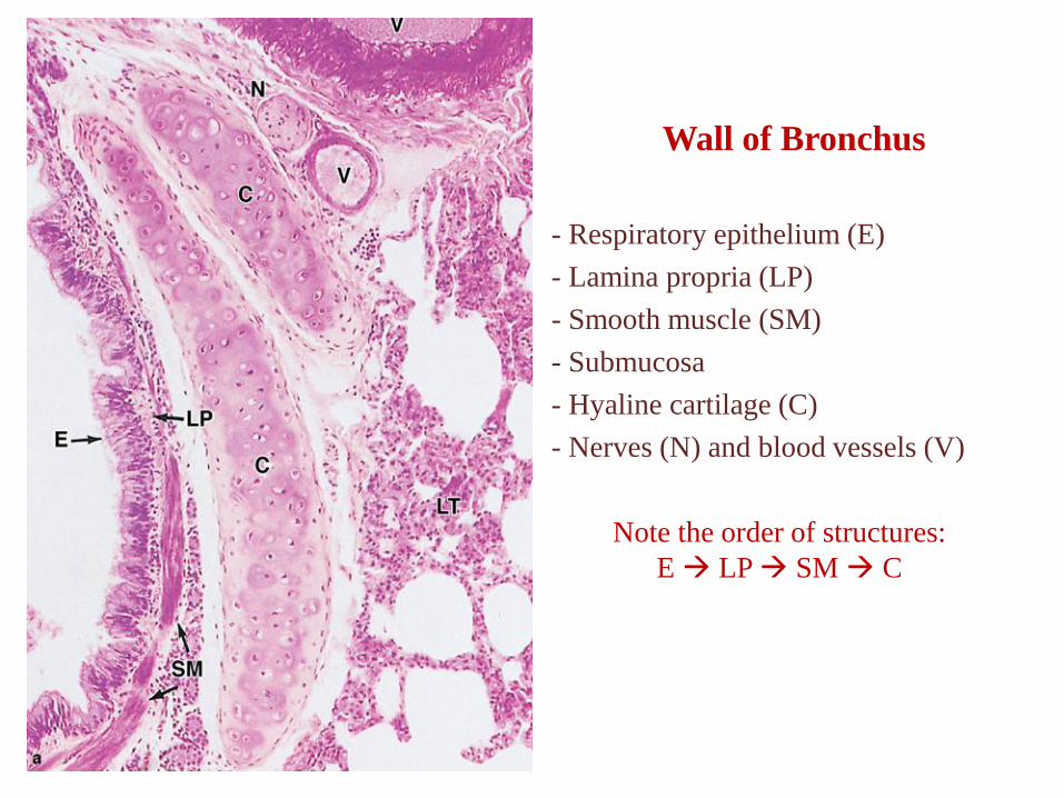
Wall of Bronchus
- Respiratory epithelium (E)
- Lamina propria (LP)
- Smooth muscle (SM)
- Submucosa
- Hyaline cartilage (C)
- Nerves (N) and blood vessels (V)
Note the order of structures:
E → LP → SM → C
Bronchus: respiratory epithelium
Goblet cellsPseudostratified
columnar
epithelium
Cilia
* NO glands or cartilage.
* Larger bronchioles have respiratory epithelium.
* Smaller bronchioles have low columnar epithelium.
* In asthma, the smooth muscle in the bronchioles constricts, causing
difficulty breathing.
Bronchioles
Bronchiole
Smooth
muscle
Respiratory
epithelium is
folded
Fibrous
connective
tissue is
present but
no cartilage
or glands!
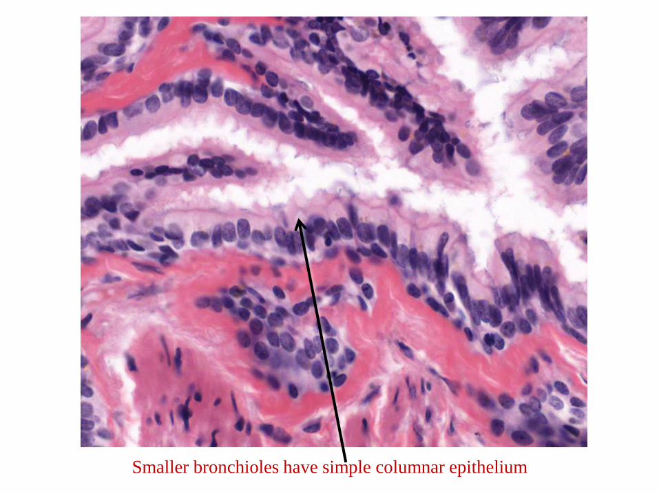
Smaller bronchioles have simple columnar epithelium
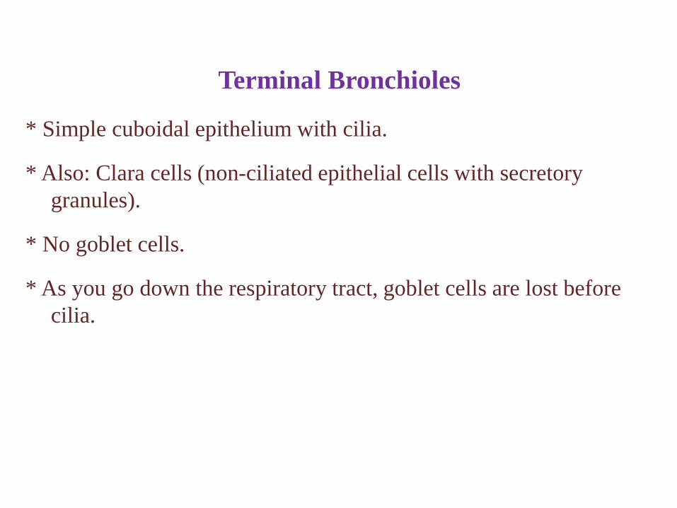
* Simple cuboidal epithelium with cilia.
* Also: Clara cells (non-ciliated epithelial cells with secretory
granules).
* No goblet cells.
* As you go down the respiratory tract, goblet cells are lost before
cilia.
Terminal Bronchioles
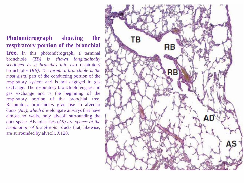
Photomicrograph showing the
respiratory portion of the bronchial
tree. In this photomicrograph, a terminal
bronchiole (TB) is shown longitudinally
sectioned as it branches into two respiratory
bronchioles (RB). The terminal bronchiole is the
most distal part of the conducting portion of the
respiratory system and is not engaged in gas
exchange. The respiratory bronchiole engages in
gas exchange and is the beginning of the
respiratory portion of the bronchial tree.
Respiratory bronchioles give rise to alveolar
ducts (AD), which are elongate airways that have
almost no walls, only alveoli surrounding the
duct space. Alveolar sacs (AS) are spaces at the
termination of the alveolar ducts that, likewise,
are surrounded by alveoli. X120.
Light micrograph of a 1 14m thick plastic section, stained with toluidine blue, showing a respiratory
bronchiole with non-ciliated, dense-granulated cells (arrows). Serous-like cells containing
relatively large dense granules are indicated by the "solid" arrows.
The "open" arrow indicates a typical Clara cell containing fewer dense secretory granules at its
protruding apex. (Scale bar•S 14m)
Clara cells
Make surfactant components, break down mucus, detoxify harmful substances, transfer
IgA, fight bacteria
produce a 16 kDa protein known as Clara cell secretory protein (CC16).
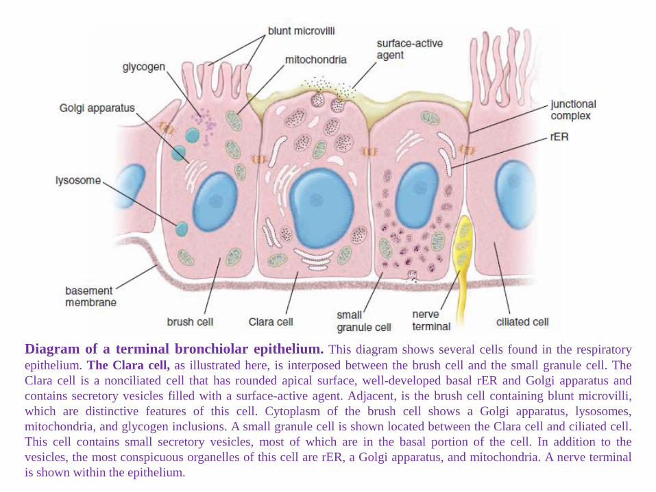
Diagram of a terminal bronchiolar epithelium. This diagram shows several cells found in the respiratory
epithelium. The Clara cell, as illustrated here, is interposed between the brush cell and the small granule cell. The
Clara cell is a nonciliated cell that has rounded apical surface, well-developed basal rER and Golgi apparatus and
contains secretory vesicles filled with a surface-active agent. Adjacent, is the brush cell containing blunt microvilli,
which are distinctive features of this cell. Cytoplasm of the brush cell shows a Golgi apparatus, lysosomes,
mitochondria, and glycogen inclusions. A small granule cell is shown located between the Clara cell and ciliated cell.
This cell contains small secretory vesicles, most of which are in the basal portion of the cell. In addition to the
vesicles, the most conspicuous organelles of this cell are rER, a Golgi apparatus, and mitochondria. A nerve terminal
is shown within the epithelium.
Scanning electron micrograph of a terminal
bronchiole. This scanning photomicrograph
shows a longitudinal
section throughout the terminal bronchiole and
surrounding alveoli (A).
Note that the apical surfaces of the Clara cells
possess no cilia and have a characteristic dome-
shaped appearance. 150. The inset shows some
of the Clara cells at a higher magnifi cation and
the cilia of a neighboring ciliated cell, which are
present in very small numbers at this level. Note
the relatively few cilia present on these small
cells. X1,200.
* Respiratory bronchioles
- As you go distally along the respiratory bronchioles, alveoli
increase in number.
- Cilia are gone by the end of the respiratory bronchiole.
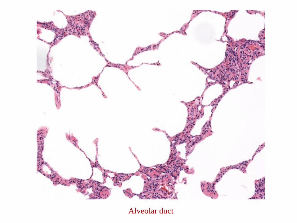
* Alveolar ducts
- Back-to-back alveolar openings along wall
- Smooth muscle between alveolar openings looks like knobs
Airways Preceding Alveoli
Respiratory bronchiole
Alveolar duct
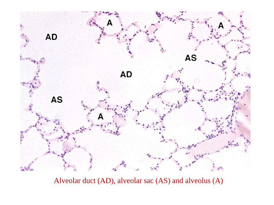
Alveolar duct (AD), alveolar sac (AS) and alveolus (A)
* Alveoli .- About 150 to 250 million alveoli are found in each adult lung;
- their combined internal surface area is approximately 75 m2.
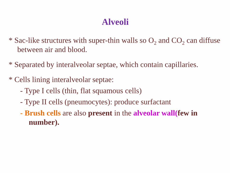
* Sac-like structures with super-thin walls so O2 and CO2 can diffuse
between air and blood.
* Separated by interalveolar septae, which contain capillaries.
* Cells lining interalveolar septae:
- Type I cells (thin, flat squamous cells)
- Type II cells (pneumocytes): produce surfactant
- Brush cells are also present in the alveolar wall(few in
number).
Alveoli
Alveolus
* Cover 95% of alveolar surface, comprise only 40% of the entire
alveolar lining cells
* Simple squamous cells with thin cytoplasm
* Blood-air barrier includes (from air to blood):
- Type I cells
- Fused basal laminae of type I cells and capillary endothelial cells
- Capillary endothelial cells
Type I Cells
* Cover 5% of alveolar surface, account for 60% of the alveolar
lining cells
* Large cuboidal cells with round nuclei.
* Typical secretory cell structure.
- Lamellar bodies in cytoplasm rich in a mixture of phospholipids, neutral
lipids, and proteins that form and store surfactant.
* Surfactant decreases surface tension in alveoli and prevents
collapse of alveoli during expiration.
Type II Cells (Pneumocytes)
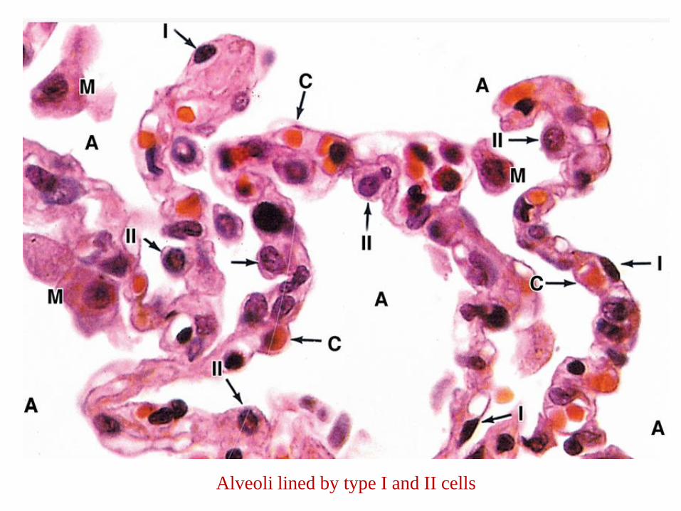
Alveoli lined by type I and II cells
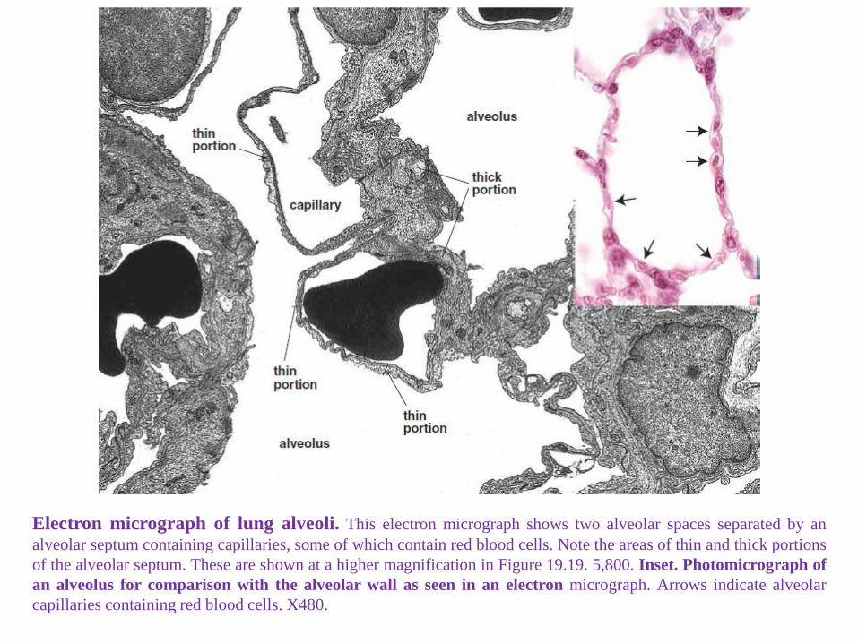
Electron micrograph of lung alveoli. This electron micrograph shows two alveolar spaces separated by an
alveolar septum containing capillaries, some of which contain red blood cells. Note the areas of thin and thick portions
of the alveolar septum. These are shown at a higher magnification in Figure 19.19. 5,800. Inset. Photomicrograph of
an alveolus for comparison with the alveolar wall as seen in an electron micrograph. Arrows indicate alveolar
capillaries containing red blood cells. X480.
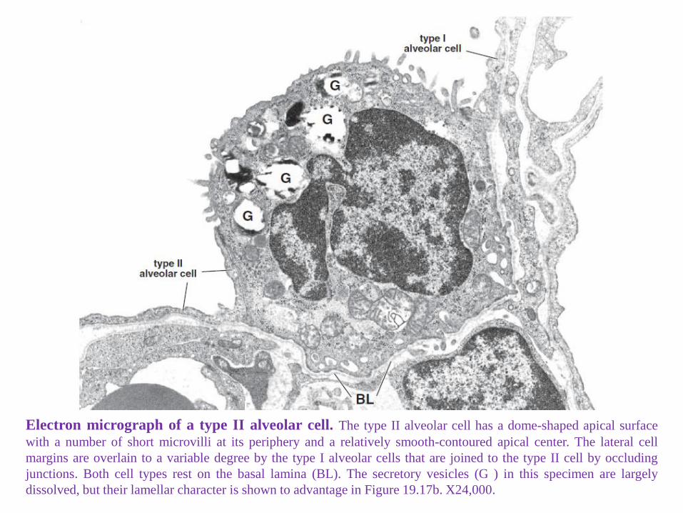
Electron micrograph of a type II alveolar cell. The type II alveolar cell has a dome-shaped apical surface
with a number of short microvilli at its periphery and a relatively smooth-contoured apical center. The lateral cell
margins are overlain to a variable degree by the type I alveolar cells that are joined to the type II cell by occluding
junctions. Both cell types rest on the basal lamina (BL). The secretory vesicles (G ) in this specimen are largely
dissolved, but their lamellar character is shown to advantage in Figure 19.17b. X24,000.
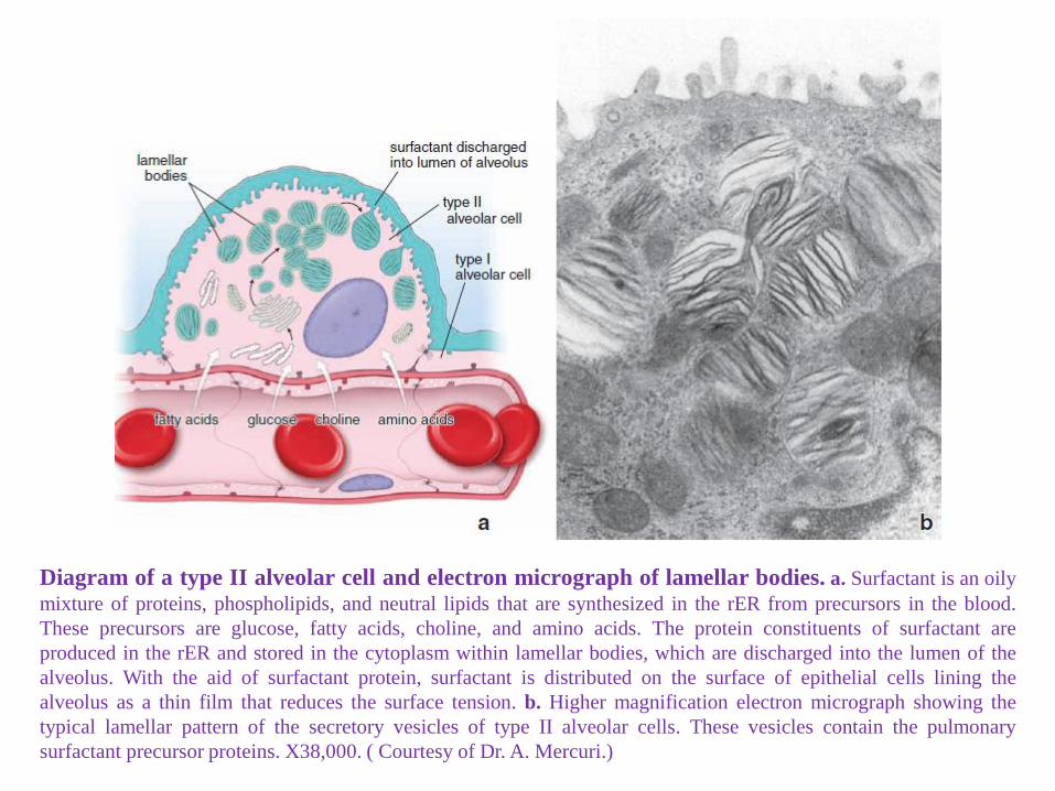
Diagram of a type II alveolar cell and electron micrograph of lamellar bodies. a. Surfactant is an oily
mixture of proteins, phospholipids, and neutral lipids that are synthesized in the rER from precursors in the blood.
These precursors are glucose, fatty acids, choline, and amino acids. The protein constituents of surfactant are
produced in the rER and stored in the cytoplasm within lamellar bodies, which are discharged into the lumen of the
alveolus. With the aid of surfactant protein, surfactant is distributed on the surface of epithelial cells lining the
alveolus as a thin film that reduces the surface tension. b. Higher magnification electron micrograph showing the
typical lamellar pattern of the secretory vesicles of type II alveolar cells. These vesicles contain the pulmonary
surfactant precursor proteins. X38,000. ( Courtesy of Dr. A. Mercuri.)
* Surfactant decreases the alveolar surface tension and actively
participates in the clearance of foreign materials.
- The most specific phospholipid called dipalmitoylphosphatidylcholine
(DPPC), which accounts for almost all surface tension.
- Surfactant synthesis in the fetus occurs after the 35th week of gestation
and is modulated by a variety of hormones, including;
. Cortisol, insulin, prolactin, and thyroxine.
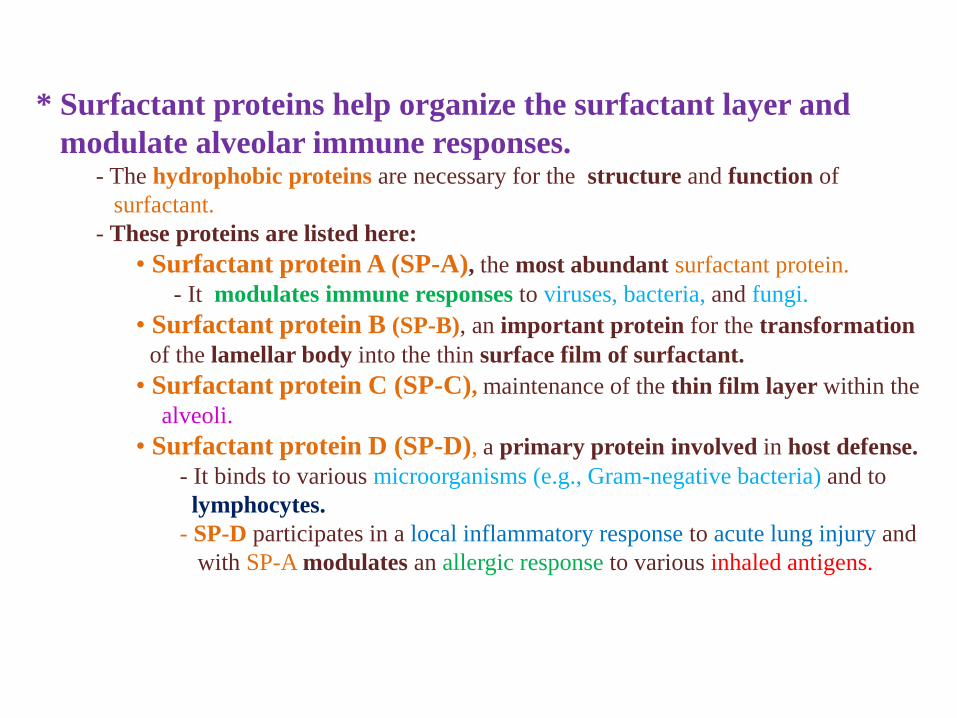
* Surfactant proteins help organize the surfactant layer and
modulate alveolar immune responses.- The hydrophobic proteins are necessary for the structure and function of
surfactant.
- These proteins are listed here:
• Surfactant protein A (SP-A), the most abundant surfactant protein.
- It modulates immune responses to viruses, bacteria, and fungi.
• Surfactant protein B (SP-B), an important protein for the transformation
of the lamellar body into the thin surface film of surfactant.
• Surfactant protein C (SP-C), maintenance of the thin film layer within the
alveoli.
• Surfactant protein D (SP-D), a primary protein involved in host defense.
- It binds to various microorganisms (e.g., Gram-negative bacteria) and to
lymphocytes.
- SP-D participates in a local inflammatory response to acute lung injury and
with SP-A modulates an allergic response to various inhaled antigens.
* The alveolar septum is the site of the air–blood barrier.- The air–blood barrier refers to the cells and cell products across which
gases must diffuse between the alveolar and capillary compartments.
- The thinnest air–blood barrier consists of
. a thin layer of surfactant,
. a type I epithelial cell and its basal lamina, and
. a capillary endothelial cell and its basal lamina.
- Often, these two basal laminae are fused.
- Connective tissue cells and fibers that may be present between the two basal
laminae.
- These two arrangements produce a thin portion and a thick portion of the
barrier.
- Most gas exchange occurs across the thin portion of the barrier.
. The thick portion is thought to be a site in which tissue fluid can
accumulate and even cross into the alveolus.
- Lymphatic vessels in the connective tissue of the terminal bronchioles drain
fluid that accumulates in the thick portion of the septum.
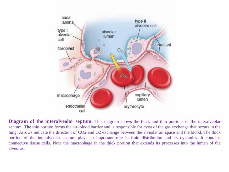
Diagram of the interalveolar septum. This diagram shows the thick and thin portions of the interalveolar
septum. The thin portion forms the air–blood barrier and is responsible for most of the gas exchange that occurs in the
lung. Arrows indicate the direction of CO2 and O2 exchange between the alveolar air space and the blood. The thick
portion of the interalveolar septum plays an important role in fluid distribution and its dynamics. It contains
connective tissue cells. Note the macrophage in the thick portion that extends its processes into the lumen of the
alveolus.
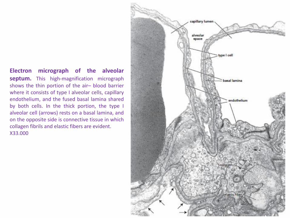
Electron micrograph of the alveolarseptum. This high-magnification micrographshows the thin portion of the air– blood barrierwhere it consists of type I alveolar cells, capillaryendothelium, and the fused basal lamina sharedby both cells. In the thick portion, the type Ialveolar cell (arrows) rests on a basal lamina, andon the opposite side is connective tissue in whichcollagen fibrils and elastic fibers are evident.X33.000
* Found on surface of alveoli, within alveoli and in interstitial
connective tissue.
* Remove debris and particles that escape mucus and cilia in
conducting portion of respiratory tract
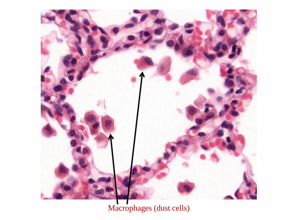
Alveolar Macrophages (Dust Cells)
Macrophages (dust cells)
Pleura
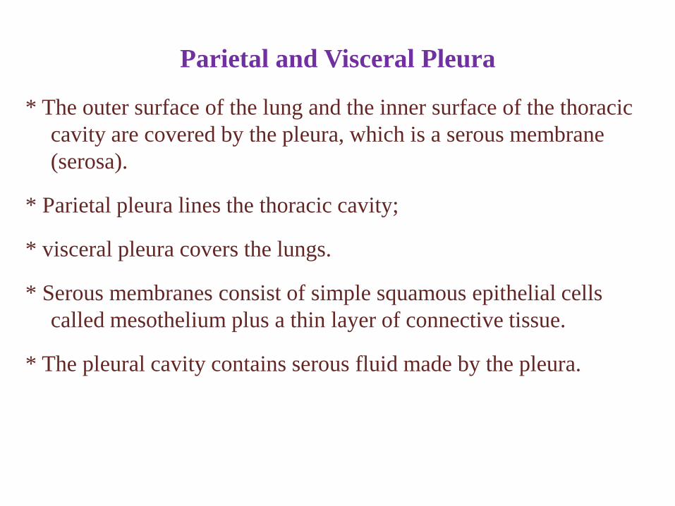
* The outer surface of the lung and the inner surface of the thoracic
cavity are covered by the pleura, which is a serous membrane
(serosa).
* Parietal pleura lines the thoracic cavity;
* visceral pleura covers the lungs.
* Serous membranes consist of simple squamous epithelial cells
called mesothelium plus a thin layer of connective tissue.
* The pleural cavity contains serous fluid made by the pleura.


![Respiratory System [โหมดความเข้ากันได้] · PATHOLOGY OF RESPIRATORY SYSTEM นพ. อรรณพ นาคะป ท Respiratory system U it](https://static.documents.pub/doc/80x56/5fa578efd4e80f055f6b3401/respiratory-system-aaaaaaaaaaaaaaaaaa-pathology.jpg)


