72 IJEDS CASE REPORT 10.5005/jp-journals-10029-1128 Restoration of Mutilated Primary Maxillary Incisors using Biological Restorations under General Anesthesia 1 R Kranthi Kumar, 2 BV Thimma Reddy, 3 Neha Nayan ABSTRACT Restoration of grossly mutilated primary maxillary incisors affected by early childhood caries has been a challenging task for the pediatric dentist due to the little amount of tooth structure available for bonding and behavioral problems of the children. A variety of treatment options ranging from extraction followed by prosthesis to the usage of intracanal pins and fiber posts have been reported in the past. The present case report depicts one such a case wherein biological restorations were used as post and core to restore the mutilated primary maxillary incisors in a 4-year-old child treated under general anesthesia. Keywords: Biological restoration, Early childhood caries (ECC), General anesthesia, Post and core. How to cite this article: Kumar KR, Reddy BVT, Nayan N. Restoration of Mutilated Primary Maxillary Incisors using Biological Restorations under General Anesthesia. Int J Experiment Dent Sci 2016;5(1):72-75. Source of support: Nil Conflict of interest: None INTRODUCTION Dental caries in very young children known as early childhood caries (ECC) is the most common chronic disease affecting the primary dentition, 1 especially the maxillary anterior teeth. The early loss of primary ante- rior teeth may result in reduced masticatory efficiency, speech problems, loss of vertical dimension, develop- ment of parafunctional habits like tongue thrusting, aesthetic functional problems, such as malocclusion and space loss, and physiological problems that can interfere in the personality and behavioral development of the child. 2,3 Various restorative options for rebuilding the muti- lated primary incisors are conventional methods like use of prefabricated crowns, direct and indirect composite restorations, omega loop post, strip crowns, and the recently proposed “biological restorations.” 4 The term “biological restoration” was coined in 1991 to describe an alternative technique that uses adhesive capabilities of materials in combination with strategic placement of parts of extracted human teeth to achieve better aesthetics and more conservation of sound dental tissues. 5 Tavares et al 6 described the first case in which tooth fragments were used to restore carious elements. Ramires-Romito et al 7 used the teeth from the Human Tooth Bank of Sao Paulo University Dental School as natural post and crown to fit into the roots and rebuild the coronal portion of the tooth. CASE REPORT A 4-year-old boy reported to the Department of Pediatric Dentistry with the chief compliant of decayed primary maxillary anterior teeth. Medical history was not contributory but the personal history revealed prolonged bottle-feeding till the age of three and a half, irregular brushing habits, and the habit of frequent snacking. On intraoral examination, it was found that all the four maxillary incisors were grossly decayed with only root stumps remaining (Fig. 1), considerably large occlusal cavities on 54 and 64, and a proximal cavity on 63 (Fig. 2). Lisping was observed with the child while producing labiodental sounds because of the missing coronal portions of the incisors for a long time. Radiographic examination revealed carious lesions involving the pulp with 54 and 64 and periapical radiolucency was seen with the incisor root stumps. Patient was extremely uncooperative during the examination and radiographic procedure. Based on the clinical and radiographic findings, a diagnosis of severe early childhood caries was made. As the patient was extremely uncooperative, complete oral rehabilitation under general anesthesia was planned. Consent was obtained from the parents after explaining the risks and benefits of general anesthesia and at the same time the concept of biological restorations was also explained to the parents and consent was obtained. The 1,3 Senior Lecturer, 2 Professor 1 Department of Pedodontics, GSL Dental College and Hospital Rajahmundry, Andhra Pradesh, India 2 Department of Pedodontics, Panineeya Dental College Hyderabad, Telangana, India 3 Department of Oral Pathology, GSL Dental College and Hospital, Rajahmundry, Andhra Pradesh, India Corresponding Author: R Kranthi Kumar, Senior Lecturer Department of Pedodontics, GSL Dental College and Hospital Rajahmundry, Andhra Pradesh, India, Phone: 088324849999 e-mail: [email protected]
Transcript
72
R Kranthi Kumar et al
IJEDS
CaSE REpoRt10.5005/jp-journals-10029-1128
Restoration of Mutilated Primary Maxillary Incisors using Biological Restorations under General Anesthesia1R Kranthi Kumar, 2 BV Thimma Reddy, 3Neha Nayan
ABSTRACTRestoration of grossly mutilated primary maxillary incisors affected by early childhood caries has been a challenging task for the pediatric dentist due to the little amount of tooth structure available for bonding and behavioral problems of the children. A variety of treatment options ranging from extraction followed by prosthesis to the usage of intracanal pins and fiber posts have been reported in the past. The present case report depicts one such a case wherein biological restorations were used as post and core to restore the mutilated primary maxillary incisors in a 4-year-old child treated under general anesthesia.
Keywords: Biological restoration, Early childhood caries (ECC), General anesthesia, Post and core.
How to cite this article: Kumar KR, Reddy BVT, Nayan N. Restoration of Mutilated Primary Maxillary Incisors using Biological Restorations under General Anesthesia. Int J Experiment Dent Sci 2016;5(1):72-75.
Source of support: Nil
Conflict of interest: None
INTRODUCTION
Dental caries in very young children known as early childhood caries (ECC) is the most common chronic disease affecting the primary dentition,1 especially the maxillary anterior teeth. The early loss of primary ante-rior teeth may result in reduced masticatory efficiency, speech problems, loss of vertical dimension, develop-ment of parafunctional habits like tongue thrusting, aesthetic functional problems, such as malocclusion and space loss, and physiological problems that can interfere in the personality and behavioral development of the child.2,3
Various restorative options for rebuilding the muti-lated primary incisors are conventional methods like use of prefabricated crowns, direct and indirect composite restorations, omega loop post, strip crowns, and the recently proposed “biological restorations.”4
The term “biological restoration” was coined in 1991 to describe an alternative technique that uses adhesive capabilities of materials in combination with strategic placement of parts of extracted human teeth to achieve better aesthetics and more conservation of sound dental tissues.5 Tavares et al6 described the first case in which tooth fragments were used to restore carious elements. Ramires-Romito et al7 used the teeth from the Human Tooth Bank of Sao Paulo University Dental School as natural post and crown to fit into the roots and rebuild the coronal portion of the tooth.
CASE REPORT
A 4-year-old boy reported to the Department of Pediatric Dentistry with the chief compliant of decayed primary maxillary anterior teeth. Medical history was not contributory but the personal history revealed prolonged bottle-feeding till the age of three and a half, irregular brushing habits, and the habit of frequent snacking.
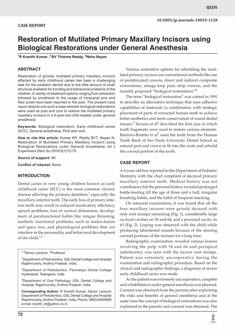
On intraoral examination, it was found that all the four maxillary incisors were grossly decayed with only root stumps remaining (Fig. 1), considerably large occlusal cavities on 54 and 64, and a proximal cavity on 63 (Fig. 2). Lisping was observed with the child while producing labiodental sounds because of the missing coronal portions of the incisors for a long time.
Radiographic examination revealed carious lesions involving the pulp with 54 and 64 and periapical radiolucency was seen with the incisor root stumps. Patient was extremely uncooperative during the examination and radiographic procedure. Based on the clinical and radiographic findings, a diagnosis of severe early childhood caries was made.
As the patient was extremely uncooperative, complete oral rehabilitation under general anesthesia was planned. Consent was obtained from the parents after explaining the risks and benefits of general anesthesia and at the same time the concept of biological restorations was also explained to the parents and consent was obtained. The
1,3Senior Lecturer, 2Professor1Department of Pedodontics, GSL Dental College and Hospital Rajahmundry, Andhra Pradesh, India2Department of Pedodontics, Panineeya Dental College Hyderabad, Telangana, India3Department of Oral Pathology, GSL Dental College and Hospital, Rajahmundry, Andhra Pradesh, India
Corresponding Author: R Kranthi Kumar, Senior Lecturer Department of Pedodontics, GSL Dental College and Hospital Rajahmundry, Andhra Pradesh, India, Phone: 088324849999 e-mail: [email protected]
International Journal of Experimental Dental Science, January-June 2016;5(1):72-75 73
IJEDS
Restoration of Mutilated Primary Maxillary Incisors using Biological Restorations
the fit of the donor teeth in the post space, they were cemented using dual cure resin cement (Fig. 6). Minor defects at the junction of the biological restorations and the root stumps were adjusted with composite resin (Fig. 7). Finally, 54 and 64 were restored with preformed stainless steel crowns and composite restoration on 63 (Fig. 8).
After the completion of the restorative procedure, extubation was done and the patient was shifted to the intensive care unit. The patient was under the care of pediatric anesthesiologist for the next 12 hours and then discharged.
DISCUSSION
Esthetic restoration of mutilated primary maxillary anterior teeth due to ECC has been a challenge to the pediatric dentist owing to the less amount of tooth structure available for bonding and the behavioral issues associated with younger children. In majority of the cases, crown parts are completely destroyed, leaving only the dentin available in the root for bonding.8 This
Fig. 3: Samples stored in Hank’s balanced salt solution after proper autoclaving and root filling
Fig. 4: Prepared coronal thirds of 51, 52, 61, and 62 to receive biological restorations
Fig. 1: Grossly decayed 51, 52, 61, and 62 Fig. 2: Large occlusal cavities on 54 and 64 and a proximal cavity on 63
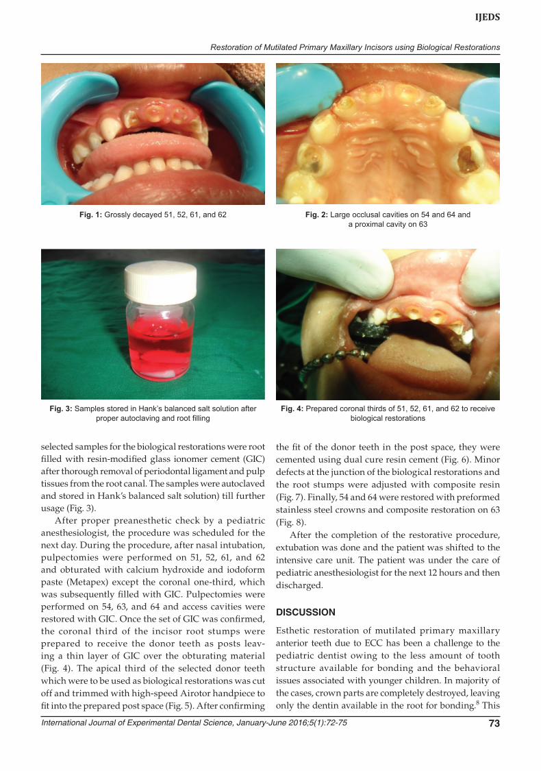
selected samples for the biological restorations were root filled with resin-modified glass ionomer cement (GIC) after thorough removal of periodontal ligament and pulp tissues from the root canal. The samples were autoclaved and stored in Hank’s balanced salt solution) till further usage (Fig. 3).
After proper preanesthetic check by a pediatric anesthesiologist, the procedure was scheduled for the next day. During the procedure, after nasal intubation, pulpectomies were performed on 51, 52, 61, and 62 and obturated with calcium hydroxide and iodoform paste (Metapex) except the coronal one-third, which was subsequently filled with GIC. Pulpectomies were performed on 54, 63, and 64 and access cavities were restored with GIC. Once the set of GIC was confirmed, the coronal third of the incisor root stumps were prepared to receive the donor teeth as posts leav- ing a thin layer of GIC over the obturating material (Fig. 4). The apical third of the selected donor teeth which were to be used as biological restorations was cut off and trimmed with high-speed Airotor handpiece to fit into the prepared post space (Fig. 5). After confirming
74
R Kranthi Kumar et al
gross destruction of the crown contemplates the use of intracanal posts to facilitate crown reconstruction. Various materials that have been tried as intracanal posts are 0.7 mm stainless steel orthodontic wire, such as Omega pin and composite resin posts. Wire posts may interfere with the physiologic root resorption if it extends a long way into the root and can increase internal stress leading to root fracture.9 And the composite resins used alone or in conjunction with fibers have low strength of loading,10 chances of polymerization shrinkage, and expensive and elaborate lab work.3
The use of “biological restorations” is gaining popu-larity because of its advantages like simple technique, preservation of excellent esthetics, as well as preserva-tion of natural tooth color compared with composite resins and stainless steel crowns, preservation of sound tooth structure, and low cost.11 The enamel of biologi-cally restored tooth offers physiologic wear, superficial smoothness, and cervical adaptation compatible with those of the surrounding teeth.12,13 Biological restorations not only mimic the missing part of the oral structures, but are also biofunctional.14
However, biological restorations have their own disadvantages, which include difficulty in obtaining teeth with required coronal dimensions and characteristics, problems inherent to indirect restorations, and matching fragment color with tooth remnant color. And also many of the patients refuse biological restorations because it is unpleasant to have other people’s teeth in their mouth.15 However, all these are not considered to be contraindications for this technique.
Even though biological crown and post restoration is a cost-effective alternative, the patient acceptance of it is an important factor which needs to be addressed. However, in the present case, parent’s consent was taken regarding the usage of biological restorations in the rehabilitation process and were informed that the donor teeth were previously submitted to rigorous sterilization that completely eliminated the risk of contamination or disease transmission to the child.
CONCLUSION
Dental Caries is a major public health concern worldwide, affecting more than 80% of the population alive in the
Fig. 5: Prepared biological restorations ready to be cemented into the post space
Fig. 6: Biological restorations after cementation into the post space
Fig. 7: Biological restorations after final finishing Fig. 8: Preformed stainless crowns placed on 54 and 64 and composite restoration on 63
International Journal of Experimental Dental Science, January-June 2016;5(1):72-75 75
IJEDS
Restoration of Mutilated Primary Maxillary Incisors using Biological Restorations
world today. However, due to limited access to profes-sional care, oral hygiene education, health status, huge population and economic limitations the total preventive programs for dental carries cannot be fully successful.16
Biological crown and post restoration is a cost-effective treatment modality for ECC when compared with con-ventional intracanal reinforced composite restorations. However, the patient acceptance of the restoration is the key factor. So, it is time to recycle the precious biological tissues which has been discarded till now.
REFERENCES 1. Mouradian WE. The face of a child: children’s oral health and
dental education. J Dent Educ 2001 Sep;65(9):821-831. 2. Ngan P, Fields H. Orthodontic diagnosis and treatment
planning in primary dentition. ASDC J Dent Child 1995 Jan-Feb;62(1):25-33.
3. Motisuki C, Santos-Pinto L, Giro EM. Restoration of severely decayed primary incisors using indirect composite resin restoration technique. Int J Pediatr Dent 2005 Jul;15(4):282-286.
4. Karina Sanches, de Carvalho FK, Nelson-Filho P, Assed S, Silva FW, de Queiroz AM. Biological restorations as a treatment option for primary molars with extensive coronal destruction—report of two cases. Braz Dent J 2007;18(3):248-252.
5. Santos J, Bianchi J. Restoration of severely damaged teeth with resin bonding systems: case reports. Quint Int 1991 Aug;22(8):611-615.
humano. Relato de caso. Rev fac odontol Sao Paulo 1992; 4(2):113-117.
7. Ramires-Romito AC, Wanderley MT, Oliveira MD, Imperato JC, Correa MS. Biological restoration of primary anterior teeth. Quintessence Int 2000;31(6):405-411.
8. Wanderley MT, Ferriora SL, Rodrigues CR, Rodrigues Filho LE. Primary anterior tooth restoration using posts with microretentive elements. Quintessence Int 1999 Jun;30(6): 432-436.
9. Rifkin A. Composite post crown in anterior teeth. J Dent Assoc S Afr 1983 Apr;38(4):225-227.
10. Rodrigues Filho LE, Bianchi J, Santos JF, Oliveira JA. Clinical evaluation of dental reinforcements by means of metallic posts with macroretentions. J Dent Res 1996;75:1095.
11. Ehrmann EH. Restoration of fractured incisor with exposed pulp using original tooth fragment: report of a case. J Am Dent Assoc 1989 Feb;118(2):183.
12. Mandroli PS. Biologic restoration of primary anterior teeth: a case report. J Indian Soc Pedo Prev Dent 2003 Sep;21(3):95-97.
13. Chosack ABDS, Edielmann EDO. Rehabilitation of a fractured incisor using the patients natural crown—case report. J Dent Child 1964;31(1):19-21.
14. Kapur A, Chawla HS, Goyal A, Guaba K. An esthetic point of view in very young children. J Clin Pediatr Dent 2005 Winter; 30(2):99-103.