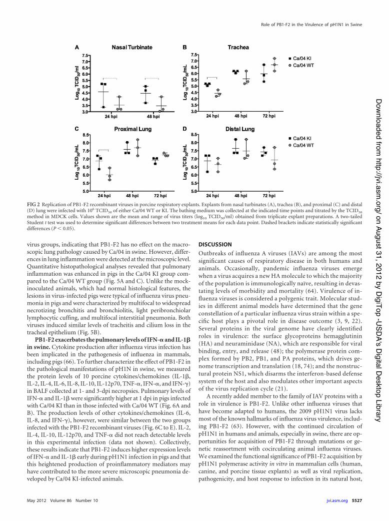
Published Ahead of Print 29 February 2012. 2012, 86(10):5523. DOI: 10.1128/JVI.00134-12. J. Virol. Perez Henningson, Kelly M. Lager, Alessio Lorusso and Daniel R. Lindomar Pena, Amy L. Vincent, Crystal L. Loving, Jamie N. Swine in H1N1 Influenza Virus Has Minimal Effects Restored PB1-F2 in the 2009 Pandemic http://jvi.asm.org/content/86/10/5523 Updated information and services can be found at: These include: REFERENCES http://jvi.asm.org/content/86/10/5523#ref-list-1 at: This article cites 77 articles, 29 of which can be accessed free CONTENT ALERTS more» articles cite this article), Receive: RSS Feeds, eTOCs, free email alerts (when new http://journals.asm.org/site/misc/reprints.xhtml Information about commercial reprint orders: http://journals.asm.org/site/subscriptions/ To subscribe to to another ASM Journal go to: on August 31, 2012 by DigiTop -USDA's Digital Desktop Library http://jvi.asm.org/ Downloaded from
Transcript
Published Ahead of Print 29 February 2012. 2012, 86(10):5523. DOI: 10.1128/JVI.00134-12. J. Virol.
PerezHenningson, Kelly M. Lager, Alessio Lorusso and Daniel R. Lindomar Pena, Amy L. Vincent, Crystal L. Loving, Jamie N. Swine
inH1N1 Influenza Virus Has Minimal Effects Restored PB1-F2 in the 2009 Pandemic
http://jvi.asm.org/content/86/10/5523Updated information and services can be found at:
These include:REFERENCES
http://jvi.asm.org/content/86/10/5523#ref-list-1at: This article cites 77 articles, 29 of which can be accessed free
CONTENT ALERTS more»articles cite this article),
Receive: RSS Feeds, eTOCs, free email alerts (when new
http://journals.asm.org/site/misc/reprints.xhtmlInformation about commercial reprint orders: http://journals.asm.org/site/subscriptions/To subscribe to to another ASM Journal go to:
Restored PB1-F2 in the 2009 Pandemic H1N1 Influenza Virus HasMinimal Effects in Swine
Lindomar Pena,a Amy L. Vincent,b Crystal L. Loving,b Jamie N. Henningson,b Kelly M. Lager,b Alessio Lorusso,b and Daniel R. Pereza
Department of Veterinary Medicine, University of Maryland College Park and Virginia-Maryland Regional College of Veterinary Medicine, College Park, Maryland, USA,a andVirus and Prion Diseases of Livestock Research Unit, National Animal Disease Center, USDA-ARS, Ames, Iowa, USAb
PB1-F2 is an 87- to 90-amino-acid-long protein expressed by certain influenza A viruses. Previous studies have shown thatPB1-F2 contributes to virulence in the mouse model; however, its role in natural hosts—pigs, humans, or birds—remains largelyunknown. Outbreaks of domestic pigs infected with the 2009 pandemic H1N1 influenza virus (pH1N1) have been detectedworldwide. Unlike previous pandemic strains, pH1N1 viruses do not encode a functional PB1-F2 due to the presence of threestop codons resulting in premature truncation after codon 11. However, pH1N1s have the potential to acquire the full-lengthform of PB1-F2 through mutation or reassortment. In this study, we assessed whether restoring the full-length PB1-F2 openreading frame (ORF) in the pH1N1 background would have an effect on virus replication and virulence in pigs. Restoring thePB1-F2 ORF resulted in upregulation of viral polymerase activity at early time points in vitro and enhanced virus yields in por-cine respiratory explants and in the lungs of infected pigs. There was an increase in the severity of pneumonia in pigs infectedwith isogenic virus expressing PB1-F2 compared to the wild-type (WT) pH1N1. The extent of microscopic pneumonia correlatedwith increased pulmonary levels of alpha interferon and interleukin-1! in pigs infected with pH1N1 encoding a functionalPB1-F2 but only early in the infection. Together, our results indicate that PB1-F2 in the context of pH1N1 moderately modulatesviral replication, lung histopathology, and local cytokine response in pigs.
Influenza A viruses (IAVs) belong to the family Orthomyxoviri-dae and represent important pathogens of humans and animals.
In the 20th century, humans experienced three IAV pandemics(1918, 1957, and 1968) that resulted in significant morbidity andmortality (62). These pandemic strains emerged through geneticreassortment between influenza viruses of avian origin and thecirculating human strain. These events resulted in antigenic shiftand the successful dissemination, in an immunologically naïvepopulation, of a virus carrying a novel hemagglutinin (HA). Inaddition to the new HA, pandemic strains inherited the PB1 genesegment from the avian influenza donor virus (and the NA gene in1957) (57). Like most avian influenza viruses, these previous pan-demic strains expressed a full-length PB1-F2 gene. However, hu-man influenza strains have invariably evolved to introduce trun-cations in the PB1-F2 open reading frame (ORF). It is tempting tospeculate that PB1-F2 function is necessary for virus survival inbirds but unnecessary or detrimental in humans. Its significancein pandemic strains remains obscure.
In the spring of 2009, a novel H1N1 IAV emerged in NorthAmerica, causing acute respiratory disease in humans. The virusquickly spread throughout most regions of the world, promptingthe World Health Organization (WHO) to declare an influenzapandemic on 11 June 2009 (13, 15). The new virus (pH1N1) re-sulted from genetic reassortment between swine influenza viruses(SIVs) circulating in North America and Eurasia. Specifically, sixof its RNA segments (PB2, PB1, PA, HA, NP, and NS) are closelyrelated to North American triple-reassortant (TR) H1N1 SIVs,whereas the NA and M gene segments are related to EurasianH1N1 SIVs (15). This unique gene constellation has never beendescribed among influenza isolates from anywhere in the world,and the precise evolutionary history of the 2009 pH1N1 is un-known (1, 13, 17, 57). Several outbreaks of pH1N1 virus infectionsin animals have been reported worldwide. These outbreaks werepredominantly documented in pigs, but incidental infection in
turkeys, cats, ferrets, dogs, and wild animals has been described (4,14, 27, 37, 46, 50, 51, 58, 59, 61). In pigs, field outbreaks of pH1N1have been reported in more than 20 countries, and epidemiolog-ical investigations have linked humans as the probable source (25,27). Experimentally, pigs are susceptible to human pH1N1 vi-ruses, and the virus is highly transmissible in swine (6, 29, 32, 70,75). Pigs inoculated with the pH1N1 virus show clinical diseasesigns and pathology similar to those seen with infection with otherSIVs (6, 29, 32, 70, 75).
Pigs are susceptible to human influenza viruses; however, andperhaps unlike humans, they appear susceptible to a wide range ofavian influenza viruses. Experimental infection studies showed thatpigs were susceptible to 13 different influenza virus subtypes (H1 toH3 and non-human-type HA types H4 to H13) (30). The mixingvessel attribute is consistent with the presence in the respiratory tractof pigs of the prototypical human-like and avian-like influenzareceptors N-acetylneuraminic acid-!2,6-galactose (!2,6Gal) and N-acetylneuraminic acid-!2,3-galactose (!2,3Gal), respectively (28).Although subsequent studies have shown both types of receptors arealso found in the respiratory tract of quail, pheasant, turkey, guineafowl (31, 73), and humans (55), pigs have been commonly associatedwith two-way transmission of influenza viruses to and from humans(43, 45, 54, 71). Pigs were undoubtedly involved in the genesis of the2009 pH1N1 (15). Since then, several reassortants between pH1N1and circulating influenza A viruses have been isolated from pigs in
Received 17 January 2012 Accepted 23 February 2012
several countries (26, 42, 51, 60, 69), raising great concerns about thepotential acquisition of virulence markers by the pH1N1 virus uponreassortment with other strains in the swine host.
Among the virulence factors that could be acquired by pH1N1is the nonstructural protein PB1-F2. In contrast to the three pre-vious pandemic influenza viruses, pH1N1 does not encode a func-tional PB1-F2 due to the presence of three stop codons that causesa premature truncation (63). PB1-F2 is an 87- to 90-amino-acid(aa)-long protein encoded by an alternate ("1) open readingframe (ORF) within the PB1 gene. Translation of PB1-F2 mRNAis likely mediated by ribosomal scanning, and the protein is ex-pressed early and transiently in infected cells (10). PB1-F2 is ex-pressed mostly by IAVs of avian origin (78), and its presence is notrequired for viral replication in embryonated eggs, in tissue cul-ture, or in vivo (11). However, PB1-F2 expression results in en-hanced apoptotic-cell death in immune cells, viral pathogenicityin mice, and immunopathology (10, 12, 39, 40, 77). Additionally,the presence of PB1-F2 in both the 1918 and the mouse-adaptedPR8 influenza A viruses enhances secondary bacterial pneumoniain the mouse model (40). Surprisingly, two recent studies investi-gating the role of PB1-F2 in the pathogenicity of clinically relevanthuman viruses and the pH1N1 concluded that the expression ofPB1-F2 has minimal effects on the virulence of these viruses inmurine and ferret models (19, 41). From these studies, it appearsthat the role of PB1-F2 in modulating influenza virus pathogenic-ity is cell type and virus strain specific and/or species dependent.The contribution of PB1-F2 to the virulence of influenza viruses inswine has yet to be determined.
Here, we restored the PB1-F2 ORF in pH1N1 and studied itseffects on viral pathogenicity and host responses in pigs. RestoringPB1-F2 in the pH1N1 virus resulted in increased virus replicationin swine respiratory explants. More importantly, in pigs, a pH1N1virus expressing a complete PB1-F2 ORF increased virus replica-tion in the lung and enhanced lung histopathology and higherpulmonary levels of alpha interferon (IFN-!) and interleukin-1#(IL-1#) than the wild-type (WT) virus but only at early timespostinfection. Although these effects were minimal and observedonly at early times postinfection, these findings suggest thatPB1-F2 can modulate pH1N1 pathogenicity and cytokine re-sponses in swine.
MATERIALS AND METHODSAnimal studies. Pig explants were prepared according to protocol in“Transmissibility of Influenza A Viruses in Swine,” approved by the In-stitutional Animal Care and Use Committee, University of Maryland,College Park, MD. Swine pathogenicity studies were conducted in thehigh containment facilities at the National Animal Disease Center inAmes, IA, under protocol in “Influenza A Virus Pathogenesis and HostResponse in Swine,” approved by the USDA-ARS Animal Care and UseCommittee. Animal studies adhered strictly to the U.S. Animal WelfareAct (AWA) laws and regulations.
Swine pathogenicity experiment. Three-week-old cross-bred pigswere obtained from a high-health herd free of SIV and porcine reproduc-tive and respiratory syndrome virus. All pigs were treated with ceftiofurcrystalline free acid (Pfizer Animal Health, New York, NY) to reducebacterial contaminants prior to the start of the experiment. Twenty pigswere randomly divided into two groups (n $ 10) and housed in separateisolation rooms. Pigs were infected intratracheally with 1 % 105 50% tissueculture infective doses (TCID50) of either influenza virus A/California/04/09 (H1N1) (Ca/04) WT or an isogenic virus expressing PB1-F2 (Ca/04KI) diluted in 2 ml of modified Eagle’s medium (MEM) using previouslydescribed protocols (70). Following inoculation, pigs were monitored
daily for clinical signs of disease, including fever, anorexia, inactivity,huddling, nasal discharge, conjunctivitis, coughing, and dyspnea. Nasalswabs were collected daily to measure viral shedding. Five pigs from eachgroup were euthanized on day 1, and the remaining five animals wereeuthanized at 3 days postinfection (dpi) for evaluation of viral lung load,pathology, and host response to infection. Five additional pigs were inoc-ulated with 2 ml MEM as described above and served as mock controls.Control pigs were euthanized at 3 dpi.
Cell lines and virus strains. Madin-Darby canine kidney (MDCK)cells were maintained in modified Eagle’s medium (MEM) (Sigma-Al-drich, St. Louis, MO) supplemented with 5% fetal bovine serum (FBS)(Sigma-Aldrich, St. Louis, MO), L-glutamine, and antibiotics. Humanembryonic kidney cells (293T) were cultured in Opti-MEM I (Gibco,Grand Island, NY) containing 5% FBS and antibiotics. A/California/04/09(H1N1) (Ca/04) was kindly provided by the Centers for Disease Controland Prevention (CDC), Atlanta, GA. Ca/04 wild type (WT) and the re-combinants thereof were propagated in MDCK cells for 3 days at 35°C toproduce viral stocks. The recombinant Ca/04 viruses used in this studywere generated from cloned cDNAs and are described below.
Mutagenesis and rescue of recombinant influenza viruses. The eightgene segments of Ca/04 were amplified by reverse transcriptase PCR (RT-PCR) and cloned in the bidirectional reverse genetics (RG) plasmid de-rived from pHW2000 (24). The QuikChange II site-directed mutagenesiskit (Stratagene, Inc., La Jolla, CA) was used according to the manufactur-er’s protocols to introduce changes in the PB1-F2 open reading frame(ORF). The PB1-F2 ORF in the PB1 segment was restored by mutating thestop codons at position 12, to code for serine, and at positions 58 and 88,to code for tryptophan, as previously described (19). The mutations didnot change the PB1 ORF. The recombinant viruses were generated bytransfecting cocultured 293T and MDCK cells as previously described(24). In order to improve virus rescue and growth in tissue culture, the HAgene from the mouse-adapted Ca/04 (76) was used in these experiments.All RG plasmids and recovered recombinant viruses were fully sequencedto confirm their identity.
Minigenome assay to study polymerase activity. The minigenomeassay was performed as described previously (49). Briefly, 1 &g of theplasmid containing the influenza virus-like NS viral RNA (vRNA) carry-ing the Gaussia luciferase (GLuc) reporter gene was transfected into 293Tcells along with 1 &g of each of the RG plasmids encoding Ca/04 PB2, PB1,PA, and NP using the TransIT-LT1 (Mirus, Madison, WI) reagent follow-ing the manufacturer’s recommendations. The Ca/04 PB1 plasmid con-tained either the WT PB1-F2 (truncated after codon position 11) or theartificially restored PB1-F2 ORF. The pCMV/SEAP (SEAP) plasmid,which expresses the secreted alkaline phosphatase, was cotransfected intocells to normalize the transfection efficiency. Alternatively, MDCK cellswere transfected with GLuc reporter under the control of the canine poly-merase I promoter (Pol I) for 12 h followed by infection with Ca/04 WT orCa/04 KI virus at a multiplicity of infection (MOI) of 1 or 0.01. At theindicated time points, supernatant from transfected cells was harvestedand assayed for both luciferase and secreted alkaline phosphatase activi-ties using the BioLux Gaussia luciferase assay kit (NEB, Ipswich, MA) andthe Phospha-Light secreted alkaline phosphatase reporter gene assay sys-tem (A&D, Foster City, CA) according to the manufacturers’ instructions.Relative polymerase activity was calculated as the ratio of luciferase andSEAP luminescence from two independent experiments performed inquadruplicate.
Isolation, culture, and infection of porcine respiratory explants.Porcine nasal turbinate (NT), tracheal, and lung explants were preparedas described previously (65), with some modifications. All respiratoryexplants were cultured at an air-liquid interface at 37°C and 5% CO2. NTand tracheal explants were cultured in 50% Dulbecco MEM (DMEM)(Gibco)-50% RPMI (Gibco) media supplemented with 100 U/ml penicil-lin (Gibco), 100 &g/ml of streptomycin (Gibco), 0.1 mg/ml gentamicin(Gibco), 25 &g/ml amphotericin B (Gibco), 0.3 mg/ml glutamine (BDHBiochemical), and nonessential amino acids (Sigma). Lung explant me-
dium consisted of M199 (Sigma) containing antibiotics (as above) andnonessential amino acids (Sigma), vitamin supplement at 10%, vol/vol(ATCC, Manassas, VA), 0.5 &g/ml hydrocortisone, and ITS (insulin,transferrin, selenium) supplement added at 10 ml/liter media. A 5-week-old swine donor obtained from a high-health-status farm whose animalsare negative for IAV was used for this study. The animal was humanelyeuthanized with Beuthanasia-D (Intervet/Schering-Plough, Summit, NJ)at a dosage of 1 ml/4.5 kg of body weight, and the respiratory tissues werecollected. NT and tracheal explants were dissected and washed 10 timeswith phosphate-buffered saline (PBS) containing antibiotics to removebacterial contamination. Tissues were then cut into squares of 25 mm2
each and placed with the epithelial surface upwards onto the filter mem-brane of the polyester tissue culture-treated inserts (Transwells; Corning,Lowell, MA) at an air-liquid interface in 12-well plates. The lower com-partment was filled with 1 ml of explant media. The right apical lung lobewas expanded with a 1% type VII-A low-gelling-temperature agarose so-lution (Sigma, St. Louis, MO) that had been dissolved in lung explantmedia and cooled down to 37°C. The expanded lung was placed at 4°C for10 min in a sterile container until the agarose solidified. The embeddedlung tissue was then cut into 1-mm-thick slices using a microtome bladeand hand microtome. Two sections of the lungs were obtained: the prox-imal lung (close to the start of the bronchial tree) and the distal lung (closeto the lung alveoli). The procedure for culturing of lung explants wassimilar to the procedure used for the NT and tracheal explants. After 24 h,lung explants were washed with warm PBS to remove most of the agarosebefore infection. At 24 h of culture, explants were washed with PBS, and106 TCID50 of the recombinant viruses diluted in 500 &l of explant mediawas deposited in the upper compartment of Transwells for 1 h at 37°C.Subsequently, explants were washed three times with PBS, and the culturewas replenished with 500 &l of explant media. One hundred microliters ofupper compartment explant bathing medium was collected at 24, 48, and72 h postinoculation to assess virus yields. Virus titers in respiratory ex-plants were determined by the standard TCID50 method in MDCK cellsusing an HA assay as the readout as described below.
Virus titration. Viral stocks and virus present in biological sampleswere titrated on MDCK cells, and the TCID50/ml was determined by themethod of Reed and Muench (52). Briefly, samples were serially diluted10-fold in serum-free medium containing antibiotics and 1 &g/ml tosyl-sulfonyl phenylalanyl chloromethyl ketone (TPCK)-trypsin (Sigma), and100 or 200 &l of the inoculum was overlaid onto confluent monolayers ofMDCK cells seeded in 96-well plates. The cells with the sample were in-cubated for 3 days, and the endpoint viral titer was determined by an HAassay using 0.5% turkey red blood cells.
Pathological examination of swine lungs. At necropsy, lungs wereremoved in toto and evaluated to determine the percentage of the lungaffected by purple-red, consolidated lesions that are typical of influenzavirus infection in pigs. The percentage of the surface affected with pneu-monia was visually estimated for each lobe, and a total percentage for theentire lung was calculated based on weighted proportions of each lobe tothe total lung volume as previously described (20). Each lung was thenlavaged with 50 ml MEM to obtain bronchoalveolar lavage fluid (BALF).A veterinary pathologist scored all lungs and was blinded to the treatmentgroups.
Histopathology and immunohistochemistry. Tissue samples fromthe trachea and right middle lung lobe were taken and fixed in 10% buff-ered formalin for histopathological examination. Tissues were routinelyprocessed and stained with hematoxylin and eosin. Microscopic lesionswere evaluated by a board-certified veterinary pathologist blinded totreatment groups. Scoring of lesions was based on scales adapted from thework of Gauger et al. (16). In brief, individual scores were assigned to fourparameters: bronchial and bronchiolar epithelial changes, bronchitis/bronchiolitis, peribronchiolar lymphocytic cuffing, and interstitial pneu-monia. Trachea sections were scored similarly to the bronchi and bron-chioles, based on epithelial changes and the degree of inflammation.
Influenza virus type A-specific antigen was detected in lung tissues
using a previously described immunohistochemical (IHC) method withminor modification (72). Briefly, tissue sections were deparaffinized andhydrated in distilled water. Slides were quenched in 3% hydrogen perox-ide for 10 min, rinsed three times in deionized water, and treated in 0.05%protease for 2 min. Slides were then rinsed three times in deionized waterand once in Tris-buffered saline (TBS). Influenza A virus-specific mono-clonal antibody (MAb) HB65 (ATCC, Manassas, VA), specific for thenucleoprotein (NP) of influenza A viruses, was applied at a 1:100 dilution,and slides were incubated at room temperature for 1 h. Bound MAbs werestained with peroxidase-labeled anti-mouse IgG followed by chromogenusing the Dako LSAB2-horseradish peroxidase (HRP) detection system(Dako, Carpinteria, CA) according to the manufacturer’s instructions.The slides were rinsed in deionized water and counterstained with Gill’shematoxylin. Antigen detection was given two scores: (i) airway epitheliallabeling and (ii) alveolar/interstitial labeling. In airway epithelium, a5-point scale was used: 0, none; 1, few cells with positive labeling; 2, mildscattered labeling; 3, moderate scattered labeling; 4, abundant scatteredlabeling (greater than 50% epithelium positive in affected airways). In theinterstitium/alveoli, a 4-point scale was used: 0, none; 1, minimal focalsignals; 2, mild multifocal signals; 3, abundant signals.
Quantification of cytokine/chemokine protein levels in bronchoal-veolar lavage fluid. Levels of nine porcine cytokines/chemokines (IL-1#,IL-2, IL-4, IL-6, IL-8, IL-10, IL-12p70, tumor necrosis factor alpha [TNF-!], and IFN-') in BALF were determined by multiplex enzyme-linkedimmunosorbent assay (ELISA) following the manufacturer’s recommen-dations (SearchLight, Aushon Biosystems, Billerica, MA). Levels of IFN-!protein were measured by ELISA using F17 monoclonal antibody, K9MAb, and recombinant porcine IFN-! (R&D Systems Inc., Minneapolis,MN) as previously described (5).
Statistical analysis. All statistical analyses were performed usingGraphPad Prism software version 5.00 (GraphPad Software Inc., San Di-ego, CA). Comparison between two treatment means was achieved usinga two-tailed Student t test, whereas multiple comparisons were carried outby two-way analysis of variance (ANOVA) considering time and virus asfactors. The differences were considered statistically significant at a Pvalue of (0.05.
RESULTSRestoring PB1-F2 in the Ca/04 pH1N1 background upregulatesearly polymerase activity. It has been shown that PB1-F2 derivedfrom the laboratory strain A/Puerto Rico/8/34 (H1N1) (PR8) in-teracts with the polymerase subunit PB1 and increases polymeraseactivity (38). We wanted to determine whether polymerase activ-ity of a pH1N1 virus would be affected when the PB1-F2 ORF wasrestored. To this end, we first developed the entire reverse geneticssystem for the Ca/04 strain. The three stop codons present in theCa/04 WT PB1-F2 were changed to code for serine (codon 12) andtryptophan (codons 58 and 88), thus allowing translation of thefull-length PB1-F2 (Ca/04 knock-in or Ca/04 KI), as previouslydescribed (19, 47). These point mutations were silent in the PB1ORF.
To study viral polymerase activity in the absence or presence ofPB1-F2, we used an influenza virus minigenome assay as previ-ously described (49). Cotransfection of 293T cells was performedwith plasmids containing the Ca/04 PB2, PB1 (with or withoutfull-length PB1-F2), PA, and NP genes and the influenza repliconcarrying GLuc. Transfection efficiency was normalized using aplasmid containing the SEAP reporter gene under the control ofan RNA Pol II promoter. Polymerase activity is monitored by theGluc/SEAP ratio. These experiments revealed that the presence ofPB1-F2 led to higher polymerase activities than in its absence (Fig.1A) but only at an early time point (12 hours posttransfection[hpt]). The minigenome assay does not reflect all of the regulatory
events that occur during the course of infection since it involvesonly the minimal components required for viral transcription andreplication (53). To overcome this drawback and study the effectof PB1-F2 on polymerase activity in the context of the viral lifecycle, we transfected MDCK cells with GLuc reporter under thecontrol of the canine Pol I reporter, followed by infection withCa/04 (WT or KI) virus at an MOI of 0.01 or 1. Consistent with theresults from the minigenome, the presence of PB1-F2 enhancedviral polymerase activity but only at early times postinfection. Theenhancement in polymerase activity was statistically significant(P ( 0.01) between 6 and 12 hpi when an MOI of 0.01 was em-ployed (Fig. 1B) and between 1 and 10 hpi when an MOI of 1 wasused (Fig. 1C). Together, these results imply that PB1-F2 pro-motes temporal regulation of pH1N1 polymerase activity.
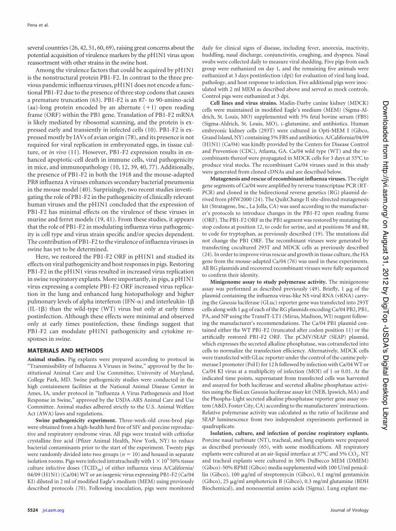
PB1-F2 enhances Ca/04 replication in porcine respiratoryexplants. It has been demonstrated that expression of full-lengthwild-type PB1-F2 by pH1N1 has no effects on viral replication inhuman A549 cells or MDCK cells (19, 47). However, cell lines donot exhibit the natural physiological conditions and cellular com-plexity present in the respiratory tract. To address this limitationand study the impact of PB1-F2 expression in a relevant biologicalsystem, we developed an ex vivo organ culture model of the pigrespiratory tract maintained at an air-liquid interface. These tissueexplants retained their cytoarchitecture (data not shown) andsupported productive replication of the recombinant Ca/04 influ-enza viruses (Fig. 2). Restoring PB1-F2 in Ca/04 improved virusreplication in explants of nasal turbinates (Fig. 2A), trachea (Fig.2B), and proximal lung (Fig. 2C). No differences were observed inexplants of the distal lung (Fig. 2D). This enhancing effect ofPB1-F2 in virus production was mainly seen at 24 hpi but not at 48or 72 hpi. These data indicate that PB1-F2 modulates moderatelyearly viral production in swine respiratory tissues infected ex vivo.
Restored PB1-F2 ORF increases virus replication in swinelungs. To evaluate whether the enhanced viral yields in porcinerespiratory explants displayed by Ca/04 KI would correlate withincreased viral replication in vivo, we inoculated groups of3-week-old pigs (n $ 10/group) with the Ca/04 WT or Ca/04 KIviruses. From each group, 5 pigs were euthanized at 1 and 3 dpiand viral titers in BALF were determined. Consistent with theexplant data, restoring the PB1-F2 ORF resulted in statisticallysignificant (P ( 0.05) increases in viral loads in the lungs at eithertime point (Fig. 3A). Increased Ca/04 KI replication in the lungswas further corroborated by the more pronounced pulmonaryexpression of influenza NP antigen by IHC analysis (Fig. 3C). Asexpected, mock-inoculated pigs had neither detectable virus norIHC influenza-positive cells in BALF or lung tissues (data notshown). Collectively, these results suggest that restoring thePB1-F2 ORF in the Ca/04 virus leads to increased viral replicationin vivo.
PB1-F2 aggravates microscopic pneumonia despite no dif-ferences in nasal virus shedding or macroscopic lesions in pigs.To determine if PB1-F2 affects the kinetics of viral shedding inpigs, nasal swabs collected from each pig from 1 to 3 dpi weretitrated in MDCK cells. Virus shedding in nasal secretions wasdetected only at 3 dpi, and there was no difference between theCa/04 WT and Ca/04 KI viruses (Fig. 4A). Infected pigs did notshow any overt clinical signs of disease regardless of the virus used.At necropsy, pigs in both the Ca/04 WT and Ca/04 KI groups hadcranioventral lung consolidation, with 5% to 10% of lung involve-ment at 3 dpi (Fig. 4B). There were no differences between the two
FIG 1 PB1-F2 upregulates early polymerase activity. (A) Minigenome assay.293T cells were transfected with plasmids encoding the minimal componentsrequired for viral transcription and replication (PB2, PB1, and PA polymerasesubunits, NP, and a vRNA influenza virus-driven luciferase reporter repliconexpressing GLuc) and with pCMV/SEAP, a plasmid to normalize transfectionefficiency, as previously described (49). At the indicated hours posttransfec-tion (hpt), the supernatant was harvested and assayed for both luciferase andphosphatase activities. (B and C) Kinetics of polymerase activity after virusinfection. MDCK cells were transfected with 1 &g of GLuc reporter under thecontrol of the canine Pol I reporter, followed by infection with Ca/04 (WT orKI) virus at an MOI of 0.01 (B) or 1 (C). Gluc activity was determined asdescribed in Materials and Methods at the indicated hours postinfection (hpi).Data are expressed as polymerase activities (mean ) standard error [SE])determined from two independent experiments performed in quadruplicate.A two-tailed Student t test was used to determine significant differences be-tween two treatment means for each data point. An asterisk and dashed brack-ets indicate statistically significant differences (P ( 0.05).
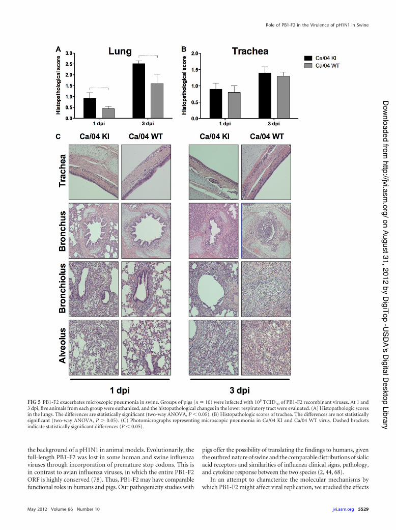
virus groups, indicating that PB1-F2 has no effect on the macro-scopic lung pathology caused by Ca/04 in swine. However, differ-ences in lung inflammation were detected at the microscopic level.Quantitative histopathological analyses revealed that pulmonaryinflammation was enhanced in pigs in the Ca/04 KI group com-pared to the Ca/04 WT group (Fig. 5A and C). Unlike the mock-inoculated animals, which had normal histological features, thelesions in virus-infected pigs were typical of influenza virus pneu-monia in pigs and were characterized by multifocal to widespreadnecrotizing bronchitis and bronchiolitis, light peribronchiolarlymphocytic cuffing, and multifocal interstitial pneumonia. Bothviruses induced similar levels of tracheitis and cilium loss in thetracheal epithelium (Fig. 5B).
PB1-F2 exacerbates the pulmonary levels of IFN-" and IL-1!in swine. Cytokine production after influenza virus infection hasbeen implicated in the pathogenesis of influenza in mammals,including pigs (66). To further characterize the effect of PB1-F2 inthe pathological manifestations of pH1N in swine, we measuredthe protein levels of 10 porcine cytokines/chemokines (IL-1#,IL-2, IL-4, IL-6, IL-8, IL-10, IL-12p70, TNF-!, IFN-!, and IFN-')in BALF collected at 1- and 3-dpi necropsies. Pulmonary levels ofIFN-! and IL-1# were significantly higher at 1 dpi in pigs infectedwith Ca/04 KI than in those infected with Ca/04 WT (Fig. 6A andB). The production levels of other cytokines/chemokines (IL-6,IL-8, and IFN-'), however, were similar between the two groupsinfected with the PB1-F2 recombinant viruses (Fig. 6C to E). IL-2,IL-4, IL-10, IL-12p70, and TNF-! did not reach detectable levelsin this experimental infection (data not shown). Collectively,these results indicate that PB1-F2 induces higher expression levelsof IFN-! and IL-1# early during pH1N1 infection in pigs and thatthis heightened production of proinflammatory mediators mayhave contributed to the more severe microscopic pneumonia de-veloped by Ca/04 KI-infected animals.
DISCUSSIONOutbreaks of influenza A viruses (IAVs) are among the mostsignificant causes of respiratory disease in both humans andanimals. Occasionally, pandemic influenza viruses emergewhen a virus acquires a new HA molecule to which the majorityof the population is immunologically naïve, resulting in devas-tating levels of morbidity and mortality (64). Virulence of in-fluenza viruses is considered a polygenic trait. Molecular stud-ies in different animal models have determined that the geneconstellation of a particular influenza virus strain within a spe-cific host plays a pivotal role in disease outcome (3, 9, 22).Several proteins in the viral genome have clearly identifiedroles in virulence: the surface glycoproteins hemagglutinin(HA) and neuraminidase (NA), which are responsible for viralbinding, entry, and release (48); the polymerase protein com-plex formed by PB2, PB1, and PA proteins, which drives ge-nome transcription and translation (18, 74); and the nonstruc-tural protein NS1, which disarms the interferon-based defensesystem of the host and also modulates other important aspectsof the virus replication cycle (21).
A recently added member to the family of IAV proteins with arole in virulence is PB1-F2. Unlike other influenza viruses thathave become adapted to humans, the 2009 pH1N1 virus lacksmost of the known hallmarks of influenza virus virulence, includ-ing PB1-F2 (63). However, with the continued circulation ofpH1N1 in humans and animals, especially in swine, there are op-portunities for acquisition of PB1-F2 through mutations or ge-netic reassortment with cocirculating animal influenza viruses.We examined the functional significance of PB1-F2 acquisition bypH1N1 polymerase activity in vitro in mammalian cells (human,canine, and porcine tissue explants) as well as viral replication,pathogenicity, and host response to infection in its natural host,
FIG 2 Replication of PB1-F2 recombinant viruses in porcine respiratory explants. Explants from nasal turbinates (A), trachea (B), and proximal (C) and distal(D) lung were infected with 106 TCID50 of either Ca/04 WT or KI. The bathing medium was collected at the indicated time points and titrated by the TCID50
method in MDCK cells. Values shown are the mean and range of virus titers (log10 TCID50/ml) obtained from triplicate explant preparations. A two-tailedStudent t test was used to determine significant differences between two treatment means for each data point. Dashed brackets indicate statistically significantdifferences (P ( 0.05).
the pig. The data demonstrated that the full-length PB1-F2 in apH1N1 virus impacts polymerase activity, viral replication effi-ciency both ex vivo and in vivo, lung inflammation, and local cy-tokine responses to infection. However, these differences are only
minor, which suggests that acquisition of the PB1-F2 ORF maynot produce significant phenotypic changes in the virulence ofpH1N1 viruses for pigs. These observations are consistent with thereport by Hai et al. (19), which shows minimal effects of PB1-F2 in
FIG 3 Replication and immunohistochemical analysis of PB1-F2 recombinant viruses in swine lungs. Groups of pigs (n $ 10) were infected with 105 TCID50 of PB1-F2recombinant viruses, and five animals from each group were euthanized either at 1 or 3 dpi. The lungs were collected and processed for virus titration and immunohis-tochemical analysis. (A) Pulmonary replication of PB1-F2 isogenic viruses in pigs. Values are means and ranges of virus titers (log10 TCID50/ml) in bronchoalveolarlavage fluid (BALF) collected at the indicated time points. Two-way ANOVA was used to determine significant differences between two treatment groups. Dashedbrackets indicate statistically significant differences (P ( 0.05). (B) Immunohistochemical staining against influenza A virus nucleoprotein (NP) in the lungs of infectedpigs. Values given are the mean ) the standard error of the mean IHC scores based on the percentage of influenza virus-positive cells in the airway and lung interstitium.(C) Representative IHC slides depicting viral antigen primarily in airway epithelium at 1 and 3 dpi. Scattered labeling in the interstitium at 3 dpi is present.
FIG 4 Viral shedding and macroscopic pneumonia are unaltered by PB1-F2 expression in swine. Groups of pigs (n $ 10) were infected with 105 TCID50 ofPB1-F2 recombinant viruses, and nasal swabs were collected from 1 to 3 dpi for measuring virus shedding. At 1 and 3 dpi, five animals from each group wereeuthanized, and the lungs were scored for gross pneumonia. (A) Viral shedding in nasal secretions of pigs. Viral titers in nasal swabs were determined by TCID50
on MDCK cells. Values are shown as the mean and range and expressed as log10TCID50/ml. (B) Percentage of macroscopic lung lesions. Values are shown as themean ) the standard error of the mean. The differences are not statistically significant (two-way ANOVA).
the background of a pH1N1 in animal models. Evolutionarily, thefull-length PB1-F2 was lost in some human and swine influenzaviruses through incorporation of premature stop codons. This isin contrast to avian influenza viruses, in which the entire PB1-F2ORF is highly conserved (78). Thus, PB1-F2 may have comparablefunctional roles in humans and pigs. Our pathogenicity studies with
pigs offer the possibility of translating the findings to humans, giventhe outbred nature of swine and the comparable distributions of sialicacid receptors and similarities of influenza clinical signs, pathology,and cytokine response between the two species (2, 44, 68).
In an attempt to characterize the molecular mechanisms bywhich PB1-F2 might affect viral replication, we studied the effects
FIG 5 PB1-F2 exacerbates microscopic pneumonia in swine. Groups of pigs (n $ 10) were infected with 105 TCID50 of PB1-F2 recombinant viruses. At 1 and3 dpi, five animals from each group were euthanized, and the histopathological changes in the lower respiratory tract were evaluated. (A) Histopathologic scoresin the lungs. The differences are statistically significant (two-way ANOVA, P ( 0.05). (B) Histopathologic scores of trachea. The differences are not statisticallysignificant (two-way ANOVA, P * 0.05). (C) Photomicrographs representing microscopic pneumonia in Ca/04 KI and Ca/04 WT virus. Dashed bracketsindicate statistically significant differences (P ( 0.05).
of Ca/04 PB1-F2 on polymerase activity using an IAV untrans-lated region (UTR)-driven GLuc reporter gene. We found thatPB1-F2 upregulates early (12-hpt) viral polymerase activity butdoes not do so at later time points (24 to 72 hpt). These resultscontrast with those reported by Chen et al., which indicated thatCa/04 PB1-F2 enhanced polymerase activity at 48 hpt (8). Thediscrepancy between this and the previous study with regard topolymerase function may be due to differences in the reportergene used as the polymerase activity readout and the length of therestored PB1-F2 ORF. Chen et al. used a chloramphenicol acetyl-transferase (CAT) reporter system and mutated only 2 stopcodons in the PB1-F2 ORF, generating an 87-aa product instead ofa 90-aa peptide product (19). It must be noted, however, thatpolymerase activity in our assay was evaluated in human and ca-nine cells; however, it appears to correlate well with the replicationof these viruses in ex vivo swine respiratory explants and in vivo inswine lungs.
As the presence of PB1-F2 augmented the replication of someviral strains in tissue culture (41, 56), we studied the growth ki-netics of PB1-F2 Ca/04 recombinant viruses in porcine respiratoryexplants. Respiratory explants have proved to be a valuable systemto study influenza-host interaction in a scalable, well-defined, andcontrolled experimental setting (7, 65). Our results are in har-mony with the hypothesis that PB1-F2 modulates viral replica-tion, since higher viral yields were observed in explants derivedfrom both the upper and lower respiratory tracts (Fig. 2). Inter-estingly, this effect of PB1-F2 in virus production (and polymeraseactivity) (Fig. 1) was mainly observed at early time points, consis-tent with the early and transient expression of PB1-F2 in thecourse of infection (10, 33, 40). Furthermore, the increased ex vivoviral replication paralleled the lung viral load in pigs infected withCa/04 KI (Fig. 4). In contrast, viral shedding in nasal secretions
was not affected by PB1-F2. This may be explained by the route ofinoculation used in our in vivo studies. Pigs were infected intra-tracheally, a route that deposits the virus in the lower respiratorytract and more consistently reproduces lung pathology (36).Shedding was detected only at 3 dpi, which could be a time pointthat makes it difficult to distinguish modest differences in shed-ding. However, we cannot rule out the possibility that PB1-F2could affect viral shedding beyond 3 dpi and/or in intranasallyinfected animals. These studies are beyond the scope of this workand warrant further investigation.
Even though PB1-F2 had no effect on the gross lung pathologyor clinical disease, Ca/04 KI-infected pigs developed more severemicroscopic pneumonia as early as 1 dpi. This finding fit withprevious reports that demonstrated that PB1-F2 increases the se-verity of pulmonary lesions in mouse models, either in the contextof viral infection (40) or simply by administration of peptides(39). Moreover, our results are in agreement with ferret studies byHai et al. that reported no changes in clinical outcome with aPB1-F2-expressing Ca/04 compared to WT controls, although theauthors did not assess lung pathology (19). Thus, it could be pos-tulated that during primary viral infection the microscopic pul-monary changes induced by PB1-F2 are not large enough to bemanifested clinically or detected macroscopically. However, theeffects of PB1-F2 enhancement of virulence might be seen underfield conditions, where immunosuppressive conditions and sec-ondary bacterial infections can aggravate the outcome of influ-enza virus infections. In contrast to previous reports that showedsignificant clinical signs in pigs infected with various strains ofpH1N1, infection of pigs with the Ca/04 WT and Ca/04 KI virusesin our studies resulted in mild clinical signs (32, 35). There areseveral factors that could account for this difference. In previousstudies, 10-fold more virus was used to inoculate pigs, and the
FIG 6 PB1-F2 exacerbates the pulmonary levels of IFN-! and IL-1# in swine. Groups of pigs (n $ 10) were infected intratracheally with 105 TCID50 of PB1-F2recombinant viruses. On days 1 and 3 postinfection, animals from each group were euthanized, and the cytokine/chemokine levels in BALF were determined byELISA. Data are shown as the mean ) the standard error of the mean for five animals in each challenge group. Two-way ANOVA was used to determinesignificant differences between groups. When the interaction between factors (time and virus) was significant, a two-tailed Student t test was used, consideringtime and virus, to determine significant differences between two treatment means for each data point. Dashed brackets indicate statistically significant differences(P ( 0.05).
clinical manifestation of the disease became obvious after 3 dpi.Our animal studies were terminated at 3 dpi, and therefore we didnot allow the clinical signs to develop. In addition, previous stud-ies used virus isolates that were not previously cloned. We havepreviously shown that that pH1N1 viruses may have contained amixed population of similar strains with different effects on viru-lence (76). In order to distinguish differences attributed to a singlefactor, we generated reverse genetics clones of Ca/04, and the onlydifference between Ca/04 WT and the Ca/04 KI is the presence ofa full-length PB1-F2 in the latter.
The proinflammatory cytokine response is critical for recruit-ing effector cells to the site of an infection. However, elevated orprolonged cytokine production can also contribute to the patho-logical changes observed during infection. To better understandthe mechanism of lesion severity in Ca/04 KI-infected pigs, wemeasured the pulmonary concentration of 10 cytokines/chemo-kines during the acute stage of infection. Increased levels of IFN-!and IL-1# proteins were found in the lungs of pigs infected withCa/04 KI compared to what was seen for the Ca/04 WT (Fig. 6).This exacerbation of the host innate response was observed at 24hpi but not at 72 hpi, adding to the notion that changes mediatedby PB1-F2 occur shortly after infection. To our knowledge, this isthe first report showing that PB1-F2 enhances type I IFN in vivo.Our results are consistent with a recent study which demonstratedthat the WSN PB1-F2 exacerbates type I IFN expression in humanrespiratory epithelial cells (33). In both pigs and humans, there isa strong positive correlation between local IFN-! levels and viraltiters in nasal secretions and clinical disease (23, 68). In addition,increased levels of IL-1# have also been linked to neutrophil infil-tration in the lungs and to the severity of pneumonia in pigs (34,67). The exacerbated inflammatory response in pigs could be adirect effect of PB1-F2 early during infection. Thus, it is possiblethat enhanced expression levels of IFN-! and IL-1# are not thecause of respiratory disease but markers of the extent of viral rep-lication. The modulation of the porcine immune system byPB1-F2 and its interaction with host factors warrant further inves-tigation and should shed light into the strain- and host-dependentmolecular mechanisms of PB1-F2. It remains to be determinedwhether the phenotypic differences observed with these two iso-genic pH1N1 viruses are common features of other swine influ-enza viruses.
In summary, we have shown that PB1-F2 modulates severalaspects of pH1N1 influenza virus interaction with swine, both exvivo and in vivo. The present study fills a critical gap regarding thevirulence determinants for influenza virus in swine. Our studiesprovide important insights into the impact that genetic changesmay have on the virulence of pH1N1 for mammalian species,including humans.
ACKNOWLEDGMENTSWe are indebted to Annabelle Pascua Crusan for her help with explantstudies. We thank Michelle Harland, Hillary Horst, Gwen Nordholm,Brian Pottebaum, Jason Crabtree, and Jason Huegel for technical assis-tance and help with the swine study.
Mention of trade names or commercial products in this article is solelyfor the purpose of providing specific information and does not implyrecommendation or endorsement by the U.S. Department of Agriculture.The opinions in this report are ours and do not necessarily represent theviews of the granting agencies. The granting agencies had no role in studydesign, data collection and analysis, decision to publish, or preparation ofthe manuscript.
This research was made possible through funding by a CDC-HHSgrant (1U01CI000355), an NIAID-NIH grant, (R01AI052155), aCSREES-USDA grant (2005-05523), and an NIAID-NIH contract(HHSN266200700010C) and by the USDA-ARS.
REFERENCES1. Al Hajjar S, McIntosh K. 2010. The first influenza pandemic of the 21st
century. Ann. Saudi Med. 30:1–10.2. Barnard DL. 2009. Animal models for the study of influenza pathogenesis
and therapy. Antiviral Res. 82:A110 –A122.3. Basler CF, Aguilar PV. 2008. Progress in identifying virulence determi-
nants of the 1918 H1N1 and the Southeast Asian H5N1 influenza A vi-ruses. Antiviral Res. 79:166 –178.
5. Brockmeier SL, et al. 2009. Adenovirus-mediated expression of interferon-alpha delays viral replication and reduces disease signs in swine challengedwith porcine reproductive and respiratory syndrome virus. Viral Immu-nol. 22:173–180.
6. Brookes SM, et al. 2009. Influenza A (H1N1) infection in pigs. Vet. Rec.164:760 –761.
7. Chan MC, et al. 2010. Tropism and innate host responses of the 2009pandemic H1N1 influenza virus in ex vivo and in vitro cultures of humanconjunctiva and respiratory tract. Am. J. Pathol. 176:1828 –1840.
8. Chen CJ, et al. 2010. Differential localization and function of PB1-F2derived from different strains of influenza A virus. J. Virol. 84:10051–10062.
9. Chen H, et al. 2007. Polygenic virulence factors involved in pathogenesisof 1997 Hong Kong H5N1 influenza viruses in mice. Virus Res. 128:159 –163.
10. Chen W, et al. 2001. A novel influenza A virus mitochondrial protein thatinduces cell death. Nat. Med. 7:1306 –1312.
11. Conenello GM, Palese P. 2007. Influenza A virus PB1-F2: a small proteinwith a big punch. Cell Host Microbe 2:207–209.
12. Conenello GM, Zamarin D, Perrone LA, Tumpey T, Palese P. 2007. Asingle mutation in the PB1-F2 of H5N1 (HK/97) and 1918 influenza Aviruses contributes to increased virulence. PLoS Pathog. 3:1414 –1421.
13. Dawood FS, et al. 2009. Emergence of a novel swine-origin influenza A(H1N1) virus in humans. N. Engl. J. Med. 360:2605–2615.
14. Dundon WG, De Benedictis P, Viale E, Capua I. 2010. Serologic evi-dence of pandemic (H1N1) 2009 infection in dogs, Italy. Emerg. Infect.Dis. 16:2019 –2021.
15. Garten RJ, et al. 2009. Antigenic and genetic characteristics of swine-origin 2009 A(H1N1) influenza viruses circulating in humans. Science325:197–201.
16. Gauger PC, et al. 2011. Enhanced pneumonia and disease in pigs vacci-nated with an inactivated human-like (delta-cluster) H1N2 vaccine andchallenged with pandemic 2009 H1N1 influenza virus. Vaccine 29:2712–2719.
17. Gibbs AJ, Armstrong JS, Downie JC. 2009. From where did the 2009‘swine-origin’ influenza A virus (H1N1) emerge? Virol. J. 6:207.
18. Graef KM, et al. 2010. The PB2 subunit of the influenza virus RNApolymerase affects virulence by interacting with the mitochondrial antivi-ral signaling protein and inhibiting expression of beta interferon. J. Virol.84:8433– 8445.
19. Hai R, et al. 2010. PB1-F2 expression by the 2009 pandemic H1N1 influ-enza virus has minimal impact on virulence in animal models. J. Virol.84:4442– 4450.
20. Halbur PG, et al. 1995. Comparison of the pathogenicity of two USporcine reproductive and respiratory syndrome virus isolates with that ofthe Lelystad virus. Vet. Pathol. 32:648 – 660.
21. Hale BG, Randall RE, Ortin J, Jackson D. 2008. The multifunctional NS1protein of influenza A viruses. J. Gen. Virol. 89:2359 –2376.
22. Hatta M, Gao P, Halfmann P, Kawaoka Y. 2001. Molecular basis for highvirulence of Hong Kong H5N1 influenza A viruses. Science 293:1840 –1842.
23. Hayden FG, et al. 1998. Local and systemic cytokine responses duringexperimental human influenza A virus infection. Relation to symptomformation and host defense. J. Clin. Invest. 101:643– 649.
24. Hoffmann E, Neumann G, Kawaoka Y, Hobom G, Webster RG. 2000.A DNA transfection system for generation of influenza A virus from eightplasmids. Proc. Natl. Acad. Sci. U. S. A. 97:6108 – 6113.
25. Hofshagen M, et al. 2009. Pandemic influenza A(H1N1)v: human to pigtransmission in Norway? Euro Surveill. 14(45). pii:19406.
26. Howard WA, et al. 2011. Reassortant pandemic (H1N1) 2009 virus inpigs, United Kingdom. Emerg. Infect. Dis. 17:1049 –1052.
27. Howden KJ, et al. 2009. An investigation into human pandemic influenzavirus (H1N1) 2009 on an Alberta swine farm. Can. Vet. J. 50:1153–1161.
28. Ito T, et al. 1998. Molecular basis for the generation in pigs of influenza Aviruses with pandemic potential. J. Virol. 72:7367–7373.
29. Itoh Y, et al. 2009. In vitro and in vivo characterization of new swine-origin H1N1 influenza viruses. Nature 460:1021–1025.
30. Kida H, et al. 1994. Potential for transmission of avian influenza virusesto pigs. J. Gen. Virol. 75:2183–2188.
31. Kimble B, Nieto GR, Perez DR. 2010. Characterization of influenza virussialic acid receptors in minor poultry species. Virol. J. 7:365.
32. Lange E, et al. 2009. Pathogenesis and transmission of the novel swine-origin influenza virus A/H1N1 after experimental infection of pigs. J. Gen.Virol. 90:2119 –2123.
33. Le Goffic R, et al. 2010. Influenza A virus protein PB1-F2 exacerbatesIFN-beta expression of human respiratory epithelial cells. J. Immunol.185:4812– 4823.
34. Loving CL, et al. 2010. Influenza virus coinfection with Bordetella bron-chiseptica enhances bacterial colonization and host responses exacerbat-ing pulmonary lesions. Microb. Pathog. 49:237–245.
35. Ma W, et al. 2011. 2009 Pandemic H1N1 influenza virus causes diseaseand upregulation of genes related to inflammatory and immune re-sponses, cell death, and lipid metabolism in pigs. J. Virol. 85:11626 –11637.
36. Maes L, Haesebrouck F, Pensaert M. 1984. Experimental reproductionof clinical disease by intratracheal inoculation of fattening pigs with swineinfluenza virus isolates. Proc. Int. Congr. Pig Vet. Soc. 8:60.
37. Mathieu C, et al. 2010. Pandemic (H1N1) 2009 in breeding turkeys,Valparaiso, Chile. Emerg. Infect. Dis. 16:709 –711.
38. Mazur I, et al. 2008. The proapoptotic influenza A virus protein PB1-F2regulates viral polymerase activity by interaction with the PB1 protein.Cell. Microbiol. 10:1140 –1152.
39. McAuley JL, et al. 2010. PB1-F2 proteins from H5N1 and 20th centurypandemic influenza viruses cause immunopathology. PLoS Pathog.6:e1001014.
40. McAuley JL, et al. 2007. Expression of the 1918 influenza A virus PB1-F2enhances the pathogenesis of viral and secondary bacterial pneumonia.Cell Host Microbe 2:240 –249.
41. McAuley JL, Zhang K, McCullers JA. 2010. The effects of influenza Avirus PB1-F2 protein on polymerase activity are strain specific and do notimpact pathogenesis. J. Virol. 84:558 –564.
42. Moreno A, et al. 2011. Novel H1N2 swine influenza reassortant strain inpigs derived from the pandemic H1N1/2009 virus. Vet. Microbiol. 149:472– 477.
43. Myers KP, Olsen CW, Gray GC. 2007. Cases of swine influenza inhumans: a review of the literature. Clin. Infect. Dis. 44:1084 –1088.
44. Nelli RK, et al. 2010. Comparative distribution of human and avian typesialic acid influenza receptors in the pig. BMC Vet. Res. 6:4.
45. Newman AP, et al. 2008. Human case of swine influenza A (H1N1) triplereassortant virus infection, Wisconsin. Emerg. Infect. Dis. 14:1470 –1472.
46. Nofs S, et al. 2009. Influenza virus A (H1N1) in giant anteaters (Myrme-cophaga tridactyla). Emerg. Infect. Dis. 15:1081–1083.
47. Ozawa M, et al. 2011. Impact of amino acid mutations in PB2, PB1-F2,and NS1 on the replication and pathogenicity of pandemic (H1N1) 2009influenza viruses. J. Virol. 85:4596 – 4601.
48. Pappas C, et al. 2008. Single gene reassortants identify a critical role forPB1, HA, and NA in the high virulence of the 1918 pandemic influenzavirus. Proc. Natl. Acad. Sci. U. S. A. 105:3064 –3069.
49. Pena L, et al. 2011. Modifications in the polymerase genes of a swine-liketriple reassortant influenza virus to generate live attenuated vaccinesagainst 2009 pandemic H1N1 viruses. J. Virol. 85:456 – 469.
50. Pereda A, et al. 2010. Pandemic (H1N1) 2009 outbreak on pig farm,Argentina. Emerg. Infect. Dis. 16:304 –307.
51. Pereda A, et al. 2011. Evidence of reassortment of pandemic H1N1 in-fluenza virus in swine in Argentina: are we facing the expansion of poten-tial epicenters of influenza emergence? Influenza Other Respi. Viruses5:409 – 412.
52. Reed LJ, Muench H. 1938. A simple method for estimating 50 percentendpoints. Am. J. Hyg. 37:493.
53. Robb NC, Smith M, Vreede FT, Fodor E. 2009. NS2/NEP proteinregulates transcription and replication of the influenza virus RNA ge-nome. J. Gen. Virol. 90:1398 –1407.
54. Robinson JL, et al. 2007. Swine influenza (H3N2) infection in a child andpossible community transmission, Canada. Emerg. Infect. Dis. 13:1865–1870.
55. Shinya K, et al. 2006. Avian flu: influenza virus receptors in the humanairway. Nature 440:435– 436.
56. Smith AM, et al. 2011. Effect of 1918 PB1-F2 expression on influenza Avirus infection kinetics. PLoS Comput. Biol. 7:e1001081.
57. Smith GJ, et al. 2009. Origins and evolutionary genomics of the 2009swine-origin H1N1 influenza A epidemic. Nature 459:1122–1125.
58. Song MS, et al. 2010. Evidence of human-to-swine transmission of thepandemic (H1N1) 2009 influenza virus in South Korea. J. Clin. Microbiol.48:3204 –3211.
59. Sponseller BA, et al. 2010. Influenza A pandemic (H1N1) 2009 virusinfection in domestic cat. Emerg. Infect. Dis. 16:534 –537.
60. Starick E, et al. 2011. Reassorted pandemic (H1N1) 2009 influenza Avirus discovered from pigs in Germany. J. Gen. Virol. 92:1184 –1188.
61. Swenson SL, et al. 2010. Natural cases of 2009 pandemic H1N1 influenzaA virus in pet ferrets. J. Vet. Diagn. Invest. 22:784 –788.
63. Trifonov V, Rabadan R. 2009. The contribution of the PB1-F2 protein tothe fitness of influenza A viruses and its recent evolution in the 2009influenza A (H1N1) pandemic virus. PLoS Curr. 1:RRN1006.
64. Tscherne DM, Garcia-Sastre A. 2011. Virulence determinants of pan-demic influenza viruses. J. Clin. Invest. 121:6 –13.
65. Van Poucke SG, Nicholls JM, Nauwynck HJ, Van Reeth K. 2010.Replication of avian, human and swine influenza viruses in porcine respi-ratory explants and association with sialic acid distribution. Virol. J. 7:38.
66. Van Reeth K. 2000. Cytokines in the pathogenesis of influenza. Vet.Microbiol. 74:109 –116.
67. Van Reeth K, Labarque G, Nauwynck H, Pensaert M. 1999. Differentialproduction of proinflammatory cytokines in the pig lung during differentrespiratory virus infections: correlations with pathogenicity. Res. Vet. Sci.67:47–52.
68. Van Reeth K, Van Gucht S, Pensaert M. 2002. Correlations between lungproinflammatory cytokine levels, virus replication, and disease after swineinfluenza virus challenge of vaccination-immune pigs. Viral Immunol.15:583–594.
69. Vijaykrishna D, et al. 2010. Reassortment of pandemic H1N1/2009 in-fluenza A virus in swine. Science 328:1529.
70. Vincent AL, et al. 2010. Experimental inoculation of pigs with pandemicH1N1 2009 virus and HI cross-reactivity with contemporary swine influ-enza virus antisera. Influenza Other Respi. Viruses 4:53– 60.
71. Vincent AL, et al. 2009. Characterization of an influenza A virus isolatedfrom pigs during an outbreak of respiratory disease in swine and peopleduring a county fair in the United States. Vet. Microbiol. 137:51–59.
72. Vincent LL, Janke BH, Paul PS, Halbur PG. 1997. A monoclonal-antibody-based immunohistochemical method for the detection of swineinfluenza virus in formalin-fixed, paraffin-embedded tissues. J. Vet. Di-agn. Invest. 9:191–195.
73. Wan H, Perez DR. 2006. Quail carry sialic acid receptors compatible withbinding of avian and human influenza viruses. Virology 346:278 –286.
74. Watanabe T, et al. 2009. Viral RNA polymerase complex promotes op-timal growth of 1918 virus in the lower respiratory tract of ferrets. Proc.Natl. Acad. Sci. U. S. A. 106:588 –592.
75. Weingartl HM, et al. 2009. Experimental infection of pigs with the hu-man 1918 pandemic influenza virus. J. Virol. 83:4287– 4296.
76. Ye J, et al. 2010. Variations in the hemagglutinin of the 2009 H1N1pandemic virus: potential for strains with altered virulence phenotype?PLoS Pathog. 6:e1001145.
77. Zamarin D, Ortigoza MB, Palese P. 2006. Influenza A virus PB1-F2protein contributes to viral pathogenesis in mice. J. Virol. 80:7976 –7983.
78. Zell R, et al. 2007. Prevalence of PB1-F2 of influenza A viruses. J. Gen.Virol. 88:536 –546.