PSYCHIATRY REVIEW ARTICLE published: 02 April 2014 doi: 10.3389/fpsyt.2014.00033 Restoring cognitive functions using non-invasive brain stimulation techniques in patients with cerebellar disorders Paul A. Pope* and R. Chris Miall School of Psychology, University of Birmingham, Birmingham, UK Edited by: Joseph M. Galea, University College London, UK Reviewed by: Masashi Hamada, University College London, UK Nick J. Davis, Swansea University, UK *Correspondence: Paul A. Pope, School of Psychology, University of Birmingham, Edgbaston, Birmingham B15 2TT, UK e-mail: [email protected]Numerous studies have highlighted the possibility of modulating the excitability of cerebro– cerebellar circuits bi-directionally using transcranial electrical brain stimulation, in a manner akin to that observed using magnetic stimulation protocols. It has been proposed that cerebellar stimulation activates Purkinje cells in the cerebellar cortex, leading to inhibition of the dentate nucleus, which exerts a tonic facilitatory drive onto motor and cognitive regions of cortex through a synaptic relay in the ventral–lateral thalamus. Some cerebellar deficits present with cognitive impairments if damage to non-motor regions of the cerebel- lum disrupts the coupling with cerebral cortical areas for thinking and reasoning. Indeed, white matter changes in the dentato–rubral tract correlate with cognitive assessments in patients with Friedreich ataxia, suggesting that this pathway is one component of the anatomical substrate supporting a cerebellar contribution to cognition. An understanding of the physiology of the cerebro–cerebellar pathway previously helped us to constrain our interpretation of results from two recent studies in which we showed cognitive enhance- ments in healthy participants during tests of arithmetic after electrical stimulation of the cerebellum, but only when task demands were high. Others studies have also shown how excitation of the prefrontal cortex can enhance performance in a variety of working memory tasks. Thus, future efforts might be guided toward neuro-enhancement in certain patient populations, using what is commonly termed “non-invasive brain stimulation” as a cognitive rehabilitation tool to modulate cerebro–cerebellar circuits, or for stimulation over the cerebral cortex to compensate for decreased cerebellar drive to this region. This arti- cle will address these possibilities with a review of the relevant literature covering ataxias and cerebellar cognitive affective disorders, which are characterized by thalamo–cortical disturbances. Keywords: tDCS, TMS, cerebellar cognitive affective syndrome, cognitive rehabilitation, spinocerebellar degeneration INTRODUCTION Clinicians have been directly exciting the cerebellar cortex with implanted electrodes in epileptic patients and in those with schiz- ophrenia and depression since the 1970s with good therapeutic results (1), demonstrating the use of constant electrical stim- ulation for the treatment of behavioral disorders and epilepsy. Today, transcranial brain stimulation techniques [often referred to as non-invasive brain stimulation (NIBS)], such as repetitive transcranial magnetic stimulation (rTMS) and transcranial direct current stimulation (tDCS), are realized to have the capacity to systematically modify behavior by inducing lasting changes in underlying brain functions, and are useful approaches to studying brain–behavior relationships in healthy participants. They have also been used to study mechanisms of cortical plasticity, and both techniques have been implicated as therapeutic tools for the treatment of motor and cognitive deficits in patients after stroke, and in cerebellar disease (2, 3). In recent years, cerebellar-tDCS has grown in popularity in various laboratories and clinics, partly because the lateral cerebellar hemispheres, which are thought to be involved in cognition, are most accessible to transcranial electri- cal stimulation, are sensitive to the effects of polarizing currents, and because the procedure is relatively inexpensive and easy to perform. MECHANISMS OF ACTION AND EFFECTS OF tDCS The mechanisms of action and effects of tDCS on the human cerebellum are inferred from animal studies, or from indirect effects on motor cortex, and from modeling data. In humans, the procedure typically involves delivering 1–2 mA of DC stimula- tion through a pair of saline-soaked electrodes: one stimulation electrode on scalp overlying the cerebellum, and the other refer- ence electrode on the ipsilateral head or shoulder. Intracerebral current flow between the two electrodes has relatively little func- tional spread to neighboring regions [e.g., visual cortex (4)] and is thought to excite or depress Purkinje cells in the cerebellar cor- tex, producing both neurophysiological and behavioral changes. The effects are polarity-specific as evidenced by the consequences of cerebellar stimulation on motor cortex excitability (5). Anodal stimulation has an excitatory effect and increases the output of Purkinje cells; increasing inhibition of the facilitatory pathway from the cerebellar nuclei to cerebral cortex. Cathodal stimulation has an opposite effect, i.e., dis-inhibition of the cerebral cortex by www.frontiersin.org April 2014 |Volume 5 | Article 33 | 1
Transcript
PSYCHIATRYREVIEW ARTICLEpublished: 02 April 2014
doi: 10.3389/fpsyt.2014.00033
Restoring cognitive functions using non-invasive brainstimulation techniques in patients with cerebellar disordersPaul A. Pope* and R. Chris Miall
School of Psychology, University of Birmingham, Birmingham, UK
Edited by:Joseph M. Galea, University CollegeLondon, UK
Reviewed by:Masashi Hamada, University CollegeLondon, UKNick J. Davis, Swansea University, UK
*Correspondence:Paul A. Pope, School of Psychology,University of Birmingham, Edgbaston,Birmingham B15 2TT, UKe-mail: [email protected]
Numerous studies have highlighted the possibility of modulating the excitability of cerebro–cerebellar circuits bi-directionally using transcranial electrical brain stimulation, in a mannerakin to that observed using magnetic stimulation protocols. It has been proposed thatcerebellar stimulation activates Purkinje cells in the cerebellar cortex, leading to inhibitionof the dentate nucleus, which exerts a tonic facilitatory drive onto motor and cognitiveregions of cortex through a synaptic relay in the ventral–lateral thalamus. Some cerebellardeficits present with cognitive impairments if damage to non-motor regions of the cerebel-lum disrupts the coupling with cerebral cortical areas for thinking and reasoning. Indeed,white matter changes in the dentato–rubral tract correlate with cognitive assessmentsin patients with Friedreich ataxia, suggesting that this pathway is one component of theanatomical substrate supporting a cerebellar contribution to cognition. An understandingof the physiology of the cerebro–cerebellar pathway previously helped us to constrain ourinterpretation of results from two recent studies in which we showed cognitive enhance-ments in healthy participants during tests of arithmetic after electrical stimulation of thecerebellum, but only when task demands were high. Others studies have also shownhow excitation of the prefrontal cortex can enhance performance in a variety of workingmemory tasks.Thus, future efforts might be guided toward neuro-enhancement in certainpatient populations, using what is commonly termed “non-invasive brain stimulation” as acognitive rehabilitation tool to modulate cerebro–cerebellar circuits, or for stimulation overthe cerebral cortex to compensate for decreased cerebellar drive to this region. This arti-cle will address these possibilities with a review of the relevant literature covering ataxiasand cerebellar cognitive affective disorders, which are characterized by thalamo–corticaldisturbances.
INTRODUCTIONClinicians have been directly exciting the cerebellar cortex withimplanted electrodes in epileptic patients and in those with schiz-ophrenia and depression since the 1970s with good therapeuticresults (1), demonstrating the use of constant electrical stim-ulation for the treatment of behavioral disorders and epilepsy.Today, transcranial brain stimulation techniques [often referredto as non-invasive brain stimulation (NIBS)], such as repetitivetranscranial magnetic stimulation (rTMS) and transcranial directcurrent stimulation (tDCS), are realized to have the capacity tosystematically modify behavior by inducing lasting changes inunderlying brain functions, and are useful approaches to studyingbrain–behavior relationships in healthy participants. They havealso been used to study mechanisms of cortical plasticity, andboth techniques have been implicated as therapeutic tools for thetreatment of motor and cognitive deficits in patients after stroke,and in cerebellar disease (2, 3). In recent years, cerebellar-tDCShas grown in popularity in various laboratories and clinics, partlybecause the lateral cerebellar hemispheres, which are thought to beinvolved in cognition, are most accessible to transcranial electri-cal stimulation, are sensitive to the effects of polarizing currents,
and because the procedure is relatively inexpensive and easy toperform.
MECHANISMS OF ACTION AND EFFECTS OF tDCSThe mechanisms of action and effects of tDCS on the humancerebellum are inferred from animal studies, or from indirecteffects on motor cortex, and from modeling data. In humans, theprocedure typically involves delivering 1–2 mA of DC stimula-tion through a pair of saline-soaked electrodes: one stimulationelectrode on scalp overlying the cerebellum, and the other refer-ence electrode on the ipsilateral head or shoulder. Intracerebralcurrent flow between the two electrodes has relatively little func-tional spread to neighboring regions [e.g., visual cortex (4)] andis thought to excite or depress Purkinje cells in the cerebellar cor-tex, producing both neurophysiological and behavioral changes.The effects are polarity-specific as evidenced by the consequencesof cerebellar stimulation on motor cortex excitability (5). Anodalstimulation has an excitatory effect and increases the output ofPurkinje cells; increasing inhibition of the facilitatory pathwayfrom the cerebellar nuclei to cerebral cortex. Cathodal stimulationhas an opposite effect, i.e., dis-inhibition of the cerebral cortex by
Pope and Miall Cognitive rehabilitation using cerebellar-tDCS
reducing Purkinje cell inhibition of the cerebellar nuclei. How-ever, the after-effects of TMS (6) and tDCS (7) over motor cortexare highly variable between individuals, and not always polarity-specific, which highlights the need to better understand individualfactors that determine the efficacy of NIBS (e.g., neural excitabil-ity and/or cognitive capacity) and to develop improved protocolsfor delivering brain stimulation. Effects of stimulation are alsodifferent depending on whether behavior is tested during (on-lineeffects) or after (off-line effects) the stimulation period, which typ-ically last 15–20 min, suggesting that on-line effects may includechanges in ion concentration gradients and cell membrane poten-tials, while off-line effects might include longer lasting changes inneural activity due to altered intracellular processes (e.g., recep-tor plasticity). Polarity-specific effects on cognitive functions aremore difficult to detect and to interpret than the direct effectsof the cerebellum on motor areas due to cerebellar-brain inhibi-tion (CBI). Nonetheless, anatomical studies in primates reveal howPurkinje cells could exert a facilitatory drive onto both motor andcognitive circuits, via a synaptic relay in the ventral–lateral thal-amus (8). And, associative plasticity induced by sensory/motorstimuli paired at 25 ms – paired associative stimulation (PAS), canbe blocked by cerebellar-tDCS, demonstrating how the cerebel-lum can exert a remote influence over excitability in the cerebralcortex (9). Thus, changes in both motor and cognitive func-tions are physiologically plausible via electrical stimulation of thecerebello–thalamo–cortical pathway.
tDCS AFTER-EFFECTS AND THE CEREBELLUMPolarizing the brain with cortical scalp electrodes as treatment forremedying cognitive deficits in human participants is not new. Inthe 1960s, Lippold and colleagues demonstrated beneficial effectsin certain psychiatric disorders caused by long duration stimula-tion (up to 10 h) at small current strengths over the forehead (10,11). The authors were able to distinguish positive and negativepolarization effects on mood in the majority of cases. Scalp-positive effects included an increase in the patients’ involvementwith the environment (e.g., alertness and cheerfulness), and scalp-negative effects included environmental inhibition and withdrawal(e.g., quietness). Due to a recent revival in this method, there isnow a better understanding of tDCS-induced effects and evidencethat cerebellar-tDCS can modulate, and in some cases, enhancecognitive functions and behavioral performance in healthy partic-ipants [reviewed in Ref. (12, 13)]. For example, in 2005, Ferrucciand colleagues measured off-line tDCS effects during a modifiedversion of the Sternberg item recognition task (i.e., identifying thepresence or absence of a digit from a list of previously presentedvisual items after a memory maintenance period) in healthy partic-ipants (14). Fifteen minutes of cerebellar stimulation (irrespectiveof electrical polarity or activity of visual cortex) impaired the usualpractice-dependent proficiency increase associated with this task.Five years later, this result was reproduced by Boehringer et al.(15). While neither study found tDCS to enhance performance,the work by Boehringer and colleagues did demonstrate that tDCScould alter performance during visual item recognition as a func-tion of task difficulty or when cognitive load is set at a specificlevel. These studies show how tDCS can alter cerebellar cognitivefunctions, and hint toward situations where tDCS is most efficient.
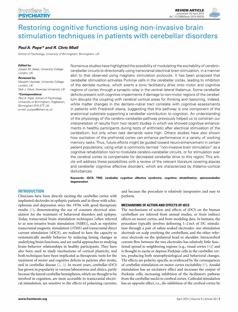
Task difficulty was a major feature of our recent study ofcerebellar functioning during tests of verbal working memory[WM; (16)], in which we applied tDCS over the right cerebellarhemisphere and showed neuro-enhancement during a demand-ing subtraction version of a mental arithmetic task [the pacedauditory serial subtraction task (PASST)] with high cognitiveload, but not during a simpler and less demanding addition ver-sion [the paced auditory serial addition task (PASAT)]. In short,cathodal stimulation improved task accuracy, response speed, andresponse variability [relative to anodal and sham stimulation (seeFigure 1)]. As both tasks share similar motor control (i.e., verbaloperations), but dissimilar cognitive load (i.e., mental effort), wespeculated that cathodal depression of the right cerebellar cor-tex might release additional cognitive resources required whendemands are high by dis-inhibition of the left prefrontal cortex towhich it projects via the cerebello–thalamo–cortical pathway (3).Supporting this view, and the emergent role for the cerebellumin cognition and emotion (17, 18), is the finding that functionalconnectivity between the cerebellum and prefrontal cortex duringmathematics is task- and difficulty-sensitive (19). This result wasdemonstrated shortly after MR signal coherence measures werefirst used to detect cerebellar–prefrontal and cerebellar–parietalconnections (20), lending further support to the idea that the cere-bellum can influence cognitive processes in the prefrontal cortex:a major site for many WM operations.
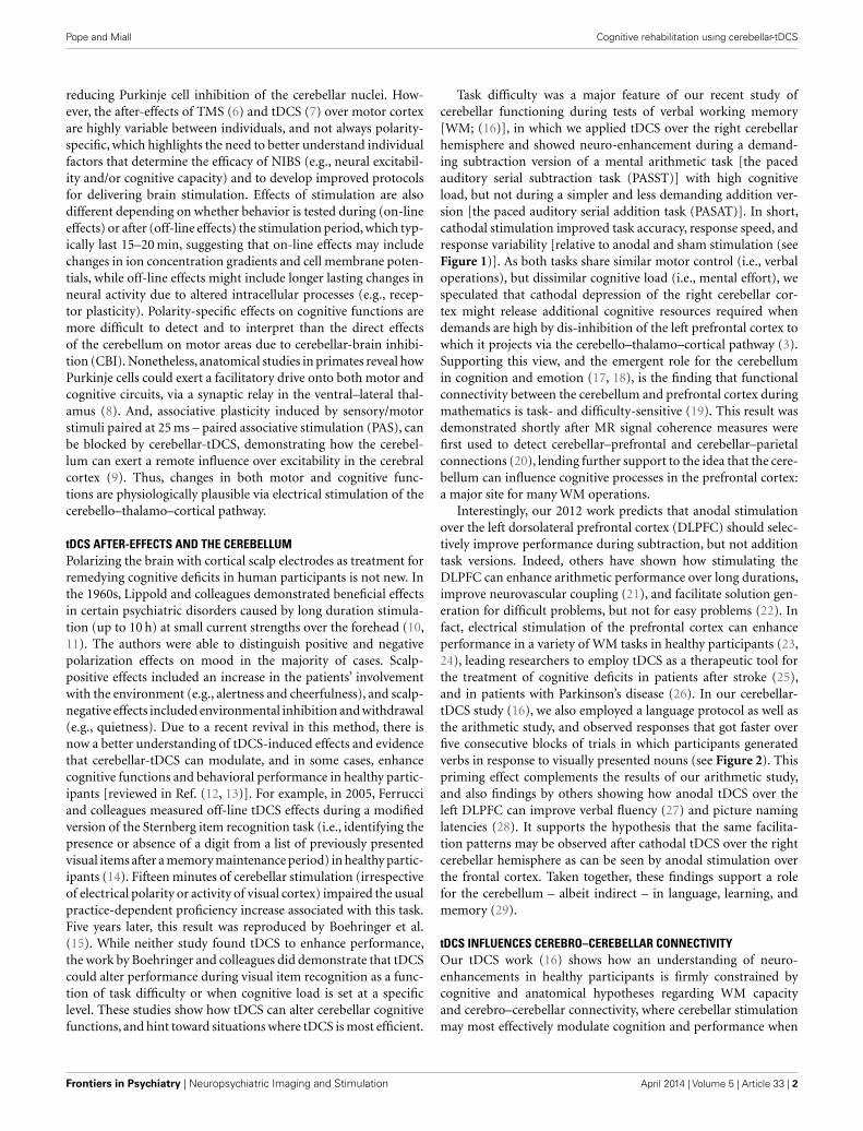
Interestingly, our 2012 work predicts that anodal stimulationover the left dorsolateral prefrontal cortex (DLPFC) should selec-tively improve performance during subtraction, but not additiontask versions. Indeed, others have shown how stimulating theDLPFC can enhance arithmetic performance over long durations,improve neurovascular coupling (21), and facilitate solution gen-eration for difficult problems, but not for easy problems (22). Infact, electrical stimulation of the prefrontal cortex can enhanceperformance in a variety of WM tasks in healthy participants (23,24), leading researchers to employ tDCS as a therapeutic tool forthe treatment of cognitive deficits in patients after stroke (25),and in patients with Parkinson’s disease (26). In our cerebellar-tDCS study (16), we also employed a language protocol as well asthe arithmetic study, and observed responses that got faster overfive consecutive blocks of trials in which participants generatedverbs in response to visually presented nouns (see Figure 2). Thispriming effect complements the results of our arithmetic study,and also findings by others showing how anodal tDCS over theleft DLPFC can improve verbal fluency (27) and picture naminglatencies (28). It supports the hypothesis that the same facilita-tion patterns may be observed after cathodal tDCS over the rightcerebellar hemisphere as can be seen by anodal stimulation overthe frontal cortex. Taken together, these findings support a rolefor the cerebellum – albeit indirect – in language, learning, andmemory (29).
tDCS INFLUENCES CEREBRO–CEREBELLAR CONNECTIVITYOur tDCS work (16) shows how an understanding of neuro-enhancements in healthy participants is firmly constrained bycognitive and anatomical hypotheses regarding WM capacityand cerebro–cerebellar connectivity, where cerebellar stimulationmay most effectively modulate cognition and performance when
Frontiers in Psychiatry | Neuropsychiatric Imaging and Stimulation April 2014 | Volume 5 | Article 33 | 2
Pope and Miall Cognitive rehabilitation using cerebellar-tDCS
FIGURE 1 | (A) Response latencies (mean + 1 SEM, n = 20) selectivelyimproved after cerebellar cathodal stimulation from session one(pre-stimulation) to session two (post-stimulation), significantly more in the
subtraction task than in the addition task. (B) The variability of participants’responses also selectively improved significantly between sessions duringsubtraction, but not during addition. Modified from Pope and Miall (16).
FIGURE 2 | (A) Response latencies (mean + 1 SEM, n = 20) decreasedacross repeated presentation of the same sets of noun–verb pairsbetween blocks 1–5 (new words were presented in block 6), andselectively improved after cathodal stimulation from session one
[pre-stimulation (left panel)] to session two [post-stimulation (rightpanel)]. (B) The variability of participants’ responses also selectivelyimproved significantly between blocks 1–5, and between sessions.Modified from Pope and Miall (16).
participants fully engage in a task, or when the task maximallyexcites the cerebellar–cortical pathway. Indeed, an fMRI study bySalmi and colleagues previously showed how a load increase in
cognition during WM tasks is associated with enhanced neuralactivity in both cerebral and cerebellar areas, which they suggestedwas involved with optimization of response speed (30). They also
Pope and Miall Cognitive rehabilitation using cerebellar-tDCS
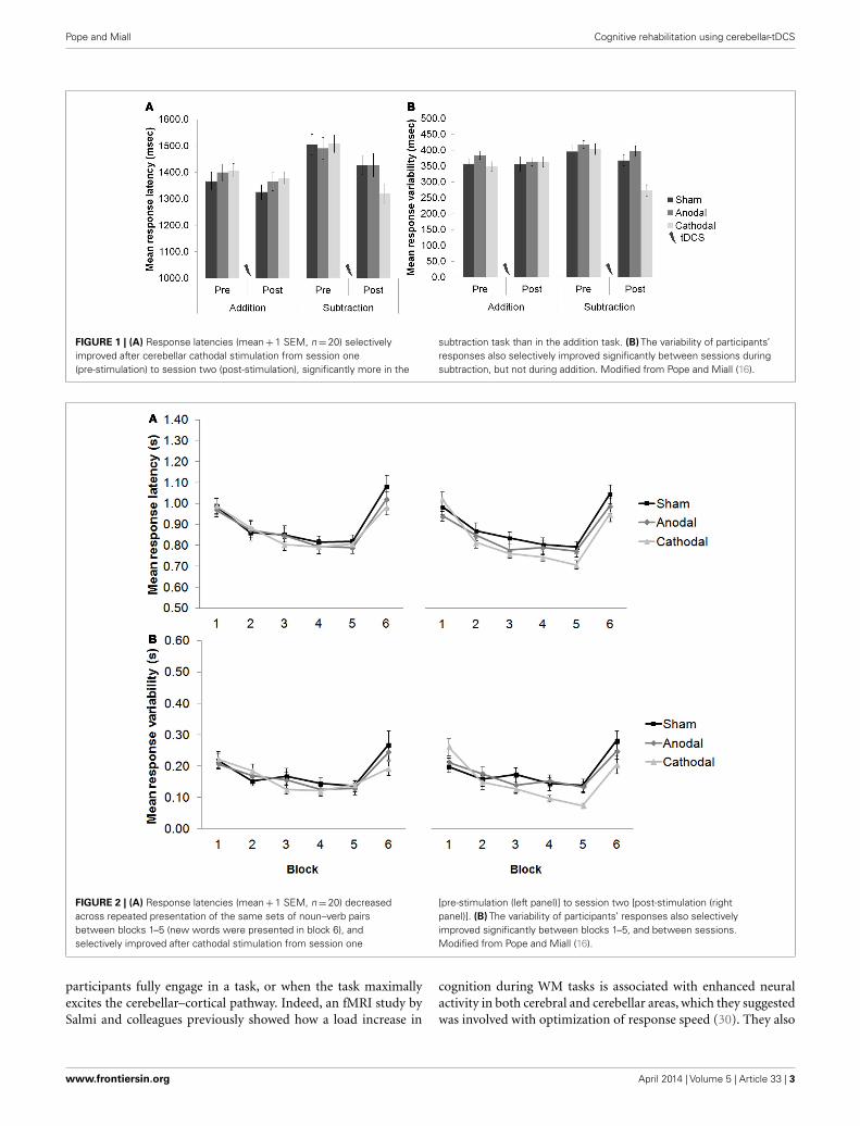
showed with MR tractography how crus I/II in the posterior lateralcerebellum was linked with the lateral prefrontal areas activated byan increase in cognitive load, whereas the anterior cerebellar lobewas not. Based on the available literature, cerebellar-tDCS could beexpected to influence cognition during certain WM tasks via exci-tation of the cerebello–thalamo–cortical pathway (see Figure 3),leading researchers to speculate on the efficacy of this techniqueas a therapeutic tool for treating cognitive deficits in patients withcerebellar disease (3).
NIBS AND CEREBELLAR ATAXIASThe cerebro–cerebellar circuits have been proposed as the anatom-ical substrate of the cerebellar involvement in executive functions,WM,and emotion in patients with the cerebellar cognitive affectivesyndrome [CCAS (17, 18)]. And the cerebellum’s role in cognition
in the context of adaptation, expertise, and giftedness is said toaccelerate information processing in WM and to make think-ing and reasoning more efficient (32). Some ataxic patients canalso present with cognitive and emotional impairments if dam-age to non-motor regions of the cerebellum disrupts the couplingwith cerebral cortical areas for thinking and reasoning. In fact,white matter changes in the dentato–rubral tract [but not thedentato–thalamic or thalamo–cortical tracts (see Figure 3)] havebeen shown to correlate with cognitive assessment in patientswith Friedreich’s ataxia (FA), suggesting that this pathway is animportant contributor to cognitive impairments in this disease(33, 34). Genetically confirmed FA patients present with impair-ments in processing speed, conceptual thinking, verbal fluency,acquisition of verbal information, use of semantic strategies inretrieval, visuoperceptive and visuoconstructive functions, and
FIGURE 3 | A schematic diagram of the main circuits and interneuronesin the cerebellar cortex and the principal white matter tracts connectingthe cerebellum to the cerebrum – cerebro–cerebellar connectivity [afterVoogd and Glickstein (31); redrawn by authors]. Inhibitory cells/synapses
are shown in black, excitatory cells/synapses are shown in gray. ML,molecular layer; PCL, Purkinje cell layer; GCL, granule cell layer; WM, whitematter layer; CN, cerebellar nuclei; IO, inferior olive; PCN, precerebellarneurones.
Frontiers in Psychiatry | Neuropsychiatric Imaging and Stimulation April 2014 | Volume 5 | Article 33 | 4
Pope and Miall Cognitive rehabilitation using cerebellar-tDCS
action naming (35). However, DC stimulation of the cerebellarcortex may not at first hand prove effective in remedying the symp-toms of this autosomal recessive inherited disease since cerebro–cerebellar circuits, including the dentato–rubral tract, becomeprogressively damaged due to atrophy of the dentate nucleus –negating possible therapeutic benefits from tDCS or TMS. But thishas not prevented clinicians from employing other interventionssuch as cognitive rehabilitation therapy (CRT), and physical andoccupational therapy to help improve/stabilize cognition, moodand motor functions in patients with spinocerebellar degenera-tion, by asking them to carry-out a battery of cognitive tests andactivities of daily living that after treatment changed the rate of dis-ease progression in some patients (36, 37). Cerebellar-tDCS couldthen be used alongside rehabilitative interventions to provide asynergistic effect – further improving the management of thesepatients. Recently, tDCS applied as an adjunct to cognitive behav-ioral therapy (CBT) has been successful in treating depression inpatients over the course of a year, whose symptoms were otherwiseresistant to other forms of treatment (38). Such a study demon-strates a favorable synergistic interaction between two very differ-ent interventions, opening up new possibilities for the use of tDCSas a cognitive neuro-rehabilitation tool. It is also worth speculatingwhether the excitatory effects of tDCS alone could prove helpful inreducing cognitive decline/stabilizing mood and motor functionsin cerebellar patients by exciting cerebro–cerebellar connectivity;preventing or slowing down further damage in analogy to theslowing of disease progression in patients with FA (36) and spin-ocerebellar ataxia (37). This might be expected if chronic cerebellardamage reduces the excitability of motor and visual cortices (18).Behavioral gains induced by tDCS do increase functional con-nectivity, for example after stroke (39). Thus, the tDCS enhancesactivity, and it seems, maintains the cerebello–thalamo–corticalpathway.
MOTOR AND COGNITIVE IMPROVEMENTS AFTER CEREBELLAR-TMSTranscranial direct current stimulation has yet to be performedin patients with cerebellar ataxia with the aim of evaluating cog-nitive functions, although improved motor symptoms (reducedamplitude of upper limb stretch reflexes) have been reported inthese patients after anodal stimulation of the cerebellum, whichincreased the inhibitory effect exerted by the cerebellar cortexupon the cerebellar nuclei (40). Anodal tDCS over the motor cor-tex can also improve gait symmetry in patients with cerebellarataxia for a short-term period by restoring motor cortex activ-ity deprived from the cerebro–cerebellar circuit (41). However,improved cognition in an ataxic patient has been demonstratedusing TMS, which uses electromagnetic induction instead of adirect electrical current to activate the brain. In a case study byFarzan and colleagues (2), a patient with a diagnosis of idiopathiclate-onset cerebellar atrophy with speech and gait difficultiesunderwent 21 daily sessions of TMS at maximum output over thecerebellum. After treatment, the authors observed improvementsin the patients’ functional mobility (postural control and walk-ing) and dual-tasking (naming supermarket items while walking).The therapeutic mechanisms were also investigated using dual-coil paired-pulse TMS to measure cerebello–thalamo–corticalconnectivity, before and after treatment. The difference between
treatments was marked by an increase (facilitation) in motorevoked potentials induced by motor cortical stimulation whenthe cerebellum was also excited a few milliseconds beforehand,demonstrating enhanced activity in contralateral motor cortexthat reflected reduced CBI. The reduction in CBI lasted 6 monthsafter treatment. The authors attributed the improvements in cog-nitive function to a consequence of enhanced motor functionand liberation of resources for the performance of the dual-task,thus enabling the patient to name more items while walking withmore ease. The TMS-induced reduction in CBI may also haveimproved prefrontal cortical function directly, through excitingcerebellar projections to this area, thus improving cognitive capac-ity. This second explanation compares well with that from ourown work showing a selective improvement in verbal WM aftercerebellar-tDCS (see above).
NIBS AND CEREBELLAR COGNITIVE AFFECTIVE DISORDERSThe cerebro–cerebellar circuits that can be enhanced by TMSin cerebellar ataxic patients presenting with cognitive impair-ments are disrupted in patients that develop the CCAS followinga lesion or damage to the non-motor cerebellum (17, 18). Adefect associated with a lesion in one cerebellar hemisphere isdecreased excitability of the contralateral prefrontal area. Thus,symptoms that are part of the CCAS extend beyond problemswith motor control, co-ordination and balance, and include prob-lems with executive functions, WM, linguistic performance, andchanges in emotion and personality (18). The use of cerebellar-tDCS to enhance cognition via its remote neuromodulatory effecton prefrontal areas can be anticipated too on the basis of exist-ing TMS studies of cerebellar cognitive functions. This shouldlead to improving mental flexibility (e.g., multi-tasking) in CCASpatients. For example, facilitatory effects of TMS have beenobserved during procedural learning [e.g., serial reaction timetask (SRTT)], which involves acquiring a skill (beyond just motorcontrol) through repeated performance and practice. This taskis thought to involve connections between the cerebellum andthe prefrontal cortex via the thalamus, and damage in any oneof these regions is likely to impair performance. Indeed, TMSover the DLPFC interferes with procedural learning when appliedover the hemisphere contralateral to the performing hand (42).A patient has also been studied with a left cerebellar lesion anda selective deficit in procedural learning, as evidenced by poorperformance with the left hand on the SSRT (43). By decreas-ing cortical excitability of the right (unaffected) cerebellum or theleft DLPFC (in separate sessions) with 1 Hz rTMS for 10 min, thedeficit recovered and task performance markedly improved. Inter-estingly, inhibition of the right DLPFC or a control fronto-parietalregion did not change the patient’s performance. It is interest-ing to speculate whether the inhibitory effects of cathodal tDCSof the same regions might produce similar results. Nonetheless,the authors explained these findings in terms of the modula-tion of a set of inhibitory and excitatory connections betweenthe lateral cerebellar hemisphere and the contralateral prefrontalarea induced by the inhibitory effect of TMS – restoring the bal-ance of cortical activation. Trains of epidural anodal/cathodal DCstimulation over the cerebellum in rats has also shown how thisstructure can exert a remote neuromodulatory effect upon the
Pope and Miall Cognitive rehabilitation using cerebellar-tDCS
excitability of the primary motor cortex – reshaping the represen-tation of muscles on motor cortex (44). In an earlier paper, thesame authors employed anodal tDCS to antagonize motor cor-tex hypoexcitability contralateral to a hemicerebellar ablation inrats, and they speculated that by setting the motor cortex at anappropriate level of excitability, tDCS might be used to modu-late motor cortex excitability after acute cerebellar dysfunction(45). In humans, neuromodulatory effects from cerebellar stimu-lation have revealed how cathodal tDCS decreases CBI, in contrastwith anodal tDCS that increases the cerebellum’s inhibitory toneover the motor cortex (5). Taken together, tDCS of the cerebellumand prefrontal cortex, either jointly or in separate sessions, mightoffer a new treatment for restoring the balance between these tworegions, which normally work together to fine-tune behavior andoptimize performance.
CONCLUSIONMany studies involving healthy participants and certain patientpopulations demonstrate the value of NIBS as the technique ofchoice for producing plastic changes in the brain, and as a researchtool for testing hypotheses about how motor and cognitive func-tions are performed and how cerebro–cerebellar circuits subservethese operations. Based on the available literature, we see five pos-sible approaches to cognitive rehabilitation using NIBS in patientswith damage at various sites in this circuit. (1) Cerebellar-tDCScould reduce cognitive decline and/or improve mood in ataxicpatients. By increasing the excitability of cerebellar projections toareas of the prefrontal cortex, this may prevent further damageand decline of this pathway and potentially enhance functionalconnectivity. (2) NIBS could also be used as an adjunct to othertypes of therapy (e.g., CRT or CBT), improving their therapeuticefficacy when treating the decline of cerebellar cognitive functions.This is because evidence suggests that NIBS enhance the neuro-plastic effects of adjunct non-stimulation therapy. And this mayapply not only in diseases primarily involving the cerebellum, butalso in those affecting interconnected regions where the cerebel-lum exerts a modulatory influence. (3) Enhancing the couplingbetween one side of the cerebellum and the contralateral regionof frontal cortex is another possibility in which the facilitatoryeffects of NIBS could be exploited: improving cognitive capac-ity and motor control in patients with pure cerebellar ataxias.This would free up more cognitive resources for dual-tasking (e.g.,talking whilst walking) – minimizing the risk of falls in aged cere-bellar patients with cognitive decline. Even in healthy individuals,NIBS may be anticipated to improve motor and cognitive func-tions and enhance performance by boosting cerebro–cerebellarconnectivity. A sedentary life does not engage this circuit much.Expert performers such as musicians (46) and athletes (47), havea significantly larger cerebellum than non-experts, suggesting thatincreased activity increases neural volume and probably neuralconnectivity. (4) The neuromodulatory effects of cerebellar stimu-lation might prove successful in restoring the balance of inhibitoryand excitatory connections in the cerebrum, which can be dysfunc-tional in patients with cerebellar damage. Studies show that thenormal effects of CBI, which typically decreases excitability of themotor cortex, are reduced or absent in patients with degeneration
or lesions of the efferent system from the cerebellum (48, 49), con-firming the clinical effectiveness of NIBS to manage motor deficitsin cerebellar ataxias (40, 41). (5) Lastly, one can foresee a pro-cedure that combines the inhibitory effects of cathodal tDCS orlow frequency rTMS to decrease CBI, with the excitatory effects ofanodal tDCS or high frequency rTMS to excite the DLPFC. Thisdual-site stimulation paradigm could be employed to enhance thedis-inhibition of the cerebral cortex, restoring system balance aftercerebellar disease and permitting improved cognitive functions.However, the type of stimulation (e.g., inhibitory versus excita-tory) and stimulation paradigm (e.g., single- versus dual-site) tobe employed as part of an effective treatment plan will be governedby understanding each patients’ specific medical condition. Futureresearch will likely explore these ideas and must be directed towardunderstanding individual factors that determine the efficacy ofNIBS, leading to better procedures and protocols for deliveringNIBS as a cognitive rehabilitation tool for neuro-enhancement.
ACKNOWLEDGMENTSThis work was funded by Wellcome Trust grant WT087554.
REFERENCES1. Heath RG, Llewellyn RC, Rouchell AM. The cerebellar pacemaker for intractable
behavioral disorders and epilepsy: follow-up report. Biol Psychiatry (1980)15(2):243–56.
2. Farzan F, Wu Y, Manor B, Anastasio EM, Lough M, Novak V, et al. CerebellarTMS in treatment of a patient with cerebellar ataxia: evidence from clinical, bio-mechanics and neurophysiological assessments. Cerebellum (2013) 12:707–12.doi:10.1007/s12311-013-0485-8
3. Block HJ, Celnik P. Can cerebellar transcranial direct current stimulationbecome a valuable neurorehabilitation intervention? Expert Rev Neurother(2012) 12:1275–7. doi:10.1586/ern.12.121
4. Parazzini M, Rossi E, Ferrucci R, Liorni I, Priori A, Ravazzani P. Modellingthe electric field and the current density generated by cerebellar transcranialDC stimulation in humans. Clin Neurophysiol (2013) 125:577–84. doi:10.1016/j.clinph.2013.09.039
5. Galea JM, Jayaram G, Ajagbe L, Celnik P. Modulation of cerebellar excitabilityby polarity-specific noninvasive direct current stimulation. J Neurosci (2009)29:9115–22. doi:10.1523/JNEUROSCI.2184-09.2009
6. Hamada M, Murase N, Hasan A, Balaratnam M, Rothwell JC. The role ofinterneuron networks in driving human motor cortical plasticity. Cereb Cortex(2013) 23:1593–605. doi:10.1093/cercor/bhs147
7. Wiethoffa S, Hamadaa M, Rothwell JC. Variability in response to transcra-nial direct current stimulation of the motor cortex. Brain Stimul (2014).doi:10.1016/j.brs.2014.02.003
8. Middleton FA, Strick PL. Basal ganglia and cerebellar loops: motor and cognitivecircuits. Brain Res Brain Res Rev (2000) 31:236–50. doi:10.1016/S0165-0173(99)00040-5
9. Hamada M,Strigaro G,Murase N,Sadnicka A,Galea JM,Edwards MJ,et al. Cere-bellar modulation of human associative plasticity. J Physiol (2012) 590:2365–74.doi:10.1113/jphysiol.2012.230540
10. Lippold OC, Redfearn JW. Mental changes resulting from the passage of smalldirect currents through the human brain. Br J Psychiatry (1964) 110:768–72.doi:10.1192/bjp.110.469.768
11. Redfearn JW, Lippold OC, Costain RA. Preliminary account of the clinical effectsof polarizing the brain in certain psychiatric disorders. Br J Psychiatry (1964)110:773–85. doi:10.1192/bjp.110.469.773
12. Tomlinson SP, Davis NJ, Bracewell RM. Brain stimulation studies of non-motor cerebellar function: a systematic review. Neurosci Biobehav Rev (2013)37(5):766–89. doi:10.1016/j.neubiorev.2013.03.001
13. Grimaldi G, Argyropoulos GP, Boehringer A, Celnik P, Edwards MJ, FerrucciR, et al. Non-invasive cerebellar stimulation – a consensus paper. Cerebellum(2014) 13(1):121–38. doi:10.1007/s12311-013-0514-7
Frontiers in Psychiatry | Neuropsychiatric Imaging and Stimulation April 2014 | Volume 5 | Article 33 | 6
Pope and Miall Cognitive rehabilitation using cerebellar-tDCS
14. Ferrucci R, Marceglia S, Vergari M, Cogiamanian F, Mrakic-Sposta S, MameliF, et al. Cerebellar transcranial direct current stimulation impairs the practice-dependent proficiency increase in working memory. J Cogn Neurosci (2008)20:1687–97. doi:10.1162/jocn.2008.20112
15. Boehringer A, Macher K, Dukart J, Villringer A, Pleger B. Cerebellar transcra-nial direct current stimulation modulates verbal working memory. Brain Stimul(2013) 6:649–53. doi:10.1016/j.brs.2012.10.001
16. Pope PA, Miall RC. Task-specific facilitation of cognition by cathodal transcra-nial direct current stimulation of the cerebellum. Brain Stimul (2012) 5:84–94.doi:10.1016/j.brs.2012.03.006
17. Schmahmann JD, Caplan D. Cognition, emotion and the cerebellum. Brain(2006) 129:290–2. doi:10.1093/brain/awh729
18. Schmahmann JD. Disorders of the cerebellum: ataxia, dysmetria of thought,and the cerebellar cognitive affective syndrome. J Neuropsychiatry Clin Neurosci(2004) 16:367–78. doi:10.1176/appi.neuropsych.16.3.367
19. Feng S, Fan Y, Yu Q, Lu Q, Tang YY. The cerebellum connectivity in mathematicscognition. BMC Neurosci (2008) 9:155. doi:10.1186/1471-2202-9-S1-P155
20. Allen G, McColl R, Barnard H, Ringe WK, Fleckenstein J, Cullum CM. Mag-netic resonance imaging of cerebellar-prefrontal and cerebellar-parietal func-tional connectivity. Neuroimage (2005) 28:39–48. doi:10.1016/j.neuroimage.2005.06.013
21. Snowball A, Tachtsidis I, Popescu T, Thompson J, Delazer M, Zamarian L, et al.Long-term enhancement of brain function and cognition using cognitive train-ing and brain stimulation. Curr Biol (2013) 23(11):987–92. doi:10.1016/j.cub.2013.04.045
22. Metuki N, Sela T, Lavidor M. Enhancing cognitive control components of insightproblems solving by anodal tDCS of the left dorsolateral prefrontal cortex. BrainStimul (2012) 5:110–5. doi:10.1016/j.brs.2012.03.002
23. Zaehle T, Sandmann P, Thorne JD, Jäncke L, Herrmann CS. Transcranial directcurrent stimulation of the prefrontal cortex modulates working memory perfor-mance: combined behavioural and electrophysiological evidence. BMC Neurosci(2011) 12:2. doi:10.1186/1471-2202-12-2
24. Fregni F, Boggio PS, Nitsche M, Bermpohl F, Antal A, Feredoes E, et al. Anodaltranscranial direct current stimulation of prefrontal cortex enhances workingmemory. Exp Brain Res (2005) 166:23–30. doi:10.1007/s00221-005-2334-6
25. Jo JM, Kim YH, Ko MH, Ohn SH, Joen B, Lee KH. Enhancing the workingmemory of stroke patients using tDCS. Am J Phys Med Rehabil (2009) 88:404–9.doi:10.1097/PHM.0b013e3181a0e4cb
26. Boggio PS, Ferrucci R, Rigonatti SP, Covre P, Nitsche M, Pascual-Leone A, et al.Effects of transcranial direct current stimulation on working memory in patientswith Parkinson’s disease. J Neurol Sci (2006) 249:31–8. doi:10.1016/j.jns.2006.05.062
27. Iyer MB, Mattu U, Grafman J, Lomarev M, Sato S, Wassermann EM. Safety andcognitive effect of frontal DC brain polarization in healthy individuals. Neurol-ogy (2005) 64:872–5. doi:10.1212/01.WNL.0000152986.07469.E9
28. Fertonani A, Rosini S, Cotelli M, Rossini PM, Miniussi C. Naming facilita-tion induced by transcranial direct current stimulation. Behav Brain Res (2010)208:311–8. doi:10.1016/j.bbr.2009.10.030
29. Desmond JE, Fiez JA. Neuroimaging studies of the cerebellum: language, learn-ing and memory. Trends Cogn Sci (1998) 2:355–62. doi:10.1016/S1364-6613(98)01211-X
30. Salmi J, Pallesen KJ, Neuvonen T, Brattico E, Korvenoja A, Salonen O, et al. Cog-nitive and motor loops of the human cerebro-cerebellar system. J Cogn Neurosci(2010) 22:2663–76. doi:10.1162/jocn.2009.21382
31. Voogd J, Glickstein M. The anatomy of the cerebellum. Trends Neurosci (1998)21:370–5. doi:10.1016/S0166-2236(98)01318-6
32. Koziol LF, Budding DE, Chidekel D. Adaptation, expertise, and giftedness:towards an understanding of cortical, subcortical, and cerebellar network con-tributions. Cerebellum (2010) 9:499–529. doi:10.1007/s12311-010-0192-7
33. Akhlaghi H,Yu J, Corben L, Georgiou-Karistianis N, Bradshaw JL, Storey E, et al.Cognitive deficits in Friedreich ataxia correlate with micro-structural changesin dentatorubral tract. Cerebellum (2013) 13:187–98. doi:10.1007/s12311-013-0525-4
34. Zalesky A, Akhlaghi H, Corben LA, Bradshaw JL, Delatycki MB, Storey E, et al.Cerebello-cerebral connectivity deficits in Friedreich ataxia. Brain Struct Funct(2013). doi:10.1007/s00429-013-0547-1
35. Nieto A, Correia R, de Nóbrega E, Montón F, Hess S, Barroso J. Cognitionin Friedreich ataxia. Cerebellum (2012) 11:834–44. doi:10.1007/s12311-012-0363-9
36. Ciancarelli I, Cofini V, Carolei A. Evaluation of neuropsychological functions inpatients with Friedreich ataxia before and after cognitive therapy. Funct Neurol(2010) 25:81–5.
37. Miyai I, Ito M, Hattori N, Mihara M, Hatakenaka M, Yagura H, et al. Cerebel-lar ataxia rehabilitation trial in degenerative cerebellar diseases. NeurorehabilNeural Repair (2012) 26:515–22. doi:10.1177/1545968311425918
38. D’Urso G, Mantovani A, Micillo M, Priori A, Muscettola G. Transcranial directcurrent stimulation and cognitive-behavioral therapy: evidence of a syner-gistic effect in treatment-resistant depression. Brain Stimul (2013) 6:465–7.doi:10.1016/j.brs.2012.09.003
39. Stagg CJ, Bachtiar V, O’Shea J, Allman C, Bosnell RA, Kischka U, et al. Corticalactivation changes underlying stimulation-induced behavioural gains in chronicstroke. Brain (2011) 135:276–84. doi:10.1093/brain/awr313
40. Grimaldi G, Manto M. Anodal transcranial direct current stimulation (tDCS)decreases the amplitudes of long-latency stretch reflexes in cerebellar ataxia. AnnBiomed Eng (2013) 41:2437–47. doi:10.1007/s10439-013-0846-y
41. Pozzi NG, Minafra B, Zangaglia R, De Marzi R, Sandrini G, Priori A, et al. Tran-scranial direct current stimulation (tDCS) of the cortical motor areas in threecases of cerebellar ataxia. Cerebellum (2014) 13:109–12. doi:10.1007/s12311-013-0524-5
42. Pascual-Leone A, Tarazona F, Keenan J, Tormos JM, Hamilton R, Catala D.Transcranial magnetic stimulation and neuroplasticity. Neuropsychologia (1999)37:207–17. doi:10.1016/S0028-3932(98)00095-5
43. Torriero S, Oliveri M, Koch G, Lo Gerfo E, Salerno S, Petrosini L, et al. Corticalnetworks of procedural learning: evidence from cerebellar damage. Neuropsy-chologia (2007) 45:1208–14. doi:10.1016/j.neuropsychologia.2006.10.007
44. Oulad Ben Taib N, Manto M. Trains of epidural DC stimulation of thecerebellum tune corticomotor excitability. Neural Plast (2013) 2013:613197.doi:10.1155/2013/613197
45. Oulad Ben Taib N, Manto M. Trains of transcranial direct current stimulationantagonize motor cortex hypoexcitability induced by acute hemicerebellectomy.J Neurosurg (2009) 111:796–806. doi:10.3171/2008.2.17679
46. Hutchinson S, Lee LH, Gaab N, Schlaug G. Cerebellar volume of musicians.Cereb Cortex (2003) 13:943–9. doi:10.1093/cercor/13.9.943
47. Park IS, Lee KJ, Han JW, Lee NJ, Lee WT, Park KA, et al. Experience-dependentplasticity of cerebellar vermis in basketball players. Cerebellum (2009) 8:334–9.doi:10.1007/s12311-009-0100-1
48. Tamburin S, Fiaschi A, Marani S,Andreoli A, Manganotti P, Zanette G. Enhancedintracortical inhibition in cerebellar patients. J Neurol Sci (2004) 217:205–10.doi:10.1016/j.jns.2003.10.011
49. Wessel K, Tegenthoff M, Vorgerd M, Otto V, Nitschke MF, Malin JP. Enhance-ment of inhibitory mechanisms in the motor cortex of patients with cerebellardegeneration: a study with transcranial magnetic brain stimulation. Electroen-cephalogr Clin Neurophysiol (1996) 101:273–80.
Conflict of Interest Statement: The authors declare that the research was conductedin the absence of any commercial or financial relationships that could be construedas a potential conflict of interest.