44
RESTRICTIVE LUNG DISEASES DR. MOHAMED SEYAM PHT.PT . ASSISTANT PROFESSOR OF PHYSICAL THERAPY
| Date post: | 23-Dec-2015 |
| Category: |
Documents |
| Upload: | winfred-montgomery |
| View: | 215 times |
| Download: | 1 times |
RESTRICTIVE LUNG DISEASESDR. MOHAMED SEYAM PHT.PT.
ASSISTANT PROFESSOR OF PHYSICAL THERAPY

•Definition
Restrictive lung disease (RDL) is a chronic disorder that causes a decrease in the ability to expand the lung (breathe in).
Disorders that inhibit normal lung excursion.


1 -Pulmonary Causes of Restrictive Lung Disease 1. Idiopathic Pulmonary Fibrosis
2. Coal Workers' Pneumoconiosis
3. Silicosis
4. Asbestosis
5. Broncho pulmonary Dysplasia
6. Bronchogenic Carcinoma
7. Pleural Effusions
8. Pneumonia

2-Neuromuscular Causes of RLD
1. Spinal Cord Injury
2. Amyotrophic Lateral Sclerosis
3. Poliomyelitis
4. Guillain-Barre Syndrome
5. Myasthenia Gravis
6. Duchenne Muscular Dystrophy
7. Other Muscular Dystrophies

3- Cardiovascular Causes
1. Pulmonary Edema
2. Pulmonary Emboli
4- Musculoskeletal Causes
3. Diaphragmatic Paralysis or Paresis
4. Kyphoscoliosis
5. Ankylosing Spondylitis
6. Pectus Excavaturn
7. Pectus Carinatum
5- Connective Tissue Causes1. Rheumatoid Arthritis
2. SystemIc Lupus Erythematosus
6- Immunologic Causes3. Goodpasture‘s Syndrome
4. Wegeners Granulomatosis

7-Pregnancy as a- Cause of Restrictive Lung.
8- Nutritional and Metabolic Causes
1. obesity
2. Diabetes Mellitus
9- Traumatfc causes
3. Crush Injuries
4. Penetrating Wounds
10- Therapeutic Causes
1. Surgical Therapy
2. Lung Transplantation
3. Drug Therapy
4. Radiation Therapy
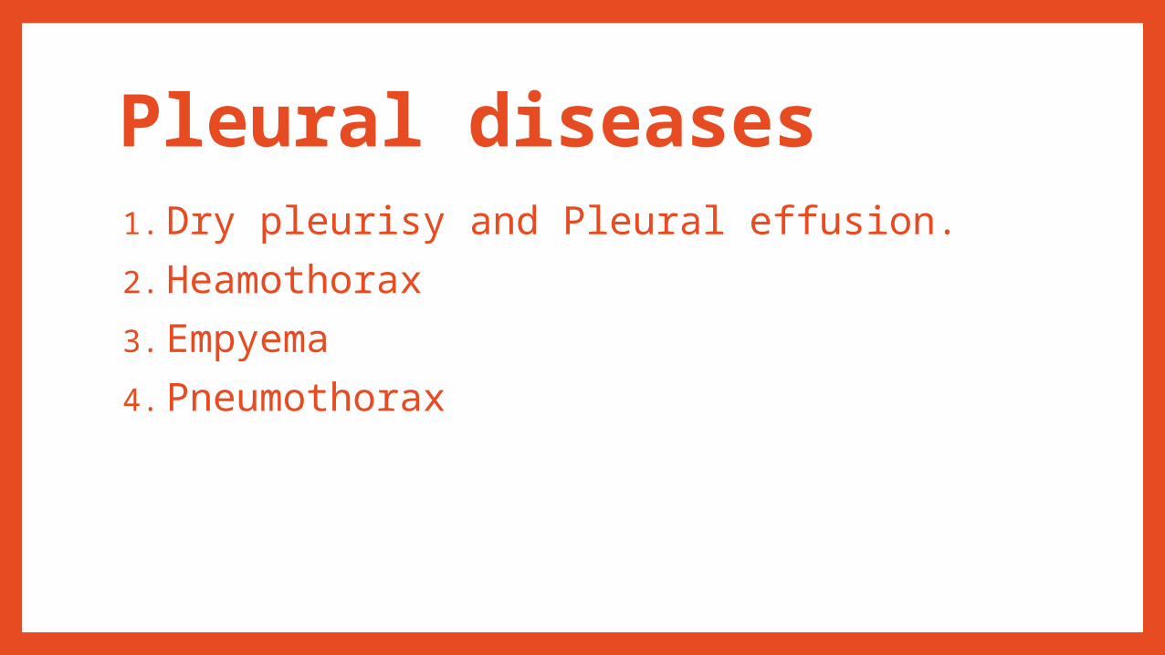
Pleural diseases1. Dry pleurisy and Pleural effusion.
2. Heamothorax
3. Empyema
4. Pneumothorax

PLEURAL EFFUSION•The pleural space normally contains less than 20 mL of fluid.
•Pleural effusion occurs when there is excess fluid in the pleural cavity.
•caused by
1. disturbed osmotic or hydrostatic pressure in the plasma,
2. changes in membrane permeability.
3. Malignancy causes 25% of pleural effusions .
4. Other causes are heart, kidney or liver failure,
5. abdominal or cardiac surgery,
6. pneumonia or TB.
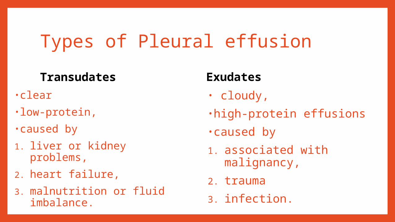
Types of Pleural effusion
Transudates
•clear
•low-protein,
•caused by
1. liver or kidney problems,
2. heart failure,
3. malnutrition or fluid imbalance.
Exudates
• cloudy,
•high-protein effusions
•caused by
1. associated with malignancy,
2. trauma
3. infection.

Clinical features •a stony dull percussion
•decreased breath sounds over the affected area, with bronchial breathing
• A small effusion of less than 500 mL creates few or no symptoms.
• A large effusion displaces the mediastinum and causes breathlessness.
•Radiologically there is a fluid line, occurs when fluid accumulates in pockets.

Medical treatment
•treatment the cause•symptomatic relief of breathlessness by•needle aspiration (thoracocentesis), performed slowly to avoid ere-expansion pulmonaryoedema‘ .
•Surgery •may be needed for a thickened restrictive pleura. •The symptoms of malignant invasion of the pleura

Bilateral pleural effusions.• The right sideshows a dense opacity with a smooth horizontal border andmeniscal edge.• The left showsa small effusion obliteratingthe costophrenic angle.

Physiotherapy •Deep breathing exercises.
•Positioning can be used to optimize gas exchange.
•People with moderate unilateral effusion may benefit from side-lying with the affected side uppermost because both ventilation and perfusion are greater in the lower lung.
•Large effusions are more likely to show improved Pa02 with the effusion downwards to minimize compression of the unaffected lung .
•Some patients require assistance with mobilization.

2 -HEAMOTHORAX
•Heamothorax is blood in the pleura as a result of malignancy or trauma,
•managed by treating the cause, plus tube drainage if necessary.

3 -EMPYEMA
•It is pus in the pleural cavity following localized infection.
•It can complicate pneumonia, bronchiectasis, chronic aspiration, abscess or chest surgery, especially oesophageal surgery.
•The patient may be asymptomatic or toxic, depending on the organism and volume of pus.
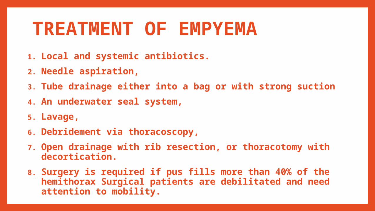
TREATMENT OF EMPYEMA1. Local and systemic antibiotics.
2. Needle aspiration,
3. Tube drainage either into a bag or with strong suction
4. An underwater seal system,
5. Lavage,
6. Debridement via thoracoscopy,
7. Open drainage with rib resection, or thoracotomy with decortication.
8. Surgery is required if pus fills more than 40% of the hemithorax Surgical patients are debilitated and need attention to mobility.

4 -PNEUMOTHORAX
•Definition
Air collects between the visceral and parietal pleura.
•Types
1. Spontaneous pneumothorax
2. Traumatic pneumothorax


Types
1.Spontaneous pneumothorax: It may be secondary to an underlying disease.
Causes
a. Asthma or bullous emphysema
b. Infections (e.g. Staphylococcal pneumonia, tuberculosis)
c. Cystic fibrosis
d. Marfan's syndrome.
2.Traumatic pneumothorax
Causes:• Penetrating injury to the chest (e.g. by stab wound or a bullet)
•Non-penetrating injury to the chest wall (e.g. Rib fracture)
• During the insertion of an intravenous (e.g. subclavian) line
•During surgery to the chest wall
•During pleural aspiration or biopsy.

Pathological changes
• As air escapes into the pleural cavity and reduces the sub
atmospheric pressure (i.e. less negative)causes the lung collapse.
• Air then enters the pleural cavity on inspiration but cannot escape during
expiration.
• The lung remains collapsed and, as air accumulates in the pleural
cavity and the pressure increases, there is displacement of the heart
together with compression of the other lung and great vessels.
• This is termed a tension pneumothorax

Clinical features• Increased respiratory rate
•Dyspnea
•Chest pain that worsens with a deep breath
•Fatigue and weakness
•Tachycardia
•Cyanosis
•Hypotension
•Decreased or absent lung sounds on affected side
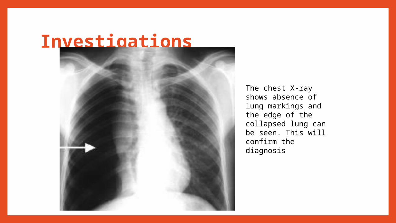
Investigations
The chest X-ray shows absence of lung markings and the edge of the collapsed lung can be seen. This will confirm the diagnosis

Medical Management
A) small pneumothorax
Requires no treatment apart from a few days bed rest until it resolves.
B) large pneumothorax (i.e. more than 25% of the pleural space is filled with air)
It is treated by needle aspiration or by an intercostal drain which connects the pleural cavity to a drainage bottle creating an underwater seal.
SURGERY: is indicated for a recurrent pneumothorax.
- PLEURODESIS: comprises the insertion of a powder into the pleural cavity to adhere both the pleura together.
- PLEURECTOMY: is the removal of the parietal pleura from the chest wall leaving a raw surface to which the visceral layer sticks.


CHEST DEFORMITIES
1.Pectus carinatum,
2.Pectus Excavatum,
3.Scoliosis,
4.Kyphosis and
5.Kyphoscoliosis

1 -Pectus Carinatum•Pectus Carinatum (pigeon chest)
•It is a Structural abnormality characterized by the sternum protruding anteriorly.
•Fifty percent of patients with atrial or ventricular septal defects have Pectus carinatum.
•It also has been associa1ed with severe prolonged childhood asthma.


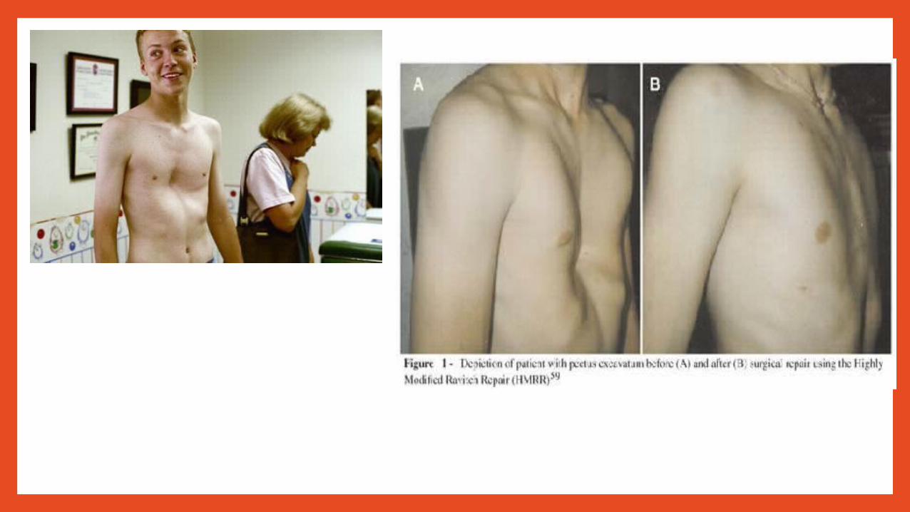
2 -PECTUS EXCAVATUM (funnel chest)
•It is a congenital abnormality characterized by sternal depression and decreased anteroposterior diameter.
•The lower portion of the sternum is displaced posteriorly, and the anterior rib5 are bowed markedly.
•If the deformity is very severe, the patient may have decreased TLC, VC and maximum voluntary ventilation and
may complain of dyspnea on exertion, precordial pain, palpitation, and dizziness.

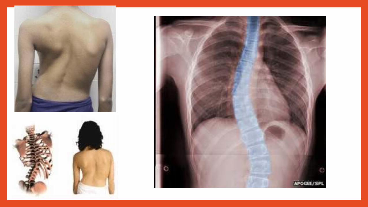
3 -scoliosis•This is characterized by lateral deviation of the spine on either side, mainly in thoracic or lumbar or both region.
•Structural scoliosis involves an irreversible lateral deviation of the spine.
•Nonstructural scoliosis is reversible and also called postural scoliosis.

4 -Kyphosis•Kyphosis may cause restrictive lung defects secondary to musculoskeletal deformity and subsequent compression of the alveoli

5 -Kyphoscoliosis•It is a combination of excessive anter0posterior and lateral curvature of the thoracic spine .
•A distorted spine increases the work of breathing because of
1. Reduced chest wall compliance
2. Micro atelectasis
3. Altered alveolar surface tension .
•The configuration of the chest wall forces the diaphragm to work inefficiently.
•Surgery is sometimes undertaken to prevent progression of scoliosis or improve body image and improve pulmonary function .
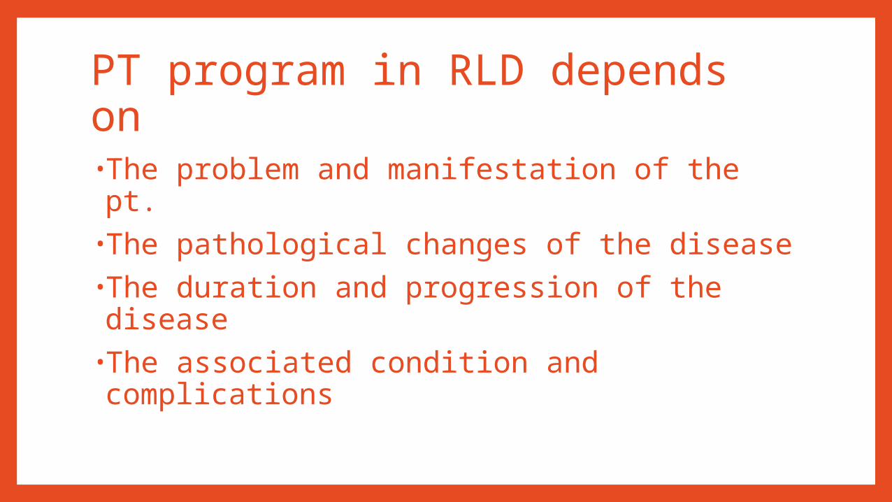
PT program in RLD depends on
•The problem and manifestation of the pt.
•The pathological changes of the disease•The duration and progression of the disease
•The associated condition and complications

Goals •Maintain and restore adequate ventilation
•Re-expand affected lung
•Prevent permanent or recurrent atelectasis
•Adequate ROM in upper limps and trunk
•Prevent deformity and maintain good posture
•Assist the removal of secretions if present
•Assist and control drainage by under water seal
•Maintain adequate peripheral circulation
•Improve exercise tolerance and endurance
•Relieve pain and anxiety
•Help in reducing dyspnea and breathing rate
•Break down any adhesion

Respiratory exercise•Nose training•Segmental breathing•Diaphragmatic breathing •Exercise connected with breathing•Belt breathing exercise•Breathing training with devices

Mobilizing exercise•Active free exercise •Gymnastic exercise•Swimming•Shoulder wheel and raw machine•Supported and unsupported exercise


Clearing lung fields - Humidification
- Clapping, shaking and percussion
- breathing exercises may all be necessary in a
- postural drainage position to the area of the lung affected.
- Modified postural drainage
- Suctioning
- Cough and huff
- Autogenic drainage


