19
Retina and Vitreous Retina
| Date post: | 02-Jan-2016 |
| Category: |
Documents |
| Upload: | tasha-harvey |
| View: | 78 times |
| Download: | 3 times |
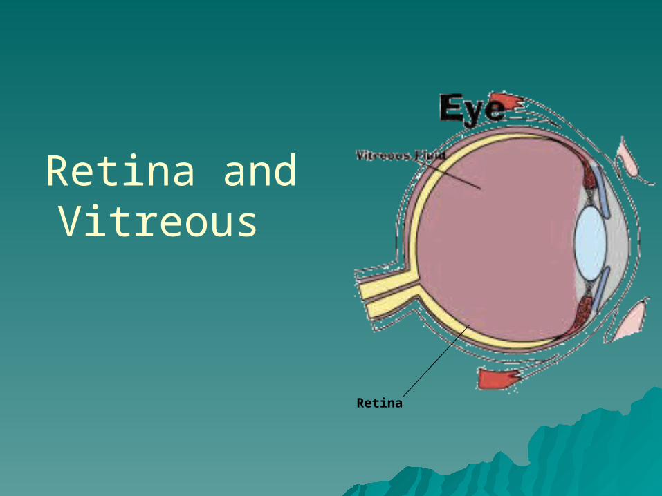
Retina and Vitreous
Retina
Retina
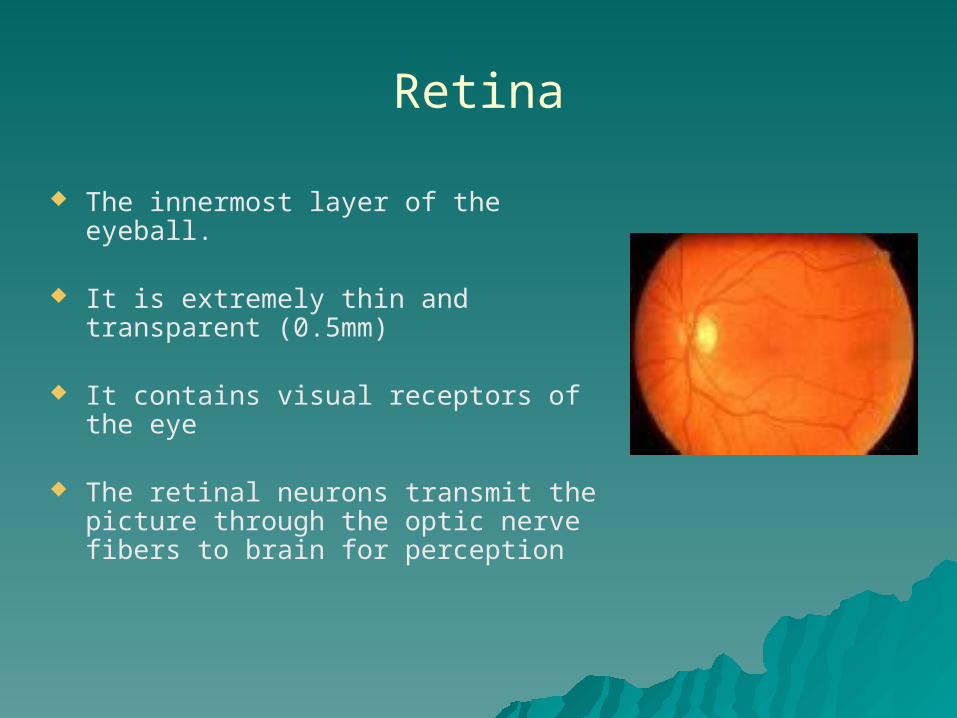
The innermost layer of the eyeball.
It is extremely thin and transparent (0.5mm)
It contains visual receptors of the eye
The retinal neurons transmit the picture through the optic nerve fibers to brain for perception
Layer of retina
There are 10 layers in the retina
Retinal pigment epithelium Layer of rods and cones External limiting membrane Outer nuclear layer Outer plexiform layer Inner nuclear layer Inner plexiform layer Ganglion cell layer Nerve fibre layer Internal limiting membrane
Retinal receptors
* The retinal receptors are divided into two main populations
* Rods* Cones
Rods
Function best in dim light There are 125million rods
in the retina Rods are relatively poor in
visual details
Cones
Function best in daylight There 6 million cones in
the retina Cones enable us to see
small visual details Helps to visualize the
colors
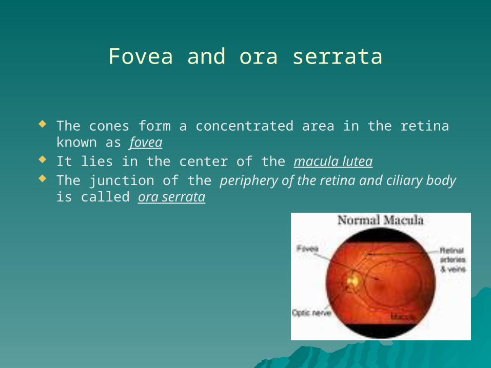
Fovea and ora serrata
The cones form a concentrated area in the retina known as fovea
It lies in the center of the macula lutea The junction of the periphery of the retina and ciliary body
is called ora serrata
Vitreous
The Vitreous humour is a transparent gel that provides a clear optical medium.
It is helps to keep the three layers apposed to each other It occupies approximately 80% of the volume of the globe. The vitreous consist of water, collagen fibrils, molecules of
hyaluronic acid, peripheral cells and mucopolysacharides forming a gel like material.
It nourishes lens, ciliary body and the retina.
Examination of vitreous
Examination of the anterior vitreous can be carried out with slit-lamp.
The vitreous should be observed for cells and any opacities.
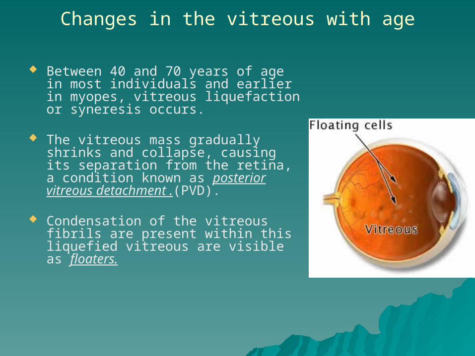
Changes in the vitreous with age
Between 40 and 70 years of age in most individuals and earlier in myopes, vitreous liquefaction or syneresis occurs.
The vitreous mass gradually shrinks and collapse, causing its separation from the retina, a condition known as posterior vitreous detachment .(PVD).
Condensation of the vitreous fibrils are present within this liquefied vitreous are visible as floaters.

Retinal Diseases
Diabetic Retinopathy
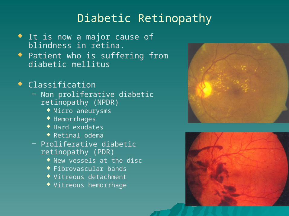
It is now a major cause of blindness in retina.
Patient who is suffering from diabetic mellitus
Classification– Non proliferative diabetic retinopathy
(NPDR) Micro aneurysms Hemorrhages Hard exudates Retinal odema
– Proliferative diabetic retinopathy (PDR) New vessels at the disc Fibrovascular bands Vitreous detachment Vitreous hemorrhage

Investigations and Treatment
– Urine and Blood Sugar examination– FFA (Fundus flourescein angiography)
Management
Medical Treatment : Good diabetic control
Laser Treatment : Photocoagulation to stop leaking from retinal vessels and bleeding
from new vesselsSurgical Treatment : Vitrectomy is done in case of
vitreous hemorrhage, traction retinal detachment
Hypertensive Retinopathy
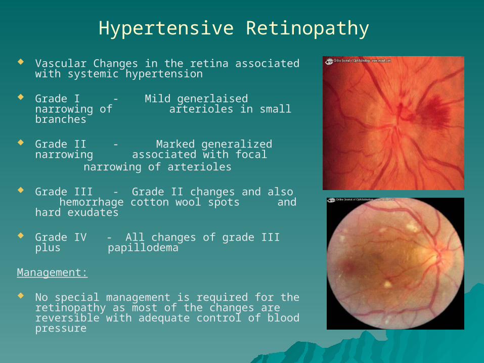
Vascular Changes in the retina associated with systemic hypertension
Grade I - Mild generlaised narrowing of arterioles in small branches
Grade II - Marked generalized narrowing associated with focalnarrowing of arterioles
Grade III - Grade II changes and also hemorrhage cotton wool spots and hard exudates
Grade IV - All changes of grade III plus papillodema
Management:
No special management is required for the retinopathy as most of the changes are reversible with adequate control of blood pressure

Retinal detachment
Separation of retina from the retinal pigment epithelial layer
Myopia Retinal Degeneration Trauma
Floaters Flashes of light Sudden painless loss of vision
Scleral buckling procedure
Retinitis pigmentosa
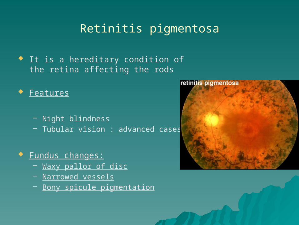
It is a hereditary condition of the retina affecting the rods
Features
– Night blindness– Tubular vision : advanced cases
Fundus changes:– Waxy pallor of disc– Narrowed vessels– Bony spicule pigmentation

Treatment
No permanent cure at present Supportive treatment
– Vitamin A– Low vision aids – Visual rehabilitation– Genetic counselling– Affected individuals discouraged to have kids
Central Serous retinopathy (CSR)
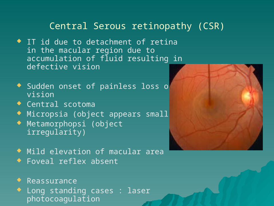
IT id due to detachment of retina in the macular region due to accumulation of fluid resulting in defective vision
Sudden onset of painless loss of vision Central scotoma Micropsia (object appears small) Metamorphopsi (object irregularity)
Mild elevation of macular area Foveal reflex absent
Reassurance Long standing cases : laser
photocoagulation
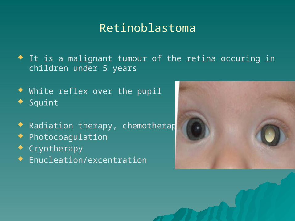
Retinoblastoma
It is a malignant tumour of the retina occuring in children under 5 years
White reflex over the pupil Squint
Radiation therapy, chemotherapy Photocoagulation Cryotherapy Enucleation/excentration
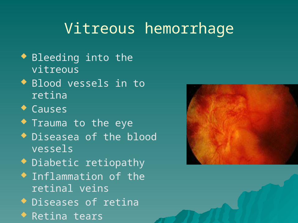
Vitreous hemorrhage
Bleeding into the vitreous Blood vessels in to retina Causes Trauma to the eye Diseasea of the blood
vessels Diabetic retiopathy Inflammation of the retinal
veins Diseases of retina Retina tears Retinal detachment