54
RETINA-VISUAL PATHWAYS
| Date post: | 22-Dec-2015 |
| Category: |
Documents |
| Upload: | kerrie-mccormick |
| View: | 223 times |
| Download: | 2 times |

RETINA-VISUAL PATHWAYS


Retina
takes the information from its 100 million photoreceptors about 1 million optic nerve axons.
Interposed between the photoreceptor layer and the layer of ganglion cells (whose axons form the optic nerve) is a layer containing three kinds of interneurons
Bipolar cells convey signals straight across this layer, receiving inputs from photoreceptors and synapsing on ganglion cells.
Horizontal cells spread laterally in the outer synaptic layer, affecting transmission from photoreceptors to bipolar cells.
Amacrine cells have a similar role in the inner synaptic layer, affecting transmission from bipolar to ganglion cells
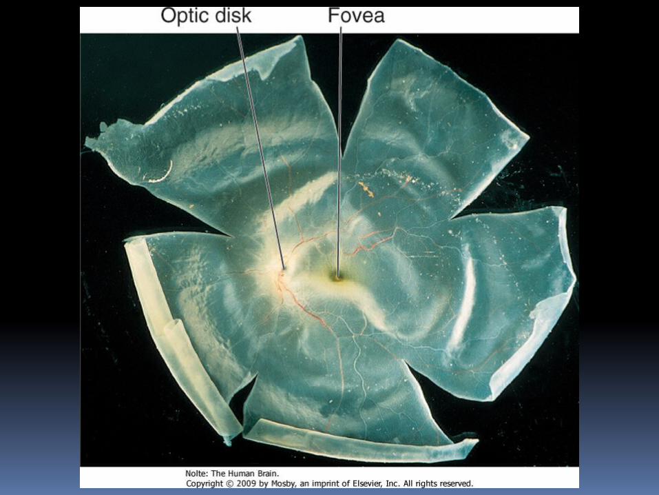
ganglion cell axons travel along its vitreal surface, so they need to cross the retina, choroid, and sclera in order to leave the eye in the optic nerve.
at the optic disc, all the axons converge and collect into groups that leave the eye through small holes in the sclera.

The sclera continues over the optic nerve as a sheath continuous with the dura mater, much as the spinal dura continues as the epineurium of spinal nerves.
The normal layers of the retina are absent at the optic disc, which results in a blind spot in the visual field of each eye

Central visual pathways
Axons of retinal ganglion cells project to a variety of places:
1. the superior colliculus, to help direct visual attention;
2. other midbrain sites, for things like the pupillary light reflex;
3. the hypothalamus, to help regulate circadian rhythms;

but mostly to the thalamus, for conscious awareness of visual stimuli.
Optic nerve fibers convey all the information we will ever get about the shape, color, location, and movement of objects in the outside world.

Different classes of ganglion cells emphasize different properties of a visual stimulus
these different properties begin to be sorted out in the six-layered lateral geniculate nucleus of the thalamus

Different layers of the lateral geniculate then project differentially to primary visual cortex above and below the calcarine sulcus (also known as striate cortex because of a stripe of myelinated fibers that run through one of its middle layers).

Primary visual cortex then picks apart these attributes a little more and parcels them out semiselectively to distinct areas of visual association cortex in the occipital and temporal lobes.


CN II
Extension of white matter of the brain- enclosed in meninges
No effective regeneration when divided
Attached to anterior part of floor of 3rd ventricle

Optic chiasma
Nasal fibres of each CN II decussate and pass to opposite optic tract
Temporal fibres pass directly to optic tract of their own side
Right tract has fibres from right ½ of each retina [nasal field of right eye and temporal field of left eye]

Optic tract
Passes around cerebral peduncle, high up against temporal lobe , reaches side of thalamus
Branches 1. Larger – lateral geniculate body [visual
fibres2. Smaller – bypasses LGB → superior
colliculus→ pretectal nuclei [re light reflexes]3. Some fibres end in hypothalamus [ circadian
rhytm]


1. Some fibres end in hypothalamus [ circadian rhythm] circa= about; diem=a day
superior colliculus also receives fibres from1. Spinotectal/spinomesencephalic tracts2. Auditory inputs via Inferior colliculus

Efferents from superior colliculus
1. Reticular formation2. Inferior colliculus3. Cervical spinal cord [tectospinal
tract]4. LGB→ pulvinar → visual association cortex

Lateral geniculate body
6 layers I, 4, 6 – crossed fibres 2, 3 ,5 – uncrossed fibres
Optic radiation/geniculocalcarine tract Run in sublentiform and
retrolentiform parts of internal capsule
Axons from LGB carrying impressions from upper ½ of contralateral visual fields spread laterally and inferiorly around anterior tip of inferior horn of lateral ventricle [Meyer’s loop]
They swing posteriorly → inferior lip of calcarine sulcus
Other fibres carrying impressions from lower ½ of visual fields go to superior lip of calcarine sulcus

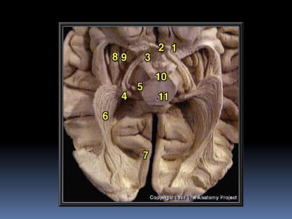
Optic Nerve, Chiasma, and Tract
visual information from one side of the world ends up in the contralateral occipital lobe.
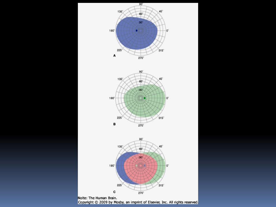
each eye looks at most of the right and left half of the total visual field
As a result, half of the output of each retina needs to cross in the optic chiasm, and half needs to stay uncrossed

the crossed and uncrossed fibers representing one half of the visual field emerge from the chiasm as an optic tract.
damage in front of the optic chiasm can cause complete blindness of the ipsilateral eye,
damage to one optic tract (or any part of the visual system behind the chiasm) can cause loss of the contralateral half of the visual field of both eyes

damage to one optic tract (or any part of the visual system behind the chiasm) can cause loss of the contralateral half of the visual field of both eyes
This deficit has the tongue-twisting name of homonymous hemianopia ("blindness in the same half of both visual fields")


Beyond primary visual cortex analysis of form and color is largely
carried out in ventral parts of the occipital and temporal lobes
analysis of location and motion takes place more dorsally, around the junction of the occipital, parietal, and temporal lobes.

dorsal stream reaching the area near the junction of the parietal, occipital, and temporal lobes is particularly important for analyzing the location and movement of visual stimuli;ventral stream reaching the occipitotemporal gyrus is particularly important for analyzing colors and shapes.

damage to the occipitotemporal gyrus can cause deficits in recognizing things visually despite visual fields being intact.

deficits can be fairly selective depending on which part of the occipitotemporal gyrus is damaged . For example
1. cortical color blindness [achromatopsia]
2. difficulty recognizing faces [prosopagnosia]).

damage near the junction of the parietal, occipital, and temporal lobes can cause difficulties in perceiving the motion of objects or in "stitching together" multiple objects in different locations into a unified scene.


