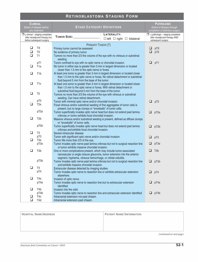
American Joint Committee on Cancer • 2010 52-1 (continued on next page) C LINICAL Extent of disease before any treatment P ATHOLOGIC Extent of disease through completion of definitive surgery y clinical – staging completed after neoadjuvant therapy but before subsequent surgery y pathologic – staging completed after neoadjuvant therapy AND subsequent surgery TX T0 T1 pT1 T1a T1b T1c T2 pT2 T2a pT2a T2b pT2b T3 pT3 T3a pT3a T3b pT3b T4 pT4 T4a pT4a T4b pT4b T4c T4d PRIMARY TUMOR (T) Primary tumor cannot be assessed No evidence of primary tumor Tumors no more than 2/3 the volume of the eye with no vitreous or subretinal seeding. Tumor confined to eye with no optic nerve or choroidal invasion. No tumor in either eye is greater than 3 mm in largest dimension or located closer than 1.5 mm to the optic nerve or fovea. At least one tumor is greater than 3 mm in largest dimension or located closer than 1.5 mm to the optic nerve or fovea. No retinal detachment or subretinal fluid beyond 5 mm from the base of the tumor. At least one tumor is greater than 3 mm in largest dimension or located closer than 1.5 mm to the optic nerve or fovea. With retinal detachment or subretinal fluid beyond 5 mm from the base of the tumor. Tumors no more than 2/3 the volume of the eye with vitreous or subretinal seeding. Can have retinal detachment. Tumor with minimal optic nerve and/or choroidal invasion Focal vitreous and/or subretinal seeding of fine aggregates of tumor cells is present, but no large clumps or “snowballs” of tumor cells. Tumor superficially invades optic nerve head but does not extend past lamina cribrosa or tumor exhibits focal choroidal invasion. Massive vitreous and/or subretinal seeding is present, defined as diffuse clumps or “snowballs” of tumor cells. Tumor superficially invades optic nerve head but does not extend past lamina cribrosa and exhibits focal choroidal invasion. Severe intraocular disease Tumor with significant optic nerve and/or choroidal invasion Tumor fills more than 2/3 of the eye. Tumor invades optic nerve past lamina cribrosa but not to surgical resection line or tumor exhibits massive choroidal invasion. One or more complications present, which may include tumor-associated neovascular or angle closure glaucoma, tumor extension into the anterior segment, hyphema, vitreous hemorrhage, or orbital cellulitis. Tumor invades optic nerve past lamina cribrosa but not to surgical resection line and exhibits massive choroidal invasion. Extraocular disease detected by imaging studies. Tumor invades optic nerve to resection line or exhibits extraocular extension elsewhere. Invasion of optic nerve. Tumor invades optic nerve to resection line but no extraocular extension identified Invasion into the orbit. Tumor invades optic nerve to resection line and extraocular extension identified Intracranial extension not past chiasm. Intracranial extension past chiasm. pTX pT0 pT1 pT2 pT2a pT2b pT3 pT3a T3b pT3b pT4 pT4a pT4b R ETINOBLASTOMA S TAGING F ORM HOSPITAL NAME /ADDRESS PATIENT NAME /INFORMATION S TAGE C ATEGORY D EFINITIONS left right bilateral LATERALITY: TUMOR SIZE:
Transcript
American Joint Committee on Cancer • 2010 52-1
(continued on next page)
CLINICAL Extent of disease before
any treatment
PATHOLOGICExtent of disease through
completion of definitive surgeryy clinical – staging completed after neoadjuvant therapy but before subsequent surgery
y pathologic – staging completed after neoadjuvant therapy AND subsequent surgery
TXT0T1
pT1T1a
T1b
T1c
T2
pT2T2a
pT2a
T2b
pT2b
T3pT3T3apT3a
T3b
pT3b
T4pT4
T4apT4a
T4bpT4bT4cT4d
PRIMARY TUMOR (T)Primary tumor cannot be assessedNo evidence of primary tumorTumors no more than 2/3 the volume of the eye with no vitreous or subretinal
seeding.Tumor confined to eye with no optic nerve or choroidal invasion.No tumor in either eye is greater than 3 mm in largest dimension or located
closer than 1.5 mm to the optic nerve or fovea.At least one tumor is greater than 3 mm in largest dimension or located closer
than 1.5 mm to the optic nerve or fovea. No retinal detachment or subretinal fluid beyond 5 mm from the base of the tumor.
At least one tumor is greater than 3 mm in largest dimension or located closer than 1.5 mm to the optic nerve or fovea. With retinal detachment or subretinal fluid beyond 5 mm from the base of the tumor.
Tumors no more than 2/3 the volume of the eye with vitreous or subretinal seeding. Can have retinal detachment.
Tumor with minimal optic nerve and/or choroidal invasionFocal vitreous and/or subretinal seeding of fine aggregates of tumor cells is
present, but no large clumps or “snowballs” of tumor cells. Tumor superficially invades optic nerve head but does not extend past lamina
cribrosa or tumor exhibits focal choroidal invasion.Massive vitreous and/or subretinal seeding is present, defined as diffuse clumps
or “snowballs” of tumor cells.Tumor superficially invades optic nerve head but does not extend past lamina
cribrosa and exhibits focal choroidal invasion.Severe intraocular disease Tumor with significant optic nerve and/or choroidal invasionTumor fills more than 2/3 of the eye.Tumor invades optic nerve past lamina cribrosa but not to surgical resection line
or tumor exhibits massive choroidal invasion.One or more complications present, which may include tumor-associated
neovascular or angle closure glaucoma, tumor extension into the anterior segment, hyphema, vitreous hemorrhage, or orbital cellulitis.
Tumor invades optic nerve past lamina cribrosa but not to surgical resection line and exhibits massive choroidal invasion.
Extraocular disease detected by imaging studies.Tumor invades optic nerve to resection line or exhibits extraocular extension
elsewhere.Invasion of optic nerve.Tumor invades optic nerve to resection line but no extraocular extension
identifiedInvasion into the orbit.Tumor invades optic nerve to resection line and extraocular extension identifiedIntracranial extension not past chiasm.Intracranial extension past chiasm.
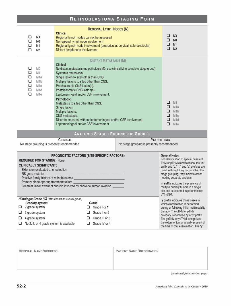
DISTANT METASTASIS (M)ClinicalNo distant metastasis (no pathologic M0; use clinical M to complete stage group)Systemic metastasis.Single lesion to sites other than CNSMultiple lesions to sites other than CNS.Prechiasmatic CNS lesion(s).Postchiasmatic CNS lesion(s).Leptomeningeal and/or CSF involvement.PathologicMetastasis to sites other than CNS.Single lesion.Multiple lesions.CNS metastasis.Discrete mass(es) without leptomeningeal and/or CSF involvement.Leptomeningeal and/or CSF involvement.
M1M1a M1bM1cM1dM1e
CLINICALNo stage grouping is presently recommended
PATHOLOGICNo stage grouping is presently recommended
PROGNOSTIC FACTORS (SITE-SPECIFIC FACTORS)REQUIRED FOR STAGING: NoneCLINICALLY SIGNIFICANT:
Extension evaluated at enucleation __________________________________RB gene mutation ________________________________________________Positive family history of retinoblastoma _______________________________Primary globe-sparing treatment failure _______________________________Greatest linear extent of choroid involved by choroidal tumor invasion _______
General Notes: For identification of special cases of TNM or pTNM classifications, the "m" suffix and "y," "r," and "a" prefixes are used. Although they do not affect the stage grouping, they indicate cases needing separate analysis.
m suffix indicates the presence of multiple primary tumors in a single site and is recorded in parentheses: pT(m)NM.
y prefix indicates those cases in which classification is performed during or following initial multimodality therapy. The cTNM or pTNM category is identified by a "y" prefix. The ycTNM or ypTNM categorizes the extent of tumor actually present at the time of that examination. The "y"
Histologic Grade (G) (also known as overall grade)Grading system2 grade system
GradeGrade I or 1
3 grade system Grade II or 2
4 grade system Grade III or 3
No 2, 3, or 4 grade system is available Grade IV or 4
R ETINOBLASTOMA S TAGING F ORM
HOSPITAL NAME/ADDRESS PATIENT NAME/ INFORMATION
A N A T O M I C S T A G E • P R O G N O S T I C G R O U P S
American Joint Committee on Cancer • 2010 52-3
(continued on next page)
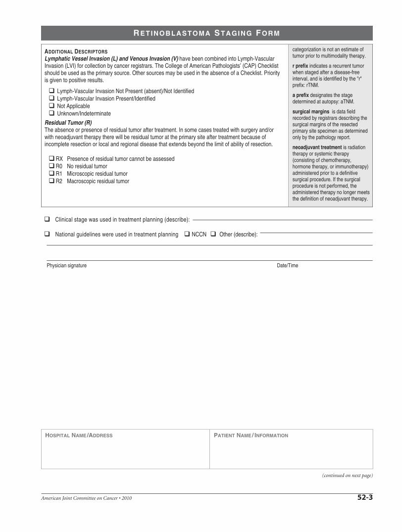
ADDITIONAL DESCRIPTORSLymphatic Vessel Invasion (L) and Venous Invasion (V) have been combined into Lymph-Vascular Invasion (LVI) for collection by cancer registrars. The College of American Pathologists’ (CAP) Checklist should be used as the primary source. Other sources may be used in the absence of a Checklist. Priority is given to positive results.
Lymph-Vascular Invasion Not Present (absent)/Not IdentifiedLymph-Vascular Invasion Present/IdentifiedNot ApplicableUnknown/Indeterminate
Residual Tumor (R)The absence or presence of residual tumor after treatment. In some cases treated with surgery and/or with neoadjuvant therapy there will be residual tumor at the primary site after treatment because of incomplete resection or local and regional disease that extends beyond the limit of ability of resection.
RX Presence of residual tumor cannot be assessedR0 No residual tumorR1 Microscopic residual tumorR2 Macroscopic residual tumor
Clinical stage was used in treatment planning (describe):
National guidelines were used in treatment planning NCCN Other (describe):
Physician signature Date/Time
categorization is not an estimate of tumor prior to multimodality therapy.
r prefix indicates a recurrent tumor when staged after a disease-free interval, and is identified by the "r" prefix: rTNM.
a prefix designates the stage determined at autopsy: aTNM.
surgical margins is data field recorded by registrars describing the surgical margins of the resected primary site specimen as determined only by the pathology report.
neoadjuvant treatment is radiation therapy or systemic therapy (consisting of chemotherapy, hormone therapy, or immunotherapy) administered prior to a definitive surgical procedure. If the surgical procedure is not performed, the administered therapy no longer meets the definition of neoadjuvant therapy.
R ETINOBLASTOMA S TAGING F ORM
HOSPITAL NAME/ADDRESS PATIENT NAME/ INFORMATION
52-4 American Joint Committee on Cancer • 2010
(continued from previous page)
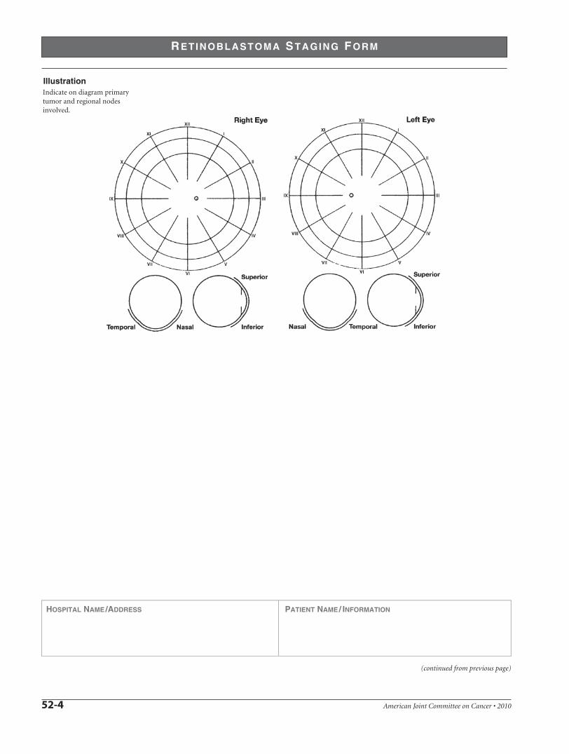
Indicate on diagram primarytumor and regional nodesinvolved.