Masthead Logo University of Pennsylvania ScholarlyCommons Master of Chemical Sciences Capstone Projects Department of Chemistry 5-2019 Reverse-Polarity Activity-Based Protein Profiling Suzanne Deling [email protected]Follow this and additional works at: hps://repository.upenn.edu/mcs_capstones is paper is posted at ScholarlyCommons. hps://repository.upenn.edu/mcs_capstones/19 For more information, please contact [email protected]. Deling, Suzanne, "Reverse-Polarity Activity-Based Protein Profiling" (2019). Master of Chemical Sciences Capstone Projects. 19. hps://repository.upenn.edu/mcs_capstones/19
Transcript
Masthead LogoUniversity of Pennsylvania
ScholarlyCommons
Master of Chemical Sciences Capstone Projects Department of Chemistry
5-2019
Reverse-Polarity Activity-Based Protein ProfilingSuzanne [email protected]
Follow this and additional works at: https://repository.upenn.edu/mcs_capstones
This paper is posted at ScholarlyCommons. https://repository.upenn.edu/mcs_capstones/19For more information, please contact [email protected].
Dettling, Suzanne, "Reverse-Polarity Activity-Based Protein Profiling" (2019). Master of Chemical Sciences Capstone Projects. 19.https://repository.upenn.edu/mcs_capstones/19
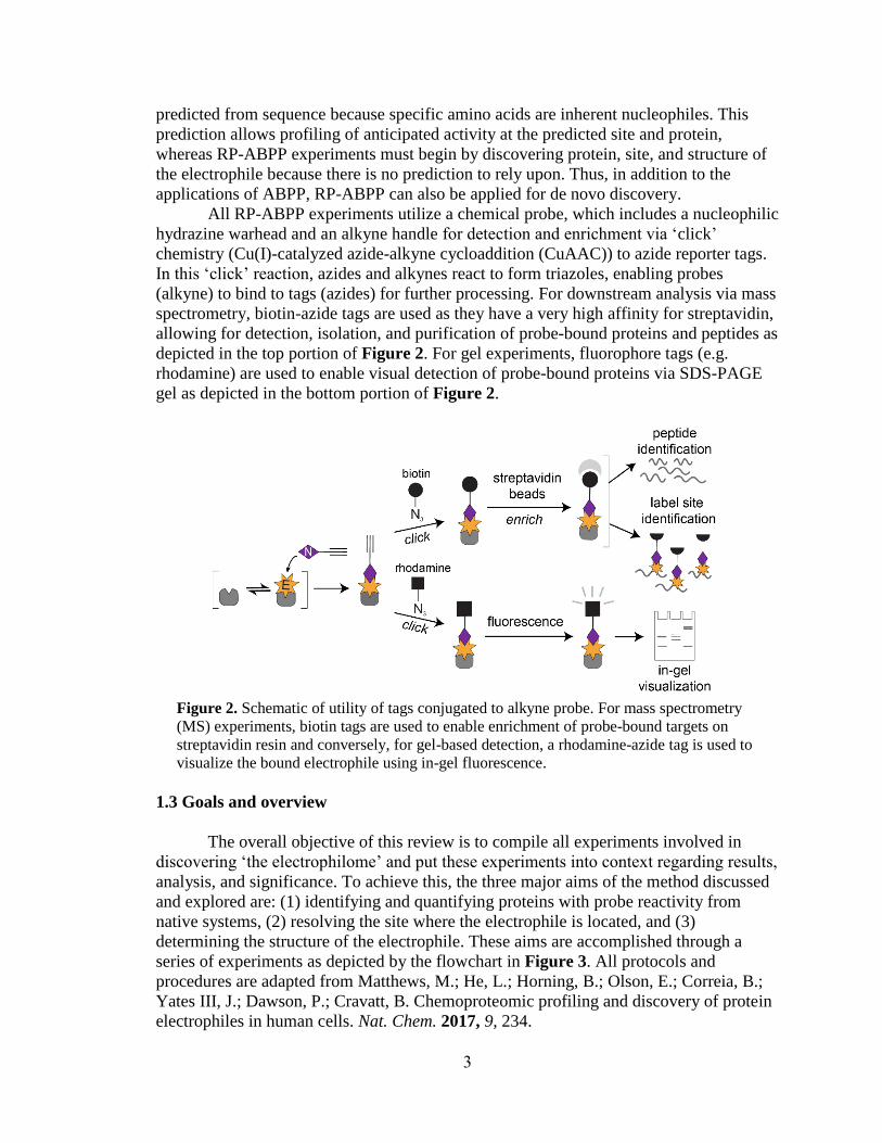
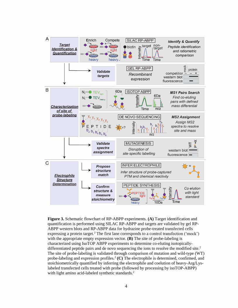
AbstractReverse-polarity activity-based protein profiling (RP-ABPP) is a chemical proteomics approach that usesclickable, nucleophilic hydrazine probes to capture and identify protein-bound electrophiles in cells. The RP-ABPP approach is used to characterize the structure and function of reactive electrophilic PTMs and theproteins that harbor them, which may uncover unknown or novel functions of proteins in an endogenoussetting. RP-ABPP has demonstrated utility as a versatile method to monitor metabolic regulation ofelectrophilic cofactors, as was done with the pyruvoyl cofactor in S-adenosyl-L- methionine decarboxylase(AMD1) and discover novel types of electrophilic modifications on proteins in human cells, as was done withthe glyoxylyl modification on secernin-3 (SCRN3). These cofactors cannot be predicted by sequence and assuch this area is relatively undeveloped. RP-ABPP is the only global unbiased approach to discover theseelectrophiles. Here, the utility of these experiments is described and a detailed protocol is provided for denovo discovery, quantitation, and global profiling of electrophilic functionality of proteins through the use ofnitrogenous nucleophilic probes deployed directly to living cells in culture.
Keywordsreverse-polarity activity-based protein profiling, chemical proteomics, enzyme activity, enzyme cofactors,post-translational modifications, de novo PTM discovery
Creative Commons LicenseCreativeCommonsLicenseThis work is licensed under a Creative Commons Attribution-Noncommercial-Share Alike 4.0 License.
This capstone report is available at ScholarlyCommons: https://repository.upenn.edu/mcs_capstones/19
~1.5−2 hours at ambient temperature on a rotator. Then, to remove unbound protein,
excess detergent, and small molecules, streptavidin beads are collected by centrifugation
(1,400 g, 1−2 min) and sequentially washed with 0.2% SDS in PBS (3 × ~10 mL),
detergent-free PBS (3 × ~10 mL) and H2O (3 × ~10 mL). To perform the digestion, the
resin is transferred to a Protein LoBind tube and bound proteins are digested on-bead
overnight at 37 °C in ~200 μL total volume containing sequencing grade porcine trypsin
(2 μg, Promega) in the presence of urea (2 M in PBS) and CaCl2 (1 mM). In the final
10
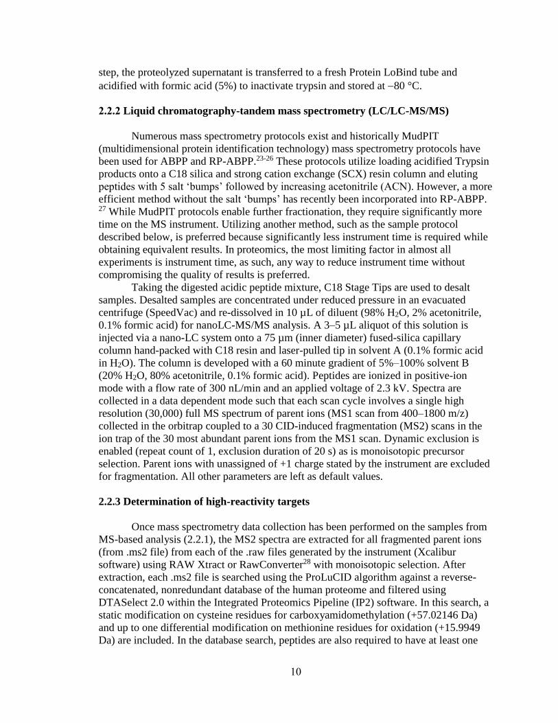
step, the proteolyzed supernatant is transferred to a fresh Protein LoBind tube and
acidified with formic acid (5%) to inactivate trypsin and stored at −80 °C.
2.2.2 Liquid chromatography-tandem mass spectrometry (LC/LC-MS/MS)
Numerous mass spectrometry protocols exist and historically MudPIT
(multidimensional protein identification technology) mass spectrometry protocols have
been used for ABPP and RP-ABPP.23-26 These protocols utilize loading acidified Trypsin
products onto a C18 silica and strong cation exchange (SCX) resin column and eluting
peptides with 5 salt ‘bumps’ followed by increasing acetonitrile (ACN). However, a more
efficient method without the salt ‘bumps’ has recently been incorporated into RP-ABPP. 27 While MudPIT protocols enable further fractionation, they require significantly more
time on the MS instrument. Utilizing another method, such as the sample protocol
described below, is preferred because significantly less instrument time is required while
obtaining equivalent results. In proteomics, the most limiting factor in almost all
experiments is instrument time, as such, any way to reduce instrument time without
compromising the quality of results is preferred.
Taking the digested acidic peptide mixture, C18 Stage Tips are used to desalt
samples. Desalted samples are concentrated under reduced pressure in an evacuated
centrifuge (SpeedVac) and re-dissolved in 10 µL of diluent (98% H2O, 2% acetonitrile,
0.1% formic acid) for nanoLC-MS/MS analysis. A 3–5 µL aliquot of this solution is
injected via a nano-LC system onto a 75 µm (inner diameter) fused-silica capillary
column hand-packed with C18 resin and laser-pulled tip in solvent A (0.1% formic acid
in H2O). The column is developed with a 60 minute gradient of 5%–100% solvent B
(20% H2O, 80% acetonitrile, 0.1% formic acid). Peptides are ionized in positive-ion
mode with a flow rate of 300 nL/min and an applied voltage of 2.3 kV. Spectra are
collected in a data dependent mode such that each scan cycle involves a single high
resolution (30,000) full MS spectrum of parent ions (MS1 scan from 400–1800 m/z)
collected in the orbitrap coupled to a 30 CID-induced fragmentation (MS2) scans in the
ion trap of the 30 most abundant parent ions from the MS1 scan. Dynamic exclusion is
enabled (repeat count of 1, exclusion duration of 20 s) as is monoisotopic precursor
selection. Parent ions with unassigned of +1 charge stated by the instrument are excluded
for fragmentation. All other parameters are left as default values.
2.2.3 Determination of high-reactivity targets
Once mass spectrometry data collection has been performed on the samples from
MS-based analysis (2.2.1), the MS2 spectra are extracted for all fragmented parent ions
(from .ms2 file) from each of the .raw files generated by the instrument (Xcalibur
software) using RAW Xtract or RawConverter28 with monoisotopic selection. After
extraction, each .ms2 file is searched using the ProLuCID algorithm against a reverse-
concatenated, nonredundant database of the human proteome and filtered using
DTASelect 2.0 within the Integrated Proteomics Pipeline (IP2) software. In this search, a
static modification on cysteine residues for carboxyamidomethylation (+57.02146 Da)
and up to one differential modification on methionine residues for oxidation (+15.9949
Da) are included. In the database search, peptides are also required to have at least one
11
tryptic terminus but allowed an unlimited number of missed cleavages. Additionally, the
exact mass shift of heavy atoms on specific amino acids from the SILAC media is
searched (e.g. if using 13C and 15N on Lysine and Arginine, K +8.0142 Da and R
+10.0082 Da). This can be done by performing a coupled ‘heavy’ search on each dataset
for both light and heavy isotopologues of the same peptide by specifying the mass shift of
heavy residues as static modifications on lysine (+8.0142 Da) and arginine (+10.0082
Da).
To further filter the data, the parent ion mass tolerance for a minimum envelope
of three isotopic peaks is set to 50 ppm, the minimum peptide length set to six residues,
the false positive rate set to 1%, and at least 2 peptides of a protein must be detected.
Heavy and light parent ion chromatograms associated with successfully identified
peptides are extracted and compared using CIMAGE software.25 Data is filtered so that at
least one ion of a co-eluting heavy-light pair must be accurately identified from a
fragmentation event that occurred within the retention time window (± 10 minutes) of
parent ion elution. Furthermore, to ensure that the correct pair of peaks is quantified,
chromatograms within a 10 ppm error tolerance of the predicted m/z, single-to-noise
ratios greater than 2.5, and ‘co-elution correlation scores’ and ‘envelope correlation
scores’ R2 values greater than or equal to 0.8 are extracted. To further eliminate false
positives and stochastic variability in the data, protein ratios are determined by median
peptide ratio derived from three or more unique qualified peptides. To provide final
values, protein ratios that comply with these criteria from a single experiment are
averaged with ratios acquired from at least three replicates.
2.3 Validation via recombinant expression
Once high-reactivity targets have been determined, targets can be validated by
demonstrating hydrazine reactivity as an intrinsic property of the protein targets that is
shared by both the endogenous and recombinant forms of these proteins. This validation
is performed by treating transfected cells with probe, followed by conjugation to a
fluorophore tag (e.g. rhodamine), and visualization of a strong fluorescent band at the
appropriate molecular weight. This band should be absent in ‘mock’ transfected cells and
in cells treated with excess non-clickable agents as this should block probe-labeling of
each protein. Additionally, recombinant expression of each protein (and lack of
expression in ‘mock’ transfected control cells) is confirmed by western blotting. In order
to perform these transfections, target genes need to be obtained in mammalian expression
vectors. These vectors can be purchased or made using cloning protocols.2 Sample
procedures for transfection and gel-based analysis (Section 2.3.1) are described below.
To begin the target validation experiment, standard growth conditions are
followed and cells are grown to ~40% confluence. The appropriate expression vector
[control cells (‘mock’) receive an equal amount of the appropriate empty vector] is added
as well as polyethyleneimine (PEI) ‘MAX’ (MW 40,000) as a transfection reagent under
standard transfection conditions [3:1 vector/PEI (w/w) ratio]. Cells are incubated with
transfection reagents for ~48 h before labeling in situ (2.1.1) and preparation of the
proteome (2.1.2).
12
2.3.1 Gel-based analysis of probe-labeled proteins
Following transfection (Section 2.3) and labeling (Section 2.1.1) and proteome
preparation (Section 2.1.2), proteomes from treated cells are diluted to 1 mg/mL. To
conjugate the fluorophore to probe-labeled proteins, 6 μL of a freshly prepared “click”
reagent mixture containing 0.1 mM TBTA (3 μL/sample, 1.7 mM in 4:1 DMSO:t-
BuOH), 1 mM CuSO4 (1 μL/sample, 50 mM in H2O), 25 μM azide-rhodamine (1
μL/sample, 1.25 mM in DMSO), and freshly prepared 1mM TCEP (1 μL/sample, 50 mM
in PBS or H2O) is added to each sample (50 μL). Immediately upon addition of the click
mixture, the samples are mixed by vortexing and the mixture is allowed to react at
ambient temperature for 1 hour. The ‘click’ reaction is quenched with SDS loading buffer
(4× stock, 17 μL) and proteins are resolved (~25 μg total protein loaded per gel lane) by
SDS-PAGE (10% acrylamide gels). Labeling is visualized using in-gel fluorescence
scanning on a flatbed fluorescence scanner (e.g. BioRad ChemiDoc MP). The same gel is
transferred to nitrocellulose membrane and western blotted using standard protocols.2
2.4 Conclusion
As seen above and will be evidenced throughout the review, most RP-ABPP
experiments build upon on another. For example, isoTOP ABPP experiments start with
in-situ labeling (Section 2.1.1) and proteome preparation (Section 2.1.2) and parts of MS-
based analysis (Section 2.2.1) are repeated as well. Additionally, mutagenic analysis
relies upon gel-based analysis (Section 2.3.1) and begins with in-situ labeling (Section
2.1.1) and proteome preparation (Section 2.1.2) as well. It is important to note that these
experiments are very rigorous. They require planning and attention to detail and have few
acceptable pause points. One of the most critical portions of target identification and
quantification is the equilibration and washing of the SILAC RP-ABPP samples on
streptavidin beads. This step is critical because if the beads are washed very vigorously,
some beads may be lost, causing low peptide signal. Conversely, if the beads are not
washed vigorously enough, too many contaminants (e.g. unbound protein, excess
detergent, small molecules, etc.) may be present, which decreases the ability to detect
proteins of interest. These risks could be mitigated through use of magnetic beads, which
are generally used for high-throughput applications. However, these beads are very
expensive and require additional equipment.
The results generated from target identification and quantification are quite
robust. When RP-ABPP was performed in two cell lines with two probes, eleven high-
reactivity targets were identified and only two of these proteins were previously known to
harbor an electrophile.2 Due to the promise of these initial results, performing
identification and quantification with other probes and cell lines is likely to widely
expand the knowledge of known functional electrophiles.
13
Chapter 3. Characterization of the probe-captured site
The second aim of RP-ABPP is to determine the site of electrophilic reactivity on
the protein. The peptide labeled is found using isoTOP-ABPP25, 29 experiments and the
specific amino-acid residue labeled is found using de novo sequencing. These results are
validated using site-specific mutagenic analysis. Characterization of the electrophilic site
yields information about where the reactivity is located within the greater context of the
protein. Once this site is determined, this paves the way for a large variety of downstream
experiments including monitoring the electrophile, examining its installation, and
potentially investigating its function. Additionally, after the site harboring the
electrophile is identified, this area can be further interrogated to determine the structure
of the electrophile.
3.1 IsoTOP ABPP
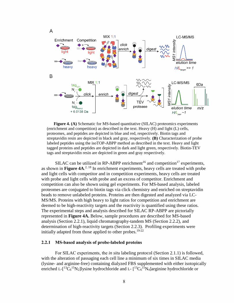
IsoTOP ABPP is used to determine the peptide labeled, which is depicted in
Figure 4B. Following probe treatment of non-SILAC cells (shown in gray in Figure 4B),
this method utilizes conjugation to heavy and light cleavable biotin-TEV tags (shown in
green in Figure 4B). After conjugation to heavy and light tags, labeled proteomes are
enriched on streptavidin beads and on bead digestion is performed. After digestion, all
unlabeled peptides are discarded as shown in Figure 4B. The remaining labeled peptides
are released from the beads through cleavage of the tag (by TEV protease), ultimately
generating probe-labeled peptides as mass differentiated pairs. Using the coeluting pair
with heavy and light tags (plotted in dark and light green, respectively, in Figure 4B), the
peptide harboring the labeled residue can be found. Determining which peptide is labeled
can give information about where the reactivity is located in the sequence of the protein
(e.g. active site, N-terminus, etc.). These searches enable differentiation between probe-
labeled peptides and other peptides in the sample regardless of identity or mass. A sample
isoTOP-ABPP procedure (Sections 3.1.1-3.1.2) is described below.
3.1.1 IsoTOP ABPP sample preparation to isolate probe-captured peptides
IsoTOP ABPP begins with probe treatment of wild-type or transfected cells in-
situ (Section 2.1.1) and proteome preparation (Section 2.1.2). After performing these
protocols, soluble proteomes (2 mg total protein) are diluted to 1 mL in PBS. To
conjugate half of the proteome (0.5 mL) to the light TEV tag and the other half to the
heavy TEV tag, click reactions are scaled accordingly to maintain final concentrations of
0.1 mM TBTA, 1 mM CuSO4, 100 μM of light or heavy biotin-TEV-azide (5 mM in
DMSO) and 1 mM TCEP. The mixture is vortexed and placed on a rotator at ambient
temperature. Once the mixture has rotated for 1 hour, it is centrifuged (16,000 g, 5
minutes, 4 °C) and resulting pellets are mildly sonicated in ice-cold methanol (0.5 mL).
The light- and heavy-labeled proteomes are combined and centrifuged once more. To
solubilize the proteomes, 1.2% SDS (1 mL in PBS) is added and samples are stored at –
80 °C overnight.
After leaving the samples in –80 °C overnight, samples are diluted to ~0.2% SDS
with PBS (~5 mL) and incubated with pre-equilibrated streptavidin agarose resin (100 μL
14
1:1 slurry) for ~2–3 hours at ambient temperature. To remove un-labeled proteins, resin
is washed as described above in the SILAC RP-ABPP MS-based analysis procedure
(Section 2.2.1), and after the washes, the resin is transferred to clean Eppendorf tubes and
resuspended in urea (500 μL, 6M in PBS). Cysteines are then reduced and alkylated with
TCEP and iodoacetamide, respectively, as described above in the SILAC RP-ABPP MS-
based analysis procedure (Section 2.2.1). To remove reagents, resin is washed once with
PBS and bound proteins are digested with sequencing grade porcine trypsin (Promega, 2
μg) for 8–12 h at 37 °C in the presence of 2 M urea (200 μL, in PBS) and CaCl2 (1 mM).
After trypsin digestion, sequential washes with PBS (5 × 0.5 mL) and H2O (5 ×
0.5 mL) are performed to remove unmodified peptides, urea, and trypsin. The resin is
transferred to fresh tubes and equilibrated with TEV buffer (50 mM Tris, pH 8). To
release the remaining immobilized peptides, TEV protease is added (~1–2 μM in ~200
μL TEV buffer at 30 °C for 3–5 hours). Heavy- and light-labeled peptides are collected
and recovered from the resin with H2O (2 × 50 μL). Samples are stored at –80 °C and
must be analyzed within several days.
3.1.2 Characterization of probe-labeled peptides by isoTOP ABPP
To analyze the samples generated from the isoTOP preparation, data is collected
from isolated probe-captured peptides using mass spectrometry protocols as described
above in the SILAC RP-ABPP procedure (Section 2.2.2). Then, the data is searched on
the MS1 level for paired spectra of mass differentiated coeluting peaks as shown on the
right in Figure 5. Every recorded monoisotopic precursor mass is searched 6.0138 Da (±
5 ppm) upstream and downstream in each total ion (MS1) spectrum for a possible
isotopic partner taking into account +2 and +3 charge states (3.0069 and 2.0046 Da,
respectively). The relative intensity of the monoisotopic peaks is required to be greater
than or equal to 5% of the base peak of each spectrum and each isotope profile (envelope)
must have at least three peaks. Additionally, the Euclidean distance between two isotope
profiles must be greater than or equal to 0.2. Pairs with the same m/z values (± 5 ppm)
and retention times (± 10 minutes) are grouped to eliminate duplicates. Pairs of parent
ions from transfected versus mock-transfected cells are analyzed and pairs of parent ions
from three biological replicates should be analyzed as well.
3.2 Fragmentation spectra assignment by de novo sequencing
To further resolve the site labeled and the mass of the probe-captured PTM, the
MS2 spectra from the isoTOP experiments is assigned via de novo sequencing by
extraction of b and y ions and assignment spectra to their respective amino acids. The
residue containing the modification can be identified as it will contain the known mass
shift of the fragmented heavy/light tag, containing covalently bound probe and
conjugated biotin-TEV tag. After all peaks are identified as either an amino acid or amino
acid with fragmented tag, the mass of the bound electrophile can be calculated. A sample
procedure is described below.
15
Figure 5. Search strategy to determine probe-labeled site from isoTOP ABPP data. To
determine the peptide containing the modification, MS1 searches for coeluting pair with
the specified mass difference are performed. To determine the residue labeled, MS2 spectra
are assigned by de novo sequencing.
Using the MS2 spectra generated from the isoTOP experiments, spectra are
assigned manually as depicted on the left in Figure 5. It may be necessary for the
instrument method to be modified such that both the parent and fragment ions are
measured in the orbitrap to gain high-resolution data for both to be certain of the peak
assignments and charge state of the tag-specific b-ions. Only the five most abundant
parent ions for fragmentation per cycle (versus 30 if the spectra are collected in the ion
trap) are selected to account for increased scan time. The number of MS2 spectra
collected per cycle is extended to ten if the data is generated on the Fusion Orbitrap. The
MS2 spectra is searched for diagnostic ions of the peptide. Highly abundant fragments
that represent diagnostic markers for the unmodified portion of the peptide and other
fragmentation products representing the portion that contains the modification and that is
differentially labeled by the isotopic tags should be found. The spectra for the heavy and
light peptides are compared and the ion shift should be observed (6.0138 Da).
RawConverter can be used to extract accurate monoisotopic m/z values for each MS2
spectra. Downstream structure determination will be based on the correct precursor mass
calculated, which will be further discussed in Chapter 4.
16
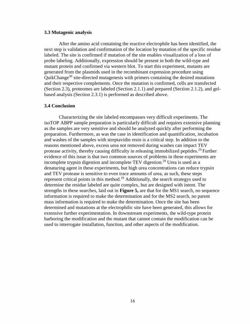
3.3 Mutagenic analysis
After the amino acid containing the reactive electrophile has been identified, the
next step is validation and confirmation of the location by mutation of the specific residue
labeled. The site is confirmed if mutation of the site enables visualization of a loss of
probe labeling. Additionally, expression should be present in both the wild-type and
mutant protein and confirmed via western blot. To start this experiment, mutants are
generated from the plasmids used in the recombinant expression procedure using
QuikChange30 site-directed mutagenesis with primers containing the desired mutations
and their respective complements. Once the mutation is confirmed, cells are transfected
(Section 2.3), proteomes are labeled (Section 2.1.1) and prepared (Section 2.1.2), and gel-
based analysis (Section 2.3.1) is performed as described above.
3.4 Conclusion
Characterizing the site labeled encompasses very difficult experiments. The
isoTOP ABPP sample preparation is particularly difficult and requires extensive planning
as the samples are very sensitive and should be analyzed quickly after performing the
preparation. Furthermore, as was the case in identification and quantification, incubation
and washes of the samples with streptavidin resin is a critical step. In addition to the
reasons mentioned above, excess urea not removed during washes can impact TEV
protease activity, thereby causing difficulty in releasing immobilized peptides.29 Further
evidence of this issue is that two common sources of problems in these experiments are
incomplete trypsin digestion and incomplete TEV digestion.29 Urea is used as a
denaturing agent in these experiments, but high urea concentrations can reduce trypsin
and TEV protease is sensitive to even trace amounts of urea, as such, these steps
represent critical points in this method.29 Additionally, the search strategys used to
determine the residue labeled are quite complex, but are designed with intent. The
strengths in these searches, laid out in Figure 5, are that for the MS1 search, no sequence
information is required to make the determination and for the MS2 search, no parent
mass information is required to make the determination. Once the site has been
determined and mutations at the electrophilic site have been generated, this allows for
extensive further experimentation. In downstream experiments, the wild-type protein
harboring the modification and the mutant that cannot contain the modification can be
used to interrogate installation, function, and other aspects of the modification.
17
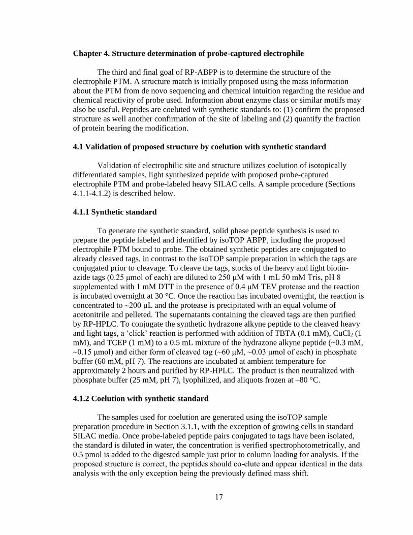
Chapter 4. Structure determination of probe-captured electrophile
The third and final goal of RP-ABPP is to determine the structure of the
electrophile PTM. A structure match is initially proposed using the mass information
about the PTM from de novo sequencing and chemical intuition regarding the residue and
chemical reactivity of probe used. Information about enzyme class or similar motifs may
also be useful. Peptides are coeluted with synthetic standards to: (1) confirm the proposed
structure as well another confirmation of the site of labeling and (2) quantify the fraction
of protein bearing the modification.
4.1 Validation of proposed structure by coelution with synthetic standard
Validation of electrophilic site and structure utilizes coelution of isotopically
differentiated samples, light synthesized peptide with proposed probe-captured
electrophile PTM and probe-labeled heavy SILAC cells. A sample procedure (Sections
4.1.1-4.1.2) is described below.
4.1.1 Synthetic standard
To generate the synthetic standard, solid phase peptide synthesis is used to
prepare the peptide labeled and identified by isoTOP ABPP, including the proposed
electrophile PTM bound to probe. The obtained synthetic peptides are conjugated to
already cleaved tags, in contrast to the isoTOP sample preparation in which the tags are
conjugated prior to cleavage. To cleave the tags, stocks of the heavy and light biotin-
azide tags (0.25 μmol of each) are diluted to 250 μM with 1 mL 50 mM Tris, pH 8
supplemented with 1 mM DTT in the presence of 0.4 μM TEV protease and the reaction
is incubated overnight at 30 °C. Once the reaction has incubated overnight, the reaction is
concentrated to ~200 μL and the protease is precipitated with an equal volume of
acetonitrile and pelleted. The supernatants containing the cleaved tags are then purified
by RP-HPLC. To conjugate the synthetic hydrazone alkyne peptide to the cleaved heavy
and light tags, a ‘click’ reaction is performed with addition of TBTA (0.1 mM), CuCl2 (1
mM), and TCEP (1 mM) to a 0.5 mL mixture of the hydrazone alkyne peptide (~0.3 mM,
~0.15 μmol) and either form of cleaved tag (~60 μM, ~0.03 μmol of each) in phosphate
buffer (60 mM, pH 7). The reactions are incubated at ambient temperature for
approximately 2 hours and purified by RP-HPLC. The product is then neutralized with
phosphate buffer (25 mM, pH 7), lyophilized, and aliquots frozen at –80 °C.
4.1.2 Coelution with synthetic standard
The samples used for coelution are generated using the isoTOP sample
preparation procedure in Section 3.1.1, with the exception of growing cells in standard
SILAC media. Once probe-labeled peptide pairs conjugated to tags have been isolated,
the standard is diluted in water, the concentration is verified spectrophotometrically, and
0.5 pmol is added to the digested sample just prior to column loading for analysis. If the
proposed structure is correct, the peptides should co-elute and appear identical in the data
analysis with the only exception being the previously defined mass shift.
18
4.2 Determination of absolute stoichiometry with synthetic isotopolgues
The fraction of modified protein can be determined by coeluting (light) synthetic
modified and internal standards with (heavy) endogenous modified and internal peptides
as depicted in Figure 6. In these experiments, the internal peptide represents the total
protein in the cell (both modified and unmodified protein). Both the endogenous
modified and internal peptides absolute quantities are able to be calculated using the
known amount of respective standards added to the sample and the relative peak areas of
the standard to the endogenous peptides as shown by fractional occupancy in Figure 6. A
sample procedure for these experiments is described below.
Figure 6. Scheme of stoichiometry measurement for modified peptide. Using a known
quantity of synthetic standards of modified and internal peptides enables absolute
quantification of the endogenous modified and total protein, allowing for calculation of
fractional occupancy.
To generate the endogenous peptides in stoichiometric quantification, cells grown
in heavy SILAC media are transfected and probe treated (Section 2.1.1) using procedures
described above. The plasmids transfected must contain genes for the protein and FLAG
tag for downstream purification. Cells are lysed in PBS (pH 7.4), fractionated by
ultracentrifugation (100,000 g, 30–45 minutes), and the samples diluted to 1 mL with 50
mM Na-HEPES buffer (pH 7.5) supplemented with 500 mM NaCl and 1% Triton X-100.
Unsolubilized protein remaining in the sample is pelleted by centrifugation and denatured
with a small volume 10% SDS for 1 hour at 37 °C. The completely resolubilized sample
is incubated at 4 °C overnight by rotation with anti-FLAG resin. The resin is washed by
19
resuspension and centrifugation with the same buffer supplemented with 500 mM NaCl
(5 × 1 mL) followed by 100 mM NaCl (2 × 1 mL). The bound protein is eluted by
incubating the beads for ~1 h at 37 °C in PBS containing 8 M urea (2 × 50 μL). Cysteines
are reduced with TCEP (10 mM pre-neutralized with 30 mM potassium carbonate for 30
minutes at 37 °C) and alkylated with iodoacetamide (20 mM under the same conditions
but protected from light). The samples are diluted to 2M urea with PBS and digested at
37 °C overnight with 2 μg trypsin supplemented with 1 mM CaCl2. Trypsin is inactivated
with 5% formic acid. The natural abundance probe-labeled peptide standard generated
above is diluted in PBS and the digested protein sample with the modified standard as
well as an internal peptide that represents the total protein (5–50 pmol of each) are doped
in just prior to analysis by the same method used for proteomic profiling. Absolute
amounts of standards should be adjusted to achieve nearly comparable peak intensities
for quantitation.
4.3 Conclusion
Throughout RP-ABPP, coelution of isotopically differentiated samples is
thoroughly utilized. This principle is used for SILAC, isoTOP, and synthetic standard
experiments. RP-ABPP has been used for de novo discovery of a new previously
unknown modification2 and there is the potential that other human proteins may harbor
previously unknown electrophilic modifications. As such, electrophilic structure
determination experiments could yield novel and far-reaching results.
20
Chapter 5. Conclusions
5.1 Previous results and significance
Electrophilic modifications are not easily predicted by sequence and sequence
predictions do not yield information about function or activity. Initial results from RP-
ABPP have yielded eleven targets and these targets are largely functional and strongly
implicated in disease.2 Additionally, nine of the eleven targets were not previously known
to harbor an electrophile and on one of these targets, SCRN3, a novel previously
undiscovered modification, the glyoxylyl, was discovered.2 The results of the RP-ABPP
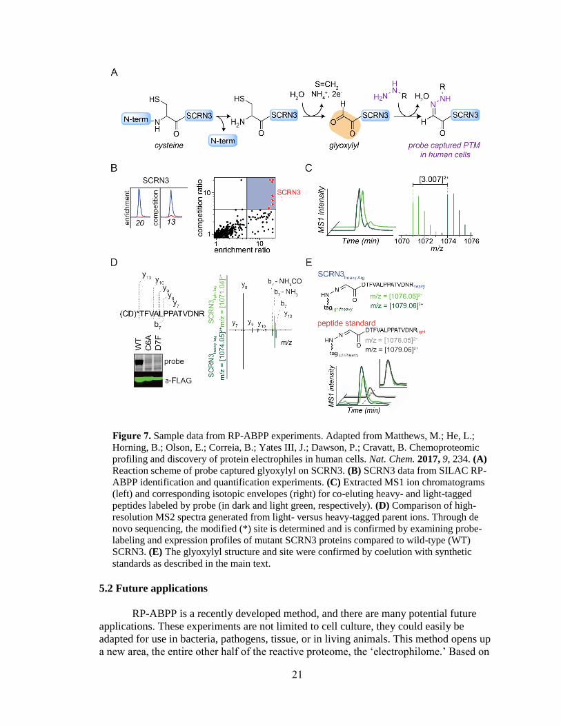
experiments used to discover the glyoxylyl are depicted in Figure 7. Figure 7A depicts
the probe reaction with the gyloxylyl. Identification and quantification of SCRN3 as a
target is shown in Figure 7B with extracted parent ion chromatograms and corresponding
heavy to light ratios for tryptic peptides of SCRN3 protein probe-treated cells quantified
in enrichment and competition (left) and quadrant plot of average competition versus
enrichment SILAC ratios from quantitative proteomics experiments (right).2 In Figure
7C, isoTOP ABPP MS1 ion chromatograms (left) and isotopic envelopes (right)
demonstrate coelution and specific mass differentiation of the labeled SCRN3 peptide.2
Figure 7D depicts de novo sequencing to determine the residue labeled and the
mutagenesis gel and western blot validate the result that both C6 and D7 must be present
for labeling to occur, because the y-ions resolve the modified site (Figure 7*) to the N-
terminal cysteine and/or adjacent aspartate and mutation profiles of Cys6-to-Ala6 (C6A)
and Asp7-to-Phe7 (D7F) mutant SCRN3 proteins compared to wild-type (WT) SCRN3
show a lack of probe labeling.2 In Figure 7E, heavy-Arg/Lys-labeled SCRN3-transfected
cells treated with probe, followed by processing by isoTOP-ABPP, yields an isotopically
differentiated probe-labeled SCRN3 peptide pair (light and dark green), which co-elutes
with a light amino acid-labeled probe-Glyoxylyl6-Arg20 standard (also an isotopically
differentiated peptide pair; light and dark gray).2 Inset chromatogram shows all four
traces scaled to the same intensity to show co-elution of endogenous and standard probe-
Glyoxylyl6-Arg20 SCRN3 peptides.2 The discovery of the glyoxylyl is significant
because it demonstrates the utility of RP-ABPP in discovering new unknown
electrophilic modifications. The results of these initial experiments indicate that other
human proteins may harbor electrophilic groups and that there are other unknown
modifications to be discovered. As such, the advent of this method is critical due to RP-
ABPP being the only global unbiased approach to discover electrophilic cofactors.
21
Figure 7. Sample data from RP-ABPP experiments. Adapted from Matthews, M.; He, L.;
Horning, B.; Olson, E.; Correia, B.; Yates III, J.; Dawson, P.; Cravatt, B. Chemoproteomic
profiling and discovery of protein electrophiles in human cells. Nat. Chem. 2017, 9, 234. (A)
Reaction scheme of probe captured glyoxylyl on SCRN3. (B) SCRN3 data from SILAC RP-
ABPP identification and quantification experiments. (C) Extracted MS1 ion chromatograms
(left) and corresponding isotopic envelopes (right) for co-eluting heavy- and light-tagged
peptides labeled by probe (in dark and light green, respectively). (D) Comparison of high-
resolution MS2 spectra generated from light- versus heavy-tagged parent ions. Through de
novo sequencing, the modified (*) site is determined and is confirmed by examining probe-
labeling and expression profiles of mutant SCRN3 proteins compared to wild-type (WT)
SCRN3. (E) The glyoxylyl structure and site were confirmed by coelution with synthetic
standards as described in the main text.
5.2 Future applications
RP-ABPP is a recently developed method, and there are many potential future
applications. These experiments are not limited to cell culture, they could easily be
adapted for use in bacteria, pathogens, tissue, or in living animals. This method opens up
a new area, the entire other half of the reactive proteome, the ‘electrophilome.’ Based on
22
targets generated from only two probes in two human cell lines,2 there are more
electrophile-bound proteins to be discovered and disease-relationships to be explored. In
addition to discovery of protein targets and electrophilic modifications, the tools in this
method can also be used to further investigate various aspects of the proteins and
modifications discovered. For example, discovery of installation mechanisms and
functions of these electrophiles could yield new information about the proteins and
mechanisms in these cells. Additionally, as many of the protein targets discovered have