+ All Categories
Home > Documents > Reversible bi -directional bending of hydrogel-based ...

Reversible bi -directional bending of hydrogel-based ...

Date post: 16-Mar-2022
Category:
Upload: others
View: 1 times
Download: 0 times
Share this document with a friend
30
Reversible bi-directional bending of hydrogel-based bilayer actuators Xue Li, Xiangbin Cai, Yongfeng Gao, Michael J. Serpe* Department of Chemistry, University of Alberta, Edmonton, AB T6G 2G2, Canada *Corresponding Author[email protected] Abstract Temperature and pH responsive semi-interpenetrating network (semi-IPN) hydrogel based bilayer actuators were fabricated by generating a poly (N-isopropylacrylamide) (pNIPAm)-based hydrogel in the presence of positively charged polyelectrolyte poly (diallyldimethylammonium chloride) (pDADMAC) on a layer of gold-coated polydimethylsiloxane (PDMS). The bilayers showed unique bidirectional bending behavior in response to solution temperature and pH, which is a result of the modulation of the hydrogel swelling state in response to solution temperature and pH changes. The behavior described here is vastly different than what is observed from bilayers composed of just pNIPAm. The direction and degree of bending of the device could be easily adjusted by tuning the composition of the hydrogel layer. We also showed that the bilayers could be used as stimulus-induced grippers and for controlled/triggered small molecule delivery, which can make the bilayers useful for various biomedical applications, among other things. Abstract: Stimuli-responsive polymers; bilayer actuators; artificial muscles; drug delivery 1. Introduction Stimuli responsive ("smart") polymer-based hydrogels are highly water swellable
Transcript

Reversible bi-directional bending of hydrogel-based bilayer actuators

Xue Li, Xiangbin Cai, Yongfeng Gao, Michael J. Serpe*

Department of Chemistry, University of Alberta, Edmonton, AB T6G 2G2, Canada

*Corresponding Author—[email protected]

Abstract

Temperature and pH responsive semi-interpenetrating network (semi-IPN) hydrogel

based bilayer actuators were fabricated by generating a poly (N-isopropylacrylamide)

(pNIPAm)-based hydrogel in the presence of positively charged polyelectrolyte poly

(diallyldimethylammonium chloride) (pDADMAC) on a layer of gold-coated

polydimethylsiloxane (PDMS). The bilayers showed unique bidirectional bending

behavior in response to solution temperature and pH, which is a result of the

modulation of the hydrogel swelling state in response to solution temperature and pH

changes. The behavior described here is vastly different than what is observed from

bilayers composed of just pNIPAm. The direction and degree of bending of the device

could be easily adjusted by tuning the composition of the hydrogel layer. We also

showed that the bilayers could be used as stimulus-induced grippers and for

controlled/triggered small molecule delivery, which can make the bilayers useful for

various biomedical applications, among other things.

Abstract: Stimuli-responsive polymers; bilayer actuators; artificial muscles; drug

delivery

1. Introduction

Stimuli responsive ("smart") polymer-based hydrogels are highly water swellable

crosslinked polymer networks,1 which undergo abrupt volume changes in response to

changes in their environment, e.g., temperature, pH, ionic strength, exposure to

specific small molecules and application of an electric field.2, 3, 4, 5 Consequently, they

play a very important role in many practical applications such as sensing,6, 7 drug

delivery,8, 9 soft actuators and robotics,4, 10 and artificial muscles.11, 12 While traditional

hydrogel networks are held together by covalent crosslinks, there are a class of

hydrogels called interpenetrating polymer networks (IPN) that are formed by

combining two polymers (at least one should be responsive) that have some physical

interactions that hold the network together.13 Additionally, there are semi-IPNs that

are composed of a covalently crosslinked hydrogel infused with another linear

polymer physically interpenetrated inside the network.14 Compared with single

network hydrogels, it is possible for the components of the IPNs to work together to

yield materials with new behavior that is not expected from the responses of the

individual components alone.15 Most research on IPNs is focused on gaining a better

understanding of their fundamental properties (e.g. their swelling/deswelling

behavior),16, 17 on their practical applications as biomaterials18 and as novel sorbents

for separations.19 Here, for the first time we describe a novel semi-IPN

hydrogel-based actuator composed of two layers. One layer is composed of a poly

(N-isopropylacrylamide) (pNIPAm)-based hydrogel interpenetrated with linear poly

(diallyldimethylammonium chloride) (pDADMAC); the other layer is

polydimethylsiloxane (PDMS).

PNIPAm is one of the most well-known temperature responsive

(thermoresponsive) polymers, which undergoes a coil-to-globule transition at

temperatures above its lower critical solution temperature (LCST) of ~32 oC.20 Like

linear pNIPAm, pNIPAm-based hydrogels undergo a transition from a swollen to a

collapsed state upon heating above pNIPAm’s LCST. PNIPAM's thermoresponsivity

has been exploited for a variety of applications, including cell culturing,21 drug

delivery,22 and soft actuators.23 Introducing ionic groups into the neutral pNIPAm

network adds an additional osmotic pressure contribution to solvation state of the

network due to the high concentration of mobile ions within the network as a

consequence of the Donnan equilibrium.24 This renders ionic hydrogel networks

responsive to dual stimuli, i.e., responsive to temperature and pH/ionic strength. For

example, a pH and temperature responsive network can be formed by copolymerizing

a weak acid with NIPAm.25 One of the most common weak acids used for this

purpose is acrylic acid (AAc), which has a pKa of ~4.25. Therefore, AAc is

deprotonated when the solution pH is higher than 4.25, which leads to a high network

osmotic pressure, and a concomitant swelling of pNIPAm-co-AAc hydrogel network.

Likewise, the network collapses when the AAc groups are protonated at low pH.

In this study we use pDADMAC as the interpenetrating species in the

pNIPAm-based network. PDADMAC is a strong polycation that exhibits a permanent

positive charge on its backbone. PDADMAC-based gels capable of absorbing several

hundred times their own volume of water,26 which makes them a good candidate for

improving water swellability of hydrogel networks. For example, a series of IPN

hydrogels were synthesized using pDADMAC and other components, such as

chitosan,27 and poly (vinyl alcohol),28 and were shown to exhibit large-scale volume

changes that were significantly greater than the pure hydrogels alone.

PNIPAm hydrogel-based bilayer actuators have been reported previously in the

literature, and their actuation relies primarily on the temperature-induced volume

change of a pNIPAm-based layer.29, 30, 31, 32 However, these actuators are generally

limited to one directional deformation, and exhibit low stiffness, which makes them

too soft for many practical applications. In order to address the limitations of these

traditional thermoresponsive bilayers, we fabricated semi-IPN pNIPAm

hydrogel-based bilayers. These devices are composed of a PDMS layer, and a

hydrogel layer formed from polymerization of a solution of NIPAm in the presence of

linear pDADMAC. Due to the extremely high water retention of pDADMAC, this

semi-IPN hydrogel layer swells to a large extent at low temperature; to a much greater

extent than the hydrogel without pDADMAC. Additionally, the semi-IPN layer

undergoes a large-scale volume change as a result of heating above the LCST of

pNIPAm. By depositing this hydrogel layer on a sheet of polydimethylsiloxane

(PDMS) with a relatively higher tensile strength, the overall stiffness of semi-IPN

hydrogel layer is increased. The PDMS layer also serves as a non-water swellable

layer, which is dissimilar to the water swellable pNIPAm-based hydrogel layer.

In this submission, we investigated the ability of the pNIPAm-based semi-IPN

bilayers to bend in response to temperature changes. Furthermore, we show that by

copolymerizing AAc into the hydrogel layer, the devices can bend in response to pH.

It is important to note that the pNIPAm-based semi-IPNs generated here exhibit

bi-directional bending, which is very different than devices composed of just

pNIPAm-based hydrogel layers. By generating these devices, more advanced

actuators can be generated that will lead to the development of more sophisticated

biomimetic actuators, manipulators and small molecule release devices.

2. Results and discussion

2.1 Strategies for design and fabrication of bilayers

PNIPAm-based hydrogels have relatively low stiffness,33 which are further

softened by addition of components that render the networks more

hydrophilic/swellable.34 In this study we attached a soft, stimuli responsive

pNIPAm-based hydrogel layer onto the surface of the more rigid PDMS; again, we

note that PDMS is not swellable with water. The active hydrogel layer exhibits

significant volume expansion/contraction by swelling/deswelling upon exposure to

certain stimuli, while the volume of PDMS remains relatively constant. It is this

differential swellability that generates the internal stress required for bi-directional

bending of the generated bilayers.

The fabrication process of the hydrogel/PDMS bilayer is shown schematically in

Figure 1. Specifically, a thin PDMS sheet (~ 1 mm) was coated with Au by thermal

evaporation. Then, devices with a defined size and shape were cut out of the PDMS

sheets. After modification of the Au layer with 1-propene-2-thiol (to allow the

hydrogel layer to covalently attach to the surface), the aqueous pre-gel solution

composed of NIPAm, AAc (if desired), N, N’-methylenebisacrylamide (BIS) as

crosslinker, pDADMAC (one of the three different molecular weights investigated),

and 2,2-diethoxyacetophenone (DEAP) as photoinitiator, was deposited on the

Au-coated PDMS. To make a thin (relative to the PDMS layer thickness) and

homogeneous hydrogel layer, a Teflon sheet (thin enough to allow light to penetrate

and irradiate the pre-gel solution) was placed on top of the pre-gel solution, as shown

in Figure 1b. Capillary forces and gravity allowed the Teflon to sandwich a layer of

the pregel solution, and the thickness of the resultant hydrogel layer could be

controlled by the amount of pre-gel solution used. The assembly was placed on a cold

plate set to maintain its temperature at 10C while the device was exposed to UV

light for 15 min to initiate polymerization. The polymerization was completed at low

temperature to ensure that the hydrogel remained in its hydrated state during

polymerization, which will allow for maximal swelling/deswelling. After

polymerization, the Teflon was removed from the hydrogel surface leaving the

hydrogel film intact and attached to the PDMS. The resultant bilayers were then

soaked in DI water for several days to remove the unreacted monomers and the excess

pDADMAC, which was not successfully trapped in the hydrogel network.

Figure 1 a) Schematic illustration of the fabrication process of cross-shaped PDMS

substrates; b) the surface of the pieces were modified with 1-propene-2-thiol and

hydrogel layers photopolymerized on top of PDMS layers by exposing the pregel

solution (blue layer) to UV-light through a piece of Teflon (grey top layer). The Teflon

was removed after formation of the hydrogel layer.

2.2 Thermoresponsive bi-directional self-bending bilayers

To investigate the thermoresponsivity of the generated bilayer system, we

prepared pNIPAm hydrogel/PDMS and pNIPAm-pDADMAC semi-IPN

hydrogel/PDMS bilayers. We used pDADMAC with three different molecular weights

(100-200 kDa (low Mw), 200-350 kDa (medium Mw), and 400-500 kDa (high Mw)).

To obtain hydrogel layers with the similar thicknesses, we deposited the same amount

of pre-gel solution on the surface of PDMS layer, followed by the placement of Teflon

sheet on top. The thickness of the thermoresponsive hydrogel layers were determined

to be ~0.8 mm. In order to investigate the structure of the hydrogel/PDMS interface

we obtained scanning electron microscopy (SEM) images of freeze-dried bilayers. As

shown in Figure 2 the hydrogel layer is extremely porous, exhibiting

micrometer-scale pores. The image clearly shows the crosslinked hydrogel network

attached to the PDMS surface. Furthermore, the two layers do not separate even after

many rounds of bending and unbending (data not shown).

Figure 2. SEM image of the interface of the two layers: the porous side is the

hydrogel layer, while the other side is the PDMS layer.

Next, the ability of the resultant devices to bend in response to temperature

changes was evaluated. Figure 3a shows that the bilayer, with pDADMAC trapped in

the pNIPAm hydrogel layer, is transparent and bends towards the PDMS layer into a

spherical capsule at low temperature (25C) in DI water. The thickness of the gel

was measured to be ~2.5 mm at these conditions. When the temperature was increased

to 40C, the hydrogel layer became white as a result of the transition of the

pNIPAm-based network from a swollen to deswollen state. The thickness of the gel

was measured to be ~0.8 mm at these conditions. This leads to an opening of the

capsule and subsequent bending of the bilayer toward the hydrogel layer. Based on the

measured hydrogel layer thicknesses at the indicated conditions, we determined that

the degree of hydrogel shrinking was ~77 % (v/v). The bilayers were able to

completely bend back to the original spherical capsule conformation when the

temperature was decreased to 25C. We point out that the bending processes were

complete within ~10 mins. It is also worth mentioning that the hydrogels generated

from the pDADMAC of three different molecular weights show very similar bending

behavior in response to temperature changes. In contrast, as shown in Figure 3b, the

bilayer system without pDADMAC failed to exhibit the same bi-directional bending

behavior. At low temperature, in the absence of pDADMAC, the mechanical force

generated from the volume expansion of the pNIPAm hydrogel layer by swelling was

not strong enough to cause the bilayer to

Figure 3. a) Actuation of the pNIPAm-pDADMAC hydrogel/PDMS bilayer system; b)

actuation of the pNIPAm hydrogel/PDMS bilayer system. The schematics in both a

and b are to clarify the bending process; the yellow side is the PDMS layer, while the

grey side is the pNIPAm-based hydrogel layer.

bend. Although at high temperature, the force generated from the shrinking of the

hydrogel network could still induce one directional bending of the bilayer system

toward the hydrogel. In this case, the degree of hydrogel shrinking was ~60%. SEM

images were also collected after temperature cycling to determine if the hydrogel

structure was affected by the bending. The images in Figure 4 (a1, a2, a3) show the

pNIPAm-pDADMAC hydrogel layer after the 1st, 2nd, and 3rd cycle of bending at 25

C, while a1’, a2’, and a3’ show the images of the same hydrogel network at 40C

for the 1st, 2nd, 3rd time. For example, a1 shows the SEM image of the hydrogel

initially at 25 oC, a2 is the image of hydrogel at 25 oC after its first heating to 40 oC

(a1'), and so on. We point out that the hydrogel structure was locked in at the specific

temperatures by freezing the bilayers "instantaneously" by dipping in liquid nitrogen

and subsequent freeze-drying. Figure 4 (b1, b2, b3) show the SEM images of the

pNIPAm hydrogels after the 1st, 2nd, and 3rd cycle of bending at 25C, while b1’, b2’,

and b3’ show the images of the same hydrogel network at 40C for the 1st, 2nd, 3rd

time. In general, we can conclude that the pore sizes of the hydrogel layers

(with/without pDADMAC) at 25C are much larger than at 40C. This is attributed

to the deswelling of the hydrogels at elevated temperature. The contraction of the

hydrogel layers explains the bending of devices with and without pDADMAC

towards the hydrogel layer at high temperature. From careful examination of the

images in Figure 4, we can also conclude that the hydrogels with pDADMAC are

more porous than those without pDADMAC. We hypothesize that the enhanced

porosity is a result of the charged quaternary ammonium groups on pDADMAC,

which generates a high osmotic pressure in the network, and a high porosity. The high

porosity leads to a high swellability for the pDADMAC-containing hydrogels at low

temperature, which allows them to bend toward the PDMS. This is in contrast to the

hydrogels without pDADMAC, which do not have sufficient porosity/swellability to

allow the bilayers to bend toward the PDMS at low temperature. As can be seen from

the images, the pores are not greatly affected by temperature cycling, which explains

the reversibility of the temperature induced bending.

Figure 4. a1, a2, a3 are SEM images for the pNIPAm-pDADMAC hydrogel layer

after the 1st, 2nd, and 3rd cycle of bending at 25C; a1’, a2’, and a3’ are images of

the same hydrogel network after the 1st, 2nd, 3rd cycle of bending at 40C; b1, b2, b3

are the SEM images for the pNIPAm hydrogel layer after the 1st, 2nd, and 3rd cycle of

bending at 25C; b1’, b2’, and b3’ are images of the same hydrogel network after the

1st, 2nd, 3rd cycle of bending at 40C;

As mentioned above, pNIPAm-based hydrogels are soft, and while they can

exhibit temperature induced bending, they are not mechanically robust enough to do

significant work. The devices here are much more mechanically robust than the

hydrogels themselves as a result of the PDMS in the structure. Figure 5 shows the

pNIPAm-pDADMAC hydrogel/PDMS bilayers being used as temperature activated

grippers. When the temperature of the solution the bilayer is exposed to is decreased

from 50C to 10C the bilayer bends towards the PDMS, allowing it to grip a

plastic bead. The bilayer opens up and releases the bead when it is exposed to water at

50C. This cycling can be repeated multiple times. We point out that the gripping

occurs at low temperature, which is quite different than most pNIPAm-based actuators,

which are usually activated to grip objects above the LCST. Finally, since the bilayers

grip the object on the PDMS side of the bilayer, it prevents the pNIPAm-based

hydrogel layer from being damaged, improving the device lifetime. This is not the

case for bilayers without pDADMAC, which only bend toward the hydrogel side of

the bilayer at elevated temperature.

Figure 5. A bilayer is able to grip an object at 10C due to the hydrogel swelling (top,

left). Note the beaker in the low temperature images appear cloudy due to water

condensation on the cold glass. At elevated temperature (50C) the bilayers open up

and release the object.

2.3 pH-responsive bi-directional self-bending bilayers

In order to render the bilayers pH responsive, we copolymerized AAc into the

hydrogel layer and evaluated the resultant pH-dependent bending behavior. These

bilayers were prepared with 15% AAc (mol %) with and without pDADMAC present

(medium Mw). We chose this MW pDADMAC as we think it allows for maximal

interaction between the linear pDADMAC and AAc in the hydrogel network. As can

be seen in Figure 6a the bilayers generated with pDADMAC bend toward the

hydrogel side of the actuator into a spherical capsule at pH 6.5 (AAc groups are

deprotonated and negatively charged). Figure 6a1 shows a photograph of the bent

bilayer, and Figure 6a2 shows the corresponding schematic diagram. When the

solution pH was changed to 3 (AAc groups are protonated), the bilayer bends in the

opposite direction, as shown in Figures 6a1’ and 6a2’. This bi-directional bending can

be repeated many times as the solution pH is switched between 6.5 and 3. In contrast,

the bilayer system with only NIPAm and AAc in the hydrogel layer (i.e., no

pDADMAC), bends toward the PDMS instead of into a capsule at pH 6.5, as shown

in Figure 6b1 and Figure 6b2. When the pH is changed to 3, the capsule opens up to

be approximately flat without bending in the other direction. We point out that all the

above pH-dependent experiments were conducted at 25 °C.

Figure 6. a1, a2, a3 are the photograph, schematic illustration, and SEM image,

respectively, of the pNIPAm-AAc (15%)-pDADMAC hydrogel layer at pH 6.5; a1’,

a2’, a3’ are images for the same hydrogel layer at pH 3.0; b1, b2, b3 are images for

the pNIPAm-AAc (15%) hydrogel layer (no pDADMAC) at pH 6.5; b1’, b2’, b3’ are

images for the same hydrogel layer at pH 3.0.

From an examination of bilayer microstructure in the SEM images of the

pNIPAm-15% AAc-pDADMAC hydrogels in Figure 6a3 (pH =6.5) and Figure 6a3’

(pH = 3), we propose that the small sized pores in Figure 6a3 are a result of the strong

electrostatic association of the negatively charged AAc (belongs to the chemically

crosslinked hydrogel network) and the positively charged pDADMAC (belongs to the

physically trapped polyelectrolyte chain) in the hydrogel network. This promotes the

shrinking of the hydrogel at pH 6.5 and leads to the bending of the bilayer towards the

hydrogel layer. On the other hand, AAc is neutralized at pH 3, which allows the

pDADMAC to be released from interaction with AAc groups, and recover its ability

to absorb a large amount of water. Consequently, the pore size of the hydrogel

network at pH 3 (Figure 6a3’) is much larger than at pH 6.5. The resulting high

osmotic pressure promotes the bending of the bilayer towards the PDMS layer. In

contrast, the pore size of the pNIPAm-15% AAc hydrogel at pH is 6.5 is relatively

large due to the osmotic pressure generated by the existence of deprotonated AAc

(shown in Figure 6b3), which leads to the bending of the bilayer towards the PDMS

layer. On the other hand, at pH 3, the protonation of AAc leads to a decrease in pore

size and shrinking of the hydrogel network (shown by Figure 6b3’), which allows the

capsule to open up to be almost flat. In addition, we further incorporated pDADMAC

with low Mw and high Mw in the pNIPAm-15% AAc hydrogel layer. It was observed

that the bilayer with high Mw pDADMAC trapped in the hydrogel layer exhibits

bending behavior that is similar to the bilayer with the medium Mw pDADMAC

trapped in response to pH switching between 6.5 and 3.0. However, the bilayer with

low Mw pDADMAC trapped shows a small degree of bending towards the PDMS

layer at pH 6.5 (photographs are not shown here), which is different from the other

two pDADMAC MWs. We hypothesize that this is a result of the low Mw

pDADMAC being unable to span as much of the network compared to the medium

and high Mw pDADMAC, thus the electrostatic interactions formed are not strong

enough to induce the shrinking of the hydrogel network at pH 6.5. However, the

bending behavior of these three types of bilayer systems is similar at pH 3. Likewise,

at pH 3 the protonation of AAc frees pDADMAC from electrostatic interaction with

the negative AAc charges, leading to a larger osmotic pressure in the hydrogel layer

and its swelling. Thus the bilayers bend towards the PDMS layer.

2.4 Dual stimuli responsive self-bending cross-shaped bilayers

We also investigated the effect of dual-stimuli on the bending behavior of the

bilayers, using the pNIPAm-15% AAc-pDADMAC (medium Mw) hydrogel layer as

an example. Figure 7a1 and Figure 7a2 show photographs and schematic

representations of the bilayer structures at pH of 6.5 and temperature of 25 °C. As we

expected, the bilayer systems bends toward the hydrogel layer.

Figure 7. Bent bilayer structures, 1: photograph, 2: schematic diagram, 3: arrangement

of polymer chains, at the indicated solution temperature and pH.

When the pH is maintained at pH 6.5 and temperature is increased to 40 oC, the

bilayer bends further toward the hydrogel layer due to the pNIPAm

thermoresponsivity leading to further contraction, as shown in Figure 7b1 and Figure

7b2. Furthermore, when the temperature was maintained at 40 oC and the pH changed

to 3.0, we found that the completely bent structure opens up to become nearly flat. We

proposed that the protonation of AAc allows the positively charged pDADMAC to

absorb water and swell, even at elevated temperatures. However, the

copolymerization of AAc with NIPAm in the hydrogel increases the LCST of

pNIPAm, which maintains the pNIPAm hydrogel network at a relatively deswollen

state compared with the hydrogel network without copolymerized AAc. When the

temperature was lowered to 25 oC, while maintaining pH at 3.0, the bilayers bend

towards the PDMS, due to the swelling of the doubly complexed hydrogel network.

Figure 7a3, Figure 7b3, Figure 7c3 and Figure 7d3 show the corresponding

hypothesized schematic diagrams of the internal structure of the bilayers at the

respective conditions. The bending states of the bilayer systems can be manipulated

by simply varying pH and temperature in a way that yields the desired structure. On

the other hand, the bending states of the bilayer can also be considered as an indicator

of the environmental conditions (pH and temperature).

2.5 Application: Small molecule release from pH-responsive bendable bilayers

Finally, we studied the ability of the semi-IPN hydrogel-based bilayers to release

small molecules in a controlled and triggered fashion. Here, we describe polymeric

actuators that actively grab objects and absorb small molecules at some specific

conditions, and subsequently release both species in a pH-triggered fashion. We

selected the small molecule tris(4-(dimethylamino)phenyl)methylium chloride

(Crystal Violet, CV) to be released from the bilayer device, which is positively

charged. The hydrogel layers we investigated were composed of pNIPAm-25% AAc

(mol%) with and without low Mw pDADMAC. As mentioned above, pDADMAC

(low Mw) in the hydrogel network is not able to form long-range strong electrostatic

interactions with deprotonated AAc throughout the hydrogel network. It is then

hypothesized that more resultant free ends of trapped polymer chains renders the

interaction between pDADMAC (low Mw) and AAc less stable and more dynamic.

Thus when CV molecules are introduced into the hydrogel networks, they are more

competitive in binding with AAc in the hydrogel in the presence of low Mw

pDADMAC compared with medium Mw species. This leads to enhanced CV loading

efficiency of the bilayer (83% for pDADMAC (low Mw) versus 70% for pDADMAC

(medium Mw)).

We point out that when loading CV in the bilayer system, the bilayer system with

pDADMAC self-bends towards the PDMS layer and is capable of gripping an object

(for instance, a polymeric bead), as shown in Figure 8b1 (the loading process is

illustrated in detail in the experimental section). This serves as a proof-of-concept that

our bilayers have the potential to grab large objects (e.g., abnormal tissues or cells),

and release small molecules (drugs) locally. The competitive binding of loaded CV

with AAc in the hydrogel renders pDADMAC free in the hydrogel, which leads to the

self-bending of the bilayer at pH 6.5. The average loading efficiency of the bilayer

with/without pDADMAC (low Mw) trapped in the hydrogel layer is 83% and 97%

respectively. The release of CV molecules from both CV-loaded bilayers was

evaluated by monitoring the absorbance at 590 nm over time. When the pH of

solution was decreased to 3.0, the protonation of AAc leads to the release of CV out

from the hydrogel layer due to the loss of electrostatic interactions between CV and

Figure 8. a) The release profiles of bilayers triggered by pH; b1, b2, b3, b4 represents

the releasing of small molecules and as well as opening up of the bent bilayer device.

AAc moieties. As indicated in Figure 8a, When the pH is adjusted to 3.0 the bilayers

with pDADMAC clearly shows faster release rate of CV than the one without

pDADMAC. This is due to the existence of positively charged pDADMAC (low Mw)

with relatively high dynamics of moving inside the hydrogel network, which

facilitates the expulsion of CV molecules out of the hydrogel network. As shown in

Figure 8b2, Figure 8b3, and Figure 8b4 the bent capsule gradually opens up as more

and more CV molecules are released.

In addition, we show that CV molecules can be released from the bilayer system

on demand by switching pH of the solution. To accomplish this, we first immersed the

CV-loaded bilayers in pH 6.5 solution and monitored the release for a specific period

of time. Then we lower the pH of the solution to 3.0 by adding hydrochloric acid (HCl)

to induce the fast release of CV, followed by soaking the bilayer in pH 6.5 solution.

This process was repeated and as shown in Figure 9, the on-off switching process for

releasing can be repeated many times. In the meantime, the bilayers show reversible

shape changes as well in responsive to pH changes.

Figure 9. Controlled release of CV from the bilayers by switching solution pH.

3. Conclusion

In summary, pNIPAm-pDADMAC semi-IPN hydrogels/PDMS bilayers were

prepared and we demonstrated that they are capable of bidirectional bending in

response to solution temperature and pH. By careful investigation of the pore size of

the hydrogel layers at the various conditions we were able to hypothesize that the

behavior was a result of the swelling and shrinking of the hydrogel layer, while the

PDMS layer remains unswellable. The bilayers exhibit reversible and repeatable

thermoresponsive and pH-responsive bending/unbending characteristics. The bilayers

were shown to act as soft grippers and were able to load and release small molecules

in response to pH while still exhibiting a bending response. The behavior of these

materials, combined with their soft mechanical properties could make them useful for

various biomedical applications.

4. Experimental

Materials: N-isopropylacrylamide (NIPAm) was purchased from TCI (Portland,

Oregon) and purified by recrystallization from hexanes (ACS reagent grade, EMD,

Gibbstown, NJ) prior to use. N, N'-methylenebisacrylamide (BIS) (99%), acrylic acid

(AAc) (99%), 2-propene-1-thiol (60% GC) and 2,2-Diethoxyacetophenone (DEAP)

were obtained from Sigma-Aldrich (Oakville, Ontario) and were used as received.

Poly (diallyldimethylammonium chloride) solution (pDADMAC) with molecular

weights of 100,000 ~ 200,000 (low Mw), 200,000 ~ 350,000 (medium Mw), and

400,000-500,000 (high Mw) (20wt % in water) were purchased from Sigma-Aldrich

(St. Louis, MO). Sylgard 184 silicone elastomer base and Sylgard 184 silicone

elastomer curing agent were purchased from Dow Corning Corporation, Midland, MI,

USA. Deionized (DI) water with a resistivity of 18.2 MΩ•cm was used. Cr/Au

annealing was done in a Thermolyne muffle furnace from Thermo Fisher Scientific

(Ottawa, Ontario). Anhydrous ethanol was obtained from Commercial Alcohols

(Brampton, Ontario). Cr was 99.999% and obtained from ESPI (Ashland, OR), while

Au was 99.99% and obtained from MRCS Canada (Edmonton, AB).

Preparation of pre-gel solution: Three kinds of solutions were prepared for the

fabrication of thermoresponsive bi-directional self-bending bilayers. In solution 1, we

firstly mixed 12 mL DI water and 3 mL pDADMAC solution (20 wt%, low Mw),

which was used as a "solvent". Then monomers mixtures were dissolved in this

solvent, with a total monomer concentration of 7.89 mol/L. The monomer mixtures

contain 95% (mol%) of NIPAm, and 5% BIS. Then, 40 μL DEAP (as a photoinitiator)

was added to the solution, followed by covering the container of the solution with

aluminum foil and shaking the solution for 1 h. Likewise, in solution 2 and 3, we used

pDADMAC solution (20 wt%) with medium Mw and high Mw, respectively. For each

solution, we made a control solution with no pDADMAC added.

Additionally, we made three solutions for the fabrication of pH responsive

bi-directional self-bending bilayers. In solution 1, we firstly mix 12 mL DI water and

3 mL pDADMAC solution (20 wt%, low Mw), which is used as a solvent. Then

monomer mixtures were dissolved in the solvent, with a total monomer concentration

of 7.89 mol/L. The monomer mixtures contain 80% (mol%) of NIPAm, 15% AAc,

and 5% BIS. Then 40 μL DEAP (as a photoinitiator) was added in the solution,

followed by covering the container of the solution by aluminum foil and shaking the

solution for 1 h. Likewise, in solution 2 and 3, we used pDADMAC solution (20 wt%)

with medium Mw and high Mw, respectively. For each solution, we made a control

solution with no pDADMAC added.

Fabrication of self-bending IPN hydrogel based bilayers: A PDMS film with a

thickness of 0.5 mm was generated by mixing silicone elastomer base and curing

agent (from Dow Corning) in a volume ratio of 10:1. Then the resulting film was

rinsed with DI water and ethanol and dried with N2 gas, and 2 nm of Cr followed by

50 nm of Au were thermally evaporated onto the PDMS at a rate of ∼0.2 Å s-1 and ∼0.1 Å s-1, respectively, using a Torr International Inc. model THEUPG thermal

evaporation system. Then the Au-coated PDMS layer was soaked in an ethanolic

solution of 1-propene-2-thiol overnight at 4C, followed by soaking in ethanol for

more than 5 h to rinse away the excess 1-propene-2-thiol. Then cross-shaped

substrates were cut out of the modified PDMS sheet. The distance between two

neighbouring vertexes was 1.25 cm. Then the cross-shaped substrates were put on the

surface of a Petri dish. After deposition of the pre-gel solution on the substrate, the

Petri dish was placed onto a cooling plate (Stir-Kool Model SK-12, Thermoelectrics

Unlimited, Inc.), which was supported by recycling cool water through the cooling

plate. The temperature of the cooling plate was set to ~ 10C. Afterwards, the

pre-gel solution was covered by a thin Teflon sheet. Then after UV irradiation for 15

min, photo-initiated polymerization led to the formation of hydrogels and IPN

hydrogels. The formed hydrogel-based bilayers were released, followed by washing

away the unreacted monomers by adding and changing DI water in the Petri dish for

several days.

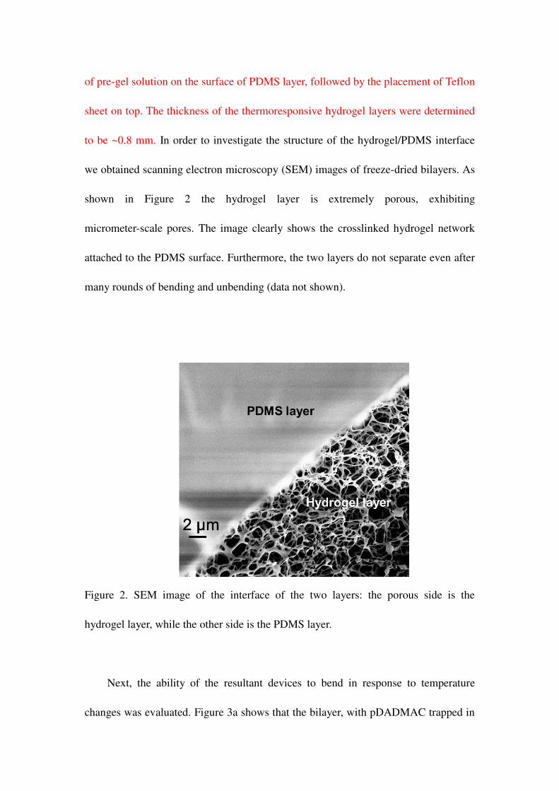
Field emission scanning electron microscopy: The bent bilayer systems were first

frozen by immersion in liquid nitrogen for 10 min. Then the bilayers were

freeze-dried overnight. (FreeZone. 4.5, LABCONCO). The dried bilayers were then

placed on a conductive copper tape coated holder and SEM images acquired on

specific areas using a Zeiss Sigma FESEM, operated at 5 kV.

Crystal violet (CV) loading and release from bilayers: Firstly, a bilayer film was

generated by mixing 12 mL of DI water and 3 mL of pDADMAC solution (20 wt%,

low Mw), which was used as a solvent. Then monomer mixtures were dissolved in the

solvent, with a total monomer concentration of 7.89 mol/L. The monomer mixtures

contain 80% (mol%) of NIPAm, 25% AAc, and 5% BIS. Then 40 μL DEAP (as a

photoinitiator) was added in the solution, followed by covering the container of the

solution by aluminum foil and shaking the solution for 1 h. The control solution

contained the same components as above except no pDADMAC was present. The

fabricated bilayers, which were generated using the same procedure as above, were

firstly soaked in a solution of pH 3, which causes the bilayers to be almost flat. Then

we placed the bilayer in a bottle containing 15 mL of CV solution (1 mg/mL, pH 6.5)

and a polymeric target particle at the bottom. After overnight soaking, the bilayer was

completely bent up towards the side of PDMS, and at the same time, the polymeric

particle was encapsulated by the bilayer. For CV release, a glass vial containing 20

mL pH 6.5 solution was placed on a plate with the temperature set as ~ 25C. The

solution was stirred continuously at 60 rpm using a magnetic stir bar and flowed

through a cuvette in an Agilent 8453 UV-vis spectrophotometer, equipped with an

89090A temperature controller and Peltier heating device, via a peristaltic pump. The

pumping speed was kept constant for the whole experiment. Then the CV loaded

bilayer with the encapsulated polymeric particle was placed into the solution, and a

timer started. After 30 mins, the pH of the solution was changed to 3. The absorbance

spectrum from the solution was collected every 2 mins.

5. Reference

1. Koetting, M. C.; Peters, J. T.; Steichen, S. D.; Peppas, N. A. Stimulus-Responsive

Hydrogels: Theory, Modern Advances, and Applications Mater. Sci. Eng. R-Rep.

2015, 93, 1-49.

2. Qiu, Y.; Park, K. Environment-Sensitive Hydrogels for Drug Delivery Adv. Drug

Deliv. Rev. 2012, 64, 49-60.

3. Ahn, S.-k.; Kasi, R. M.; Kim, S.-C.; Sharma, N.; Zhou, Y. Stimuli-Responsive

Polymer Gels Soft Matter 2008, 4, 1151-1157.

4. Tokarev, I.; Minko, S. Stimuli-Responsive Hydrogel Thin Films Soft Matter 2009,

5, 511-524.

5. Roy, D.; Cambre, J. N.; Sumerlin, B. S. Future Perspectives and Recent Advances

in Stimuli-Responsive Materials Prog. Polym. Sci. 2010, 35, 278-301.

6. Bashir, R.; Hilt, J.; Elibol, O.; Gupta, A.; Peppas, N. Micromechanical Cantilever

as an Ultrasensitive Ph Microsensor Appl. Phys. Lett. 2002, 81, 3091-3093.

7. Stieger, M.; Richtering, W.; Pedersen, J. S.; Lindner, P. Small-Angle Neutron

Scattering Study of Structural Changes in Temperature Sensitive Microgel Colloids J.

Chem. Phys. 2004, 120, 6197-6206.

8. Bajpai, A.; Shukla, S. K.; Bhanu, S.; Kankane, S. Responsive Polymers in

Controlled Drug Delivery Prog. Polym. Sci. 2008, 33, 1088-1118.

9. Hamidi, M.; Azadi, A.; Rafiei, P. Hydrogel Nanoparticles in Drug Delivery Adv.

Drug Deliv. Rev. 2008, 60, 1638-1649.

10. Wang, E.; Desai, M. S.; Lee, S.-W. Light-Controlled Graphene-Elastin Composite

Hydrogel Actuators Nano Lett. 2013, 13, 2826-2830.

11. Liu, Z.; Calvert, P. Multilayer Hydrogels as Muscle‐ Like Actuators Adv. Mater.

2000, 12, 288-291.

12. Fuhrer, R.; Athanassiou, E. K.; Luechinger, N. A.; Stark, W. J. Crosslinking Metal

Nanoparticles into the Polymer Backbone of Hydrogels Enables Preparation of Soft,

Magnetic Field‐ Driven Actuators with Muscle‐ Like Flexibility Small 2009, 5,

383-388.

13. Myung, D.; Waters, D.; Wiseman, M.; Duhamel, P. E.; Noolandi, J.; Ta, C. N.;

Frank, C. W. Progress in the Development of Interpenetrating Polymer Network

Hydrogels Polymer. Adv. Tech. 2008, 19, 647-657.

14. Kozhunova, E. Y.; Makhaeva, E. E.; Khokhlov, A. R. Collapse of

Thermosensitive Polyelectrolyte Semi-Interpenetrating Networks Polymer 2012, 53,

2379-2384.

15. Tominaga, T.; Tirumala, V. R.; Lee, S.; Lin, E. K.; Gong, J. P.; Wu, W.-l.

Thermodynamic Interactions in Double-Network Hydrogels J. Phys. Chem. B 2008,

112, 3903-3909.

16. Liu, M.; Su, H.; Tan, T. Synthesis and Properties of Thermo-and pH-Sensitive

Poly (N-Isopropylacrylamide)/Polyaspartic Acid Ipn Hydrogels Carbohydr. Polym.

2012, 87, 2425-2431.

17. Chen, J.; Liu, M.; Liu, H.; Ma, L.; Gao, C.; Zhu, S.; Zhang, S. Synthesis and

Properties of Thermo-and pH-Sensitive Poly (Diallyldimethylammonium

Chloride)/Poly (N, N-Diethylacrylamide) Semi-Ipn Hydrogel Chem. Eng. J. 2010,

159, 247-256.

18. Haque, M. A.; Kurokawa, T.; Gong, J. P. Super Tough Double Network

Hydrogels and Their Application as Biomaterials Polymer 2012, 53, 1805-1822.

19. Dragan, E. S. Design and Applications of Interpenetrating Polymer Network

Hydrogels. A Review Chem. Eng. J. 2014, 243, 572-590.

20. Schild, H. G. Poly (N-Isopropylacrylamide): Experiment, Theory and Application

Prog. Polym. Sci. 1992, 17, 163-249.

21. Stoychev, G.; Puretskiy, N.; Ionov, L. Self-Folding All-Polymer

Thermoresponsive Microcapsules Soft Matter 2011, 7, 3277-3279.

22. de las Heras Alarcón, C.; Pennadam, S.; Alexander, C. Stimuli Responsive

Polymers for Biomedical Applications Chem. Soc. Rev. 2005, 34, 276-285.

23. Ionov, L. Biomimetic Hydrogel‐ Based Actuating Systems Adv. Funct. Mater.

2013, 23, 4555-4570.

24. Flory, P. J. Principle of Polymer Chemistry Cornell University Press: London

1986.

25. Sorrell, C. D.; Carter, M. C.; Serpe, M. J. Color Tunable Poly (N‐

Isopropylacrylamide)‐ Co‐ Acrylic Acid Microgel–Au Hybrid Assemblies Adv.

Funct. Mater. 2011, 21, 425-433.

26. Wandrey, C.; Hernandez-Barajas, J.; Hunkeler, D. Diallyldimethylammonium

Chloride and Its Polymers. In Radical Polymerisation Polyelectrolytes, Springer:

1999; pp 123-183.

27. Kim, S. J.; Yoon, S. G.; Kim, I. Y.; Kim, S. I. Swelling Characterization of the

Semiinterpenetrating Polymer Network Hydrogels Composed of Chitosan and Poly

(Diallyldimethylammonium Chloride) Appl. Polym. Sci. 2004, 91, 2876-2880.

28. Kim, S. J.; Yoon, S. G.; Lee, Y. M.; An, K. H.; Kim, S. I. Water Sorption of Poly

(Vinyl Alcohol)/Poly (Diallyldimethylammonium Chloride) Interpenetrating Polymer

Network Hydrogels J. Appl. Polym. Sci. 2003, 90, 1389-1392.

29. Bassik, N.; Abebe, B. T.; Laflin, K. E.; Gracias, D. H. Photolithographically

Patterned Smart Hydrogel Based Bilayer Actuators Polymer 2010, 51, 6093-6098.

30. Maeda, S.; Hara, Y.; Sakai, T.; Yoshida, R.; Hashimoto, S. Self‐ Walking Gel Adv.

Mater. 2007, 19, 3480-3484.

31. Malachowski, K.; Breger, J.; Kwag, H. R.; Wang, M. O.; Fisher, J. P.; Selaru, F.

M.; Gracias, D. H. Stimuli‐ Responsive Theragrippers for Chemomechanical

Controlled Release Angew. Chem. Int. Ed. 2014, 126, 8183-8187.

32. Yoon, C.; Xiao, R.; Park, J.; Cha, J.; Nguyen, T. D.; Gracias, D. H. Functional

Stimuli Responsive Hydrogel Devices by Self-Folding Smart Mater. Struct. 2014, 23,

094008.

33. Wang, J.; Lin, L.; Cheng, Q.; Jiang, L. A Strong Bio‐ Inspired Layered Pnipam–

Clay Nanocomposite Hydrogel Angew. Chem. Int. Ed. 2012, 51, 4676-4680.

34. Ma, C.; Shi, Y.; Pena, D. A.; Peng, L.; Yu, G. Thermally Responsive Hydrogel

Blends: A General Drug Carrier Model for Controlled Drug Release Angew. Chem.

Int. Ed. 2015, 127, 7484-7488.


Recommended