Scheimpflug photography is the basis for a variety of imaging devices that are highly versatile. The applications of Scheimpflugimaging are wide in scope, spanning from evaluation of corneal ectasia to quantifying density in nuclear sclerotic cataracts. Thepotential uses for Scheimpflug-based devices are expanding and a number of them are relevant in glaucoma. In particular, theycan provide three-dimensional image reconstruction of the anterior segment which includes assessment of the iridocorneal angle.Photographic analyses allow also for a noncontact method of estimating central corneal thickness (CCT) and intraocular pressure(IOP), as well as the study of various corneal biomechanical properties, which may be useful for stratifying glaucoma risk.
1. Introduction
The clinical utility of anterior segment imaging continues tobe refined as newer investigations reveal novel applicationsfor their use. As a complement to the slit lamp examination,anterior segment imaging offers qualitative information aswell as objective, quantifiable data [1]. Scheimpflug photog-raphy is the basis for a number of devices that can imagethe anterior segment.The technology is highly versatile, withpotential applications in the areas of keratorefractive surgery,corneal biomechanics, corneal ectasia evaluation, anteriorsegment imaging, cataract grading, and surgical planning forfemtosecond laser-assisted cataract surgery [2–7].
Herein we review Scheimpflug photography and someof its applications that are relevant to the managementof glaucoma patients. Like other anterior segment imagingtechnologies, Scheimpflug-based devices can provide three-dimensional image representations of the anterior segment,which may be useful for screening narrow angles [1–3].Intraocular pressure (IOP) is a modifiable and independentrisk factor for predicting glaucomatous progression [8]. Itsmeasurement can be affected by corneal parameters like cen-tral corneal thickness (CCT). Despite this, there is accruingevidence to suggest that CCT and corneal biomechanicalproperties are associated with glaucoma independently of
their effect on tonometry [6].TheCorvis ST (Corneal Visual-ization Scheimpflug Technology, Oculus, Wetzlar, Germany)is an ultra-high speed Scheimpflug device that offers a highlyreproducible, noncontactmethod of performing pachymetry,estimating intraocular pressure (IOP), and obtaining cornealbiomechanical data [6, 7].
2. The Scheimpflug Principle
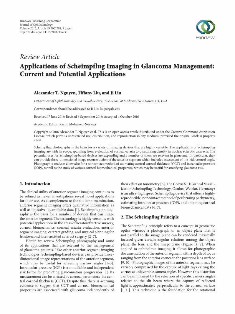
The Scheimpflug principle refers to a concept in geometricoptics whereby a photograph of an object plane that isnot parallel to the image plane can be rendered maximallyfocused given certain angular relations among the objectplane, the lens, and the image plane (Figure 1) [2]. Whenapplied to ophthalmic imaging, it allows for photographicdocumentation of the anterior segment with a depth of focusranging from the anterior cornea to the posterior lens surface[9, 10]. Photographic images of the anterior segment may bevariably compressed by the capture of light rays exiting thecornea at unfavorable camera angles.However, this distortioncan be minimized by the selection of specific camera anglesrelative to the slit beam where the capture of reflectedlight is approximately perpendicular to the corneal surface[1, 11]. This technique is the foundation for the rotational
Hindawi Publishing CorporationJournal of OphthalmologyVolume 2016, Article ID 3062381, 9 pageshttp://dx.doi.org/10.1155/2016/3062381
2 Journal of Ophthalmology
Image Lens Lens axis
Image plane
Lens plane
Object plane (plane of focus)
Scheimpflug intersection
Figure 1: Depiction of the Scheimpflug principle as it applies tophotography. When an oblique tangent is extended from the imageplane and the lens plane, they intersect at a point that is alsointersected by a line extended from the plane of focus. An object thatlies on this plane can be captured in focus despite not being parallelwith the image plane.
Table 1: Select Scheimpflug imaging systems.
Device Manufacturer Image acquisitionOrbscan II Bausch & Lomb, USA Horizontal cross sectionPentacam Oculus, Germany Single rotating cameraGalilei Ziemer, Switzerland Dual rotational cameraSirius CSO, Italy Single rotating cameraTMS-5 Tomey, Japan Single rotating cameraPrecisio Ivis, Italy Single rotating camera
Scheimpflug camera. A select list of Scheimpflug devices ispresented in Table 1.
3. Anterior Chamber and IridocornealAngle Assessment
Recent investigations have explored the emerging role ofanterior segment imaging in screening for angle closure [1].Gonioscopy has served as the diagnostic standard for eval-uating narrow angles and related entities [9]. However, thistechnique requires a contact lens and a proficient examinerto provide a confident diagnosis [3]. And even amongexperienced clinicians, there is variability in angle gradingdue to the subjective nature of the assessment. Anteriorsegment imaging devices hold the potential for a noncontactmethod of angle closure screening [1, 12, 13]. It may enablepractitioners that are less familiar with gonioscopy to effec-tively screen for angle closure or patients suspected ofhaving angle closure. For experienced clinicians, noncontactimaging may serve as a useful supplement to gonioscopy. Itmay be particularly valuable in situations where a routineexamination is difficult, such as with patients who poorlytolerate gonioscopy or who have difficulty with the requiredpositioning [1, 2, 8]. Several technologies have capabilitiesthat may be useful in the evaluating narrow angles, which areoverviewed in Table 2 [1, 3, 12].
The Orbscan (Bausch & Lomb Surgical, Salt Lake City,USA) was commercially introduced in 1995. It is based ona concept referred to as slit-scan triangulation to obtaintopographic data. It projects 40 slit beams (20 nasal and 20temporal) at the anterior segment at an angle of 45∘ fromthe axis of the camera [13]. In order for the cornea, the iris,and the lens to be captured in focus, the image plane of thecamera is tilted to satisfy the Scheimpflug condition. Themeasurements obtained by triangulation can then be inte-grated to provide three-dimensional information regardingthe anterior segment. Relevant to the evaluation of angleclosure, the Orbscan can estimate the iridocorneal angleand the anterior chamber depth (ACD) [14]. In normalsubjects, these measurements have been shown to be highlyreproducible [14]. However, studies validating the utility ofthe Orbscan in assessing angle closure are still needed.
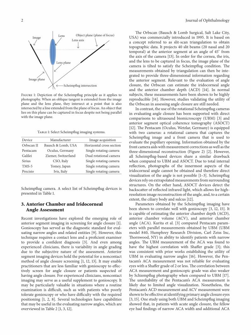
In contrast, the use of the rotational Scheimpflug camerasin evaluating angle closure has been supported with directcomparisons to ultrasound biomicroscopy (UBM) [3] andanterior segment optical coherence tomography (ASOCT)[12]. The Pentacam (Oculus, Wetzlar, Germany) is equippedwith two cameras: a rotational camera that captures theScheimpflug image and a front camera that is used toevaluate the pupillary opening. Information obtained by thefront camera aidswithmeasurement corrections aswell as thethree-dimensional reconstruction (Figure 2) [2]. However,all Scheimpflug-based devices share a similar drawbackwhen compared to UBM and ASOCT. Due to total internalreflection, photographs of the innermost aspects of theiridocorneal angle cannot be obtained and therefore directvisualization of the angle is not possible [1–3]. Scheimpflugdevices rely on extrapolatedmeasurements from surroundingstructures. On the other hand, ASOCT devices detect thebackscatter of reflected infrared light, which allows for high-resolution image reconstruction of the angle, and, to a certainextent, the ciliary body and sulcus [12].
Parameters obtained by the Scheimpflug imaging havebeen shown to correlate well with gonioscopy [3, 12, 15]. Itis capable of estimating the anterior chamber depth (ACD),anterior chamber volume (ACV), and anterior chamberangle (ACA). Kurita et al. [3] compared Pentacam param-eters with parallel measurements obtained by UBM (UBMmodel 840, Humphrey Research Division, Carl Zeiss Inc,Thornwood, NY) in ability to identify patients with narrowangles. The UBM measurement of the ACA was found tohave the highest correlation with Shaffer grade [3]; thiswas consistent with prior works establishing the utility ofUBM in evaluating narrow angles [16]. However, the Pen-tacam’s ACA measurement was not reliable for evaluatingeyes with a Shaffer grade of 2 or less.The correlation betweenACA measurement and gonioscopic grade was also weakerby Schiempflug photography when compared to UBM [17].The unreliability of the Pentacam’s ACA measurement islikely due to limited angle visualization. Nonetheless, thePentacam’s ACD measurement and ACV measurement wereshown to be effective at identifying primary angle closure eyes[3, 15]. One study using both UBM and Scheimpflug imagingshowed that, in patients with acute angle closure, the felloweye had findings of narrow ACA width and additional ACA
Journal of Ophthalmology 3
Table 2: Comparison of anterior segment imaging modalities for assessing narrow angles.
Imaging system Correlation withgonioscopy
Quantitativeparameters Advantages Limitations
Slit scantopography N/A Iridocorneal angle
ACD Noncontact No visualization of angle, ciliarybody or sulcus
RotationalScheimpflugcamera
++∗ACDACVACA
Noncontact No visualization of angle, ciliarybody or sulcus
ASOCT +++ AOD500TISA500
NoncontactDirect angle visualizationSome visualization of ciliarybody and sulcus
Requires identification of scleralspurNoncontact assessment limitedto temporal and nasal angles
UBM +++
ACDACVACA
AOD500TISA500
Excellent visualization of angle,ciliary body and sulcus
Requires contact, identificationof scleral spur
ACD: anterior chamber depth, ACV: anterior chamber volume, ASOCT: anterior segment OCT, UBM: ultrasound biomicroscopy, AOD: angle openingdistance, and TISA: trabecular iris area.N/A: not available, validating studies required.∗ indicates that it may be as useful as ASOCT for partitioning narrow angles but it does not provide direct angle visualization.
Figure 2:Three-dimensional image representation of the anterior segment obtained by the Pentacam (Oculus, Wetzlar, Germany). Note thatvisualization of the iridocorneal angle is obscured by total internal reflection. Various parameters obtained by extrapolatedmeasurementsmaybe useful for angle closure screening (red box). These include anterior chamber angle (ACA), anterior chamber depth (ACD), and anteriorchamber volume (ACV).
narrowing in response to a light-to-dark luminance changeand also pilocarpine-induced pupillary constriction [18].
Rotational Scheimpflug imaging has also been shownto be comparable to ASOCT in partitioning patients withnarrow angles. Grewal et al. [12] matched the Pentacam’sACD and ACV measurement against parameters obtainedby spectral domain ASOCT. The RTVue 100 (Optovue Inc,Fremont, CA,USA)ASOCTused in their studywas equippedwith a corneal adaptor module that allows for software tocalculate the angle opening distance at 500 microns from thescleral spur (AOD500). The AOD500 is a parameter previ-ously defined using UBM [17]. The Pentacam’s estimation ofthe ACV and ACD outperformed the parameters obtainedby the RTVue 100 ASOCT in the detection of narrow angles
with gonioscopy as the reference standard [12]. Although thePentacam cannot directly visualize the angle, it is advantagedby the breadth of three-dimensional data incorporated in itsanalyses. In contrast, noncontact ASOCT assessment limitedto cross sections of only the nasal and temporal angles mayexclude representative information regarding the angle. Toimage the superior and inferior angles, contact would berequired to move the eyelids obscuring visualization [9].
Scheimpflug systems such as the Pentacam appear to beviable technologies for evaluating angle closure. Although thePentacam was better able to predict angle anatomy than theRTVue 100 ASOCT in one study [12], further investigationsare needed to discern how rotational Scheimpflug imagingmeasures up against ASOCT. Presently, both technologies
4 Journal of Ophthalmology
appear comparable in their ability to deliver a noncontactmethod for screening angle closure. The value of havingreproducible, quantifiable parameters is desirable for screen-ing as well as monitoring treatment effect. The Pentacam,for instance, is capable of demonstrating a posttreatmentincrease in ACV following laser iridotomy [17, 19–23]. How-ever, one clear advantage held by ASOCT and UBM imagingis direct visualization of the angle, ciliary body, and sulcus.This is of particular importance for evaluating mass lesionsor conditions such as plateau iris where visualization of ciliarybody anatomy is essential to the diagnosis [9, 24].
4. Central Corneal Thickness
Goldmann applanation tonometry (GAT) is often regardedas reference standard for measurement of intraocular pres-sure (IOP). When Goldmann and Schmidt introduced theirtonometer in 1957, they acknowledged sources of possibleerror including CCT. The Imbert-Fick law serves as the basisfor GAT. The concept assumes a perfectly thin cornea thatbehaves like a membrane where the IOP is equal to theapplanating pressure. In actuality, the cornea is variably thickrather than perfectly thin, and the tear film contributes aconfounding force from surface tension [25, 26].
One of the strongest independent risk factors for devel-oping primary open-angle glaucoma (POAG) is CCT [27–29]. Another independent risk factor is IOP, a measurementwhich can be influenced byCCT. For this reason, it is believedby some that the CCT is a risk factor for developing POAGonly by virtue of its influence on IOP measurement [29].If this is correct, then we would not expect CCT to be anindependent risk factor for developing glaucoma if predictivemodels corrected for the IOP measurement error attributedto CCT [29]. However, the evidence to date suggests thata thin CCT is associated with an increased risk of POAGbeyond its artefactual effect on tonometry [27–30]. Forthis reason, an interest in corneal properties like CCT willremain relevant regardless of the error it confers towards IOPmeasurement.
Although ultrasound pachymetry is widely used to mea-sure CCT, it is disadvantaged in several ways [31, 32].Measurement accuracy and repeatability depend on accurateplacement of the probe onto the cornea, which is done man-ually. Corneal indentation can occur with contact betweenthe cornea and the probe, which may falsely underestimatethe actual CCT. And because this technique relies on theassumption that the speed of sound is similar throughhealthy and diseased corneal tissue, the measurement may beinaccurate in certain pathologic disease states. For these rea-sons, noncontact methods for estimating CCT are desirable.Devices capable of measuring CCT in this fashion includeScheimpflug imaging devices such as the Pentacam, Galilei(Ziemer, Port, Switzerland), Sirius (Costruzione StrumentiOftalmici, Florence, Italy), TMS-5 (Tomey, Nagoya, Japan),and the Corvis ST.
Scheimpflug devices are able to provide highly repeatableCCT measurements that are comparable to, but not likelyinterchangeable with, ultrasound pachymetry CCT [32–36].Prior studies have shown that highly reproducible CCT
measurements can be obtained by the Pentacam, Sirius,Galilei, and Corvis ST. Of these devices, the Galilei has thehighest reported intraoperator repeatability. This may be inpart attributable to its dual-rotational camera design, whichcan average the CCT estimate from two different Scheimpflugcameras [32]. However, studies vary widely in reportinghow similar CCT measurements are between the differentdevices. A study comparing CCT measurements obtained byScheimpflug systems with ultrasound pachymetry has beenpublished previously [32]. Some investigations have notedno difference in mean CCT obtained by either ultrasoundpachymetry or with the Pentacam [31, 37]. In contrast,several other studies have noted a significant difference inthe mean CCT measured by Pentacam and by ultrasoundpachymetry [38–41]. Similarly, the Sirius-CCT measurementis comparable but significantly different from the ultrasoundpachymetry CCT [42, 43]. Despite sharing a common imag-ing technology, the various Scheimpflug devices appear toobtain CCTmeasurements that are statistically different fromeach other. Even though these differences may be small,caution is generally advised in comparing CCT values acrossdifferent measurement platforms. It remains to be clarifiedwhether these differences may be sufficiently small for themto be negligible in clinical contexts.
5. Corneal Biomechanics and IOP
CCT is but one dimension of a multifaceted area of studythat comprises corneal biomechanics. Interest in the biome-chanical properties of the cornea parallels our interest inCCT: it may help explain the source of measurement errorin tonometry and how structural features of the cornea canpredict the risk of glaucomatous progression independent oftheir effect on IOP. However, the mechanisms underpinninghow CCT and corneal properties confer a risk towardsdeveloping glaucoma remain unclear.
Theoretical models predict that optic nerve head biome-chanics can be influenced by the structure of the adjacentsclera [44, 45]. The optic nerve head is a site of discontinuitywithin the cornealscleral shell [45] and is the location ofretinal ganglion cell injury due to glaucoma [45]. Fromtheoretical models, the lamina cribrosa is predicted to expe-rience an increase in tensile strain with increasing eye radius[44, 46]. These models also predict, though, that scleralstiffness may mitigate this increase in strain expected fromhaving a longer eye [44, 46]. It has been hypothesized thatcorneal parameters likeCCTmay serve as surrogatemeasuresfor some biomechanical property of the sclera [44, 46].Lanzagorta-Aresti et al. [47] showed that changes in thedisplacement of the lamina cribrosa after medical treatmentof IOP was significantly correlated corneal hysteresis. Fur-thermore, Wells et al. [48] demonstrated that low cornealhysteresis, but not CCT, was associated with reduced opticnerve head compliance. However, efforts to correlate CCTwith lamina cribrosa thickness have yielded no significantassociation between the two [49].
The two devices capable of quantifying biomechanicalfeatures of the cornea include the Ocular Response Ana-lyzer or ORA (Richert Ophthalmic Instruments, Depew,
Journal of Ophthalmology 5
New York, USA) and the Scheimpflug-based noncontacttonometer Corvis ST [7, 50]. To date, most of the studiesinvestigating corneal biomechanics have utilized the ORA,which was released in 2005; the Corvis STwasmade availablein 2012.The ORA can measure corneal hysteresis, which maybe an indicator of the cornea’s viscoelasticity [51]. Patientswith POAG and normal tension glaucoma have been shownto have lower-than-average corneal hysteresis values [52].Furthermore, low corneal hysteresis has also been implicatedin glaucomatous field progression [53, 54].
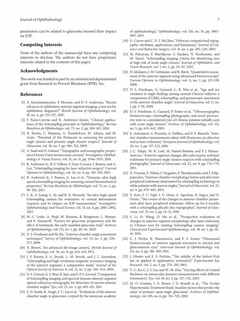
The Corvis ST provides a noncontact method for eval-uating IOP, CCT, and the cornea’s biomechanical responseto a collimated puff of air. It is equipped with an ultrahigh-speed Scheimpflug camera that is capable of recording 4330frames/second. The air puff is delivered with a fixed pressureat the corneal surface over 31ms, allowing for the digitalcapture of 140 images. The corneal response to the air puffis initially marked by an inward conformational change inthe corneal curvature. This initial flattening of the cornea isreferred to as the first applanation (Figure 3) [50, 55]. Thecornea eventually deforms to a point where it is maximallyconcave prior to returning to its original shape. This eventdefines the corneal deformation amplitude at the highestconcavity (Figure 3). The cornea then naturally returns to itsoriginal shape, which is referred to as the second applanation[50, 55]. Various aspects of the corneal deformation responseto the air-puff can be quantified, which are reviewed in detailelsewhere [55].
Recently, Lee et al. [50] published a cross-sectional studydemonstrating a parameter obtained by the Corvis ST thatappeared to be independently associated with glaucoma risk.They identified three Corvis ST parameters that could parti-tion patients with either POAG or normal tension glaucomafrom normal controls: (1) outward applanation velocity, (2)time to highest concavity, and (3) peak distance. However,two of these parameters, namely, outward applanation veloc-ity and peak distance, were associated with Corvis-IOP [50].On the other hand, the highest concavity time was associatedwith the glaucomatous group and not dependent on otherestablished risk factors for glaucoma such as CCT or IOP. Itshould be noted, though, that their results contrast with thoseof Leung et al. [7] in that the latter study group did not findany of the Corvis ST parameters to be capable of partitioningglaucomatous patients from normal subjects.
While various corneal parameters may be associatedwith glaucomatous risk independently of their effect on IOPmeasurement, the study of corneal biomechanics may aid inbetter understanding sources of measurement error. IOP isa modifiable risk factor associated with glaucomatous pro-gression [27–30]. A coveted goal in glaucoma managementis approximating IOP, as close as possible, to the “true”IOP. An intracameral measurement of IOP may be the mostaccurate method for achieving this end, but it is clinicallyimpractical [56]. Because of this, various approaches havebeen developed and described to help correct for the effectsthat biomechanical features of the cornea have on IOPmeasurement. Regression analyses have been proposed withformulaic corrections for values obtained by applanationtonometry, taking into account the effects of CCT. There
is growing evidence, though, that corneal biomechanicalproperties, such as corneal hysteresis, may better explain thesource of measurement error in tonometry than CCT [56].
The corneal deformation amplitude measured by theCorvis ST has been implicated as source of measurementerror of GAT. In a study population containing normaland glaucomatous eyes, Leung et al. [7] compared IOPmeasurements obtained by GAT with dynamic contourtonometry (DCT, Pascal, Swiss Microtechnology AG, Port,Switzerland). IOP measurement by DCT is theoretically lessdependent on corneal biomechanical properties and by thisvirtue it may better approximate the “true” IOP. Indeed, IOPmeasurements obtained by DCT are highly concordant withintracameralmeasurements [57]. To evaluate factors thatmayexplain the difference in IOPmeasurements obtained byDCTand GAT, Leung et al. [7] also studied the biomechanicalproperties obtained by the ORA and the Corvis ST. Mea-surement of the corneal deformation amplitude was shownto be dependent on IOP and CCT (measured by ultrasoundpachymetry). From their univariate analysis, the differencein DCT and GAT measurements was associated with CCTand corneal deformation amplitude after adjusting for theeffect of IOP. But with the multivariate analysis, the onlyparameter significantly associated with this measurementdifference was corneal deformation amplitude. The authorscomment that the influence of CCT on GAT likely stemsfrom the effect of CCT on corneal deformation amplitude [7].Of note, the corneal hysteresis and corneal resistance factormeasurements obtained by the ORA were not significantlyassociated with the measurement discrepancy between DCTand GAT in this investigation [7]. The ORA functions in avery different way from the Corvis ST; while the air puff ofthe Corvis ST is applied with a fixed force, the ORA air puff isdelivered with a variable force. Although both instrumentsassess corneal biomechanics, it is difficult to compare themetrics obtained by these two devices [55, 58].
Although the Corvis ST is capable of providing anestimate of IOP, Leung et al. [7] did not compare the accuracyof this measurement with DCT. The IOP estimate by theCorvis ST has been shown to be highly reproducible [59]and comparable to GAT [60, 61]. It should be noted, though,that the degree of correlation between Corvis-IOP [59] andGAT was relatively weak compared to other techniques suchas DCT [62] and iCare rebound tonometry [62, 63]. The sig-nificance of this finding is unclear absent data validatingthe accuracy of Corvis-IOP measurements [59, 61]. Futureinvestigations comparing intracameral IOPmeasurements tothe Corvis ST would be helpful for this end [60]. There is noconsensus agreement among studies on whether the Corvis-IOP tends to be higher [60, 63] or lower [61, 64] than the IOPestimated by GAT.
6. Cataract Evaluation
A brief section on cataract evaluation is reviewed givenits relevance in glaucoma management and especially inlight of the rising interest in combining cataract extractionwith minimally invasive glaucoma surgery (MIGS). MIGSprocedures offer the potential for modest reductions in IOP
6 Journal of Ophthalmology
Applanation length1 Applanation length2
D
T1T2
Figure 3: Diagramatic representation of the biomechanical response of the cornea to the metered air puff delivered by the CorneaVisual Scheimpflug Technology (Corvis ST). The first phase (left) is marked by corneal surface flattening and the initiation of an inwardconformational change in the corneal curvature (referred to as the first applanation 𝑇
1). Further deformation produces a concave corneal
surface. The moment it reaches the maximally deformed state (middle) is referred to as the time of highest concavity. The distance, 𝐷, is thepeak distance or corneal deformation amplitude. After reaching its maximally concave shape, the cornea recoils into its original shape. Whenthe surface is similarly flattened compared to 𝑇
1, this moment marks the second applanation or 𝑇
2(right).
in patients with mild-moderate glaucoma. They are notalways performed as standalone procedures given that theirIOP-lowering effect cannot rival that of traditional filteringsurgeries. Because of this, it is often preferable for MIGSprocedures to be done conveniently at the time of cataractsurgery [65].
Traditionally, the appraisal of a visually significantcataract involves two main factors: (1) a functional deficit,such as Snellen acuity, and (2) the clinician’s assessment oflens opacification. There are shortcomings associated withboth arms of the traditional approach. Patients with relativelygood Snellen acuity may actually have poor vision qualityand the subjective grading of lens opacification is subjectto interrater variability. The most widely recognized lens-grading schema is the Lens Opacification Classification III(LOCS III), which was last updated in 1993 [66].
Scheimpflug systems such as the Pentacam may be ableto enhance our assessment of cataractogenesis. The Penta-cam can measure lens densitometry with specific metricsthat include average density and maximum density. Basedon these measurements, the Pentacam can be equippedwith software that then assigns a grade of nuclear sclerosison a scale of 1–5 (Pentacam Nuclear Staging or PNS).The PNS score has been shown to correlate well withSnellen acuity and LOCS III grade, validating the Pentacam’sautomated assessment of nuclear sclerosis [67]. Furthermore,the Pentacam’s densitometric parameters have been positivelycorrelated with higher-order aberrations (HOAs) obtainedfrom wavefront analyses [67]. A consideration of HOAsmay enable clinicians to better appreciate why patients withrelatively good Snellen acuity may complain of poor visionquality. Similar such innovations are likely to increase oursensitivity in identifying visually significant lens opacities,thereby potentially expanding our indications for cataractextraction.
The use of Scheimpflug systems as surgical planning toolshas also been suggested. Measurements of lens densitometryenable a quantitative evaluation of a nuclear sclerosis; this
may help guide the selection of phacoemulsification tech-nique or use of a femtosecond laser for lens fragmentation.It should be mentioned, however, that lens densitometrymeasurements are currently less precise for higher-gradenuclear cataracts [67]. Scheimpflug imaging has been shownto be helpful for evaluating intraocular lens tilt and decen-tration following cataract extraction [68, 69]. The LENSAR(LENSAR Inc., Winter Park, USA) is a femtosecond laserthat is equipped with Scheimpflug imaging capabilities [70].Similar to the Pentacam, the device can automatically gradelens density on a scale of 1–5. It special features an imagingsystem that enables the detection of any tilt that may beexhibited by the native crystalline lens; this is important formaximizing the likelihood of a producing a precise, free-floating capsulotomy [70].
7. Summary
TheScheimpflug principle is the basis for a number of devicesand imaging systems. The technology is extraordinarilyversatile, with applications spanning from laser keratore-fractive surgery to quantifying cataractogenesis. Scheimpflugdevices have several relevant applications for glaucomamanagement. Currently, Scheimpflug-based imaging systemshave formidable capabilities to ASOCT for predicting angleclosure, despite their inability to visualize the iridocornealangle. Noncontact methods of assessment have multipleadvantages including sanitary considerations, patient com-fort, and in some cases, less operator-dependent. Because ofthis, technologies like Scheimpflug-based devices are likelyto be increasingly used as they become more accessible.The clinician should heed caution, though, in interchangingmeasurements obtained by different technologies as they canbe slightly different. With the introduction of the Corvis ST,investigators have 2 available devices for the study of cornealbiomechanics. Relevant investigations with these devices areneeded to provide unanswered questions for how corneal
Journal of Ophthalmology 7
parameters can be related to glaucoma beyond their impacton IOP.
Competing Interests
None of the authors of the manuscript have any competinginterests to disclose. The authors do not have proprietaryinterests related to the contents of this paper.
Acknowledgments
Thisworkwas funded in part by anunrestricted departmentalgrant from Research to Prevent Blindness (RPB), Inc.
References
[1] A. Konstantopoulos, P. Hossain, and D. F. Anderson, “Recentadvances in ophthalmic anterior segment imaging: a new era forophthalmic diagnosis?” British Journal of Ophthalmology, vol.91, no. 4, pp. 551–557, 2007.
[2] F. Faria-Correia and R. Ambrosio Junior, “Clinical applica-tions of the Scheimpflug principle in Ophthalmology,” RevistaBrasileira de Oftalmologia, vol. 75, no. 2, pp. 160–165, 2016.
[3] N. Kurita, C. Mayama, A. Tomidokoro, M. Aihara, and M.Araie, “Potential of the Pentacam in screening for primaryangle closure and primary angle closure suspect,” Journal ofGlaucoma, vol. 18, no. 7, pp. 506–512, 2009.
[4] A. Saad andD. Gatinel, “Topographic and tomographic proper-ties of forme fruste keratoconus corneas,” Investigative Ophthal-mology & Visual Science, vol. 51, no. 11, pp. 5546–5555, 2010.
[5] R. Ambrosio Jr., B. F. Valbon, F. Faria-Correia, I. Ramos, and A.Luz, “Scheimpflug imaging for laser refractive surgery,” CurrentOpinion in Ophthalmology, vol. 24, no. 4, pp. 310–320, 2013.
[6] R. Ambrosio Jr., I. Ramos, A. Luz et al., “Dynamic ultra highspeed scheimpflug imaging for assessing corneal biomechanicalproperties,” Revista Brasileira de Oftalmologia, vol. 72, no. 2, pp.99–102, 2013.
[7] C. K.-S. Leung, C. Ye, and R. N. Weinreb, “An ultra-high-speedScheimpflug camera for evaluation of corneal deformationresponse and its impact on IOP measurement,” InvestigativeOphthalmology and Visual Science, vol. 54, no. 4, pp. 2885–2892,2013.
[8] M. C. Leske, A. Heijl, M. Hussein, B. Bengtsson, L. Hyman,and E. Komaroff, “Factors for glaucoma progression and theeffect of treatment: the early manifest glaucoma trial,” Archivesof Ophthalmology, vol. 121, no. 1, pp. 48–56, 2003.
[9] D. S. Friedman andM. He, “Anterior chamber angle assessmenttechniques,” Survey of Ophthalmology, vol. 53, no. 3, pp. 250–273, 2008.
[10] N. Brown, “An advanced slit-image camera,” British Journal ofOphthalmology, vol. 56, no. 8, pp. 624–631, 1972.
[11] J. F. Koretz, S. A. Strenk, L. M. Strenk, and J. L. Semmlow,“Scheimpflug and high-resolution magnetic resonance imagingof the anterior segment: a comparative study,” Journal of theOptical Society of America A, vol. 21, no. 3, pp. 346–354, 2004.
[12] D. S. Grewal, G. S. Brar, R. Jain, and S. P. S. Grewal, “Comparisonof Scheimpflug imaging and spectral domain anterior segmentoptical coherence tomography for detection of narrow anteriorchamber angles,” Eye, vol. 25, no. 5, pp. 603–611, 2011.
[13] S. D. Smith, K. Singh, S. C. Lin et al., “Evaluation of the anteriorchamber angle in glaucoma: a report by the american academy
of ophthalmology,” Ophthalmology, vol. 120, no. 10, pp. 1985–1997, 2013.
[14] G. Cairns and C. N. J. McGhee, “Orbscan computerized topog-raphy: attributes, applications, and limitations,” Journal of Cat-aract and Refractive Surgery, vol. 31, no. 1, pp. 205–220, 2005.
[15] M. Pakravan, F. Sharifipour, S. Yazdani, N. Koohestani, andM. Yaseri, “Scheimpflug imaging criteria for identifying eyesat high risk of acute angle closure,” Journal of Ophthalmic andVision Research, vol. 7, no. 2, pp. 111–117, 2012.
[16] H. Ishikawa, J.M. Liebmann, and R. Ritch, “Quantitative assess-ment of the anterior segment using ultrasound biomicroscopy,”Current Opinion in Ophthalmology, vol. 11, no. 2, pp. 133–139,2000.
[17] D. S. Friedman, G. Gazzard, C. B. Min et al., “Age and sexvariation in angle findings among normal Chinese subjects: acomparison of UBM, scheimpflug, and gonioscopic assessmentof the anterior chamber angle,” Journal of Glaucoma, vol. 17, no.1, pp. 5–10, 2008.
[18] D. S. Friedman, G. Gazzard, P. Foster et al., “Ultrasonographicbiomicroscopy, scheimpflug photography, and novel provoca-tive tests in contralateral eyes of Chinese patients initially seenwith acute angle closure,” Archives of Ophthalmology, vol. 121,no. 5, pp. 633–642, 2003.
[19] E. Antoniazzi, S. Pezzotta, A. Delfino, and P. E. Bianchi, “Ante-rior chamber measurements taken with Pentacam: an objectivetool in laser iridotomy,”European Journal of Ophthalmology, vol.20, no. 3, pp. 517–522, 2010.
[20] J. C. Talajic, M. R. Lesk, M. Nantel-Battista, and P. J. Harasy-mowycz, “Anterior segment changes after pilocarpine and laseriridotomy for primary angle-closure suspects with scheimpflugphotography,” Journal of Glaucoma, vol. 22, no. 9, pp. 776–779,2013.
[21] N. Vryonis, E. Nikita, I. Vergados, P.Theodossiadis, and T. Filip-popoulos, “Anterior chambermorphology before and after laserperipheral iridotomy determined by scheimpflug technology inwhite patients with narrow angles,” Journal of Glaucoma, vol. 22,no. 9, pp. 679–683, 2013.
[22] Y. Acet, F. U. Yigit, I. U. Onur, A. Agachan, B. Tugcu, and O.Orum, “The course of the changes in anterior chamber param-eters after laser peripheral iridotomy: follow-up for 6 monthswith a scheimpflug-placido disc topographer,” Journal of Glau-coma, vol. 25, no. 1, pp. 14–21, 2016.
[23] S. Li, H. Wang, D. Mu et al., “Prospective evaluation ofchanges in anterior segment morphology after laser iridotomyin Chinese eyes by rotating Scheimpflug camera imaging,”Clinical and Experimental Ophthalmology, vol. 38, no. 1, pp. 10–14, 2010.
[24] C. J. Pavlin, K. Harasiewicz, and F. S. Foster, “Ultrasoundbiomicroscopy of anterior segment structures in normal andglaucomatous eyes,” American Journal of Ophthalmology, vol.113, no. 4, pp. 381–389, 1992.
[25] J. Gloster and E. S. Perkins, “The validity of the Imbert-Ficklaw as applied to applanation tonometry,” Experimental EyeResearch, vol. 2, no. 3, pp. 274–283, 1963.
[26] Y.-C. Ko, C. J.-I. Liu, andW.-M. Hsu, “Varying effects of cornealthickness on intraocular pressure measurements with differenttonometers,” Eye, vol. 19, no. 3, pp. 327–332, 2005.
[27] M. O. Gordon, J. A. Beiser, J. D. Brandt et al., “The OcularHypertension Treatment Study: baseline factors that predict theonset of primary open-angle glaucoma,” Archives of Ophthal-mology, vol. 120, no. 6, pp. 714–720, 2002.
8 Journal of Ophthalmology
[28] European Glaucoma Prevention Study Group, “Predictive fac-tors for open-angle glaucoma among patients with ocularhypertension in the European Glaucoma Prevention Study,”Ophthalmology, vol. 114, no. 1, pp. 3–9, 2007.
[29] J. D. Brandt, M. O. Gordon, F. Gao, J. A. Beiser, J. P. Miller, andM. A. Kass, “Adjusting intraocular pressure for central cornealthickness does not improve prediction models for primaryopen-angle glaucoma,” Ophthalmology, vol. 119, no. 3, pp. 437–442, 2012.
[30] B. A. Francis, R. Varma, V. Chopra, M.-Y. Lai, C. Shtir, and S.P. Azen, “Intraocular pressure, central corneal thickness, andprevalence of open-angle glaucoma: the los angeles Latino EyeStudy,” American Journal of Ophthalmology, vol. 146, no. 5, pp.741–746, 2008.
[31] M. Fujioka, M. Nakamura, Y. Tatsumi, A. Kusuhara, H.Maeda, and A. Negi, “Comparison of Pentacam Scheimpflugcamera with ultrasound pachymetry and noncontact specularmicroscopy in measuring central corneal thickness,” CurrentEye Research, vol. 32, no. 2, pp. 89–94, 2007.
[32] A. Yu, W. Zhao, G. Savini et al., “Evaluation of central cornealthickness using corneal dynamic scheimpflug analyzer corvisST and comparisonwith pentacam rotating scheimpflug systemandultrasound pachymetry in normal eyes,” Journal of Ophthal-mology, vol. 2015, Article ID 767012, 8 pages, 2015.
[33] N.Maresca, F. Zeri, P. Palumbo, andA. Calossi, “Agreement andreliability in measuring central corneal thickness with a rotat-ing Scheimpflug-Placido system and ultrasound pachymetry,”Contact Lens and Anterior Eye, vol. 37, no. 6, pp. 442–446, 2014.
[34] J. Huang, X. Ding, G. Savini et al., “Central and midperipheralcorneal thickness measured with Scheimpflug imaging andoptical coherence tomography,” PLoS ONE, vol. 9, no. 5, ArticleID e98316, 2014.
[35] M. Lanza, E. Paolillo, U. A. Gironi Carnevale et al., “Centralcorneal thickness evaluation in healthy eyes with three differentoptical devices,”Contact Lens andAnterior Eye, vol. 38, no. 6, pp.409–413, 2015.
[36] J. C. Hernandez-Camarena, P. Chirinos-Saldana, A. Navas etal., “Repeatability, reproducibility, and agreement between threedifferent scheimpflug systems in measuring corneal and ante-rior segment biometry,” Journal of Refractive Surgery, vol. 30,no. 9, pp. 616–621, 2014.
[37] N. Nassiri, K. Sheibani, S. Safi et al., “Central corneal thicknessin highly myopic eyes: inter-device agreement of ultrasonicpachymetry, Pentacam and Orbscan II before and after pho-torefractive keratectomy,” Journal of Ophthalmic and VisionResearch, vol. 9, no. 1, pp. 14–21, 2014.
[38] C. O’Donnell and C. Maldonado-Codina, “Agreement and re-peatability of central thickness measurement in normal corneasusing ultrasound pachymetry and the OCULUS Pentacam,”Cornea, vol. 24, no. 8, pp. 920–924, 2005.
[39] W. Buehl, D. Stojanac, S. Sacu, W. Drexler, and O. Findl, “Com-parison of three methods of measuring corneal thickness andanterior chamber depth,” American Journal of Ophthalmology,vol. 141, no. 1, pp. 7–21, 2006.
[40] Y. Barkana, Y. Gerber, U. Elbaz et al., “Central corneal thicknessmeasurement with the Pentacam Scheimpflug system, opti-cal low-coherence reflectometry pachymeter, and ultrasoundpachymetry,” Journal of Cataract and Refractive Surgery, vol. 31,no. 9, pp. 1729–1735, 2005.
[41] L.-Y. Tai, K.-W. Khaw, C.-M. Ng, and V. Subrayan, “Centralcorneal thickness measurements with different imaging devices
and ultrasound pachymetry,”Cornea, vol. 32, no. 6, pp. 766–771,2013.
[42] J. Jorge, J. L. Rosado, J. A. Dıaz-Rey, and J. M. Gonzalez-Meijome, “Central corneal thickness and anterior chamberdepth measurement by sirius� Scheimpflug tomography andultrasound,” Clinical Ophthalmology, vol. 7, pp. 417–422, 2013.
[43] J. Huang, G. Savini, L. Hu et al., “Precision of a new Scheimpflugand Placido-disk analyzer in measuring corneal thickness andagreement with ultrasound pachymetry,” Journal of Cataractand Refractive Surgery, vol. 39, no. 2, pp. 219–224, 2013.
[44] I. A. Sigal and C. R. Ethier, “Biomechanics of the optic nervehead,” Experimental Eye Research, vol. 88, no. 4, pp. 799–807,2009.
[45] A. J. Bellezza, R. T. Hart, and C. F. Burgoyne, “The opticnerve head as a biomechanical structure: initial finite elementmodeling,” Investigative Ophthalmology and Visual Science, vol.41, no. 10, pp. 2991–3000, 2000.
[46] I. A. Sigal, J. G. Flanagan, and C. R. Ethier, “Factors influencingoptic nerve head biomechanics,” Investigative Ophthalmologyand Visual Science, vol. 46, no. 11, pp. 4189–4199, 2005.
[47] A. Lanzagorta-Aresti, M. Perez-Lopez, E. Palacios-Pozo, andJ. Davo-Cabrera, “Relationship between corneal hysteresis andlamina cribrosa displacement after medical reduction of intra-ocular pressure,” British Journal of Ophthalmology, 2016.
[48] A. P. Wells, D. F. Garway-Heath, A. Poostchi, T. Wong, K. C.Y. Chan, and N. Sachdev, “Corneal hysteresis but not cornealthickness correlates with optic nerve surface compliance inglaucoma patients,” Investigative Ophthalmology and VisualScience, vol. 49, no. 8, pp. 3262–3268, 2008.
[49] E. J. Lee, T.-W. Kim, R. N. Weinreb, M. H. Suh, and H.Kim, “Lamina cribrosa thickness is not correlated with centralcorneal thickness or axial length in healthy eyes: central cornealthickness, axial length, and lamina cribrosa thickness,” Graefe’sArchive for Clinical and Experimental Ophthalmology, vol. 251,no. 3, pp. 847–854, 2013.
[50] R. Lee, R. T. Chang, I. Y. H. Wong, J. S. M. Lai, J. W. Y. Lee,and K. Singh, “Novel parameter of corneal biomechanics thatdifferentiate normals from glaucoma,” Journal of Glaucoma, vol.25, no. 6, pp. e603–e609, 2016.
[51] K. E. Brown andN.G. Congdon, “Corneal structure and biome-chanics: impact on the diagnosis and management of glau-coma,”CurrentOpinion inOphthalmology, vol. 17, no. 4, pp. 338–343, 2006.
[52] D. Luce and D. Taylor, “Reichert ocular response analyzer mea-sures corneal biomechanical properties and IOP provides newindicators for corneal specialties and glaucoma management,”Reichert Ocular Response Analyzer White Paper, 2005.
[53] N. G. Congdon, A. T. Broman, K. Bandeen-Roche, D. Grover,and H. A. Quigley, “Central corneal thickness and corneal hys-teresis associated with glaucoma damage,” American Journal ofOphthalmology, vol. 141, no. 5, pp. 868–875, 2006.
[54] F. A. Medeiros, D. Meira-Freitas, R. Lisboa, T.-M. Kuang, L. M.Zangwill, and R. N.Weinreb, “Corneal hysteresis as a risk factorfor glaucoma progression: A Prospective Longitudinal Study,”Ophthalmology, vol. 120, no. 8, pp. 1533–1540, 2013.
[55] M. Lanza, S. Iaccarino, and M. Bifani, “In vivo human cornealdeformation analysis with a Scheimpflug camera, a criticalreview,” Journal of Biophotonics, vol. 9, pp. 464–477, 2016.
[56] A.-Y. Yu, S.-F. Duan, Y.-E. Zhao et al., “Correlation betweencorneal biomechanical properties, applanation tonometry anddirect intracameral tonometry,” British Journal of Ophthalmol-ogy, vol. 96, no. 5, pp. 640–644, 2012.
Journal of Ophthalmology 9
[57] A. G. Boehm, A. Weber, L. E. Pillunat, R. Koch, and E. Spoerl,“Dynamic contour tonometry in comparison to intracameralIOP measurements,” Investigative Ophthalmology and VisualScience, vol. 49, no. 6, pp. 2472–2477, 2008.
[58] Z. Han, C. Tao, D. Zhou et al., “Air puff induced cornealvibrations: theoretical simulations and clinical observations,”Journal of Refractive Surgery, vol. 30, no. 3, pp. 208–213, 2014.
[59] G. Nemeth, Z. Hassan, A. Csutak, E. Szalai, A. Berta, and L.Modis Jr., “Repeatability of ocular biomechanical datameasure-ments with a scheimpflug-based noncontact device on normalcorneas,” Journal of Refractive Surgery, vol. 29, no. 8, pp. 558–563, 2013.
[60] L. Reznicek, D. Muth, A. Kampik, A. S. Neubauer, andC. Hirneiss, “Evaluation of a novel Scheimpflug-based non-contact tonometer in healthy subjects and patients with ocularhypertension and glaucoma,” British Journal of Ophthalmology,vol. 97, no. 11, pp. 1410–1414, 2013.
[61] J. Hong, J. Xu, A. Wei et al., “A new tonometer—the corvisST tonometer: clinical comparison with noncontact and gold-mann applanation tonometers,” Investigative Ophthalmologyand Visual Science, vol. 54, no. 1, pp. 659–665, 2013.
[62] G. Johannesson, P. Hallberg, A. Eklund, and C. Linden, “Pascal,ICare and Goldmann applanation tonometry—A ComparativeStudy,” Acta Ophthalmologica, vol. 86, no. 6, pp. 614–621, 2008.
[63] A. Smedowski, B. Weglarz, D. Tarnawska, K. Kaarniranta, andE. Wylegala, “Comparison of three intraocular pressure mea-surement methods including biomechanical properties of thecornea,” Investigative Ophthalmology and Visual Science, vol. 55,no. 2, pp. 666–673, 2014.
[64] M. L. Salvetat,M. Zeppieri, C. Tosoni,M. Felletti, L. Grasso, andP. Brusini, “Corneal deformation parameters provided by thecorvis-ST pachy-tonometer in healthy subjects and glaucomapatients,” Journal of Glaucoma, vol. 24, no. 8, pp. 568–574, 2015.
[65] L. M. Brandao and M. C. Grieshaber, “Update on minimallyinvasive glaucoma surgery (MIGS) and new implants,” Journalof Ophthalmology, vol. 2013, Article ID 705915, 12 pages, 2013.
[66] L. T. Chylack Jr., J. K. Wolfe, D. M. Singer et al., “The lensopacities classification system III. The Longitudinal study ofcataract study group,” Archives of Ophthalmology, vol. 111, no.6, pp. 831–836, 1993.
[67] F. Faria-Correia, B. Lopes, T. Monteiro, N. Franqueira, and R.Ambrosio Jr., “Scheimpflug lens densitometry and ocular wave-front aberrations in patients withmild nuclear cataract,” Journalof Cataract&Refractive Surgery, vol. 42, no. 3, pp. 405–411, 2016.
[68] K. Kranitz, K. Mihaltz, G. L. Sandor, A. Takacs, M. C. Knorz,and Z. Z. Nagy, “Intraocular lens tilt and decentrationmeasuredby scheimpflug camera following manual or femtosecond laser-created continuous circular capsulotomy,” Journal of RefractiveSurgery, vol. 28, no. 4, pp. 259–263, 2012.
[69] K. Hayashi, H. Hayashi, F. Nakao, and F. Hayashi, “Intraocularlens tilt and decentration after implantation in eyes with glau-coma,” Journal of Cataract and Refractive Surgery, vol. 25, no. 11,pp. 1515–1520, 1999.
[70] M. Packer, S. D. Klyce, and C. Smith, “The LENSAR� lasersystem-fs 3D for femtosecond cataract surgery,”US OphthalmicReview, vol. 7, no. 2, pp. 89–94, 2014.