Review ArticleDifferential Effects of Leptin and Adiponectin inEndothelial Angiogenesis
Raghu Adya,1 Bee K. Tan,1,2 and Harpal S. Randeva1
1Division of Translational and Systems Medicine-Metabolic and Vascular Health, Warwick Medical School,University of Warwick, Coventry CV4 7AL, UK2Department of Obstetrics and Gynaecology, Birmingham Heartlands Hospital, Birmingham B9 5SS, UK
Obesity is amajor health burdenwith an increased risk of cardiovascularmorbidity andmortality. Endothelial dysfunction is pivotalto the development of cardiovascular disease (CVD). In relation to this, adipose tissue secreted factors termed “adipokines” havebeen reported to modulate endothelial dysfunction. In this review, we focus on two of the most abundant circulating adipokines,that is, leptin and adiponectin, in the development of endothelial dysfunction. Leptin has been documented to influence amultitudeof organ systems, that is, central nervous system (appetite regulation, satiety factor) and cardiovascular system (endothelialdysfunction leading to atherosclerosis). Adiponectin, circulating at a much higher concentration, exists in different molecularweight forms, essentially made up of the collagenous fraction and a globular domain, the latter being investigated minimally forits involvement in proinflammatory processes including activation of NF-𝜅𝛽 and endothelial adhesion molecules. The opposingactions of the two forms of adiponectin in endothelial cells have been recently demonstrated. Additionally, a local and systemicchange to multimeric forms of adiponectin has gained importance. Thus detailed investigations on the potential interplay betweenthese adipokines would likely result in better understanding of the missing links connecting CVD, adipokines, and obesity.
1. Introduction
Obesity is a global epidemic with serious health complica-tions. In particular, obesity is associatedwith elevated free fat-ty acid levels, leading to the development of insulin resis-tance, diabetes, and cardiovascular disease (CVD) [1–3]. Thedevelopment of CVD is characterised by impaired nitricoxide (NO) release from vascular endothelium and decreasedblood flow to insulin target tissues resulting in insulin resis-tance, termed as endothelial dysfunction [4]. However, themechanisms by which obesity causes both insulin resistanceand vascular dysfunction are not fully understood. In thisrespect, increasing attention has been paid to the directvascular effects of adipose tissue (AT) derived factors termed“adipokines” or “adipocytokines” which have been docume-nted to affect endothelial function [5]. A few of these adipok-ines are characterised by their favourable effects to main-tain the body’s energy and vascular homeostasis; however,
adipokines have also been implicated in the pathogenesis ofobesity-related disorders, such as atherosclerosis, specifically,by increasing the expression of proangiogenic/proatheroge-nic factors like endothelial gelatinases (matrix metallopro-teinases 2 and 9) and vascular endothelial growth factor(VEGF) [6]. Leptin, adiponectin, monocyte chemoattractantprotein- (MCP-) 1, plasminogen activator inhibitor- (PAI-)1, tumour necrosis factor (TNF) 𝛼, interleukin- (IL-) 6, andresistin are a fewof these adipokines implicated in endothelialdysfunction [5].
2. Obesity and Molecular Aspects ofEndothelial Dysfunction
Endothelial dysfunction in obesity is characterised by increa-sed generation of oxygen-derived free radicals (ROS) [7].Thisis contributed by vascular cells and inflamed hypertrophiedadipocytes as a result of endoplasmic reticulum (ER) stress
Hindawi Publishing CorporationJournal of Diabetes ResearchVolume 2015, Article ID 648239, 12 pageshttp://dx.doi.org/10.1155/2015/648239
2 Journal of Diabetes Research
and mitochondrial dysfunction [8]. Enzymes of mitochon-drial electron transport chain, xanthine oxidase, cyclooxy-genases, lipoxygenases, myeloperoxidases, cytochrome P450monooxygenase, heme oxygenases, peroxidases, and NAD-(P)Hoxidases contribute to endothelial dysfunction [7]. Unc-oupling of endothelial nitric oxide synthase (eNOS) is amajorcontributor to ROS production [9]. This results in decreasedNO (nitric oxide) bioavailability, increased O
2
− production,and formation of peroxynitrite (ONOO−), a key mediator oflipid peroxidation and foam cell formation in atheroscleroticlesions [10]. Additionally, ROS accumulation results in acti-vation of signalling cascades that regulate transcription fac-tors, including NF-𝜅𝛽 (nuclear factor kappa beta) adhesionmolecules, chemotactic factors, antioxidant enzymes, andvasoactive substances promoting adhesion and migration ofcirculating monocytes initiating atherosclerotic lesions [11].Dysregulated adipokine production leading to increasedROSgeneration forms a major feedback loop in initiation, main-tenance, and progression of endothelial dysfunction [12].
Adiponectin and leptin are the two widely studied most-abundant, circulating adipokines. In this review, we discussthe diverse roles of leptin and adiponectin in endothelial dys-function with emphasis on proangiogenic/proatherogenicfactors in the endothelial cells.
3. Adiponectin
Adiponectin, the most abundantly secreted adipokine (2–20𝜇g/mL in circulating plasma), was first identified asAcrp30—adipose complement-related protein of 30 kDa[13]—because of its high similarity to complement protein.Adiponectin exerts its insulin-sensitizing effects by increas-ing 𝛽-oxidation of fatty acids, in the process reducing serumtriglyceride and levels of free-fatty acids, and thus indirectlyimproving insulin sensitivity of the liver [14]. In addition toits metabolic actions, adiponectin is also reported to pos-sess antiatherogenic and anti-inflammatory properties [15].Circulating low adiponectin levels (hypoadiponectinemia) isconsidered an independent risk factor for endothelial dysfun-ction and modulating vessel wall health [16].
Adiponectin is a 247-amino acid protein with four doma-ins, an amino-terminal signal sequence, a variable region, acollagenous domain, and a carboxy-terminal globular dom-ain [13, 17–19], and undergoes posttranslational modifica-tions within the adipocytes into multimeric forms includingtrimers, hexamers, and high-molecular-weight (HMW) olig-omers [20]. More importantly, cleavage of globular domainof full-length adiponectin (fAD) by activated monocytes hasbeen reported to conversely affect the protective role of fAD[21].
Both globular adiponectin (gAD) and fAD exert theireffects via transmembrane G-protein coupled receptors, adi-ponectin receptor 1 (AdipoR1), and adiponectin receptor2 (AdipoR2) [22]. These receptors have been described asstructurally related integral plasma membrane proteins withseven transmembrane domains having their extracellular Cterminus and intracellular N terminus regions [depicted inFigure 1]. AdipoR1 is abundantly synthesised and expressed
NH2 NH2
COOHCOOH
AdipoR1 AdipoR2
Extracellular
IntracellularHHHH2 NH2
COOOOCCCCCOOOOOOOHExtr
Intra
Figure 1: Structure of adiponectin receptors—AdipoR1 and Adi-poR2 (66.7% amino acid homology).
in skeletal muscle, whereas AdipoR2 is found predominantlyin liver [22]. Both receptors have also been described in pan-creatic 𝛽-cells, macrophages, endothelial cells, and smoothmuscle cells within atherosclerotic plaques [23, 24]. C-terminus of AdipoR1 possesses high affinity for gAD, whereasadipoR2 exhibits intermediate affinity for both the gAD andfAD. Overexpression and gene knockout experiments inrodents have demonstrated the ability of these receptors toactivate AMP-activated protein kinase (AMPK), p38mitogenactivated protein kinase (p38 MAPK), and peroxisome-proliferator-activated receptor-𝛼 (PPAR-𝛼) and to stimulatefatty acid oxidation and glucose uptake in murine hepato-cytes and C2C12myocytes [22]. Globular, trimeric, and high-molecular-weight (HMW) adiponectin forms activate differ-ent signal transduction pathways [25]. Additionally, osmotin,a plant protein with structural similarities to mammalianglobular adiponectin, binds to adiponectin receptors andactivates AMPK in C2C12 myocytes [26].
Exercise training increases AdipoR1/R2 mRNA expres-sion in human skeletal muscle [27], whereas no significantchange has been reported in human subcutaneous adiposetissue during calorie restriction [28]. The expression levels ofAdipoR1 and AdipoR2 in skeletal muscle, as well as plasmaadiponectin concentrations, have been described to be lowerin individuals with a family history of type 2 diabetes mellitus(T2DM) than in those with no family history [29]. The exp-ression level of both receptors correlated positively with ins-ulin sensitivity [29]. A study byZhang et al., 2010, haddemon-strated significant reduction in expression of AdipoR2 inboth coronary arterioles and aortas of diabetic mice, with nochanges in AdipoR1 expression levels [30].
Another adiponectin-binding protein with a preferenceforHMWadiponectinmultimers and no affinity for the trim-eric adiponectin has been identified as T-cadherin [45]. Sincethis protein is a glycosylphosphatidylinositol-anchored extra-cellular protein devoid of any intracellular domain, themech-anism explaining its role in adiponectin intracellular signall-ing has not been fully clarified.
4. Leptin
Leptin, a 16 kDa protein, is an adipose-tissue specific adipo-kine involved in regulation of food intake and energy haemo-stasis [46]. Leptin also has multiple roles in carbohydrate andlipid metabolism, reproductive system, and inflammatoryand immune reactions [47]. Leptin has been shown to exert
Journal of Diabetes Research 3
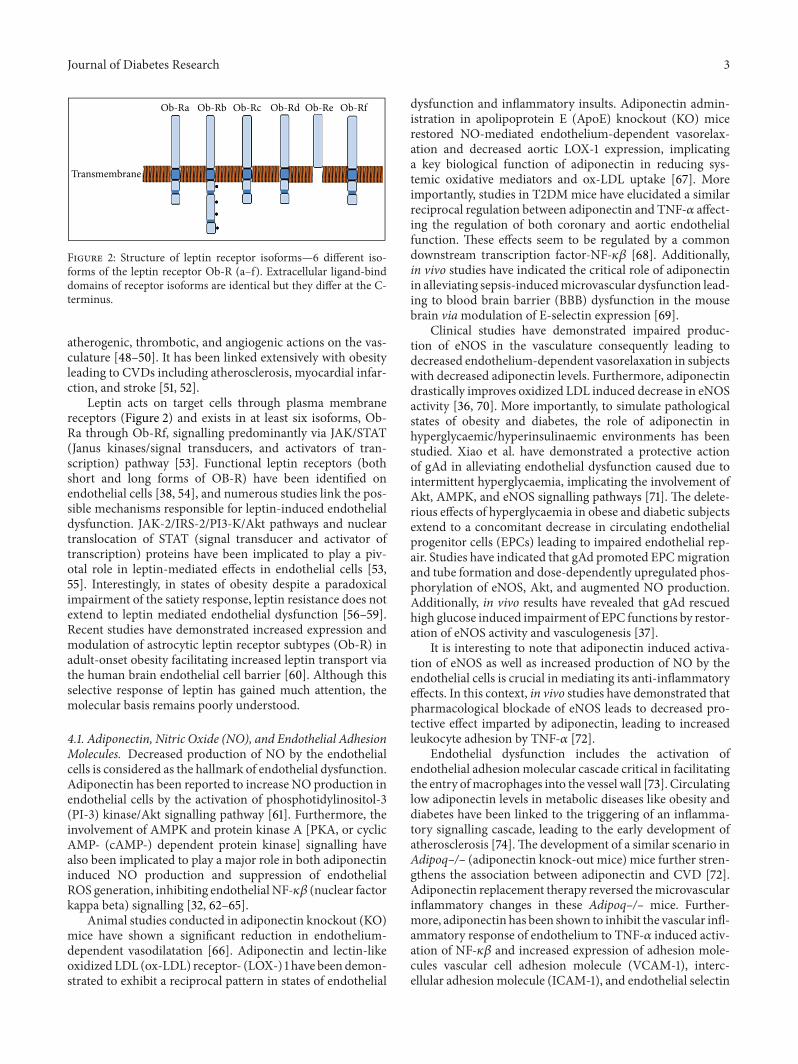
Transmembrane
Ob-Ra Ob-Rb Ob-Rc Ob-Rd Ob-Re Ob-Rf
Figure 2: Structure of leptin receptor isoforms—6 different iso-forms of the leptin receptor Ob-R (a–f). Extracellular ligand-binddomains of receptor isoforms are identical but they differ at the C-terminus.
atherogenic, thrombotic, and angiogenic actions on the vas-culature [48–50]. It has been linked extensively with obesityleading to CVDs including atherosclerosis, myocardial infar-ction, and stroke [51, 52].
Leptin acts on target cells through plasma membranereceptors (Figure 2) and exists in at least six isoforms, Ob-Ra through Ob-Rf, signalling predominantly via JAK/STAT(Janus kinases/signal transducers, and activators of tran-scription) pathway [53]. Functional leptin receptors (bothshort and long forms of OB-R) have been identified onendothelial cells [38, 54], and numerous studies link the pos-sible mechanisms responsible for leptin-induced endothelialdysfunction. JAK-2/IRS-2/PI3-K/Akt pathways and nucleartranslocation of STAT (signal transducer and activator oftranscription) proteins have been implicated to play a piv-otal role in leptin-mediated effects in endothelial cells [53,55]. Interestingly, in states of obesity despite a paradoxicalimpairment of the satiety response, leptin resistance does notextend to leptin mediated endothelial dysfunction [56–59].Recent studies have demonstrated increased expression andmodulation of astrocytic leptin receptor subtypes (Ob-R) inadult-onset obesity facilitating increased leptin transport viathe human brain endothelial cell barrier [60]. Although thisselective response of leptin has gained much attention, themolecular basis remains poorly understood.
4.1. Adiponectin, Nitric Oxide (NO), and Endothelial AdhesionMolecules. Decreased production of NO by the endothelialcells is considered as the hallmark of endothelial dysfunction.Adiponectin has been reported to increase NO production inendothelial cells by the activation of phosphotidylinositol-3(PI-3) kinase/Akt signalling pathway [61]. Furthermore, theinvolvement of AMPK and protein kinase A [PKA, or cyclicAMP- (cAMP-) dependent protein kinase] signalling havealso been implicated to play a major role in both adiponectininduced NO production and suppression of endothelialROS generation, inhibiting endothelial NF-𝜅𝛽 (nuclear factorkappa beta) signalling [32, 62–65].
Animal studies conducted in adiponectin knockout (KO)mice have shown a significant reduction in endothelium-dependent vasodilatation [66]. Adiponectin and lectin-likeoxidized LDL (ox-LDL) receptor- (LOX-) 1 have beendemon-strated to exhibit a reciprocal pattern in states of endothelial
dysfunction and inflammatory insults. Adiponectin admin-istration in apolipoprotein E (ApoE) knockout (KO) micerestored NO-mediated endothelium-dependent vasorelax-ation and decreased aortic LOX-1 expression, implicatinga key biological function of adiponectin in reducing sys-temic oxidative mediators and ox-LDL uptake [67]. Moreimportantly, studies in T2DMmice have elucidated a similarreciprocal regulation between adiponectin and TNF-𝛼 affect-ing the regulation of both coronary and aortic endothelialfunction. These effects seem to be regulated by a commondownstream transcription factor-NF-𝜅𝛽 [68]. Additionally,in vivo studies have indicated the critical role of adiponectinin alleviating sepsis-inducedmicrovascular dysfunction lead-ing to blood brain barrier (BBB) dysfunction in the mousebrain viamodulation of E-selectin expression [69].
Clinical studies have demonstrated impaired produc-tion of eNOS in the vasculature consequently leading todecreased endothelium-dependent vasorelaxation in subjectswith decreased adiponectin levels. Furthermore, adiponectindrastically improves oxidized LDL induced decrease in eNOSactivity [36, 70]. More importantly, to simulate pathologicalstates of obesity and diabetes, the role of adiponectin inhyperglycaemic/hyperinsulinaemic environments has beenstudied. Xiao et al. have demonstrated a protective actionof gAd in alleviating endothelial dysfunction caused due tointermittent hyperglycaemia, implicating the involvement ofAkt, AMPK, and eNOS signalling pathways [71]. The delete-rious effects of hyperglycaemia in obese and diabetic subjectsextend to a concomitant decrease in circulating endothelialprogenitor cells (EPCs) leading to impaired endothelial rep-air. Studies have indicated that gAd promoted EPCmigrationand tube formation and dose-dependently upregulated phos-phorylation of eNOS, Akt, and augmented NO production.Additionally, in vivo results have revealed that gAd rescuedhigh glucose induced impairment of EPC functions by restor-ation of eNOS activity and vasculogenesis [37].
It is interesting to note that adiponectin induced activa-tion of eNOS as well as increased production of NO by theendothelial cells is crucial in mediating its anti-inflammatoryeffects. In this context, in vivo studies have demonstrated thatpharmacological blockade of eNOS leads to decreased pro-tective effect imparted by adiponectin, leading to increasedleukocyte adhesion by TNF-𝛼 [72].
Endothelial dysfunction includes the activation ofendothelial adhesionmolecular cascade critical in facilitatingthe entry ofmacrophages into the vessel wall [73]. Circulatinglow adiponectin levels in metabolic diseases like obesity anddiabetes have been linked to the triggering of an inflamma-tory signalling cascade, leading to the early development ofatherosclerosis [74].The development of a similar scenario inAdipoq–/– (adiponectin knock-out mice) mice further stren-gthens the association between adiponectin and CVD [72].Adiponectin replacement therapy reversed themicrovascularinflammatory changes in these Adipoq–/– mice. Further-more, adiponectin has been shown to inhibit the vascular infl-ammatory response of endothelium to TNF-𝛼 induced activ-ation of NF-𝜅𝛽 and increased expression of adhesion mole-cules vascular cell adhesion molecule (VCAM-1), interc-ellular adhesionmolecule (ICAM-1), and endothelial selectin
4 Journal of Diabetes Research
(E-selectin) [32]. Functional effects induced by mediators ofsystemic inflammation like TNF-𝛼 and subsequent intera-ctions with adipokines have a significant influence in eitherpromoting or downregulating vascular insult.
4.2. Adiponectin and Endothelial Angiogenesis. Adiponectinhas been shown to induce in vitro angiogenesis in endothe-lial cells via AMPK-eNOS pathway [31]. More importantly,adiponectin replacement rectified ischemic stress inducedimpaired angiogenesis in Adipoq–/– mice [75]. Studies con-ducted in adiponectin-overexpressed mice brain (followingtransfection with adenoassociated viral vector (AAV) con-taining adiponectin gene) have shown a significant benefitinduced by adiponectin following ischemic insult. This pro-tective action was related to adiponectin induced focal angio-genesis involving VEGF and AMPK pathways [76]. On theother hand, other groups have demonstrated the potentinhibition of endothelial angiogenic events like migrationand proliferation by adiponectin [33], involving MAPK andcAMP-PKA pathways [34]. Similar antiangiogenic effects ofadiponectin have been studied in tumour growth suppressioninvolving Rho kinase/IFN-inducible protein 10 and matrixmetalloproteinase 9 (MMP-9) [77].
4.3. Differential Effects of fAD and gAD. Numerous studieshave implicated the vasoprotective actions of fAD by reduc-ing the expression of endothelial adhesion molecules andinhibiting TNF-𝛼 induced cytokine production from macr-ophages via NF-𝜅𝛽/cAMP-dependent pathway [32, 78–80].Animal studies in Ad−/− (mice completely lacking adiponec-tin) and Ad+/− (adiponectin-hemizygous mice) mice showedan increased expression of E-selectins and VCAMs. More-over, administration of gAd attenuated VCAM expression inAd−/− mice [72]. Adiponectin has been demonstrated to sup-press VEGF-stimulated HCAEC migration via cAMP/PKA-dependent signalling [34]. Clinical studies in patients withacute coronary syndromes have shown a negative correlationbetween circulating adiponectin levels and MMP-9/TIMP-1ratio, an independent predictor of atherosclerotic plaque sta-bility [80]. However, three independent studies have demon-strated that gAd activates NF-𝜅𝛽 leading to activation ofthe proinflammatory adhesion cascade, proliferation, andincreased procoagulability in endothelial cells and cardiacfibroblasts [39, 40, 81].
Furthermore, studies by Hattori et al. have indicated thesuppression of cytokine induced inflammatory cascade viaNF-𝜅𝛽 by gAD, albeit with a prolonged response time. Theauthors attribute this to desensitisation of the receptor, seenin instances of cytokine overload. More recently, colocaliza-tion studies conducted by Xu et al. have demonstrated thatadiponectin induces interaction between lymphotoxin- (LT-)b receptor (LTBR) and human AdipoR1, subsequently result-ing in inhibition of the NF-𝜅𝛽 pathway [82].
Studies have demonstrated the ability of leukocyte elasta-se secreted by activated monocytes and neutrophils to cleavethe globular domain of adiponectin [21].This local generationof gAd at sites of inflammation, namely, in atheroscleroticlesions, could be having pathophysiological relevance given
the differential actions of multimeric forms of adiponectin.In a study comparing the differential effects of fAd and gAdin human aortic endothelial cells (HAEC), both peptidesupregulated NO production by AMPK-dependent pathways.However, in contrast to fAd, gAd activated NF-𝜅𝛽 and p38MAPK signalling pathways, resulting in cyclooxygenase-2(COX-2) production and subsequently prostacyclin 2 [PGI]release. This study further demonstrated that monocyte-endothelial adhesion enhanced by gAD remained unaffectedwith either abrogation of AdipoR1 [siRNA] signalling orCOX-2 [siRNA] downregulation, thereby suggesting indepe-ndent mechanisms governing actions of fAd and gAd [41].
The obvious discrepancies between the experimental out-comes could be due to the differences in the forms of adipo-nectin used (Table 1). Additionally, endogenous productionof adiponectin by the endothelial cells needs to be considered[83].
Recently, we undertook a study to investigate the effectof gAD and fAD (Figure 3) on endothelial cell proliferationas well as in vitro migration and angiogenesis in relation tothe induction of endothelial angiogenic factors, specifically,MMP-2, MMP-9, and VEGF; furthermore, we examined theinvolvement of the adiponectin receptors, that is, adiponectinreceptor 1 (AdipoR1) and adiponectin receptor 2 (AdipoR2),within this context [35]. More importantly, given the connec-tion between the coexistence of hyperglycaemia and systemicinflammation with vascular disease in pathological statessuch as diabetes mellitus, we also studied the interaction bet-ween glucose and C-reactive protein (CRP) [a potent proi-nflammatory protein], respectively, with gAD and fAD.Finally, since AMP-activated protein kinase (AMPK), astress-activated protein kinase, and Akt have been implicatedas critical mediators of adiponectin induced angiogenesisin both normoxic and ischemic tissues, we examined therole of these signalling pathways in gAd induced endothelialangiogenesis [31]. We found that gAd led to a significantincrease in in vitro endothelial proliferation, migration, andangiogenesis with concomitant increase in MMP-2, MMP-9,and VEGF gene and protein production, as well as MMP-2andMMP-9 activation.The effect of gAd onVEGF appears tobe mediated by AdipoR1 whereas the effect of gAd on MMP-2 and MMP-9 appears to be mediated by AdipoR1 and Adi-poR2. On the other hand, only endothelial cell proliferationwas significantly increased by fAd and appears to bemediatedby AdipoR2; no significant effects on MMP-2, MMP-9, andVEGF were observed. Ouchi et al. 2004 had reported thatmouse fAD stimulates in vitro migration and angiogenesisand suggested that this effect may be beneficial in line withthe report by Shibata et al., 2004, who demonstrated thatadiponectin promotes ischemia-mediated revascularizationin adiponectin-knockout mice. It is important to note thatalthough in vitro angiogenic assays have been merited asuseful reporters in deciphering specific steps, they howeverlack the complex interplay of multiple factors vital for in vivoprocesses [31, 33]. Taken together, it remains unclear as towhether our observations reflect on balance a beneficial ordetrimental effect of adiponectin.
Thus it seems imperative to study the local effects ofvarious multimers of adiponectin in situ, for instance, in
Journal of Diabetes Research 5
Table 1: Differential effects of fAD, gAD, and leptin in endothelial cells.
Induced effect inendothelial cells [EC]
fAD (dose and time duration ofresponse)
gAD (dose and time duration ofresponse)
Leptin (dose and time durationof response)
Receptors AdipoR1 and AdipoR2 Predominantly AdipoR1 OB-R (both short and longforms)
[(HUVECs)-10-ng/mL-1 hr]NF-𝜅𝛽 activation, MCP-1production, and ↑ROSproduction [42][(HCAECs)-10 ng/mL-↑TFexpression and activity,↑VCAM-1, ICAM-1, andE-selectin expression andEC-monocyte adhesion [43]]
eNOS and NO production[(HUVECs)-30 ug/mL-]-AMPK-eNOS phosphorylation[31]
[(EPCs)-5𝜇g/mL-eNOSphosphorylation and NOproduction [37]]
[(HAECs)-10 ng/mL-eNOSphosphorylation and ↑NOproduction [44]].
The in vitro effects of fAD, gAD, and leptin differ on the concentration, time duration of peptide exposure, and the type of endothelial cells. BAECs: bovineaortic endothelial cells, EC: endothelial cell, eNOS: endothelial nitric oxide synthase, EPCs: endothelial progenitor cells, E-selectin: endothelial selectin, fAD:full length adiponectin, gAD: globular adiponectin, HAECs: human aortic endothelial cells, HCAECs: human coronary artery endothelial cells, HMECs:human microvascular endothelial cells, HUVECs: human umbilical vein endothelial cells, ICAM-1: intercellular cell adhesion molecule, MCP-1: monocytechemoattractant protein-1, PAEs: porcine aortic endothelial cells, ROS: reactive oxygen species, TF: tissue factor, TNF𝛼: tumour necrosis factor alpha, VCAM-1: vascular cell adhesion molecule, NF-𝜅𝛽: nuclear factor kappa beta, and VEGF: vascular endothelial growth factor.
atherosclerotic plaques, to ascertain the potential pro/anti-inflammatory actions of this adipokine.
4.4. Leptin and Endothelial Cell Dysfunction. Leptin hasmul-tiple proinflammatory and immune mediated effects on thevasculature. On engagement with leptin receptors expressedon vascular cell walls, leptin induces oxidative stress respon-ses, increases MCP-1, TNF-𝛼, IL-6, and endothelin-1, andpotentiates proliferation, along with the expression of otherendothelial cell adhesion molecules, MMPs, VEGF, and imp-aired smooth-muscle cell function, resulting in impairedendothelium-dependent vasodilatation promoting hyperten-sion and atherosclerosis [84]. Clinical studies have reported apositive correlation between circulating leptin, plasma thro-mbomodulin, and VCAM-1 levels [85].
4.5. Leptin Induced Endothelium Dependent and Indepen-dent Vasodilation. Endothelium dependent leptin inducedvasorelaxation observed in rat arterial rings was promptlyinhibited by increasing extracellular calcium [86] and inhibi-tion of NO synthase.Moreover, leptin has been demonstratedto phosphorylate eNOS leading to NO release [44]. Intra-arterial administration of leptin showed a similar vasoactive
response independent of NO in humans [87]. Additionally,a direct vasorelaxive effect of leptin on smooth muscle cellsindependent of endothelium was also observed in both ratand human arterial samples [88, 89]. Acute hyperleptinemiainduced vasodilatory effects and this seemingly contradictsthe coexisting hypertension and increased leptin levels onobesity. A plausible explanation for this could be attributedto the acute and chronic effects of leptin on the vasculature.Recent in vivo studies have revealed additional induction ofendothelial nNOS (neuronal nitric oxide synthase) expres-sion by leptin as a compensatory mechanism to induceendothelium-dependent relaxation in eNOS (−/−) mice [90].More importantly, hyperleptinemia induced endothelial dys-function may play a crucial role in the differential actions ofleptin.
4.6. Leptin Induced Endothelial Dysfunction and NO Pro-duction. Experiments by Naseem have indicated that leptininitiated upregulation of inducible NO synthase (iNOS),which may or may not lead to net increased NO productionand paradoxically impairs endothelial function by inducingoxidative stress [91]. Furthermore, a significant vasodilatoryresponse induced by leptin in lean Zucker rats failed to do soin obese hyperleptinemic Zucker rats [92].
Figure 3: Differential effects of leptin and adiponectin in vascular endothelium. Dual effects of gAD and fAD on endothelium with andwithout inflammatory stimuli. Circulating fAD gets cleaved by leucocyte elastase (secreted from neutrophils) releasing globular domain(gAD) fraction. AdipoR1 and AdipoR2 receptors following engagement with fAD, signals downstream activating the following pathways(a) AMPK, (b) cAMP-PKA, (c) MAPK, and (d) PI3K-Akt. Activation of cAMP-PKA/AMPK causes increased NO production, decreasedROS generation, suppression of NF-𝜅𝛽 pathway leading to reduction in IL-18, and endothelial adhesion molecule expression. These eventscollectively lead to a decrease in EC permeability, motility, and migration. Activation of AMPK/PI-3k/Akt signalling pathway specificallyleads to eNOS phosphorylation and NO release. In vitro studies have shown that gAD independently activates NF-𝜅𝛽 via AdipoR1/AMPK-Akt pathway. Proangiogenic/inflammatory effects of gAD have been shown to involve AMPK-Akt pathways. However, these pathways(AMPK-Akt) also contribute to an opposite effect of gAD in coexisting states of hyperglycaemia and inflammation. In hyperglycaemic andhyperinsulinaemic states, gAD improves endothelial dysfunction via activation of Akt-AMP-eNOS pathways and suppression of endothelialROS generation via inhibition of NF-𝜅𝛽 signalling. The binding of leptin to its receptor (OB-Rb) leads to the phosphorylation of Ob-R/JAK2 complex. Subsequent activation of downstream signalling cascades including PI3k/Akt-STAT3 activation results in transcription ofgenes [MCP-1, TNF-𝛼, IL-6/-2, and endothelin-1] involved in proatherogenic/angiogenic and inflammatory effects, potentiating endothelialproliferation. Additionally, leptin signalling in ECs also activates endothelial cell adhesionmolecules,MMPs, andVEGF resulting in impairedendothelium-dependent vasodilatation promoting hypertension and atherosclerosis.
As mentioned, leptin has been shown to induce oxidativestress by increasing the formation of reactive oxygen species(ROS), a key mediator of endothelial dysfunction [42, 93].This generated ROS has potent peroxidant effects and therebyreduces the bioavailability of NO in aortic endothelial cells[93], vascular smooth muscle cells [84], and macrophages[94]. Additionally, ROS further contributes to endothelialdysfunction by upregulating proinflammatory cascades incl-uding adhesion and chemotactic pathways in endothelial cells[95].
It is interesting to note that genetically modified (ob/ob-leptin knock out) mice maintain a relative hypotensive statusin comparison with their wild types. Leptin administrationin these mice promptly induces hypertension. This could beattributed to the disturbance in the fine balance between thesympathetic nervous system and endothelial cell mediatedregulation of vasomotor tone [96]. With respect to the regu-lation of leptin receptors and endothelial dysfunction, a studyby Park et al. 2012 has revealed that leptin receptors in coro-nary arterioles are downregulated in high-fat fed sedentary
Journal of Diabetes Research 7
mice leading to endothelial dysfunction. However, when sub-jected to exercise, the expression of leptin receptors in coro-nary arterioles was restored along withmaintenance of eNOSphosphorylation, leptin sensitivity, and redox balance [97].
4.7. Leptin and Endothelial Angiogenesis. As mentioned pre-viously, leptin-mediated actions in endothelial cells, includ-ing angiogenesis, primarily occur via the activation of Ob-R. It is interesting to note the increased expressions of bothOb-R and MMPs in atherosclerotic plaques, particularly theendothelial lining of neointimal regions, suggesting the roleof leptin in mediating aberrant angiogenesis [98]. Both invivo and in vitro studies have demonstrated the activationof endothelial Ob-R by leptin, leading to capillary tube for-mation, a prerequisite for angiogenesis [38]. Bouloumie et al.showed that leptin induced activation of mitogen-activatedprotein kinase family ERK1/2 leads to an increase in endothe-lial cell viability in serum-free media. Leptin has been shownto upregulate key proangiogenic molecules like the gelati-nases (MMPs, MMP-2/-9) and TIMPs. Additionally, leptinhas been shown to upregulate and act synergistically with thekey angiogenic mediators like FGF-2, VEGF, and its receptorVEGFR1, stimulating vascular permeability, consequentlyresulting in functional angiogenesis [99].
It is important to note that wound healing disorder (dueto deficient angiogenesis) in ob/ob mice is corrected byboth topical and systemic leptin administration but not infa/fa Zucker rats (rats with a recessive trait of the leptinreceptor), due to the absence of functional leptin receptors[100]. In a recent study involving an obese NZO (mice withphosphatidylcholine transfer protein mutation leading toabnormal lipid homeostasis)micemodel, the angiogenic pot-ential of leptin was found to be insignificant, perhaps dueto the relative inactivity of its receptor in these mice [101].Studies in HUVECs have implicated the involvement of afunctional endothelial p38 (MAPK)/Akt/COX-2 signallingaxis for leptin’s proangiogenic effects and more importantlythis signalling pathway is regulated upstream by ObRb-dependent activation of VEGFR2 receptor [102]. In vivofindings have implicated increased mobilisation of vascularprogenitor cells mobilized from the bonemarrow in responseto leptin stimulation leading to angiogenesis. These effects ofleptin seem to be mediated via Ob-R induced activation ofNOX2 and MMP9 [103]. Additional studies have evidencedthe importance of an ObR-Src kinase-alpha v beta 5 crosstalk in leptin mediated functional effects in enhancing theangiogenic potential of circulating angiogenic cells (CACs).More importantly, CACs derived from obese, hyperleptine-mic individuals were associated with relative insensitivity tothe angiogenic effects of leptin [104]. Leptin induced EPCsand NO production has been shown to play critical rolesin melanoma tumour growth induction [105]. Extendingthese findings to tumour angiogenesis, recent studies haveimplicated intratumoral leptin to exert proangiogenic effectsstimulating tube formation and proliferation of endothelialcells. More importantly, the authors have also demonstra-ted the therapeutic potential of a peptide ObR antago-nist in inhibiting these proangiogenic effects of leptin via
the VEGF pathway [106]. Interestingly, leptin induced pro-liferation/migration as well as expression of proangiogenicmolecules in breast cancer has been recently demonstrated toinvolve extensive crosstalk between Notch and interleukin-1(NILCO) pathways [107].
4.8. Leptin and Vascular Inflammation. Leptin has beenshown to upregulate variousmediators of vascular inflamma-tion like TNF-𝛼, IL-2, IL-6, MCP-1, ROS,Th1-type cytokines,and TGF-𝛽 from endothelial cells and PBMCs [56, 108–110].In vitro studies have demonstrated leptin induced increases intissue factor (TF) and cellular adhesion molecules (CAMs)expression in human coronary endothelial cells (HCAECs)via NF-𝜅𝛽 leading to increased procoagulant activity andleukocyte adhesion [111]. Additional molecules pivotal invascular inflammation including PAI-1 (plasminogen acti-vator inhibitor-1) and P-selectin have been documented tobe induced upon leptin treatment [112, 113]. Clinical studieshave shown a positive correlation with PAI-1, vWf, tPA, andplasma fibrinogen levels and an inverse relationship withprotein C and tissue factor pathway inhibitor. These findingsclearly demonstrate a strong link with circulating leptinand increased platelet activity observed in the metabolicsyndrome [114–117]. It is therefore not surprising to notethe decreased incidence of atherosclerosis in hyperlipidaemicmice (ob/ob; apoE−/−) [118].
4.9. Leptin-Adiponectin Ratio and Interactions. As discussedpreviously, converse actions of leptin and adiponectin in thevascular system have been widely studied. In obesity and dia-beticmetabolic abnormalities, coexistence of hypoadiponect-inemia and hyperleptinemia is observed. Thereby, the leptinto adiponectin ratio (L : A) is higher in these subjects. Variousclinical studies have been conducted to elucidate the rela-tionship between L : A ratio and markers of atheroscleroticdisease including carotid intimamedia thickness (CIMT) andpulse wave velocity [119–121]. In yet another clinical study,L : A ratio has been demonstrated as a useful biomarker forthe prevalence of metabolic syndrome, in comparison witheither leptin or adiponectin levels on their own. Additionally,visceral fatmass and cardiorespiratory fitness levels have beendocumented to influence this ratio [122]. Subjects with eNOSpolymorphisms with or without hyperinsulinemia have ahigher L : A ratio and are more prone for cardiovascularevents, suggesting a genetic link in the associated risk factors[123]. Labruna et al. have demonstrated that high serum L/Aratio is positively correlated with serum triglyceride levels,serving as surrogate markers of vascular inflammation in “at-risk” young severely obese individuals, which is independentof waist circumference (WC) and BMI [124]. Additionally,L : A ratio represented a powerful independent predictor ofintima media thickness (IMT), correlating with anthropo-metric, metabolic, and clinical parameters. Moreover thecorrelation with this ratio was much stronger than whencompared individually [120]. Furthermore, components ofthe metabolic syndrome were correlated positively with lep-tin/HMW adiponectin ratio, independent of parameters
8 Journal of Diabetes Research
including age, smoking status, exercise, low-density lipopro-tein (LDL) cholesterol, and BMI [125].
However, in contrary to the above mentioned findings,L : A ratio failed to establish any significant differences in dis-ease parameters, in a study conducted in patients with severecoronary heart disease [126]. It is noteworthy to mentionthat these above mentioned studies do differ in patient char-acteristics and pathological parameters leading to opposingresults.
5. Conclusions/Future Directions
Adipose tissue secreted factors or adipokines have been impl-icated in facilitating communication between adipose tissueand vasculature comprising the adipovascular axis. Bothproinflammatory and anti-inflammatory activities of thesesecreted adipokines seem to be crucial in creating a hom-eostatic responsewhich remains disturbed in states of adiposetissue expansion. In addition to alterations in the circulatinglevels, the local (i.e., tissue concentration) availability of theactivated forms of these adipokines has a significant bearingin influencing vascular function. For example, it is importantto consider the actions of locally available gAD fragmentof adiponectin, which could potentially drive leptin-inducedeffects. In-depth understanding of themechanisms and prop-erties of adipokine-receptor interactions and downstreamsignalling cascadesmay help in a clearer understanding of thepathogenesis of obesity-linked disorders. Studies investigat-ing the vascular effects of various multimeric/cleaved formsof adipokines will help in developing novel therapeutic strate-gies and targets in counteracting obesity-related metabolicand CVDs. Additionally large multicentric clinical studieswith strict inclusion-exclusion metabolic criteria need to beperformed.
Abbreviations
AdipoR1: Adiponectin receptor 1AdipoR2: Adiponectin receptor 2AMPK: AMP-activated protein kinasecAMP: Cyclic adenosine monophosphateeNOS: Endothelial nitric oxide synthasefAD: Full length adiponectinFGF-2: Fibroblast growth factor-2gAD: Globular adiponectiniNOS: Inducible nitric oxide synthaseIL-2/-6: Interleukin-2/-6MCP-1: Monocyte chemoattractant protein-1MMP-2/-9: Matrix metalloproteinase 2/9NF-𝜅𝛽: Nuclear factor 𝜅𝛽NO: Nitric oxidePI3K: Phosphatidylinositol 3-kinasePKA: Protein kinase AROS: Reactive oxygen speciesSTAT3: Signal transducer and activator of
No potential conflict of interests relevant to this paper wasreported.
References
[1] E. S. Gordon, “Non-esterified fatty acids in blood of obese andlean subjects,”TheAmerican Journal of Clinical Nutrition, vol. 8,pp. 740–747, 1960.
[2] G. Boden and G. I. Shulman, “Free fatty acids in obesity andtype 2 diabetes: defining their role in the development of insulinresistance and 𝛽-cell dysfunction,” European Journal of ClinicalInvestigation, vol. 32, no. 3, pp. 14–23, 2002.
[3] A. H. Lichtenstein, “Trans fatty acids, plasma lipid levels,and risk of developing cardiovascular disease: a statement forhealthcare professionals from theAmericanHeart Association,”Circulation, vol. 95, no. 11, pp. 2588–2590, 1997.
[4] J. A. Kim,M.Montagnani, K. K.Kwang, andM. J.Quon, “Recip-rocal relationships between insulin resistance and endothelialdysfunction—molecular and pathophysiological mechanisms,”Circulation, vol. 113, no. 15, pp. 1888–1904, 2006.
[5] W. Aldhahi and O. Hamdy, “Adipokines, inflammation, and theendothelium in diabetes,” Current Diabetes Reports, vol. 3, no.4, pp. 293–298, 2003.
[6] D. C. W. Lau, B. Dhillon, H. Yan, P. E. Szmitko, and S. Verma,“Adipokines: molecular links between obesity and atheroslcero-sis,” American Journal of Physiology—Heart and CirculatoryPhysiology, vol. 288, no. 5, pp. H2031–H2041, 2005.
[7] A. Avogaro and S. V. de Kreutzenberg, “Mechanisms of endo-thelial dysfunction in obesity,” Clinica Chimica Acta, vol. 360,no. 1-2, pp. 9–26, 2005.
[8] Y. B. Tripathi and V. Pandey, “Obesity and endoplasmic reticu-lum (ER) stresses,” Frontiers in Immunology, vol. 3, article 240,2012.
[9] A. Bouloumie, J. Bauersachs, W. Linz et al., “Endothelialdysfunction coincides with an enhanced nitric oxide synthaseexpression and superoxide anion production,” Hypertension,vol. 30, no. 4, pp. 934–941, 1997.
[10] R. Stocker and J. F. Keaney Jr., “Role of oxidative modificationsin atherosclerosis,”Physiological Reviews, vol. 84, no. 4, pp. 1381–1478, 2004.
[11] D. Cooper, K. Y. Stokes, A. Tailor, andD. N. Granger, “Oxidativestress promotes blood cell-endothelial cell interactions in themicrocirculation,” Cardiovascular Toxicology, vol. 2, no. 3, pp.165–180, 2002.
[12] A. R. Subauste and C. F. Burant, “Role of FoxO1 in FFA-inducedoxidative stress in adipocytes,” The American Journal of Phys-iology—Endocrinology andMetabolism, vol. 293, no. 1, pp. E159–E164, 2007.
[13] P. E. Scherer, S. Williams, M. Fogliano, G. Baldini, and H.F. Lodish, “A novel serum protein similar to C1q, producedexclusively in adipocytes,” The Journal of Biological Chemistry,vol. 270, no. 45, pp. 26746–26749, 1995.
[14] T. Yamauchi, J. Kamon, H. Waki et al., “The fat-derived hor-mone adiponectin reverses insulin resistance associated withboth lipoatrophy and obesity,”Nature Medicine, vol. 7, no. 8, pp.941–946, 2001.
Journal of Diabetes Research 9
[15] M. E. Trujillo and P. E. Scherer, “Adiponectin—journey froman adipocyte secretory protein to biomarker of the metabolicsyndrome,” Journal of Internal Medicine, vol. 257, no. 2, pp. 167–175, 2005.
[16] H. Okui, S. Hamasaki, S. Ishida et al., “Adiponectin is a betterpredictor of endothelial function of the coronary artery thanHOMA-R, body mass index, immunoreactive insulin, or trig-lycerides,” International Journal of Cardiology, vol. 126, no. 1, pp.53–61, 2008.
[17] E. Hu, P. Liang, and B. M. Spiegelman, “AdipoQ is a noveladipose-specific gene dysregulated in obesity,” Journal of Bio-logical Chemistry, vol. 271, no. 18, pp. 10697–10703, 1996.
[18] K. Maeda, K. Okubo, I. Shimomura, T. Funahashi, Y. Mat-suzawa, and K. Matsubara, “cDNA cloning and expression ofa novel adipose specific collagen-like factor, apM1 (AdiPoseMost abundant Gene transcript 1),” Biochemical and BiophysicalResearch Communications, vol. 221, no. 2, pp. 286–289, 1996.
[19] Y. Nakano, T. Tobe, N. H. Choi-Miura, T. Mazda, and M.Tomita, “Isolation and characterization of GBP28, a novelgelatin-binding protein purified from human plasma,” Journalof Biochemistry, vol. 120, no. 4, pp. 803–812, 1996.
[20] Y.Wang, K. S. L. Lam,M.-H. Yau, andA. Xu, “Post-translationalmodifications of adiponectin: mechanisms and functionalimplications,” Biochemical Journal, vol. 409, no. 3, pp. 623–633,2008.
[21] H. Waki, T. Yamauchi, J. Kamon et al., “Generation of globularfragment of adiponectin by leukocyte elastase secreted bymonocytic cell line THP-1,” Endocrinology, vol. 146, no. 2, pp.790–796, 2005.
[22] T. Yamauchi, J. Kamon, Y. Ito et al., “Cloning of adiponectinreceptors that mediate antidiabetic metabolic effects,” Nature,vol. 423, no. 6941, pp. 762–769, 2003.
[23] I. Kharroubi, J. Rasschaert, D. L. Eizirik, and M. Cnop,“Expression of adiponectin receptors in pancreatic beta cells,”Biochemical and Biophysical Research Communications, vol. 312,no. 4, pp. 1118–1122, 2003.
[24] G. Chinetti, C. Zawadski, J. C. Fruchart, and B. Staels, “Expres-sion of adiponectin receptors in humanmacrophages and regu-lation by agonists of the nuclear receptors PPAR𝛼, PPAR𝛾, andLXR,” Biochemical and Biophysical Research Communications,vol. 314, no. 1, pp. 151–158, 2004.
[25] T.-S. Tsao, E. Tomas, H. E. Murrey et al., “Role of disulfidebonds in Acrp30/adiponectin structure and signaling speci-ficity: different oligomers activate different signal transductionpathways,”The Journal of Biological Chemistry, vol. 278, no. 50,pp. 50810–50817, 2003.
[26] M. L. Narasimhan, M. A. Coca, J. B. Jin et al., “Osmotin is ahomolog of mammalian adiponectin and controls apoptosis inyeast through a homolog of mammalian adiponectin receptor,”Molecular Cell, vol. 17, no. 2, pp. 171–180, 2005.
[27] M. Bluher, J. W. Bullen Jr., J. H. Lee et al., “Circulating adipone-ctin and expression of adiponectin receptors in human skeletalmuscle: associations with metabolic parameters and insulinresistance and regulation by physical training,” Journal of Clini-cal Endocrinology and Metabolism, vol. 91, no. 6, pp. 2310–2316,2006.
[28] N. Viguerie, H. Vidal, P. Arner et al., “Adipose tissue gene expr-ession in obese subjects during low-fat and high-fat hypocaloricdiets,” Diabetologia, vol. 48, no. 1, pp. 123–131, 2005.
[29] A. E. Civitarese, C. P. Jenkinson, D. Richardson et al., “Adip-onectin receptors gene expression and insulin sensitivity in
non-diabetic Mexican Americans with or without a familyhistory of Type 2 diabetes,” Diabetologia, vol. 47, no. 5, pp. 816–820, 2004.
[30] H. Zhang, Y. Park, and C. Zhang, “Coronary and aorticendothelial function affected by feedback between adiponectinand tumor necrosis factor 𝛼 in type 2 diabeticmice,”Arterioscle-rosis, Thrombosis, and Vascular Biology, vol. 30, no. 11, pp. 2156–2163, 2010.
[31] N.Ouchi,H.Kobayashi, S. Kihara et al., “Adiponectin stimulatesangiogenesis by promoting cross-talk between AMP-activatedprotein kinase and Akt signaling in endothelial cells,” TheJournal of Biological Chemistry, vol. 279, no. 2, pp. 1304–1309,2004.
[32] N. Ouchi, S. Kihara, Y. Arita et al., “Adiponectin, an adipocyte-derived plasma protein, inhibits endothelial NF-𝜅B signalingthrough a cAMP-dependent pathway,” Circulation, vol. 102, no.11, pp. 1296–1301, 2000.
[33] E. Brakenhielm, N. Veitonmaki, R. Cao et al., “Adiponectin-induced antiangiogenesis and antitumor activity involvecaspase-mediated endothelial cell apoptosis,” Proceedings of theNational Academy of Sciences of the United States of America,vol. 101, no. 8, pp. 2476–2481, 2004.
[34] K. Mahadev, X. Wu, S. Donnelly, R. Ouedraogo, A. D. Eckhart,and B. J. Goldstein, “Adiponectin inhibits vascular endothelialgrowth factor-induced migration of human coronary arteryendothelial cells,” Cardiovascular Research, vol. 78, no. 2, pp.376–384, 2008.
[35] R. Adya, B. K. Tan, J. Chen, and H. S. Randeva, “Protectiveactions of globular and full-length adiponectin on humanendothelial cells: novel insights into adiponectin-inducedangiogenesis,” Journal of Vascular Research, vol. 49, no. 6, pp.534–543, 2012.
[36] H.Motoshima, X.Wu, K.Mahadev, and B. J. Goldstein, “Adipo-nectin suppresses proliferation and superoxide generation andenhances eNOS activity in endothelial cells treated with oxi-dized LDL,” Biochemical and Biophysical Research Communica-tions, vol. 315, no. 2, pp. 264–271, 2004.
[37] P.-H. Huang, J.-S. Chen, H.-Y. Tsai et al., “Globular adiponectinimproves high glucose-suppressed endothelial progenitor cellfunction through endothelial nitric oxide synthase dependentmechanisms,” Journal of Molecular and Cellular Cardiology, vol.51, no. 1, pp. 109–119, 2011.
[38] A. Bouloumie, H. C. A. Drexler, M. Lafontan, and R. Busse,“Leptin, the product of Ob gene, promotes angiogenesis,”Circulation Research, vol. 83, no. 10, pp. 1059–1066, 1998.
[39] Y. Hattori, S. Hattori, K. Akimoto et al., “Globular adiponectinactivates nuclear factor-kappaB and activating protein-1 andenhances angiotensin II-inducedproliferation in cardiac fibrob-lasts,” Diabetes, vol. 56, no. 3, pp. 804–808, 2007.
[40] P. Bobbert, S. Antoniak, H. P. Schultheiss, and U. Rauch, “Glob-ular adiponectin but not full-length adiponectin induces incre-ased procoagulability in human endothelial cells,” Journal ofMolecular and Cellular Cardiology, vol. 44, no. 2, pp. 388–394,2008.
[41] F. Addabbo, C. Nacci, L. de Benedictis et al., “Globular adipone-ctin counteracts VCAM-1-mediated monocyte adhesion viaadipoR1/NF-𝜅B/COX-2 signaling in human aortic endothe-lial cells,” American Journal of Physiology—Endocrinology andMetabolism, vol. 301, no. 6, pp. E1143–E1154, 2011.
[42] A. Bouloumie, T. Marumo, M. Lafontan, and R. Busse, “Leptininduces oxidative stress in human endothelial cells,”The FASEBJournal, vol. 13, no. 10, pp. 1231–1238, 1999.
10 Journal of Diabetes Research
[43] P. Cirillo, V. Angri, S. de Rosa et al., “Pro-atherothromboticeffects of leptin in human coronary endothelial cells,” Throm-bosis and Haemostasis, vol. 103, no. 5, pp. 1065–1075, 2010.
[44] C. Vecchione, A. Maffei, S. Colella et al., “Leptin effect on endo-thelial nitric oxide is mediated through Akt-endothelial nitricoxide synthase phosphorylation pathway,” Diabetes, vol. 51, no.1, pp. 168–173, 2002.
[45] C. Hug, J. Wang, N. S. Ahmad, J. S. Bogan, T.-S. Tsao, and H.F. Lodish, “T-cadherin is a receptor for hexameric and high-molecular-weight forms of Acrp30/adiponectin,” Proceedings ofthe National Academy of Sciences of the United States of America,vol. 101, no. 28, pp. 10308–10313, 2004.
[46] Y. Y. Zhang, R. Proenca,M.Maffei,M. Barone, L. Leopold, and J.M. Friedman, “Positional cloning of the mouse obese gene andits human homolog,” Nature, vol. 372, pp. 425–432, 1994.
[47] S. Margetic, C. Gazzola, G. G. Pegg, and R. A. Hill, “Leptin: areview of its peripheral actions and interactions,” InternationalJournal of Obesity, vol. 26, no. 11, pp. 1407–1433, 2002.
[48] J. Bełtowski, G. Wojcicka, and A. Jamroz, “Leptin decreasesplasma paraoxonase 1 (PON1) activity and induces oxidativestress: the possible novel mechanism for proatherogenic effectof chronic hyperleptinemia,” Atherosclerosis, vol. 170, no. 1, pp.21–29, 2003.
[49] M. L. G. Correia andK. Rahmouni, “Role of leptin in the cardio-vascular and endocrine complications of metabolic syndrome,”Diabetes, Obesity &Metabolism, vol. 8, no. 6, pp. 603–610, 2006.
[50] N. Werner and G. Nickenig, “From fat fighter to risk factor: thezigzag trek of leptin,” Arteriosclerosis, Thrombosis, and VascularBiology, vol. 24, no. 1, pp. 7–9, 2004.
[51] A.M.Wallace, A. D.McMahon, C. J. Packard et al., “Plasma lep-tin and the risk of cardiovascular disease in theWest of ScotlandCoronary Prevention Study (WOSCOPS),”Circulation, vol. 104,no. 25, pp. 3052–3056, 2001.
[52] R. Wolk, P. Berger, R. J. Lennon, E. S. Brilakis, B. D. Johnson,and V. K. Somers, “Plasma leptin and prognosis in patients withestablished coronary atherosclerosis,” Journal of the AmericanCollege of Cardiology, vol. 44, no. 9, pp. 1819–1824, 2004.
[53] G. Fruhbeck, “Intracellular signalling pathways activated byleptin,”The Biochemical Journal, vol. 393, no. 1, pp. 7–20, 2006.
[54] M. R. Sierra-Honigmann, A. K. Nath, C. Murakami et al.,“Biological action of leptin as an angiogenic factor,” Science, vol.281, no. 5383, pp. 1683–1686, 1998.
[55] K. Rahmouni andW.G.Haynes, “Leptin and the cardiovascularsystem,” Recent Progress in Hormone Research, vol. 59, pp. 225–244, 2004.
[56] A. Avogaro and S. V. de Kreutzenberg, “Mechanisms of endo-thelial dysfunction in obesity,” Clinica Chimica Acta, vol. 360,no. 1-2, pp. 9–26, 2005.
[57] J. M. Friedman and J. L. Halaas, “Leptin and the regulation ofbody weight in mammals,” Nature, vol. 395, no. 6704, pp. 763–770, 1998.
[58] J. M. Friedman, “Modern science versus the stigma of obesity,”Nature Medicine, vol. 10, no. 6, pp. 563–569, 2004.
[59] J. Ren, “Leptin and hyperleptinemia—from friend to foe forcardiovascular function,”The Journal of Endocrinology, vol. 181,no. 1, pp. 1–10, 2004.
[60] H. Hsuchou, A. J. Kastin, H. Tu, N. Joan Abbott, P.-O. Couraud,andW. Pan, “Role of astrocytic leptin receptor subtypes on lep-tin permeation across hCMEC/D3 human brain endothelialcells,” Journal of Neurochemistry, vol. 115, no. 5, pp. 1288–1298,2010.
[61] H. Chen, M. Montagnani, T. Funahashi, I. Shimomura, and M.J. Quon, “Adiponectin stimulates production of nitric oxide invascular endothelial cells,” The Journal of Biological Chemistry,vol. 278, no. 45, pp. 45021–45026, 2003.
[62] R. Ouedraogo, X. D. Wu, S. Q. Xu et al., “Adiponectin sup-pression of high-glucose-induced reactive oxygen species invascular endothelial cells: evidence for involvement of a cAMPsignaling pathway,”Diabetes, vol. 55, no. 6, pp. 1840–1846, 2006.
[63] B. J. Goldstein and R. Scalia, “Adiponectin: a novel adipokinelinking adipocytes and vascular function,”The Journal of Clini-cal Endocrinology andMetabolism, vol. 89, no. 6, pp. 2563–2568,2004.
[64] Y. Hattori, Y. Nakano, S. Hattori, A. Tomizawa, K. Inukai, andK. Kasai, “High molecular weight adiponectin activates AMPKand suppresses cytokine-induced NF-𝜅B activation in vascularendothelial cells,” FEBS Letters, vol. 582, no. 12, pp. 1719–1724,2008.
[65] S.-Q. Xu, K. Mahadev, X. Wu et al., “Adiponectin protectsagainst angiotensin II or tumor necrosis factor 𝛼-inducedendothelial cell monolayer hyperpermeability: role of cAMP/PKA signaling,” Arteriosclerosis, Thrombosis, and Vascular Biol-ogy, vol. 28, no. 5, pp. 899–905, 2008.
[66] N. Ouchi, M. Ohishi, S. Kihara et al., “Association of hypoad-iponectinemia with impaired vasoreactivity,”Hypertension, vol.42, no. 3, pp. 231–234, 2003.
[67] X. P. Chen, H. R. Zhang, S. McAfee, and C. H. Zhang, “Thereciprocal relationship between adiponectin and LOX-1 inthe regulation of endothelial dysfunction in ApoE knockoutmice,” American Journal of Physiology—Heart and CirculatoryPhysiology, vol. 299, no. 3, pp. H605–H612, 2010.
[68] H. R. Zhang, Y. Park, and C. H. Zhang, “Coronary and aorticendothelial function affected by feedback between adiponectinand tumor necrosis factor alpha in type 2 diabetic mice,”Arteriosclerosis, Thrombosis, and Vascular Biology, vol. 30, no.11, pp. 2156–2163, 2010.
[69] V. Vachharajani, C. Cunningham, B. Yoza, J. Carson Jr., T. J.Vachharajani, and C.McCall, “Adiponectin-deficiency exagger-ates sepsis-induced microvascular dysfunction in the mousebrain,” Obesity, vol. 20, no. 3, pp. 498–504, 2012.
[70] K. C. B. Tan, A. Xu, W. S. Chow et al., “Hypoadiponectinemiais associated with impaired endothelium-dependent vasodila-tion,” Journal of Clinical Endocrinology and Metabolism, vol. 89,no. 2, pp. 765–769, 2004.
[71] X. Xiao, Y. Dong, J. Zhong et al., “Adiponectin protects endothe-lial cells from the damages induced by the intermittent highlevel of glucose,” Endocrine, vol. 40, no. 3, pp. 386–393, 2011.
[72] R. Ouedraogo, Y. Gong, B. Berzins et al., “Adiponectin defi-ciency increases leukocyte-endothelium interactions via upreg-ulation of endothelial cell adhesion molecules in vivo,” TheJournal of Clinical Investigation, vol. 117, no. 6, pp. 1718–1726,2007.
[73] E. S. Biegelsen and J. Loscalzo, “Endothelial function and ather-osclerosis,” Coronary Artery Disease, vol. 10, no. 4, pp. 241–256,1999.
[74] C. Weyer, T. Funahashi, S. Tanaka et al., “Hypoadiponectine-mia in obesity and type 2 diabetes: close association withinsulin resistance and hyperinsulinemia,” Journal of ClinicalEndocrinology and Metabolism, vol. 86, no. 5, pp. 1930–1935,2001.
[75] R. Shibata, N. Ouchi, S. Kihara, K. Sato, T. Funahashi, andK. Walsh, “Adiponectin stimulates angiogenesis in response to
Journal of Diabetes Research 11
tissue ischemia through stimulation of AMP-activated proteinkinase signaling,” Journal of Biological Chemistry, vol. 279, no.27, pp. 28670–28674, 2004.
[76] L. Shen, J. Miao, F. Yuan et al., “Overexpression of adiponectinpromotes focal angiogenesis in the mouse brain followingmiddle cerebral artery occlusion,” Gene Therapy, vol. 20, no. 1,pp. 93–101, 2013.
[77] K. Man, K. T. P. Ng, A. Xu et al., “Suppression of liver tumorgrowth and metastasis by adiponectin in nude mice throughinhibition of tumor angiogenesis and downregulation of rhokinase/IFN-inducible protein 10/matrix metalloproteinase 9signaling,” Clinical Cancer Research, vol. 16, no. 3, pp. 967–977,2010.
[78] N. Ouchi, S. Kihara, Y. Arita et al., “Novel modulator forendothelial adhesionmolecules: adipocyte-derived plasma pro-tein adiponectin,” Circulation, vol. 100, no. 25, pp. 2473–2476,1999.
[79] N. Ouchi, S. Kihara, Y. Arita et al., “Adipocyte-derived plasmaprotein, adiponectin, suppresses lipid accumulation and classA scavenger receptor expression in human monocyte-derivedmacrophages,” Circulation, vol. 103, no. 8, pp. 1057–1063, 2001.
[80] M. Cheng, S. Hashmi, X.Mao, andQ. T. Zeng, “Relationships ofadiponectin and matrix metalloproteinase-9 to tissue inhibitorof metalloproteinase-1 ratio with coronary plaque morphologyin patients with acute coronary syndrome,” The CanadianJournal of Cardiology, vol. 24, no. 5, pp. 385–390, 2008.
[81] A. Tomizawa, Y. Hattori, K. Kasai, and Y. Nakano, “Adiponectininduces NF-𝜅B activation that leads to suppression of cytokine-inducedNF-𝜅B activation in vascular endothelial cells: globularadiponectin vs. high molecular weight adiponectin,” Diabetesand Vascular Disease Research, vol. 5, no. 2, pp. 123–127, 2008.
[82] Y. Xu, C. Zhang, N. Wang et al., “Adiponectin inhibitslymphotoxin-𝛽 receptor-mediated NF-𝜅B signaling in humanumbilical vein endothelial cells,” Biochemical and BiophysicalResearch Communications, vol. 404, no. 4, pp. 1060–1064, 2011.
[83] R. Natarajan, F. N. Salloum, B. J. Fisher, R. C. Kukreja, and A. A.Fowler III, “Hypoxia inducible factor-1 upregulates adiponectinin diabetic mouse hearts and attenuates post-ischemic injury,”Journal of Cardiovascular Pharmacology, vol. 51, no. 2, pp. 178–187, 2008.
[84] L. Li, J.-C. Mamputu, N. Wiernsperger, and G. Renier, “Sig-naling pathways involved in human vascular smooth musclecell proliferation and matrix metalloproteinase-2 expressioninduced by leptin: inhibitory effect of metformin,”Diabetes, vol.54, no. 7, pp. 2227–2234, 2005.
[85] E. Porreca, C. Di Febbo, L. Fusco, V. Moretta, M. Di Nisio,and F. Cuccurullo, “Soluble thrombomodulin and vascularadhesion molecule-1 are associated to leptin plasma levels inobese women,” Atherosclerosis, vol. 172, no. 1, pp. 175–180, 2004.
[86] G. Lembo, C. Vecchione, L. Fratta et al., “Leptin inducesdirect vasodilation through distinct endothelial mechanisms,”Diabetes, vol. 49, no. 2, pp. 293–297, 2000.
[87] K. Matsuda, H. Teragawa, Y. Fukuda, K. Nakagawa, Y. Higashi,andK. Chayama, “Leptin causes nitric-oxide independent coro-nary artery vasolidation in humans,”HypertensionResearch, vol.26, no. 2, pp. 147–152, 2003.
[88] A. Fortuno, A. Rodrıguez, J. Gomez-Ambrosi et al., “Leptininhibits angiotensin II-induced intracellular calcium increaseand vasoconstriction in the rat aorta,” Endocrinology, vol. 143,no. 9, pp. 3555–3560, 2002.
[89] A.U.Momin,N.Melikian, A.M. Shah et al., “Leptin is an endot-helial-independent vasodilator in humans with coronary artery
disease: evidence for tissue specificity of leptin resistance,”European Heart Journal, vol. 27, no. 19, pp. 2294–2299, 2006.
[90] S. Benkhoff, A. E. Loot, I. Pierson et al., “Leptin potentiatesendothelium-dependent relaxation by inducing endothelialexpression of neuronal no synthase,” Arteriosclerosis, Thrombo-sis, and Vascular Biology, vol. 32, pp. 1605–1612, 2012.
[91] K. M. Naseem, “The role of nitric oxide in cardiovasculardiseases,”Molecular Aspects of Medicine, vol. 26, no. 1-2, pp. 33–65, 2005.
[92] J. D. Knudson, U. D. Dincer, C. Zhang et al., “Leptin receptorsare expressed in coronary arteries, and hyperleptinemia causessignificant coronary endothelial dysfunction,”American Journalof Physiology—Heart and Circulatory Physiology, vol. 289, no. 1,pp. H48–H56, 2005.
[93] S. I. Yamagishi, D. Edelstein, X. L. Du, Y. Kaneda, M. Guzman,and M. Brownlee, “ Leptin induces mitochondrial superoxideproduction and monocyte chemoattractant protein-1 expres-sion in aortic endothelial cells by increasing fatty acid oxidationvia protein kinase A,” The Journal of Biological Chemistry, vol.276, pp. 25096–25100, 2001.
[94] F.Maingrette andG. Renier, “Leptin increases lipoprotein lipasesecretion by macrophages: involvement of oxidative stress andprotein kinase C,” Diabetes, vol. 52, no. 8, pp. 2121–2128, 2003.
[95] D. Cooper, K. Y. Stokes, A. Tailor, andD. N. Granger, “Oxidativestress promotes blood cell-endothelial cell interactions in themicrocirculation,” Cardiovascular Toxicology, vol. 2, no. 3, pp.165–180, 2002.
[96] Y.-M. Leung and C.-Y. Kwan, “Dual vascular effects of leptin viaendothelium: hypothesis and perspective,”The Chinese Journalof Physiology, vol. 51, no. 1, pp. 1–6, 2008.
[97] Y. Park, F. W. Booth, S. Lee, M. J. Laye, and C. H. Zhang, “Phys-ical activity opposes coronary vascular dysfunction inducedduring high fat feeding in mice,”The Journal of Physiology, vol.590, no. 17, pp. 4255–4268, 2012.
[98] H.-Y. Park, H. M. Kwon, H. J. Lim et al., “Potential role of leptinin angiogenesis: Leptin induces endothelial cell proliferationand expression of matrix metalloproteinases in vivo and invitro,” Experimental and Molecular Medicine, vol. 33, no. 2, pp.95–102, 2001.
[99] R. Cao, E. Brakenhielm, C. Wahlestedt, J. Thyberg, and Y.Cao, “Leptin induces vascular permeability and synergisticallystimulates angiogenesis with FGF-2 and VEGF,” Proceedings ofthe National Academy of Sciences of the United States of America,vol. 98, no. 11, pp. 6390–6395, 2001.
[100] B. Stallmeyer, J. Pfeilschifter, and S. Frank, “Systemically andtopically supplemented leptin fails to reconstitute a normalangiogenic response during skin repair in diabetic ob/ob mice,”Diabetologia, vol. 44, no. 4, pp. 471–479, 2001.
[101] L. Wator, U. Razny, A. Balwierz et al., “Impaired leptin activityin New Zealand Obese mice: model of angiogenesis,”Genes andNutrition, vol. 3, no. 3-4, pp. 177–180, 2008.
[102] E. Garonna, K. M. Botham, G. M. Birdsey, A. M. Randi,R. R. Gonzalez-Perez, and C. P. D. Wheeler-Jones, “Vascularendothelial growth factor receptor-2 couples cyclo-oxygenase-2 with pro-angiogenic actions of leptin on human endothelialcells,” PLoS ONE, vol. 6, no. 4, Article ID e18823, 2011.
[103] M. R. Schroeter, S. Stein, N.-M. Heida et al., “Leptin promotesthe mobilization of vascular progenitor cells and neovascular-ization by NOX2-mediated activation of MMP9,” Cardiovascu-lar Research, vol. 93, no. 1, pp. 170–180, 2012.
[104] N.-M. Heida, M. Leifheit-Nestler, M. R. Schroeter et al., “Lep-tin enhances the potency of circulating angiogenic cells via
12 Journal of Diabetes Research
src kinase and integrin 𝛼v𝛽5: implications for angiogenesisin human obesity,” Arteriosclerosis, Thrombosis, and VascularBiology, vol. 30, no. 2, pp. 200–206, 2010.
[105] F. Amjadi, S. H. Javanmard, H. Zarkesh-Esfahani, M. Khazaei,and M. Narimani, “Leptin promotes melanoma tumor growthin mice related to increasing circulating endothelial progenitorcells numbers and plasma NO production,” Journal of Experi-mental and Clinical Cancer Research, vol. 30, no. 1, article 21,2011.
[106] R. Ferla, M. Bonomi, L. Otvos Jr., and E. Surmacz, “Glioblas-toma -derived leptin induces tube formation and growth ofendothelial cells: comparison with VEGF effects,” BMC Cancer,vol. 11, article 303, 2011.
[107] S. Guo and R. R. Gonzalez-Perez, “Notch, IL-1 and leptincrosstalk outcome (NILCO) is critical for leptin-induced pro-liferation, migration and VEGF/VEGFR-2 expression in breastcancer,” PLoS ONE, vol. 6, no. 6, Article ID e21467, 2011.
[108] S. Loffreda, S. Q. Yang, H. Z. Lin et al., “Leptin regulatesproinflammatory immune responses,” The FASEB Journal, vol.12, no. 1, pp. 57–65, 1998.
[109] V. Sanchez-Margalet, C. Martın-Romero, J. Santos-Alvarez, R.Goberna, S. Najib, and C. Gonzalez-Yanes, “Role of leptin as animmunomodulator of bloodmononuclear cells: mechanisms ofaction,” Clinical and Experimental Immunology, vol. 133, no. 1,pp. 11–19, 2003.
[110] G. Wolf, A. Hamann, D. C. Han et al., “Leptin stimulates prolif-eration and TGF-𝛽 expression in renal glomerular endothelialcells: potential role in glomerulosclerosis,”Kidney International,vol. 56, no. 3, pp. 860–872, 1999.
[111] H. Hsuchou, A. J. Kastin, H. Tu, N. Joan Abbott, P. O. Couraud,and W. Pan, “Role of astrocytic leptin receptor subtypes onleptin permeation across hCMEC/D3 human brain endothelialcells,” Journal of Neurochemistry, vol. 115, no. 5, pp. 1288–1298,2010.
[112] P. Singh, T. E. Peterson, K. R. Barber et al., “Leptin upregulatesthe expression of plasminogen activator inhibitor-1 in humanvascular endothelial cells,”Biochemical and Biophysical ResearchCommunications, vol. 392, no. 1, pp. 47–52, 2010.
[113] H. Wallaschofski, A. Kobsar, O. Sokolova et al., “Differencesin platelet activation by prolactin and leptin,” Hormone andMetabolic Research, vol. 36, no. 7, pp. 453–457, 2004.
[114] A.M.Thøgersen, S. Soderberg, J.-H. Jansson et al., “Interactionsbetween fibrinolysis, lipoproteins and leptin related to a firstmyocardial infarction,” European Journal of CardiovascularPrevention and Rehabilitation, vol. 11, no. 1, pp. 33–40, 2004.
[115] N.-F. Chu, D. Spiegelman, G. S. Hotamisligil, N. Rifai, M.Stampfer, and E. B. Rimm, “Plasma insulin, leptin, and solubleTNF receptors levels in relation to obesity-related atherogenicand thrombogenic cardiovascular disease risk factors amongmen,” Atherosclerosis, vol. 157, no. 2, pp. 495–503, 2001.
[116] S. Soderberg, B. Ahren, J.-H. Jansson et al., “Leptin is associatedwith increased risk ofmyocardial infarction,” Journal of InternalMedicine, vol. 246, no. 4, pp. 409–418, 1999.
[117] J. Małyszko, S. Wołczynski, J. Małyszko, and M. Mysliwiec,“Leptin correlates with some hemostatic parameters in CAPDpatients,” Nephron, vol. 92, no. 3, pp. 721–724, 2002.
[118] T. Chiba, S. Shinozaki, T. Nakazawa et al., “Leptin deficiencysuppresses progression of atherosclerosis in apoE-deficientmice,” Atherosclerosis, vol. 196, no. 1, pp. 68–75, 2008.
[119] K. Kotani, N. Sakane, K. Saiga, and Y. Kurozawa, “Leptin:adiponectin ratio as an atherosclerotic index in patients with
type 2 diabetes: relationship of the index to carotid intima-media thickness,” Diabetologia, vol. 48, no. 12, pp. 2684–2686,2005.
[120] G. D. Norata, S. Raselli, L. Grigore et al., “Leptin: adiponectinratio is an independent predictor of intima media thickness ofthe common carotid artery,” Stroke, vol. 38, no. 10, pp. 2844–2846, 2007.
[121] N. Satoh, M. Naruse, T. Usui et al., “Leptin-to-adiponectinratio as a potential atherogenic index in obese type 2 diabeticpatients,” Diabetes Care, vol. 27, no. 10, pp. 2488–2490, 2004.
[122] S. Kumagai, H. Kishimoto, M. Suwa, B. Zou, and H. Sasaki,“The leptin to adiponectin ratio is a good biomarker for theprevalence of metabolic syndrome, dependent on visceral fataccumulation and endurance fitness in obese patients withdiabetes mellitus,” Metabolic Syndrome and Related Disorders,vol. 3, no. 2, pp. 85–94, 2005.
[123] E. Galluccio, P. Piatti, L. Citterio et al., “Hyperinsulinemia andimpaired leptin-adiponectin ratio associate with endothelialnitric oxide synthase polymorphisms in subjects with in-stentrestenosis,”American Journal of Physiology—Endocrinology andMetabolism, vol. 294, no. 5, pp. E978–E986, 2008.
[124] G. Labruna, F. Pasanisi, C. Nardelli et al., “High leptin/adipo-nectin ratio and serum triglycerides are associated with an ‘at-risk’ phenotype in young severely obese patients,” Obesity, vol.19, no. 7, pp. 1492–1496, 2011.
[125] J. E. Yun, S. Won, Y. Mok, W. Cui, H. Kimm, and S. H. Jee,“Association of the leptin to high-molecular-weight adiponectinratio with metabolic syndrome,” Endocrine Journal, vol. 58, no.9, pp. 807–815, 2011.
[126] J. I. Hall, N. Vora, R. Langworthy et al., “Leptin/adiponectinratio in patients with coronary heart disease: comparing sub-jects with and without metabolic syndrome,” Annals of ClinicalBiochemistry, vol. 48, no. 4, pp. 327–331, 2011.


![Involvement of adiponectin in age-related increases in ... · adiponectin levels in humans [12]. Adiponectin is a 30-kDa multimeric protein that is mainly secreted by white adipose](https://static.documents.pub/doc/80x56/5fd0b8fc0e3ec754280fd3af/involvement-of-adiponectin-in-age-related-increases-in-adiponectin-levels-in.jpg)


