Hindawi Publishing CorporationInternational Journal of MicrobiologyVolume 2010, Article ID 148178, 14 pagesdoi:10.1155/2010/148178
Review Article
Similarities and Differences in the Glycosylation Mechanisms inProkaryotes and Eukaryotes
Anne Dell,1 Alaa Galadari,2 Federico Sastre,1 and Paul Hitchen1
1 Division of Molecular Biosciences and Centre for Integrative Systems Biology, Faculty of Natural Sciences,Imperial College London, London SW7 2AZ, UK
2 Faculty of Medicine and Health Sciences, United Arab Emirates University, P.O. BOX 17666, Al-Ain, UAE
Recent years have witnessed a rapid growth in the number and diversity of prokaryotic proteins shown to carry N- and/or O-glycans, with protein glycosylation now considered as fundamental to the biology of these organisms as it is in eukaryotic systems.This article overviews the major glycosylation pathways that are known to exist in eukarya, bacteria and archaea. These are (i)oligosaccharyltransferase (OST)-mediated N-glycosylation which is abundant in eukarya and archaea, but is restricted to a limitedrange of bacteria; (ii) stepwise cytoplasmic N-glycosylation that has so far only been confirmed in the bacterial domain; (iii) OST-mediated O-glycosylation which appears to be characteristic of bacteria; and (iv) stepwise O-glycosylation which is common ineukarya and bacteria. A key aim of the review is to integrate information from the three domains of life in order to highlightcommonalities in glycosylation processes. We show how the OST-mediated N- and O-glycosylation pathways share cytoplasmicassembly of lipid-linked oligosaccharides, flipping across the ER/periplasmic/cytoplasmic membranes, and transferring “en bloc”to the protein acceptor. Moreover these hallmarks are mirrored in lipopolysaccharide biosynthesis. Like in eukaryotes, stepwiseO-glycosylation occurs on diverse bacterial proteins including flagellins, adhesins, autotransporters and lipoproteins, with O-glycosylation chain extension often coupled with secretory mechanisms.
1. Introduction
Protein glycosylation is a phenomenon shared by all domainsof life. Over 70% of the eukaryotic proteome is thoughtto be glycosylated. Although it is too early to predict thefull extent of prokaryotic glycosylation, it is clear from thediversity of prokaryotic glycoproteins discovered in recentyears that glycosylation in these organisms is the normrather than the exception. A great deal of progress has beenmade in understanding prokaryotic glycosylation since theseminal review of Szymanski and Wren in 2005 [1] whichfocused on the discovery, five years earlier, of a generalN-glycosylation system in Campylobacter jejuni. The bestunderstood prokaryotic glycoproteins are S-layers, pilins,and flagellins plus a selection of cell surface and secretedproteins which are known to be involved in adhesion and/orbiofilm formation. Notably, novel general O-glycosylationsystems have recently been uncovered in both pathogenic
and symbiotic bacteria. In this paper, our primary aim is toarticulate commonalities and differences in eukaryotic andprokaryotic glycosylation rather than provide full coverageof specific areas. There are many excellent specialist reviewsreferred to throughout our paper which the reader shouldconsult for in depth coverage of particular topics.
2. Oligosaccharyltransferase-MediatedN-Glycosylation Occurs in All ThreeDomains of Life
2.1. Overview. Until recently it was widely believed thatN-glycosylation of proteins is a eukaryotic phenomenon.Nevertheless, it was as long ago as 1976 that Mescherand Strominger reported that the S-layer protein from anarchaeal prokaryote, Halobacterium salinarum, containedglycans covalently linked to asparagine residues [2]. Over theensuing three decades sporadic evidence emerged suggesting
2 International Journal of Microbiology
that N-glycosylation was likely to be common in the S-layers of archaea. In contrast, bacterial S-layers seemed toexclusively carry O-glycans. Then, in the early years ofthe 21st century, groundbreaking research on the bacterialpathogen Campylobacter jejuni, showed that this prokaryotehas a general N-glycosylation system [3, 4]. It soon becameclear that all three domains of life (Eukarya, Bacteria, andArchaea) perform N-glycosylation in a similar manner. Thus,all engage in stepwise assembly of sugars in the cytoplasm,donated by soluble nucleotide-activated sugars, to form anoligosaccharide precursor attached via pyrophosphate (alldomains) or phosphate (archaea) to a lipid carrier (the so-called lipid-linked oligosaccharide or LLO). After assemblyof the oligosaccharide, the LLO is flipped from the cytoplasmto face the lumen of the endoplasmic reticulum (ER), orthe periplasmic face of the inner membrane, in eukaryotesand Gram-negative bacteria, respectively (Figures 1(b) and1(c)). Thus far N-glycosylation has not been observed inGram-positive bacteria. In the case of archaea, which donot have a compartment equivalent to the ER or periplasm,flipping across the cytoplasmic membrane will position theLLO on the exterior surface of the cell where the subsequenttransfer to proteins is believed to occur (Figure 1(a)) [5]. Inall three cases, the oligosaccharide is subsequently transferred“en bloc” from the lipid carrier onto the acceptor protein ina step catalysed by the ubiquitous oligosaccharyltransferaseenzyme (N-OST).
Shared and unique aspects of the three hallmark events ofN-glycosylation (cytoplasmic assembly of the LLO, flippingacross the ER/periplasmic/cytoplasmic membranes, and “enbloc” transfer of the oligosaccharide to the protein acceptor)within the three domains are examined in more detail in thenext sections. Interestingly these hallmark processes are mir-rored in the biosynthesis of bacterial lipo-oligosaccharides(LOS) and lipo-polysaccharides (LPS) [6] (compare Figures1 and 2).
2.2. LLOs in the Three Domains. In eukarya and archaea,the lipid constituent of the LLOs is dolichol, which isa polymer of isoprene units (CH3–C(CH3)=CH–CH2–)numbering about 12 in archaea, 14 in yeast, and up to 19in mammals. Bacteria also have a polyisoprene as their LLOlipid but, instead of dolichol, they use undecaprenol (11isoprene units) which has one more double bond than thesame length dolichol. This double bond is located betweencarbons 2 and 3 with respect to the alcohol group (seeFigure 3). The absence of this specific double bond willconfer greater rotational mobility to the oligosaccharidicchain in the dolichol LLOs, compared with the undecaprenolLLOs, which might facilitate chain extension after flipping.
The LLO biosynthetic pathway has been exhaustivelycharacterized in eukaryotes and is very well understood[7, 8]. Remarkably it is highly conserved in all eukaryotes.Thus the cytoplasmic LLO carries a unique heptasaccharide(Man5GlcNAc2; Figure 1(b)) which is further elaboratedin all higher eukaryotes, after flipping to the lumen ofthe ER, by the stepwise addition of 4 additional man-noses plus 3 glucoses, donated by dolichol-phosphate-linkedsugars, to form Glc3Man9GlcNAc2-P-P-Dol (Figure 1(b)).
The glucoses play a pivotal role in lectin-mediated qualitycontrol of glycoprotein folding in the ER and are removedby glucosidases during the folding process. In protozoa,however, there is some divergence from the conserved 14sugar LLO [9, 10]. It has been discovered that these primitiveeukaryotes are characterized by LLOs that lack glucose andsome are further deficient in the four ER-derived mannoses.This lack of LLO processing in the protist’s ER is reminiscentof periplasmic events in bacteria which do not appear toinvolve the addition of further sugars to their translocatedLLOs (see below).
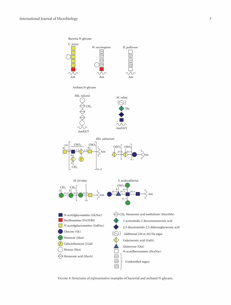
Although the N-biosynthetic pathways of the threedomains have much in common, the archaeal and bacterialLLO processes differ from eukaryotes in two key respects.Firstly, there is no evidence for oligosaccharide sequencesbeing conserved amongst the archaea and bacteria, incontrast to the conserved Glc3Man9GlcNAc2 sequence ofall higher eukaryotes. Indeed, as shown in Figure 4, a greatdiversity of glycans are known to be transferred by N-OSTsto bacterial and archaeal proteins. Despite this diversity, thereis some commonality with respect to the type of linkingsugar utilized in the three domains. This issue is discussedfurther in the OST section, below. Secondly, the bacterialand archaeal LLOs do not appear to be further elaboratedafter flipping. However, it should be borne in mind thatknowledge of bacterial and archaeal N-linked glycosylationis only just emerging, and very few biosynthetic pathwayshave been investigated thus far. Therefore, it remains an openquestion as to whether LLOs can be extended by stepwiseaddition of extra sugars in the periplasmic and cell surfacecompartments.
2.3. Flippases in the Three Domains. Although there existsa very substantial body of evidence, assembled over morethan three decades, demonstrating unequivocally that ineukaryotes the LLO precursor is assembled in the cytoplasmand then flipped across the ER membrane to the lumen,remarkably no ER flippase has yet been biochemicallyidentified [11]. Hence comparisons of flippase structures andmechanisms between the three domains are not yet possible.Fortunately, genetic tools have enabled considerable progressto be made in uncovering likely candidates for flippases.Thus, genetic experiments in yeast have provided very goodevidence that the RFT1 protein is involved in transfer ofthe Man5GlcNAc2-LLO across the ER membrane [12]. Inaccordance with the conclusion that they play a role intranslocation, RFT1 proteins are conserved in eukaryoticorganisms, although it is still not clear whether they areactually the elusive flippases [11].
The elucidation of archaeal N-biosynthetic pathways isstill in its infancy (see Calo et al. [5] for an in-depthreview of recent discoveries) and thus far flippases havenot been studied in this domain. In contrast, bacterialflippases are quite well understood, not the least becauseLLO translocation is integral to LPS biosynthetic pathwayswhich have been intensively studied for many years. It isknown, for example, that the product of the Wzx gene, a non-ABC-type transporter, mediates transport of undecaprenol-linked O-antigen subunits across the plasma membrane in
Figure 1: This figure highlights similarities between the biosynthetic pathways of N-linked glycosylation in archaea (a) compared to eukarya(b) and bacteria (c). The elongation steps flagged in yellow in (b) do not have counterparts in (a) and (c).
4 International Journal of Microbiology
Cytoplasm
Periplasm
-P
Ligase Waal
-PP-
-PP-
Inner membrane
Outer membrane
-P
To cell surface
Wzx
O antigenUndecaprenyl phosphate (UndP) Inner/outer core
Lipid A
Figure 2: This figure depicts key steps in the LOS and LPS biosynthetic pathway in Gram-negative bacteria which have their parallels in N-and O-glycosylation (Figures 1 and 5). For simplicity, other key steps such as the polymerization of the O-antigen prior to transfer to lipid Ainvolved in LPS biosynthesis are not shown.
CH CH2 (CH2 CH2)10–17C CH3C
CH3CH3 CH3
CH CHCH2 CH2 CH2 O P
O
Dolichol phosphate
O−
O−
(a)
CH CH2 (CH2 CH2)9C CH3C
CH3CH3 CH3
CH CCH2 CH CH2 O P
O
Undecaprenol phosphate
O−
O−
(b)
Figure 3: Structures of Dolichol phosphate and Undecaprenol phosphate.
LPS biosynthesis [13]. With respect to the bacterial N-glycosylation pathway, which has been rigorously studiedin the “paradigm” organism, C. jejuni, Aebi, and coworkershave shown that PglK (previously called wlaB), which is anABC-type transporter, is responsible for flipping the LLO[14]. Interestingly, these workers found that PglK has arelaxed substrate specificity exemplified by its ability to com-plement a Wzx deficiency in O-antigen biosynthesis in E. coli.
Notably, all bacterial N-glycans identified to date haveseven or fewer sugar residues, with many archaeal structuresbeing of a similar size (Figure 4). As described earlier,the eukaryal cytoplasmic LLO contains seven sugars (seeFigure 1(b)). These observations suggest that a maximum ofseven sugars might be optimal for the flipping mechanism,though it has also been suggested that the flipping processmight be affected by monosaccharide composition at thereducing end of the glycan [14]. In this context, it could besignificant that the large archaeal N-linked polysaccharide
shown in Figure 4 is composed of tandem repeats of ashort oligosaccharide. This type of structure is reminiscentof bacterial LPS and could therefore be assembled fromshort LLO precursors, after flipping across the cytoplasmicmembrane, in a similar way to Wzx/Wzy-dependent O-antigen polymerization in the periplasm of bacteria [13].Alternatively, it possible that this N-linked polysaccharidemight be flipped across the membrane in an ATP-bindingcassette (ABC) transporter-dependent manor [15].
2.4. N-OSTs in the Three Domains. The transfer of oligosac-charides from the LLOs to asparagine acceptors in N-linkedglycoproteins is catalysed by homologous oligosaccharyl-transferase enzymes (N-OSTs) in the three domains of life.In eukaryotes and archaea; N-OSTs are ubiquitous. Con-sequently, N-linked glycoproteins are found in abundancethroughout both domains. On the other hand, bacteria have
International Journal of Microbiology 5
Hfx. volcaniiM. voltae
C. jejuniH. pullorumW. succinogenes
216
216
216
232
217
216
217
Bacteria N-glycans
Archaea N-glycans
N-acetylglucosamine (GlcNac)
Bacillosamine (DATDH)
N-acetylgalactosamine (GalNac)
Glucose (Glc)
Mannose (Man)
Hexose (Hex)
Hexuronic acid (HexA)
Hexuronic acid methylester (HexAMe)
2-acetoamido-2-deoxymannuronic acid
2,3-diacetoamido-2,3-dideoxyglucuronic acid
Galacturonic acid (GalA)Galactofuranose (f Gal)
Quinovose (Qui)
217
216
N-acetylhexosamine (HexNac)
232
Additional 220 or 262 Da sugar
Unidentified sugars
4OSO3
6
3
3 3
3
6
OSO3 OSO3 OSO3 OSO3
OSO3
2
AsnAsn
AsnAsn
Asn Asn Asn
AsnXS/TAsnXS/T
Hbt. salinarum
M. fervidus S. acidocaldarius
4
CH3Thr
CH3
CH3
CH3
f
f
CH3
α1–6
α1–6
α1–2 α1–4
α1–4
1–41–4
10–15
β1–4
β1–4 β1–4β1–3
β1–41–3
Figure 4: Structures of representative examples of bacterial and archaeal N-glycans.
Figure 5: This figure shows key steps in the O-oligosaccharyltransferase-mediated O-glycosylation pathways in Neisseria and Pseudomonas.Note the similarities to N-glycosylation (Figure 1) and LPS biosynthesis (Figure 2).
probably not evolved N-OSTs of their own (see below) andN-glycosylation is restricted to a limited number of species.The first bacterial N-OST gene was identified about a decadeago. This is the pglB gene of Campylobacter jejuni whichwas found to be highly homologous to the catalytic subunit(called Stt3) of eukaryotic N-OSTs. A similar degree ofhomology was found in archaeal N-OST genes (which are
called aglB) when their identity was confirmed a few yearslater [16, 17].
When the general N-glycosylation system was first dis-covered in C. jejuni, it was thought to be unique, and it waspostulated that this organism might have acquired the pglBgene by lateral gene transfer from either the archaeal or theeukaryal domains [4]. It is now considered most likely that
Figure 6: Comparison of mucin-like sequences in bacteria with mammalian mucins. Partial sequences of Fap1 in S. parasanguinis and MUC1 in Homo sapiens are shown.
GS VS IVTD S QGGGA S GPKVVQWGKDNIVTGTAIAGEHMWQEGGNLTTNVARGQL T AV TDT NVV T EADPGGEAG
DGN T SNVVTDSATGGNHVYQYGGNLTTDLAHGHVTQDGGAYVVTTNATGEARVIQRGNKNITTRVADGRVFQG
GGADVQLRGKQNVTTNGAVGGNKVIQWGD TAT VN T VLAGGQKL TST AA TVT FAGLRNIG S NELVVNDAKGEVV
LVDLRGG T HG TATNT GGDDVR T GGNRVDL TA TT VS DALLVGGNGM S VAAG S RTG S ELMLADAQGGGISFAKGD
KGSLTLIAGNNLTTTGALTGGRATFLGGNVTTDTATDGANLTFSSGK T DNNV TLT GGQLLADGERI S LT GNG S KEV
S GS GTLTVS N TT LT DIAGNLVTSGTLKLATGRQAIVDTTVTSDNLTLTGENLNVAKQ
Figure 7: Sequence of the passenger domain of the autotransporter protein Ag43 from E. coli with heptosylation sites indicated in red.
pglB originated from archaea rather than eukarya (BrendanWren, London School of Hygiene and Tropical Medicine,personal communication). This conclusion is based onknowledge emerging from searches of bacterial genomes forpglB orthologues. Thus far, bacterial N-OST candidates havebeen found exclusively in a subset of species belonging to thephylogenetic grouping known as the epsilon subdivision ofthe Proteobacteria, which include Campylobacter, Helicobac-ter, and Wolinella genera. Amongst these, N-glycosylationhas been rigorously confirmed by mass spectrometry forC. jejuni, W. succinogenes, and H. pullorum (Figure 4) [1,18, 19]. Note, however, that although H. pullorum has themachinery for N-glycosylation, the pglB gene is absent inrelated mammalian pathogens such as H. pylori and H.hepaticus. It may be significant that in primordial deep seavents, which are the homes for many archaea, the majorityof bacteria are epsilon proteobacteria. So it is tempting tospeculate that these extreme environments have provided theconditions for N-OST gene transfer between the prokaryoticdomains (Brendan Wren, personal communication).
The preceding section has focused on the genes encodingthe N-OST enzymes. We now overview current understand-ing of the biochemistry of N-OSTs across the three domainsof life. N-OSTs in archaea, bacteria, and primitive eukaryotes(protozoa) are comprised of a single subunit (the catalytic
subunit) which is the product of the aforementioned aglB,pglB, and Stt3 genes, respectively. In contrast, all N-OSTs ofhigher eukaryotes are multi-subunit complexes in which thecatalytic subunit (Stt3) is accompanied by a total of sevenadditional proteins whose roles remain poorly understood[14, 20–22]. Suggested functions of these accessory proteinsinclude regulating substrate specificity, possibly by expand-ing the range of acceptor sequences, and assisting in proteintranslocation and/or folding. Why primitive eukaryotes donot require a multiprotein complex remains enigmatic, buteven more enigmatic is the observation that a single Stt3from Leishmania major can substitute for the whole N-OSTcomplex in yeast [23, 24].
It is quite normal for eukaryotes to have more thanone Stt3 gene. The highest number has been found inprimitive eukaryotes. For example, L. major expresses fourStt3 paralogs, whilst Trypanosoma brucei has three. The latterhas been shown to have distinct LLO and glycosylation sitepreferences [10]. Yeast, however, has only a single Stt3 gene(called Stt3p), whilst vertebrates, insects, and plants have two,encoding for Stt3A and Stt3B, respectively. It has been shown,via siRNA knockdown experiments in mammalian cells,that Stt3A glycosylates cotranslationally, whilst Stt3B, whichis normally coexpressed, acts posttranslationally, althoughthe protein must not be folded. Also, Stt3B is required
8 International Journal of Microbiology
for efficient glycosylation adjacent to the N-terminal signalsequence [25]. Thus the two isoforms appear to function inconcert to ensure maximal efficiency of N-glycosylation.
Information is only just beginning to emerge concerningthe number of N-OST genes in prokaryotes. Campylobacterhas only a single pglB gene but H. pullorum has two unrelatedgenes, denoted pglB1 and pglB2, the first of which has beenproven to mediate glycosylation [18]. The role of the pglB2protein is not known. Bioinformatic searches for aglB genesin archaea have suggested multiple candidates in individualorganisms but confirmation of expression and activity hasnot yet been determined experimentally [5].
N-OSTs from all domains have been found to exhibitquite relaxed specificity with respect to the oligosaccharidedonor. Thus each is capable of transferring short glycansfrom biosynthetic LLO intermediates in addition to the fulllength glycans of the mature LLOs. Eukaryotic and bacterialN-OSTs transfer glycans whose reducing sugar carries atleast one acetamido (NAc) group. Thus the eukaryoticN-glycan has a chitobiose core (GlcNAcb1-4GlcNAc) andthe linking sugar in characterized bacterial N-glycans iseither 2,4-diacetamido-2,4,6-trideoxyglucopyranose (bacil-losamine; Campylobacter and Wolinella) or HexNAc (H. pul-lorum). Interestingly, the pglB protein of C. jejuni is capableof transferring a variety of O-antigen oligosaccharides ontoprotein acceptors in engineered E. coli cells, provided theirreducing sugar has an NAc moiety [26]. This discovery hasimportant implications for the development of O-antigencontaining neoglycoprotein vaccines. Archaea appear to havea greater diversity of linking sugars, including Glc as wellas GlcNAc and GalNAc [5, 17]. Interestingly, a Sulfolobusarchaeal species, which is very close phylogenetically toprimitive eukaryotes, has mannose rich chitobiose-linked N-glycans reminiscent of the eukaryotic core sequence [27].
When comparing mechanisms of prokaryotic andeukaryotic N-glycosylation, it is important to remember thatthe folding status of their proteins is very different at thetime of glycosylation. Thus, eukaryotic oligosaccharides aretransferred to nascent proteins before they are folded, whilstin prokaryotes the proteins are presumably fully folded,having already been transported from the cytoplasm, wheretranslation occurs, into the periplasm or onto the surface,where glycosylation takes place. In all three domains, theasparagine acceptor must normally be located in a consensussequence (Asn-X-Ser/Thr or, rarely, Asn-X-Cys, where Xcannot be proline); however, not all consensus sequences areglycosylated. Sequence motifs contributing to specificity ofsite occupancy are not yet fully understood, but it is alreadyclear that bacterial glycosylation is much more restrictedthan eukaryotic glycosylation. For example, consensus sitesin C. jejuni require an upstream Glu or Asp residue in theextended consensus sequence D/EZNXS/T, where neitherZ nor X can be proline [28]. High throughput glycopro-teomic efforts are beginning to provide comprehensive site-occupancy data in eukaryotic systems [29]. It is hoped thatthese and similar experiments will facilitate the developmentof algorithms that will be capable of accurately predictingwhich consensus sequences in eukaryotic proteomes arelikely to be occupied.
In contrast to eukaryotes, very few prokaryotic glycopro-teins have had their glycosylation sites determined. Basedon a limited body of data, some predictions have beenmade for sequences favouring archaeal glycosylation [5] butemerging data from studies of Sulfolobus S-layers suggestthat these rules probably will not be universally applicable(see [27] and unpublished work from our laboratory).Bearing in mind that glycosylation in prokaryotes occursposttranslationally, it is conceivable that general rules forsite occupancy may not prevail in these organisms, becauseof the unique nature of individual proteins with respectto accessible consensus sequences. This could be especiallyrelevant in S-layer glycosylation, because these proteins self-assemble into crystalline monolayers and all their consensussequences are therefore likely to be in exposed locations[30, 31].
In concluding this section on N-OSTs, we draw attentionto the fact that crystal structures are now available forthe C-terminal domains, that include the WWDYG motifimplicated in the catalytic mechanism, of both an archaealAglB and the PglB of C. jejuni, although not so farfor the eukaryotic Stt3, despite many valiant efforts [20].Mechanistic and evolutionary understanding provided bythe crystal structures has been reviewed very recently [5] sowill not be covered here.
2.5. Glycoprotein Remodelling. All eukaryotic glycoproteinsare subjected to extensive remodelling in the Golgi apparatusafter they exit the ER, resulting in heterogeneous mixtures ofglycoforms exhibiting a great variety of peripheral structures,many of which are rich in functionally important sugarssuch as fucose and sialic acid [7, 8]. Prokaryotes have nocounterpart to the Golgi apparatus, and there is no evidenceso far that they remodel their N-linked glycoproteins.
3. Haemophilus influenzae Can PerformCytoplasmic N-Glycosylation
About seven years ago a study of the HMW1 adhesin ofH. influenzae uncovered a potentially novel N-glycosylationpathway occurring in the cytoplasm of this bacterium[32]. This intriguing discovery has now been confirmedby rigorous structure analyses which, remarkably, haveidentified 31 glycosylated Asn residues within the HMW1protein [33, 34]. All sites carry either Hex or Hex-Hex,where Hex can be Gal or Glc, and all but one of theglycosylation sites has the normal N-glycosylation consensussequence (Asn-X-Ser/Thr, see Section 2). The cytoplasmicenzyme responsible for glycosylation has been confirmed asHMW1C. Interestingly it transfers glucose to all glycosylatedasparagines but only transfers galactose to a subset of thesesites. Moreover, the same enzyme appears to be responsiblefor the Hex-Hex glycoforms as well as those carrying a singleGlc or Gal. The mechanisms of these processes remain tobe established. Homology analysis suggests that a variety ofother bacteria possess HMW1C-like proteins, so it is likelythat this type of cytoplasmic N-glycosylation will be foundelsewhere. Whether similar glycosylation occurs in archaea is
International Journal of Microbiology 9
not known. As shown in Figure 4, glucose has been observedas a linking sugar in some archaeal N-glycans, but it is likelythat the N-OST pathway is employed in their biosynthesis[5]. The presence of a Glc-Asn moiety was reported ineukaryotic laminin in 1994 [35] but this observation has notbeen independently confirmed.
4.1. Overview. During the past five years, intensive researchon Neisseria and Pseudomonas pilin glycosylation has uncov-ered a general O-glycosylation pathway that, remarkably,has all the hallmarks of N-linked glycosylation (Figure 5).Moreover, this general pathway does not appear to berestricted to a few pilin proteins in a handful of pathogens.Thus, very interesting data are emerging from researchon Bacteroides species that suggests that these bacteriaare capable of glycosylating a great number of proteinsin this way. So far oligosaccharyltransferase-mediated O-glycosylation has only been found in Gram-negative bacteria,which is perhaps not surprising, bearing in mind that itmirrors LPS biosynthesis.
4.2. Neisseria and Pseudomonas Pilins. Much of our knowl-edge of bacterial O-linked glycosylation pathways has beenelucidated from studies in Neisseria species. O-linked gly-cosylation was first characterised in Neisseria meningitidis,where the pilin protein was shown to be modified bya trisaccharide [36], with a similar glycan being foundon N. gonorrhoeae [37]. Subsequent bioinformatics anddirected mutagenesis led to the identification of an O-OST, called PglL, in N gonorrhoeae [38, 39]. PglL O-OSTsbelong to a family of bacterial OSTs responsible for O-linkedglycosylation of type IV pilins. This family appears to bewidespread amongst pathogenic bacteria, including somestrains of Pseudomonas aeruginosa, where it is called PilO[40, 41]. Moreover, it has recently been demonstrated thatNeisseria are able to decorate a diverse set of proteins via theO-OST pathway [42, 43].
Research using Neisseria and Pseudomonas glycosylationsystems in engineered E. coli cells has demonstrated thatthe biosynthesis of the O-linked glycan has a number ofsimilarities to its N-linked counterpart (compare Figures 1and 5). The O-linked glycosylation pathway involves LLOs,and the glycans are transferred en bloc by the O-OSTs fromthe LLOs carrier onto the protein [39]. The translocation ofthe LLO substrate into the periplasm is required for activityand it has been shown that PglF, a protein with homologyto O-antigen “flippase,” is required for pilin glycosylationwhich is thought to occur in an analogous manner to theWzy-dependent addition of O-antigen to the core-LPS [38].In a similar fashion to the N-OSTs in archaea, bacteria, andlower eukaryotes, the O-OST’s catalytic subunit is sufficientfor glycosylation. As with O-glycosylation in eukaryotes,there appears to be no consensus sequence for definingsites of O-glycan attachment. Interestingly, the Neisseria
O-OSTs display a pronounced substrate promiscuity whencompared to N-OSTs, as demonstrated by their ability totransfer virtually any glycan from an LLO carrier onto pilinin engineered E. coli cells. For example, it was shown thatthe Neisseria PglL could transfer peptidoglycan subunitsonto pilin, highlighting the potential for exploitation ofsuch pathways for biotechnological purposes [44]. As such itappears that the substrate specificity of the O-OSTs is foundin the lipid carrier, a hypothesis nicely demonstrated using anin vitro glycosylation system that utilised purified NeisseriaPglL, pilin, and the lipid farnesyl pyrophosphate carrying asynthetic pentasaccharide that was successfully transferredonto the pilin protein [44].
4.3. Bacteroides Species Have an OST-Mediated O-Glycosylation Pathway. Bacteroides comprise one of themost abundant genera of commensals in the human colon.Exciting recent research suggests that these bacteria arenot only capable of O-glycosylating many of their proteinsbut, unusually, they exploit a host-like pathway to addfucose (apparently acquired from their host glycans and/orfrom plant polysaccharides present in the gut) onto theirglycoproteins and polysaccharides [45]. A combinationof cell biology and molecular biology experiments hasprovided convincing evidence for the existence of a generalO-glycosylation system in these symbiotic bacteria whichhas all the hallmarks of the pilin O-OST-mediated pathogenpathway described earlier [46]. Notably, O-glycosylationappears to be central to the physiology of B. fragilis as wellas its ability to colonise its ecological niche. Although thestructures of the B. fragilis O-glycans remain to be defined,many elements of the biosynthetic pathway are beginningto be unraveled. Thus five glycosyltransferases, plus anunrelated fucosyltransferase, have been proposed to beinvolved in assembly of the LLO on the cytoplasmic faceof the inner membrane. Translocation to the periplasm isthought to be mediated by the O-antigen flippase (Wzx, seeFigure 2). However, there is no candidate gene as yet for theputative O-OST.
Interestingly, very recently it has been reported thatfucosylated O-glycans are present on the fimbriae of Por-phyromonas gingivalis [47]. Like B. fragilis, this oral mucosalpathogen is a member of the Bacteroides genus. It is conceiv-able, therefore, that P. gingivalis glycosylates its proteins via asimilar pathway to B. fragilis. Monosaccharide compositionalanalysis has shown that the P. gingivalis glycans are likely tobe complex (Fuc, Xyl, Man, Gal, Glc, GalNAc and GlcNAchave all been detected) but so far no sequences are availablefor this glycoprotein.
5. Processive O-Glycosylation Systems inEukaryotes and Prokaryotes
5.1. Overview. All eukaryotic O-glycosylation is processivethat is, it is a stepwise process which begins with theattachment of the linking monosaccharide to the acceptorserine or threonine. Further sugars are added one at atime to form the mature glycan. Many eukaryotic O-glycans
10 International Journal of Microbiology
are of the mucin-type which are linked via GalNAc, butother classes exist which are attached to proteins via avariety of sugars including Fuc, Man, Glc, Gal, Ara, Xyl,and GlcNAc. Most eukaryotic O-biosynthetic events takeplace in the Golgi, although some classes of O-glycans, forexample, O-Man linked glycans (see later), are initiated inthe ER. An enormous variety of sequences can be attachedto these linking sugars. Thus there is a great diversity of O-glycosylation in the eukaryotic domain [6, 7].
Archaeal O-glycosylation has rarely been investigated.The only substantive study was about twenty years agowhen the S-layers of Halobacterium salinarum and Haloferaxvolcanii were shown to carry several O-linked disaccharidesof sequence Glc1-2Gal [48]. In contrast, there is a large bodyof evidence pointing to a rich diversity of O-glycans in thebacterial domain. The most complex structures have beenfound on bacterial S-layers which have been investigatedin many species of bacteria over the last thirty years.Their structures and biosynthesis have been comprehensivelyreviewed on several occasions [5, 31, 49, 50] and the readeris referred to these articles for further information. Below wediscuss emerging understanding of other families of bacterialcell surface and secreted O-linked glycoproteins, many ofwhich, in contrast to the S-layers, have structural and/orfunctional counterparts in the eukaryotic domain.
5.2. Mucin-Like Glycoproteins. Mucins are high molecularweight eukaryotic glycoproteins, produced in abundance byepithelial and goblet cells, whose polypeptide chains arecoded by the MUC genes [7]. Mucins are characterized bythe presence of tandem repeats of serine/threonine/proline-rich sequences which are extensively O-glycosylated. Mucinsreadily form gels and are a key component of most gel-likesecretions in eukaryotes where they have functions rangingfrom lubrication to serving as receptors for microbes. Inrecent years, it has become evident that many bacterialbiofilms contain glycoproteins whose compositions indicatethat they are mucin-like molecules [51–53]. The best charac-terized are the serine-rich repeat (SRR) glycoproteins belong-ing to the Fap1 family, which are conserved in Streptococci,Staphylococci, and Lactobacilli, and are required for bacterialbiofilm formation and pathogenesis.
The polypeptides of SRR family members are comprisedof a long signal peptide followed in turn by a short serine-richdomain, an acidic or basic region, a long serine-rich domainand a C-terminal anchoring motif. The SRR sequences arereminiscent of the eukaryotic mucins in that the majorityof the polypeptide is comprised of tandem repeats ofshort motifs which have related sequences (see Figure 6for comparison of a portion of an SRR domain comparedwith part of the human MUC-1 sequence). The serine-rich domains have the key hallmark of the mucins, namely,variable repeated sequences rich in potential O-glycosylationsites (see Figure 6), but in contrast to the mammalianmucins, proline is absent from the SRR domains. Eukaryoticmucin glycosylation is initiated in the Golgi after the proteinis fully folded. Thus their proline residues are importantfor ensuring that exposed, accessible sites are available for
glycosylation. Currently little is known about the process ofglycan attachment to the bacterial SRRs, other than the factthat attachment of the linking sugar appears to occur veryrapidly in the cytoplasm, before transport to the cell surfacevia the accessory Sec transporter. It has been proposed thatthe glycosylation mechanism is a two-step process, withthe second step requiring several accessory secretion com-ponents and thus is probably coupled with secretion [53].Recent electron microscopy structural studies have indicatedthat the serine dipeptide repeat domains have a super-helicalextended structure with exposed serine side-chains, whichare expected to be readily accessible to O-glycosylation [54].
The glycan content of the SRRs has been explored infive species, Streptococcus parasanguinis, S gordonii [55],S. agalactiae [56], S. pneumoniae [57], and Staphylococcalaureus. This has been largely done using lectins such aswheat germ agglutinin (WGA) which recognize terminalGlcNAc, and by sugar compositional analyses [53]. Nocomplete glycan structures have been defined so far. Lectinblotting, supplemented by sugar composition data, hasindicated that the linking sugar in the SRRs is probablyGlcNAc. Interestingly two glycosyltransferases, called Gtf1and Gtf2, are required for this initial glycosylation stepby S. parasanguinis [58]. Gtf1 and Gtf2 homologs from S.pneumoniae also form an enzyme complex that catalyzesthe transfer of GlcNAc to serine-rich sites of PsrP [59]. Asimilar requirement for two glycosyltransferases adding asingle sugar occurs in the O-mannosyl glycans of highereukaryotes (see later) whose biosynthesis is initiated bya heterodimer enzyme complex composed of protein O-mannosyltransferases (POMT) 1 and 2 [7]. It should benoted that although O-linked GlcNAc is found on eukaryoticcytoplasmic and nuclear proteins, it is probably not analo-gous to SRR O-glycosylation, because eukaryotic O-linkedGlcNAc residues are not further extended. Moreover, thisubiquitous eukaryotic glycosylation is unusual because it isdynamic and involves cross-talk with phosphorylation [60].
A glucosyltransferase has recently been identified in S.parasanguinis that transfers glucose to the GlcNAc-modifiedFap1 [61]. Although the structure of the product of glycosy-lation remains to be determined, it is tempting to speculatethat these bacterial proteins might carry glycans whose coresequences are glucosyl analogues of the core type 1 sequence,Galβ1-3GalNAc, that is ubiquitous in mammalian mucins.Sugars additional to Glc and GlcNAc, including GalNAc andRha, have been observed at low levels in sugar analyses of theFap1 glycoproteins [53]. It remains to be established whetherthe putative Glc-GlcNAc moieties are further elongated orwhether other glycans account for the compositional data.
5.3. O-Linked Mannose Glycosylation. The title of a recentreview “Protein O-mannosylation: conserved from bacteriato humans” [62] encapsulates the importance of this classof glycosylation. In eukaryotes, O-mannosyl glycans areabundant in yeast and fungi, whilst in mammals they occuron a restricted number of proteins, such as α-dystroglycanwhere their impairment is a cause of congenital musculardystrophy [63, 64]. Yeast and fungi express short mannosyl
International Journal of Microbiology 11
oligomers, with galactose being present on terminal sitesin some species. In contrast, mammalian O-mannosylglycans carry sialylated and fucosylated N-acetyllactosaminesequences similar to those found in mucins. EukaryoticO-mannosylation is initiated in the ER by the concertedaction of two protein O-mannosyltransferases (POMT1 and2) which employ Dol-P-Man as the mannose donor. Chainextension subsequently takes place in the Golgi. The reviewcited above gives a comprehensive account of these events.
O-mannosyl glycans analogous to those found in yeastand fungi have been found on glycoproteins from membersof the Actinomycetes class of Gram-positive bacteria whichinclude the Mycobacteria and Streptomyces genera [62, 65].The first to be characterised were the surface glycoproteins ofMycobacterium tuberculosis [66, 67]. Subsequently M. bovis[68], Corynebacterium glutamicum [69], and Streptomycescoelicolor [70] were shown to be similarly glycosylated. Allcontain O-glycans whose sequences are restricted to shortstretches of mannose (usually three residues or less). Like ineukaryotes, the mannosyl donor is a polyprenol phosphate,and their protein O-mannosyltransferases (POMTs) aremembrane associated. The activities of the products ofcandidate POMT genes in M. tuberculosis, C. glutamicum,and S. coelicolor have been genetically and biochemicallyconfirmed. In contrast to eukaryotes, heterodimeric enzymesdo not appear to be required. The fact that bioinformaticscreening has uncovered a plethora of POMT homologs inother species, indicates that O-mannosylation constitutes ageneral O-glycosylation pathway in Actinomycetes.
Steps equivalent to the eukaryotic Golgi processes of O-mannose extension have not been determined thus far inbacteria. There are, nevertheless, candidate mannosyltrans-ferases, for example, those involved in the biosynthesis of themannan core of cell wall lipomannan/lipoarabinomannan inMycobacteria [62]. It has been suggested that O-mannoseextension in Mycobacteria is coupled with Sec-dependentsecretion in a manner akin to that proposed for the serine-rich proteins in Streptococci and Staphylococci describedearlier [53].
5.4. O-Linked Heptose in E. coli. Autotransporters constitutethe biggest group of secreted proteins in Gram-negativebacteria. AIDA-I, TibA, and Ag43 are three autotransporterproteins in pathogenic E. coli which are associated withvirulence phenotypes, such as the formation of biofilms andaggregates. All three are extensively glycosylated with O-linked heptose on their so-called “passenger domains” [71–74]. Glycosylation occurs in the cytoplasm and the heptosesare derived from ADP-glycero-manno-heptopyranose whichis recruited from the LPS biosynthetic pathway. The pas-senger domains are secreted to the extracellular environ-ment where their glycosylation appears to enhance bacterialattachment to human cells. The heptoses are attached atmultiple sites in Ser/Thr rich domains (Figure 7) that arereminiscent of eukaryotic mucin sequences (see earlier)although they lack the hallmark tandem repeats of the latter.
5.5. O-Glycosylation of Bacterial Flagellins. Flagellin O-linked glycosylation has been widely reported in a number of
bacteria, where it appears to be restricted, with the archaealcounterparts being N-linked. Current knowledge of the O-linked sugars involved in flagellin glycosylation has beencovered in recent reviews [75, 76]. Probably the best studiedflagellin glycans are found to be glycosylated with a family of“sialic acid-like” monosaccharides, based around the sugarspseudaminic acid and legionaminic acid, with a diversitybeing generated by variation in their decorating appendages.These sugars have been found in several species includingCampylobacter jejuni, Helicobacter pylori, and Aeromonascaviae. In C. jejuni, the O-linked glycosylation gene clusterhas been identified and the function of a number of geneproducts involved in the pseudaminic acid biosyntheticpathway has been elucidated. It appears to have evolvedto share some of the same biosynthetic machinery as theN-linked glycosylation pathway, allowing the organism tomaintain a compact genome and avoid redundancy [1].Flagellin glycosylation in P aeruginosa has also been shown toshare biosynthetic machinery, in this case with the O-antigenpathway [77].
The most complex flagellin O-glycans identified thusfar have been found in hypervirulent strains of Clostridiumdifficile [75, 78]. The C. difficile flagellins carry HexNAc-linked oligosaccharides up to at least five sugars in length.
In contrast to the “en bloc” transfer in the N-linkedpathway, it is apparent that flagellin O-linked glycosylationis likely to proceed in a sequential fashion. Given that asingle sugar residue is often added to sites of attachment,specific glycosyltransferases are thought to be involved inthe glycosylation process, but our present understanding ofthe glycosyltransferases involved in the glycosylation processremains limited. The current proposed model for O-linkedflagellin glycosylation occurs at the cytoplasmic face of theinner membrane in the vicinity of the type III secretioncomplex [76]. Nucleotide activated sugars are utilised byspecific glycosyltransferases in the glycosylation machineryand are added to exposed serine and threonine residues. Theglycosylated flagellin monomers are then secreted to the tipof the growing flagellin filament.
Unlike N-glycosylation, there is no consensus sequencefor O-glycosylation. However, it is well known that certainsequence motifs are preferred in mucin type O-glycosylation.Indeed the NetOglyc open access tool, which can be veryhelpful for predicting possible sites of eukaryotic mucin O-glycosylation, was developed using knowledge of preferredsequence motifs [79]. The rapid progress that is being madein defining O-glycosylation sites in diverse prokaryoticglycoproteins, coupled with the fact that some researchersare beginning to employ the NetOglyc tool to guidethem in the choice of targets for mutation in searches forprokaryotic glycosylation [67, 80], have made it timely toassess the applicability of the NetOglyc tool to prokaryoticglycoprotein research.
In a preliminary unpublished study, we have ascertainedNetOglyc predictions for selected members of each of the
12 International Journal of Microbiology
families of prokaryotic O-glycoproteins described in theprevious sections, for which there is published experimentaldata on site occupancy. The outputs send the very clearmessage that NetOglyc does not, in fact, correctly predict O-glycosylation in most families of prokaryotic glycoproteins.Thus we found that no sites were correctly predicted in thepilins, flagellins, serine-rich proteins, the autotransporters,or the BF2494 glycoprotein of B. fragilis. Experimental data[46] suggests that the latter has an essential three-residueglycosylation site motif (D)(S/T)(A/I/L/V), which is nottypical of eukaryotes, so it is therefore not surprising thatNetOglyc appears to be not suitable for predicting this typeof O-glycosylation. In conclusion, NetOglyc is likely to onlybe useful for predicting O-glycosylation in prokaryotic glyco-proteins like mycobacterial lipoproteins whose Pro/Ser/Thr-rich glycosylation domains have sequence characteristicswhich they share with the mammalian mucins [67]. Predic-tion in other families will require the development of newalgorithms which take account of their specific glycosylationdomain characteristics.
7. Conclusions
Although much remains to be uncovered concerning proteinglycosylation in prokaryotes, several themes are emergingfrom the discoveries of the past decade. Firstly, like ineukaryotes, N-glycosylation is largely restricted to Asn-X-Ser/Thr consensus sequences, even when the canonicaloligosaccharyltransferase pathway is not involved. SecondlyO-glycosylation is far more abundant in bacteria than inarchaea whilst the reverse is true for N-glycosylation. Thirdly,oligosaccharyltransferase-mediated O-glycosylation is likelyto be widespread in Gram-negative bacteria. In contrast thistype of O-glycosylation has not thus far been proven tooccur experimentally in Gram-positive bacteria or archaea.Finally cytoplasmic O-glycosylation appears to be both morecommon and more diverse in Gram-positive compared withGram-negative bacteria, possibly because of the existence ofthe alternative periplasmic O-OST pathway of the latter.
Acknowledgment
Research conducted in the authors’ laboratory was supportedby the Biotechnology and Biological Sciences ResearchCouncil (Grants BBF0083091 and BBC5196701).
References
[1] C. M. Szymanski and B. W. Wren, “Protein glycosylation inbacterial mucosal pathogens,” Nature Reviews Microbiology,vol. 3, no. 3, pp. 225–237, 2005.
[2] M. F. Mescher and J. L. Strominger, “Purification andcharacterization of a prokaryotic glycoprotein from the cellenvelope of Halobacterium salinarium,” Journal of BiologicalChemistry, vol. 251, no. 7, pp. 2005–2014, 1976.
[3] N. M. Young, J. R. Brisson, J. Kelly et al., “Structure ofthe N-linked glycan present on multiple glycoproteins in thegram-negative bacterium, Campylobacter jejuni,” Journal ofBiological Chemistry, vol. 277, no. 45, pp. 42530–42539, 2002.
[4] M. Wacker, D. Linton, P. G. Hitchen et al., “N-linked glycosy-lation in Campylobacter jejuni and its functional transfer intoE. coli,” Science, vol. 298, no. 5599, pp. 1790–1793, 2002.
[5] D. Calo, L. Kaminski, and J. Eichler, “Protein glycosylation inArchaea: sweet and extreme,” Glycobiology, vol. 20, no. 9, pp.1065–1076, 2010.
[6] I. Hug and M. F. Feldman, “Analogies and homologies inlipopolysaccharide and glycoprotein biosynthesis in bacteria,”Glycobiology, vol. 21, no. 2, pp. 138–151, 2011.
[7] A. Varki, R. D. Cummings, J. D. Esko et al., Essentials ofGlycobiology, Cold Spring Harbor Laboratory Press, New York,NY, USA, 2nd edition, 2009.
[8] M. T. Taylor and K. Drickamer, Introduction to Glycobiology,Oxford University Press, Oxford, UK, 2nd edition, 2006.
[9] D. J. Kelleher, S. Banerjee, A. J. Cura, J. Samuelson, and R.Gilmore, “Dolichol-linked oligosaccharide selection by theoligosaccharyltransferase in protist and fungal organisms,”Journal of Cell Biology, vol. 177, no. 1, pp. 29–37, 2007.
[10] L. Izquierdo, B. L. Schulz, J. A. Rodrigues et al., “Distinctdonor and acceptor specificities of Trypanosoma bruceioligosaccharyltransferases,” The EMBO Journal, vol. 28, no. 17,pp. 2650–2661, 2009.
[11] S. Sanyal and A. K. Menon, “Stereoselective transbilayertranslocation of mannosyl phosphoryl dolichol by an endo-plasmic reticulum flippase,” Proceedings of the NationalAcademy of Sciences of the United States of America, vol. 107,no. 25, pp. 11289–11294, 2010.
[12] J. Helenius, D. T. W. Ng, C. L. Marolda, P. Walter, M. A.Valvano, and M. Aebi, “Translocation of lipid-linked oligosac-charides across the ER membrane requires Rft1 protein,”Nature, vol. 415, no. 6870, pp. 447–450, 2002.
[13] C. R. H. Raetz, C. M. Reynolds, M. S. Trent, and R. E. Bishop,“Lipid a modification systems in gram-negative bacteria,”Annual Review of Biochemistry, vol. 76, pp. 295–329, 2007.
[14] C. Alaimo, I. Catrein, L. Morf et al., “Two distinct but inter-changeable mechanisms for flipping of lipid-linked oligosac-charides,” The EMBO Journal, vol. 25, no. 5, pp. 967–976,2006.
[15] L. Cuthbertson, V. Kos, and C. Whitfield, “ABC transportersinvolved in export of cell surface glycoconjugates,” Microbiol-ogy and Molecular Biology Reviews, vol. 74, no. 3, pp. 341–362,2010.
[16] M. Abu-Qarn and J. Eichler, “Protein N-glycosylation inArchaea: defining Haloferax volcanii genes involved in S-layerglycoprotein glycosylation,” Molecular Microbiology, vol. 61,no. 2, pp. 511–525, 2006.
[17] M. Abu-Qarn, S. Yurist-Doutsch, A. Giordano et al.,“Haloferax volcanii AglB and AglD are involved in N-glycosylation of the S-layer glycoprotein and proper assemblyof the surface layer,” Journal of Molecular Biology, vol. 374, no.5, pp. 1224–1236, 2007.
[18] A. J. Jervis, R. Langdon, P. Hitchen et al., “Characterizationof N-linked protein glycosylation in Helicobacter pullorum,”Journal of Bacteriology, vol. 192, no. 19, pp. 5228–5236, 2010.
[19] P. G. Hitchen, K. Twigger, E. Valiente, R. H. Langdon, B. W.Wren, and A. Dell, “Glycoproteomics: a powerful tool forcharacterizing the diverse glycoforms of bacterial pilins andflagellins,” Biochemical Society Transactions, vol. 38, no. 5, pp.1307–1313, 2010.
[20] W. J. Lennarz, “Studies on oligosaccharyl transferase in yeast,”Acta Biochimica Polonica, vol. 54, no. 4, pp. 673–677, 2007.
[21] D. J. Kelleher and R. Gilmore, “An evolving view of theeukaryotic oligosaccharyltransferase,” Glycobiology, vol. 16,no. 4, pp. 47R–62R, 2006.
International Journal of Microbiology 13
[22] B. L. Schulz and M. Aebi, “Analysis of glycosylation siteoccupancy reveals a role for Ost3p and Ost6p in site-specificN-glycosylation efficiency,” Molecular and Cellular Proteomics,vol. 8, no. 2, pp. 357–364, 2009.
[23] F. P. Nasab, B. L. Schulz, F. Gamarro, A. J. Parodi, and M. Aebi,“All in one: leishmania major STT3 proteins substitute for thewhole oligosaccharyltransferase complex in Saccharomycescerevisiae,” Molecular Biology of the Cell, vol. 19, no. 9, pp.3758–3768, 2008.
[24] K. Hese, C. Otto, F. H. Routier, and L. Lehle, “The yeastoligosaccharyltransferase complex can be replaced by STT3from Leishmania major,” Glycobiology, vol. 19, no. 2, pp. 160–171, 2009.
[25] C. Ruiz-Canada, D. J. Kelleher, and R. Gilmore, “Cotransla-tional and posttranslational N-glycosylation of polypeptidesby distinct mammalian OST isoforms,” Cell, vol. 136, no. 2,pp. 272–283, 2009.
[26] M. F. Feldman, M. Wacker, M. Hernandez et al., “Engineer-ing N-linked protein glycosylation with diverse O antigenlipopolysaccharide structures in Escherichia coli,” Proceedingsof the National Academy of Sciences of the United States ofAmerica, vol. 102, no. 8, pp. 3016–3021, 2005.
[27] E. Peyfoon, B. Meyer, P. G. Hitchen et al., “The S-layerglycoprotein of the crenarchaeote Sulfolobus acidocaldariusis glycosylated at multiple sites with chitobiose-linked N-glycans,” Archaea. In press.
[28] M. Kowarik, N. M. Young, S. Numao et al., “Definition of thebacterial N-glycosylation site consensus sequence,” The EMBOJournal, vol. 25, no. 9, pp. 1957–1966, 2006.
[29] D. F. Zielinska, F. Gnad, J. R. Wisniewski, and M. Mann,“Precision mapping of an in vivo N-glycoproteome revealsrigid topological and sequence constraints,” Cell, vol. 141, no.5, pp. 897–907, 2010.
[30] H. Claus, E. Akca, T. Debaerdemaeker et al., “Molecular orga-nization of selected prokaryotic S-layer proteins,” CanadianJournal of Microbiology, vol. 51, no. 9, pp. 731–743, 2005.
[31] P. Messner, K. Steiner, K. Zarschler, and C. Schaffer, “S-layernanoglycobiology of bacteria,” Carbohydrate Research, vol.343, no. 12, pp. 1934–1951, 2008.
[32] S. Grass, A. Z. Buscher, W. E. Swords et al., “TheHaemophilus influenzae HMW1 adhesin is glycosylated ina process that requires HMW1C and phosphoglucomutase,an enzyme involved in lipooligosaccharide biosynthesis,”Molecular Microbiology, vol. 48, no. 3, pp. 737–751, 2003.
[33] J. Gross, S. Grass, A. E. Davis, P. Gilmore-Erdmann, R.R. Townsend, and J. W. St Geme III, “The Haemophilusinfluenzae HMW1 adhesin is a glycoprotein with an unusualN-linked carbohydrate modification,” Journal of BiologicalChemistry, vol. 283, no. 38, pp. 26010–26015, 2008.
[34] S. Grass, C. F. Lichti, R. R. Townsend, J. Gross, and J. W. StGeme III, “The haemophilus influenzae HMW1c protein is aglycosyltransferase that transfers hexose residues to asparaginesites in the HMW1 adhesin,” PLoS Pathogens, vol. 6, no. 5,Article ID e1000919, 9 pages, 2010.
[35] R. Schreiner, E. Schnabel, and F. Wieland, “Novel N-glycosylation in eukaryotes: laminin contains the linkage unitβ-glucosylasparagine,” Journal of Cell Biology, vol. 124, no. 6,pp. 1071–1081, 1994.
[36] E. Stimson, M. Virji, K. Makepeace et al., “Meningococ-cal pilin: a glycoprotein substituted with digalactosyl 2,4-diacetamido-2,4,6-trideoxyhexose,” Molecular Microbiology,vol. 17, no. 6, pp. 1201–1214, 1995.
[37] F. T. Hegge, P. G. Hitchen, F. E. Aas et al., “Unique modifica-tions with phosphocholine and phosphoethanolamine define
alternate antigenic forms of Neisseria gonorrhoeae type IVpili,” Proceedings of the National Academy of Sciences of theUnited States of America, vol. 101, no. 29, pp. 10798–10803,2004.
[38] P. M. Power, K. L. Seib, and M. P. Jennings, “Pilin glycosylationin Neisseria meningitidis occurs by a similar pathway towzy-dependent O-antigen biosynthesis in Escherichia coli,”Biochemical and Biophysical Research Communications, vol.347, no. 4, pp. 904–908, 2006.
[39] A. Faridmoayer, M. A. Fentabil, D. C. Mills, J. S. Klassen,and M. F. Feldman, “Functional characterization of bacterialoligosaccharyltransferases involved in O-linked protein glyco-sylation,” Journal of Bacteriology, vol. 189, no. 22, pp. 8088–8098, 2007.
[40] M. Qutyan, M. Paliotti, and P. Castric, “PilO of Pseudomonasaeruginosa 1244: subcellular location and domain assign-ment,” Molecular Microbiology, vol. 66, no. 6, pp. 1444–1458,2007.
[41] J. G. Smedley III, E. Jewell, J. Roguskie et al., “Influence ofpilin glycosylation on Pseudomonas aeruginosa 1244 pilusfunction,” Infection and Immunity, vol. 73, no. 12, pp. 7922–7931, 2005.
[42] A. Vik, F. E. Aas, J. H. Anonsen et al., “Broad spectrum O-linked protein glycosylation in the human pathogen Neisseriagonorrhoeae,” Proceedings of the National Academy of Sciencesof the United States of America, vol. 106, no. 11, pp. 4447–4452,2009.
[43] S. C. Ku, B. L. Schulz, P. M. Power, and M. P. Jennings, “Thepilin O-glycosylation pathway of pathogenic Neisseria is ageneral system that glycosylates AniA, an outer membranenitrite reductase,” Biochemical and Biophysical Research Com-munications, vol. 378, no. 1, pp. 84–89, 2009.
[44] A. Faridmoayer, M. A. Fentabil, M. F. Haurat et al., “Extremesubstrate promiscuity of the Neisseria oligosaccharyl trans-ferase involved in protein O-glycosylation,” Journal of Biologi-cal Chemistry, vol. 283, no. 50, pp. 34596–34604, 2008.
[45] M. J. Coyne, B. Reinap, M. M. Lee, and L. E. Comstock,“Human symbionts use a host-like pathway for surfacefucosylation,” Science, vol. 307, no. 5716, pp. 1778–1781, 2005.
[46] C. M. Fletcher, M. J. Coyne, O. F. Villa, M. Chatzidaki-Livanis,and L. E. Comstock, “A general O-glycosylation systemimportant to the physiology of a major human intestinalsymbiont,” Cell, vol. 137, no. 2, pp. 321–331, 2009.
[47] A. E. Zeituni, W. McCaig, E. Scisci, D. G. Thanassi, andC. W. Cutler, “The native 67-kilodalton minor fimbria ofPorphyromonas gingivalis is a novel glycoprotein with DC-SIGN-targeting motifs,” Journal of Bacteriology, vol. 192, no.16, pp. 4103–4110, 2010.
[48] M. Sumper, E. Berg, R. Mengele, and I. Strobel, “Primarystructure and glycosylation of the S-layer protein of Haloferaxvolcanii,” Journal of Bacteriology, vol. 172, no. 12, pp. 7111–7118, 1990.
[49] C. Schaffer, M. Graninger, and P. Messner, “Prokaryoticglycosylation,” Proteomics, vol. 1, no. 2, pp. 248–261, 2001.
[50] M. Abu-Qarn, J. Eichler, and N. Sharon, “Not just for Eukaryaanymore: protein glycosylation in Bacteria and Archaea,”Current Opinion in Structural Biology, vol. 18, no. 5, pp. 544–550, 2008.
[51] H. Wu, M. Zeng, and P. Fives-Taylor, “The glycan moieties andthe N-terminal polypeptide backbone of a fimbria-associatedadhesin, Fap1, play distinct roles in the biofilm developmentof Streptococcus parasanguinis,” Infection and Immunity, vol.75, no. 5, pp. 2181–2188, 2007.
14 International Journal of Microbiology
[52] Z. Peng, P. Fives-Taylor, T. Ruiz et al., “Identification of criticalresidues in Gap3 of Streptococcus parasanguinis involved inFap1 glycosylation, fimbrial formation and in vitro adhesion,”BMC Microbiology, vol. 8, article 52, 2008.
[53] M. Zhou and H. Wu, “Glycosylation and biogenesis of a familyof serine-rich bacterial adhesins,” Microbiology, vol. 155, no. 2,pp. 317–327, 2009.
[54] S. Ramboarina, J. A. Garnett, M. Zhou et al., “Structuralinsights into serine-rich fimbriae from gram-positive bac-teria,” Journal of Biological Chemistry, vol. 285, no. 42, pp.32446–32457, 2010.
[55] B. A. Bensing, B. W. Gibson, and P. M. Sullam, “The Strep-tococcus gordonii platelet binding protein GspB undergoesglycosylation independently of export,” Journal of Bacteriology,vol. 186, no. 3, pp. 638–645, 2004.
[56] N. M. van Sorge, D. Quach, M. A. Gurney, P. M. Sullam, V.Nizet, and K. S. Doran, “The group B streptococcal serine-rich repeat 1 glycoprotein mediates penetration of the blood-brain barrier,” Journal of Infectious Diseases, vol. 199, no. 10,pp. 1479–1487, 2009.
[57] P. Shivshankar, C. Sanchez, L. F. Rose, and C. J. Orihuela, “TheStreptococcus pneumoniae adhesin PsrP binds to Keratin 10on lung cells,” Molecular Microbiology, vol. 73, no. 4, pp. 663–679, 2009.
[58] SU. Bu, Y. Li, M. Zhou et al., “Interaction between two putativeglycosyltransferases is required for glycosylation of a serine-rich streptococcal adhesin,” Journal of Bacteriology, vol. 190,no. 4, pp. 1256–1266, 2008.
[59] R. Wu, M. Zhou, and H. Wu, “Purification and characteri-zation of an active N-acetylglucosaminyltransferase enzymecomplex from Streptococci,” Applied and EnvironmentalMicrobiology, vol. 76, no. 24, pp. 7966–7971, 2010.
[60] Q. Zeidan and G. W. Hart, “The intersections between O-GlcNAcylation and phosphorylation: implications for multi-ple signaling pathways,” Journal of Cell Science, vol. 123, no. 1,pp. 13–22, 2010.
[61] M. Zhou, F. Zhu, S. Dong, D. G. Pritchard, and H. Wu, “Anovel glucosyltransferase is required for glycosylation of aserine-rich adhesin and biofilm formation by Streptococcusparasanguinis,” Journal of Biological Chemistry, vol. 285, no.16, pp. 12140–12148, 2010.
[62] M. Lommel and S. Strahl, “Protein O-mannosylation: con-served from bacteria to humans,” Glycobiology, vol. 19, no. 8,pp. 816–828, 2009.
[63] T. Endo, “O-Mannosyl glycans in mammals,” Biochimica etBiophysica Acta, vol. 1473, no. 1, pp. 237–246, 1999.
[64] N. Nakamura, D. Lyalin, and V. M. Panin, “Protein O-mannosylation in animal development and physiology: fromhuman disorders to Drosophila phenotypes,” Seminars in Celland Developmental Biology, vol. 21, no. 6, pp. 622–630, 2010.
[65] C. Espitia, L. Servın-Gonzalez, and R. Mancilla, “New insightsinto protein O-mannosylation in actinomycetes,” MolecularBioSystems, vol. 6, no. 5, pp. 775–781, 2010.
[66] K. M. Dobos, K. H. Khoo, K. M. Swiderek, P. J. Brennan, and J.T. Belisle, “Definition of the full extent of glycosylation of the45-kilodalton glycoprotein of Mycobacterium tuberculosis,”Journal of Bacteriology, vol. 178, no. 9, pp. 2498–2506, 1996.
[67] M. J. Sartain and J. T. Belisle, “N-Terminal clustering ofthe O-glycosylation sites in the Mycobacterium tuberculosislipoprotein SodC,” Glycobiology, vol. 19, no. 1, pp. 38–51,2009.
[68] S. L. Michell, A. O. Whelan, P. R. Wheeler et al., “TheMPB83 antigen from Mycobacterium bovis contains O-linked
mannose and (1 → 3)-mannobiose moieties,” Journal ofBiological Chemistry, vol. 278, no. 18, pp. 16423–16432, 2003.
[69] M. Mahne, A. Tauch, A. Puhler, and J. Kalinowski, “TheCorynebacterium glutamicum gene pmt encoding a glycos-yltransferase related to eukaryotic protein-O-mannosyltra-nsferases is essential for glycosylation of the resuscitationpromoting factor (Rpf2) and other secreted proteins,” FEMSMicrobiology Letters, vol. 259, no. 2, pp. 226–233, 2006.
[70] S. Wehmeier, A. S. Varghese, S. S. Gurcha et al., “Glycosylationof the phosphate binding protein, PstS, in Streptomyces coeli-color by a pathway that resembles protein O-mannosylationin eukaryotes,” Molecular Microbiology, vol. 71, no. 2, pp. 421–433, 2009.
[71] I. Benz and M. A. Schmidt, “Glycosylation with heptoseresidues mediated by the aah gene product is essential foradherence of the AIDA-I adhesin,” Molecular Microbiology, vol.40, no. 6, pp. 1403–1413, 2001.
[72] O. Sherlock, U. Dobrindt, J. B. Jensen, R. M. Vejborg, andP. Klemm, “Glycosylation of the self-recognizing Escherichiacoli Ag43 autotransporter protein,” Journal of Bacteriology, vol.188, no. 5, pp. 1798–1807, 2006.
[73] S. Reidl, A. Lehmann, R. Schiller, A. Salam Khan, and U.Dobrindt, “Impact of O-glycosylation on the molecular andcellular adhesion properties of the Escherichia coli auto-transporter protein Ag43,” International Journal of MedicalMicrobiology, vol. 299, no. 6, pp. 389–401, 2009.
[74] S. K. Knudsen, A. Stensballe, M. Franzmann, U. B. West-ergaard, and D. E. Otzen, “Effect of glycosylation on theextracellular domain of the Ag43 bacterial autotransporter:enhanced stability and reduced cellular aggregation,” Biochem-ical Journal, vol. 412, no. 3, pp. 563–577, 2008.
[75] P. G. Hitchen, K. Twigger, E. Valiente, R. H. Langdon, B. W.Wren, and A. Dell, “Glycoproteomics: a powerful tool forcharacterizing the diverse glycoforms of bacterial pilins andflagellins,” Biochemical Society Transactions, vol. 38, no. 5, pp.1307–1313, 2010.
[76] S. M. Logan, “Flagellar glycosylation—a new component ofthe motility repertoire?” Microbiology, vol. 152, no. 5, pp.1249–1262, 2006.
[77] W. L. Miller, M. J. Matewish, D. J. McNally et al., “Flagellinglycosylation in Pseudomonas aeruginosa PAK requires theO-antigen biosynthesis enzyme WbpO,” Journal of BiologicalChemistry, vol. 283, no. 6, pp. 3507–3518, 2008.
[78] S. M. Twine, C. W. Reid, A. Aubry et al., “Motility and flagellarglycosylation in Clostridium difficile,” Journal of Bacteriology,vol. 191, no. 22, pp. 7050–7062, 2009.
[79] K. Julenius, A. Mølgaard, R. Gupta, and S. Brunak, “Predic-tion, conservation analysis, and structural characterization ofmammalian mucin-type O-glycosylation sites,” Glycobiology,vol. 15, no. 2, pp. 153–164, 2005.
[80] J. L. Herrmann, R. Delahay, A. Gallagher, B. Robertson,and D. Young, “Analysis of post-translational modificationof mycobacterial proteins using a cassette expression system,”FEBS Letters, vol. 473, no. 3, pp. 358–362, 2000.


![Towards multidimensional genome annotation · Towards multidimensional genome annotation ... golgi aparatus [x]: peroxisome [p]: periplasm [v]: vacuole [h]: chloroplast [l]: lysosome](https://static.documents.pub/doc/80x56/5b8685b17f8b9a8f318cac35/towards-multidimensional-genome-annotation-towards-multidimensional-genome-annotation.jpg)


