Review ArticleInflammatory Cardiomyopathy: A Current View onthe Pathophysiology, Diagnosis, and Treatment
Jan Krejci,1,2 Dalibor Mlejnek,1,2 Dana Sochorova,2 and Petr Nemec2,3
1Department of Cardiovascular Diseases, St. Anne’s University Hospital-International Clinical Research Center,Pekarska 53, 65691 Brno, Czech Republic2International Clinical Research Center and Masaryk University, Brno, Czech Republic3Centre for Cardiovascular Surgery and Transplantation, Brno, Czech Republic
Inflammatory cardiomyopathy is defined as inflammation of the heart muscle associated with impaired function of themyocardium. In our region, its etiology is most often viral. Viral infection is a possible trigger of immune and autoimmunemechanisms which contributed to the damage ofmyocardial function.Myocarditis is considered themost common cause of dilatedcardiomyopathy. Typical manifestation of this disease is heart failure, chest pain, or arrhythmias. The most important noninvasivediagnostic method is magnetic resonance imaging, but the gold standard of diagnostics is invasive examination, endomyocardialbiopsy. In a significant proportion of cases with impaired left ventricular systolic function, recovery occurs spontaneously in severalweeks and therefore it is appropriate to postpone critical therapeutic decisions about 3–6 months after start of the treatment.Therapy is based on standard heart failure treatment; immunosuppressive or antimicrobial treatment may be considered in somecases depending on the results of endomyocardial biopsy. If severe dysfunction of the left ventricle persists, device therapy may beneeded.
1. Introduction
Inflammatory cardiomyopathy (ICM) is defined as inflam-mation of the heart muscle associated with impaired functionof the myocardium, which has most often the morphologyof dilated cardiomyopathy. Inflammation of the heart muscleitself, that is, myocarditis, may have many infectious (viral,bacterial, and protozoal infections) and noninfectious causes(e.g., myocarditis accompanying autoimmune disease orhypersensitivity to certain noxious substances). According tothe 1995 WHO/ISCF definition, myocarditis is an inflamma-tion of the heart muscle and is diagnosed by using histolog-ical, immunological, and immunohistochemical criteria [1].In 2013, the Position Statement of the European Society ofCardiology Working Group on Myocardial and PericardialDiseases was published. It is stressed that histological andimmunohistochemical evidence of myocardial inflamma-tion is absolutely crucial, and therefore endomyocardialbiopsy (EMB) is necessary for the final in vivo confirmation
of myocarditis. Assessment of the bioptic samples of themyocardium allows beside the diagnosis of myocarditis itselfalso its accurate classification by typing of infiltrating cellsor histological character of lesions (e.g., lymphocyte oreosinophilic infiltration, giant cell myocarditis (GCM) (seeFigure 1), granulomatous or necrotizing process, and autoim-mune features) with all important prognostic and therapeuticconsequences. An integral and key part of EMB samplesevaluation is the search for potential infectious agents inthe myocardium, usually using reverse polymerase chainreaction (PCR) [2].
Clinical picture of myocarditis can vary, which may bringdifficulties in the diagnosis of this disease, but it has beenshown that the most frequent manifestation is heart failure[3].
It usually occurs due to a dysfunction of the left ventricle(LV), which is an integral part of the diagnosis of inflamma-tory cardiomyopathy.
Hindawi Publishing CorporationBioMed Research InternationalVolume 2016, Article ID 4087632, 11 pageshttp://dx.doi.org/10.1155/2016/4087632
2 BioMed Research International
Figure 1: Giant cell myocarditis, hematoxylin eosin, magnification200x. Massive inflammatory myocardial lesions with regressivecardiomyocytes (yellow arrows) and mixed reactive cellulisationwith the giant multinuclear elements (green arrows) (from thearchive of V. Zampachova, MD).
The most common etiological cause of myocarditis inWestern civilization is considered to be viral infection. Inrecent decades there has been a shift in viral spectrum;previously dominating adenovirus and enteroviruses werecurrently replaced by parvovirus B19 (PVB19) and humanherpes virus 6 (HHV-6) [4]. This has been also convincinglyconfirmed by the results coming from the Marburg Registry,the largest database of patients with suspected myocarditiswho underwent EMB [5].
In Central and South America, Chagas disease is oftenfound. It is caused by the protozoan Trypanosoma cruzi andone of the disease symptoms is myocarditis [6]. In someendemic regions, Borrelia burgdorferi is relatively frequentlydetected in patients with myocarditis [7, 8].
Contemporary view on the pathophysiology of myocardi-tis is based on animal models of enteroviral myocarditis andassumes the three-phase evolution of the disease [9–11]. Thefirst acute phase is associated with viral entry into myocytesover the virus-specific receptor (CAR coxsackie-adenoviralreceptor) with the participation of coreceptors (DAF, decayaccelerating factor, for enteroviruses and integrins 𝛼]𝛽 3 and5 for adenoviruses) [12]. In this phase, which lasts several daysto weeks, viral replication and inflammatory mediators pro-duction associated with nonspecific immunity are predom-inantly responsible for myocytes impairment (and thus thefunction of the myocardium). In clinical practice, this periodmay often be asymptomatic. The second phase starts usually2–4 weeks after onset of the disease and is characterized bya specific immune reaction. This includes both cellular andantibody-mediated immune response which often could haveautoimmune features.These autoimmune reactions are basedon two main mechanisms: the first is the cross-reactivityof viral epitopes and some cardiac structures (molecularmimicry phenomenon); another option is the exposure oforiginally intracellular structures to the immune system thatoccurs after the virus-induced damage of myocytes. Sucha situation is seen in the production of antibodies againstalpha and beta myosin-heavy-chains, wherein the antibodyagainst alpha chains is considered organ (heart) specific.Antibodies against myosin have a negative effect on myocyte
contractility, which was confirmed in vitro and also in animalexperiments. They also affect calcium channels, leading tocalcium overload of myocytes. In patients with ICM, a num-ber of other antibodies was captured, for example, antibodiesagainst beta-adrenoceptors, against M2 muscarin-receptors,or against troponin [5, 13–16]. The third phase of the diseaseoccurs after several weeks or months and may include eitherretreat of inflammation and improvement in LV function(in 50–70% of cases, usually after removal of viruses frommyocardium) or persistent LV dysfunction associated withdevelopment of postinflammatory dilated cardiomyopathy(DCM). A number of factors play an important role in thedisease course, for example, degree of initial damage of themyocardium, the intensity and duration of inflammation, orthe persistence of viral replication [17, 18].
Whether the described course takes place in everycase of myocarditis caused by various viruses (i.e., thosethat are primarily not invading myocytes, but endothelialcells of blood vessels as is the case of PVB19 or HHV6infection) is not entirely clear [19]. It seems likely that anecessary condition for the creation of myocarditis is certaingenetic predisposition; the vast majority of individuals willnot develop myocarditis, even after meeting the so-calledcardiotropic virus. This theory is also supported by morefrequent occurrence of myocarditis in some families [2, 11].
Epidemiology. The real incidence of myocarditis is difficult todetermine exactly due to the complex definitive diagnosis inroutine clinical practice. In young adults who died suddenly,myocarditis was found post mortem in a wide range between2 and 42%; other studies indicate up to 46% incidence ofmyocarditis in children with unexplained DCM [2]. Previousworks using the Dallas Criteria reported incidence of biopsy-provenmyocarditis in 9–16% ofDCMcases [20].More recentstudies [21, 22] demonstrate that almost 50% of patientswith clinical diagnosis of DCMhave immunohistochemicallydetectable myocarditis and thus could be classified as ICM.Myocarditis (or ICM in particular) is considered as the mostcommon cause of dilated cardiomyopathy [23].
Another interesting fact is the frequent detection of viralnucleic acids in the myocardium (up to 60–80% of cases)[21, 22, 24]. Given the fact that some viruses (e.g., PVB19)are often found even in individuals with normal LV function,their real significance is not elucidatedwith certainty, and thisissue is the subject of intense research [24–27]. According tosome authors, the presence of any virus in the myocardiumis a negative prognostic factor [28, 29], while others havenot confirmed that presence of a virus has negative effecton the prognosis and evolution of LV function [22, 30]. Alot of controversies are about the most frequently detectedPVB19 or HHV6, respectively, because not only is theirpresence in the myocardium that plays an important role inthe pathogenicity, but there are also other factors such as viralload, active virus replication, coinfection with other viruses,genetic background, sex differences, and others influencingtheir etiological role [31–35]. InMarburg Registry comprisingdata from almost 12,500 patients it was shown that prevalenceof PVB19 in patients with myocardial inflammation and LVdysfunction was higher than in the group with inflammation
BioMed Research International 3
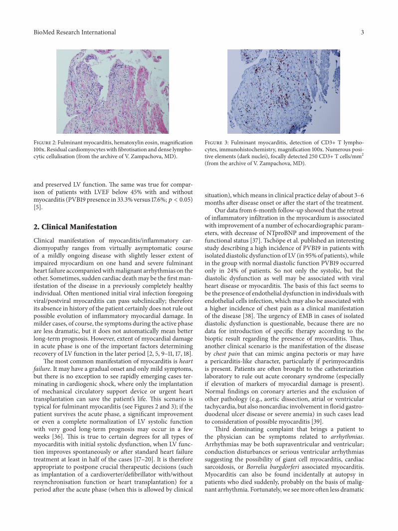
Figure 2: Fulminantmyocarditis, hematoxylin eosin, magnification100x. Residual cardiomyocytes with fibrotisation and dense lympho-cytic cellulisation (from the archive of V. Zampachova, MD).
and preserved LV function. The same was true for compar-ison of patients with LVEF below 45% with and withoutmyocarditis (PVB19 presence in 33.3% versus 17.6%;𝑝 < 0.05)[5].
2. Clinical Manifestation
Clinical manifestation of myocarditis/inflammatory car-diomyopathy ranges from virtually asymptomatic courseof a mildly ongoing disease with slightly lesser extent ofimpaired myocardium on one hand and severe fulminantheart failure accompaniedwithmalignant arrhythmias on theother. Sometimes, sudden cardiac deathmay be the firstman-ifestation of the disease in a previously completely healthyindividual. Often mentioned initial viral infection foregoingviral/postviral myocarditis can pass subclinically; thereforeits absence in history of the patient certainly does not rule outpossible evolution of inflammatory myocardial damage. Inmilder cases, of course, the symptoms during the active phaseare less dramatic, but it does not automatically mean betterlong-term prognosis. However, extent of myocardial damagein acute phase is one of the important factors determiningrecovery of LV function in the later period [2, 5, 9–11, 17, 18].
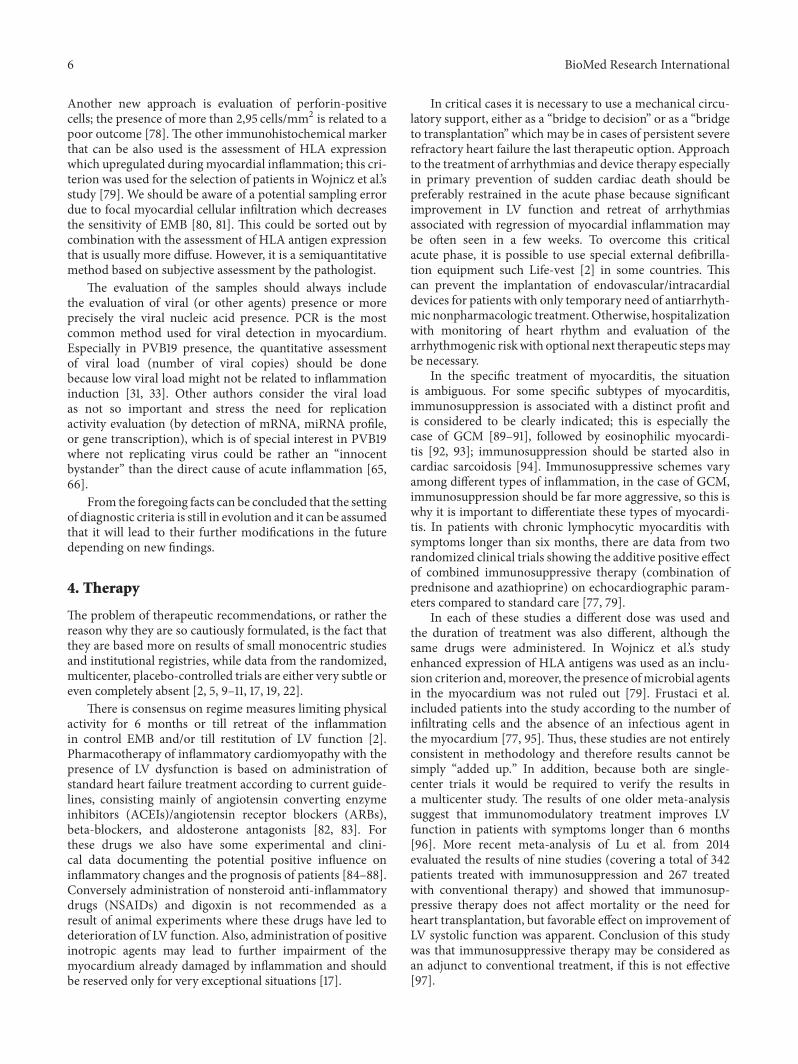
The most common manifestation of myocarditis is heartfailure. It may have a gradual onset and only mild symptoms,but there is no exception to see rapidly emerging cases ter-minating in cardiogenic shock, where only the implantationof mechanical circulatory support device or urgent hearttransplantation can save the patient’s life. This scenario istypical for fulminant myocarditis (see Figures 2 and 3); if thepatient survives the acute phase, a significant improvementor even a complete normalization of LV systolic functionwith very good long-term prognosis may occur in a fewweeks [36]. This is true to certain degrees for all types ofmyocarditis with initial systolic dysfunction, when LV func-tion improves spontaneously or after standard heart failuretreatment at least in half of the cases [17–20]. It is thereforeappropriate to postpone crucial therapeutic decisions (suchas implantation of a cardioverter/defibrillator with/withoutresynchronisation function or heart transplantation) for aperiod after the acute phase (when this is allowed by clinical
Figure 3: Fulminant myocarditis, detection of CD3+ T lympho-cytes, immunohistochemistry, magnification 100x. Numerous posi-tive elements (dark nuclei), focally detected 250 CD3+ T cells/mm2(from the archive of V. Zampachova, MD).
situation), whichmeans in clinical practice delay of about 3–6months after disease onset or after the start of the treatment.
Our data from 6-month follow-up showed that the retreatof inflammatory infiltration in the myocardium is associatedwith improvement of a number of echocardiographic param-eters, with decrease of NTproBNP and improvement of thefunctional status [37]. Tschope et al. published an interestingstudy describing a high incidence of PVB19 in patients withisolated diastolic dysfunction of LV (in 95%of patients), whilein the group with normal diastolic function PVB19 occurredonly in 24% of patients. So not only the systolic, but thediastolic dysfunction as well may be associated with viralheart disease or myocarditis. The basis of this fact seems tobe the presence of endothelial dysfunction in individuals withendothelial cells infection, which may also be associated witha higher incidence of chest pain as a clinical manifestationof the disease [38]. The urgency of EMB in cases of isolateddiastolic dysfunction is questionable, because there are nodata for introduction of specific therapy according to thebioptic result regarding the presence of myocarditis. Thus,another clinical scenario is the manifestation of the diseaseby chest pain that can mimic angina pectoris or may havea pericarditis-like character, particularly if perimyocarditisis present. Patients are often brought to the catheterizationlaboratory to rule out acute coronary syndrome (especiallyif elevation of markers of myocardial damage is present).Normal findings on coronary arteries and the exclusion ofother pathology (e.g., aortic dissection, atrial or ventriculartachycardia, but also noncardiac involvement in florid gastro-duodenal ulcer disease or severe anemia) in such cases leadto consideration of possible myocarditis [39].
Third dominating complaint that brings a patient tothe physician can be symptoms related to arrhythmias.Arrhythmias may be both supraventricular and ventricular;conduction disturbances or serious ventricular arrhythmiassuggesting the possibility of giant cell myocarditis, cardiacsarcoidosis, or Borrelia burgdorferi associated myocarditis.Myocarditis can also be found incidentally at autopsy inpatients who died suddenly, probably on the basis of malig-nant arrhythmia. Fortunately, we seemore often less dramatic
4 BioMed Research International
course with the presence of palpitations, dizziness, or evensyncope, which always have to alert attending physician tothe possible presence of serious arrhythmias.
Of course, it is not unique that all described symptomsmay be present in one patient, either simultaneously or atdifferent time phases of the disease. In terms of prognosis, ithas been reported that cases with symptoms of heart failure,namely, those which meet the criteria of inflammatory car-diomyopathy, have a poorer prognosis than casesmanifestingby chest pain or arrhythmias [28].
Another disease that should bementionedwhile speakingabout ICM is peripartum cardiomyopathy (PPCM) [40–42].PPCM is manifested by systolic heart failure in previouslyhealthy women at the end of pregnancy or in the first monthsafter the birth. The causes of the disease are not definitivelyclarified, but according to some of the authors myocarditiscould play an important role in pathogenesis of this quitemysterious disease [43, 44]. It affects more often African-Americanwomen (relative risk is almost 16 times higher) [45]and has a relatively high frequency of recovery of LV function(on the contrary, particularly among white women) [46, 47].Nevertheless, in about 10% of women, it progresses to severeheart failure, when the only solution may be urgent hearttransplantation or LVAD implantation. In less developedcountries where these treatment options are not available(and where PPCM is unfortunately relatively more frequent),the mortality is in comparison with European countries andthe USA not negligible [47–49].
3. Diagnostics
In the past, the diagnostics of myocarditis was a difficultand challenging task. Even today, despite various imagingmodalities that are available nowadays myocarditis oftenremains a diagnosis per exclusionem. The Position Statementof ESC Working Group on myocardial and pericardial dis-eases based clinical suspicion for myocarditis on the presenceof typical clinical presentation (heart failure, chest pain,and arrhythmia) and noninvasive imagining techniques (seeDiagnostic Criteria for Clinically SuspectedMyocarditis) [2].Endomyocardial biopsy is recommended for all patients whofulfil clinical diagnostic criteria and remains the standard toolfor definitive confirmation of the diagnosis [2, 5, 10, 11, 17, 19].However, this procedure is the method of first choice onlyin specialized centers with experience in performing EMBwith advanced laboratory equipment needed for complexevaluation of EMB samples.
Diagnostic Criteria for Clinically Suspected Myocarditis. Diag-nosis of myocarditis is suspected in presence of
(i) ≥1 clinical presentation and ≥1 diagnostic criterion,(ii) ≥2 diagnostic criteria, if the patient is asymptomatic.
Diagnostic Criteria. Diagnostic criteria are as follows:
(I) Electrocardiogram (ECG) test features (atrioventric-ular block, bundle branch block, ST/T-wave changes,supraventricular or ventricular arrhythmias, low volt-age of QRS complex, and abnormal Q waves).
(II) Markers of myocardial necrosis (elevated cardiactroponins or CK-MB).
(III) Functional and structural abnormalities on echocar-diography or CMR imaging (impaired left or rightventricle function, with or without left or right ven-tricle dilatation, increased ventricle wall thickness,pericardial effusion, and intracardiac thrombi).
(IV) Tissue characteristics by CMR (presence of at leasttwo of three Lake Louise criteria, myocardial oedemaand early and late gadolinium enhancement).
Similarly to diagnostics of other diseases in cardiology,the process starts with simply conventional examinationssuch as ECG, which can have very variable and also non-specific findings (presence of arrhythmias, changes of PQand ST interval, prolongation of QRS complex, and thepresence of Q waves), although some findings (especially thepresence of rhythm disorders, i.e., ventricular tachycardia oratrioventricular block of 2nd or 3rd degree)may be suggestiveof special types of myocarditis (giant cell myocarditis orcardiac sarcoidosis).
Another basic diagnostic method is echocardiography.Here as well, there is not any typical finding allowing diag-nosis with some of nonspecific echocardiographic featuresincluding both global and regional kinetic disorders of theleft or right ventricle, diastolic dysfunction, left ventriclehypertrophy, and pericardial effusion. But even a normalfinding does not rule the diagnosis out. The value ofechocardiography lies rather in excluding other causes of thesymptoms (valvular or pericardial disease, aortic dissection)and also in risk stratification based on evaluation of leftventricle systolic dysfunction [2, 9, 11, 17].
The most important noninvasive diagnostic method ismagnetic resonance imaging (MRI) which is a routinely avail-able technique in last years and is suited for the evaluation ofboth morphological and functional myocardial impairmentand tissue characterization [50–53]. The clinical suspicion ofmyocarditis is one of the most common indications for MRIstudy in cardiology because it is an accurate modality for theassessment of a number of common features in myocarditis:myocardial oedema and hyperemia, capillary leak, necrosisand fibrosis, and contraction abnormalities or pericardialeffusion [54]. The Lake Louise criteria have been proposedto standardize the evaluation of findings and to improvethe diagnostic accuracy [50]. The criteria are based on theevaluation of myocardial oedema (T2-weighted sequences)frequently present in acute inflammation, early gadoliniumenhancement (EGE) related to hyperemia, and in particu-lar the assessment of the presence of the late gadolinium
BioMed Research International 5
enhancement (LGE)with the presence of a characteristic typeof gadolinium accumulation in areas of myocardial necrosisor fibrotic reparative changes. If two of these three criteriaare present, the MRI imaging demonstrates 67% sensitivity,91% specificity, and 78% diagnostic accuracy [2, 50]. TheLGE was shown as important for prognostic stratification; ifnot present, the outcome is very good; on the contrary thepresence of LGE is considered to be a significant predictor ofoverall and cardiovascular mortality (OR 8.4 and 12.8, resp.)[55].
The diagnostic sensitivity of MRI is higher in acute sce-narios than in chronic cases with less intensive inflammatorychanges. The sensitivity is higher also in cases with clinicalmanifestation by chest pain (“infarction-like” symptoms)than in patients with arrhythmias or heart failure [56].Despite several technical difficulties in evaluation of, forexample, early gadolinium enhancement, MRI is definitelyone of the leading diagnostic modalities if myocarditis issuspected. However, especially in fulminant forms, MRIshould not delay EMB performance representing the goldstandard with more significant additive information regard-ing treatment decision [2, 19, 34].
Some of the laboratory testsmay be useful in myocarditisdiagnostics; most useful is detection of myocardial damagein acute phase by troponin and CK-MB elevation; elevationof troponin was identified as a negative prognostic factorand may be also used for long-term monitoring of diseaseactivity [57]. Elevated levels of natriuretic peptides are neitherdiagnostic nor specific but they can identify patients withworse prognosis [58]. Also the detection of certain anti-bodies against the myocardial structures (see above) relatedto autoimmune impairment showed to be contributive todiagnostics but standardized commercial kits are currentlynot available [15, 19]. The presence of antibodies could beone of the markers of positive response to immunosuppres-sive treatment [59]. If antibodies are detected in healthyrelatives of patients with dilated cardiomyopathy, the riskof disease manifestation in these individuals is higher [2,15]. Inflammatory markers can be elevated but this is nota rule. The diagnostic approach based on serological testsfrom peripheral blood often used in the past did not showsignificant correlation with EMB results [60].
Recently, the development of new sophisticated methodshighlights the tendency for less invasive or even noninvasivediagnosis of myocarditis using modern approaches. One ofthese methods is detection of different gene transcriptionwhich seems to be promising due to high specificity andsensitivity to distinguish myocarditis and dilated cardiomy-opathy [61, 62]. Another method is evaluation of the miRNAlevels. MiRNAs are small noncoding RNAs regulating post-transcription gene expression. Their levels differ in variousphysiologic and pathologic conditions and the first studiesbased on animal models showed that some of them areupregulated (e.g., miRNA-155, miRNA-146b, and miRNA-21)in myocarditis and can distinguish inflammatory and non-inflammatory myocardial impairment [63]. Similar upreg-ulation was also proved in patients with viral myocarditisfor miRNA-155 and miRNA-148a [64]. Different expressionof miRNAs [65] and different gene transcription [66] were
recently published comparing individuals with replicationactive and latent myocardial PVB19 infection. The PVB19replication activity seems to be the crucial factor in theunderstanding of PVB19 role in pathogenesis of myocarditis.The study by Kuhl et al. including 415 patients with thePVB19 myocardial presence showed that only in 15,9%patients the virus was replicating and it was in relation tochanges in cardiac gene expression, for example, INF- 𝛽1 (up-regulation), FOXP3, ADIPOR2, and IL-10 (downregulation),and with elevated mRNA levels. These methods could beused for prognostic stratification and personalised treatmentdecisions [66].
Coronary angiography is indicated to exclude coronaryartery disease (CAD) as one of the possible causes of thesymptoms and should be done in all patients in risk of CADregardless of the symptoms, which means also in patientswithout chest pain.
Endomyocardial biopsy is still considered as gold standardand the onlymethod for definitive diagnosis in vivo.The sam-ple can be obtained from left or right ventricle (or both); thediagnostic yield probably depends on the number of samples,not on the particular site of EMB [2, 67] despite the fact thatsome studies showed higher sensitivity in left ventricular andbiventricular biopsy than only right ventricular one [68, 69].Unlike the presence of infiltrating cells, which is comparablein both ventricles, some characteristics differ between the twochambers; for example, degree of fibrosis ismore pronouncedin LV [67].
The endomyocardial biopsy use in diagnosing ofmyocarditis is not a completely new trend. The beginningdates back to the 80s when Dallas criteria were set tostandardize the histology evaluation of biopsy samples [70].When these criteria were found to be of low sensitivity andhigh interobserver variability on histology assessment, it wasnecessary to set new, more sensitive and precise criteria thatcould be used in routine practice [71, 72]. There is also anoticeable difference in indication of the EMB between theUS and European countries. In the US, it is recommended touse EMB only in specific clinical scenarios [73, 74]; the ESCrecommended approach is, however, more aggressive andEMB should be performed in all cases when myocarditis isclinically suspected [2]. The consensus of European pathol-ogists published in 2013 also came to the same conclusion[75]. The addition of immunohistochemistry used for typingof infiltrative leucocytes constitutes a breakthrough due tohigher sensitivity of EMB for detection of myocarditis [72].At the turn of themillennium,Marburg’s criteria were set andwere based on the presence of more than 14 mononuclearleucocytes/mm2 of bioptic sample [5, 76]. The inclusioncriteria of the TIMIC study added the alternative presence ofmore than 7 T-lymphocytes per mm2 as a second criterion[77]. The current position statement requires the simultane-ous presence of both and, moreover, excluded patients withthe presence of more than 4 monocytes per mm2 [2]. Recentstudies from Berlin showed that setting novel cut-off valuesfor the number of the infiltrating cells (e.g., more than 10CD3+ cells per 10mm2 or more than 30 CD45+ per mm2)could make the prognostic stratification even more precise.
6 BioMed Research International
Another new approach is evaluation of perforin-positivecells; the presence of more than 2,95 cells/mm2 is related to apoor outcome [78]. The other immunohistochemical markerthat can be also used is the assessment of HLA expressionwhich upregulated duringmyocardial inflammation; this cri-terion was used for the selection of patients in Wojnicz et al.’sstudy [79]. We should be aware of a potential sampling errordue to focal myocardial cellular infiltration which decreasesthe sensitivity of EMB [80, 81]. This could be sorted out bycombination with the assessment of HLA antigen expressionthat is usually more diffuse. However, it is a semiquantitativemethod based on subjective assessment by the pathologist.
The evaluation of the samples should always includethe evaluation of viral (or other agents) presence or moreprecisely the viral nucleic acid presence. PCR is the mostcommon method used for viral detection in myocardium.Especially in PVB19 presence, the quantitative assessmentof viral load (number of viral copies) should be donebecause low viral load might not be related to inflammationinduction [31, 33]. Other authors consider the viral loadas not so important and stress the need for replicationactivity evaluation (by detection of mRNA, miRNA profile,or gene transcription), which is of special interest in PVB19where not replicating virus could be rather an “innocentbystander” than the direct cause of acute inflammation [65,66].
From the foregoing facts can be concluded that the settingof diagnostic criteria is still in evolution and it can be assumedthat it will lead to their further modifications in the futuredepending on new findings.
4. Therapy
The problem of therapeutic recommendations, or rather thereason why they are so cautiously formulated, is the fact thatthey are based more on results of small monocentric studiesand institutional registries, while data from the randomized,multicenter, placebo-controlled trials are either very subtle oreven completely absent [2, 5, 9–11, 17, 19, 22].
There is consensus on regime measures limiting physicalactivity for 6 months or till retreat of the inflammationin control EMB and/or till restitution of LV function [2].Pharmacotherapy of inflammatory cardiomyopathy with thepresence of LV dysfunction is based on administration ofstandard heart failure treatment according to current guide-lines, consisting mainly of angiotensin converting enzymeinhibitors (ACEIs)/angiotensin receptor blockers (ARBs),beta-blockers, and aldosterone antagonists [82, 83]. Forthese drugs we also have some experimental and clini-cal data documenting the potential positive influence oninflammatory changes and the prognosis of patients [84–88].Conversely administration of nonsteroid anti-inflammatorydrugs (NSAIDs) and digoxin is not recommended as aresult of animal experiments where these drugs have led todeterioration of LV function. Also, administration of positiveinotropic agents may lead to further impairment of themyocardium already damaged by inflammation and shouldbe reserved only for very exceptional situations [17].
In critical cases it is necessary to use a mechanical circu-latory support, either as a “bridge to decision” or as a “bridgeto transplantation” which may be in cases of persistent severerefractory heart failure the last therapeutic option. Approachto the treatment of arrhythmias and device therapy especiallyin primary prevention of sudden cardiac death should bepreferably restrained in the acute phase because significantimprovement in LV function and retreat of arrhythmiasassociated with regression of myocardial inflammation maybe often seen in a few weeks. To overcome this criticalacute phase, it is possible to use special external defibrilla-tion equipment such Life-vest [2] in some countries. Thiscan prevent the implantation of endovascular/intracardialdevices for patients with only temporary need of antiarrhyth-mic nonpharmacologic treatment.Otherwise, hospitalizationwith monitoring of heart rhythm and evaluation of thearrhythmogenic riskwith optional next therapeutic stepsmaybe necessary.
In the specific treatment of myocarditis, the situationis ambiguous. For some specific subtypes of myocarditis,immunosuppression is associated with a distinct profit andis considered to be clearly indicated; this is especially thecase of GCM [89–91], followed by eosinophilic myocardi-tis [92, 93]; immunosuppression should be started also incardiac sarcoidosis [94]. Immunosuppressive schemes varyamong different types of inflammation, in the case of GCM,immunosuppression should be far more aggressive, so this iswhy it is important to differentiate these types of myocardi-tis. In patients with chronic lymphocytic myocarditis withsymptoms longer than six months, there are data from tworandomized clinical trials showing the additive positive effectof combined immunosuppressive therapy (combination ofprednisone and azathioprine) on echocardiographic param-eters compared to standard care [77, 79].
In each of these studies a different dose was used andthe duration of treatment was also different, although thesame drugs were administered. In Wojnicz et al.’s studyenhanced expression of HLA antigens was used as an inclu-sion criterion and,moreover, the presence ofmicrobial agentsin the myocardium was not ruled out [79]. Frustaci et al.included patients into the study according to the number ofinfiltrating cells and the absence of an infectious agent inthe myocardium [77, 95]. Thus, these studies are not entirelyconsistent in methodology and therefore results cannot besimply “added up.” In addition, because both are single-center trials it would be required to verify the results ina multicenter study. The results of one older meta-analysissuggest that immunomodulatory treatment improves LVfunction in patients with symptoms longer than 6 months[96]. More recent meta-analysis of Lu et al. from 2014evaluated the results of nine studies (covering a total of 342patients treated with immunosuppression and 267 treatedwith conventional therapy) and showed that immunosup-pressive therapy does not affect mortality or the need forheart transplantation, but favorable effect on improvement ofLV systolic function was apparent. Conclusion of this studywas that immunosuppressive therapy may be considered asan adjunct to conventional treatment, if this is not effective[97].
According to amajority of experts, but based ondata froma single study, viral presence in the myocardium is associatedwith the absence of a positive response to immunosup-pression (data from 17 patients, but only one was positivefor PVB19) [69, 95]. Viral presence in the myocardiumhas not been determined in Myocarditis Treatment Trial(with neutral effect of immunosuppression) nor in the Pol-ish study (with positive effect of immunosuppression onechocardiographic parameters), which makes the situationin this regard even more confusing [79, 98]. In the TIMICstudy [77], worsening of echocardiographic parameters wasobserved in patients in the placebo group. Indeed, this is incontradiction with the results of other studies, including ourown experience [22, 37, 79]. Because of this, CZECH-ICITstudy was initiated with the ambition to bring more light tothe uncertainties in the use of immunosuppressive therapy inmyocarditis [99]. Recruitment of patients in the study is stillin progress and the results are expected in coming years.
Treatment with intravenous immunoglobulins has a log-ical theoretical basis, which was confirmed in several smallstudies with quite favorable results [100, 101], but the largest
multicenter trial by McNamara et al. showed no profit versusplacebo [102].Therefore, the administration of immunoglob-ulins is not currently considered as routinely indicated [2].Similarly unclear is the position of immunoadsorption,wheresome studies have shown little effect on improvement ofLV function, reduction of biomarkers levels, and retreatof inflammatory changes in the myocardium [5, 103, 104].However, until these subtle data are confirmed by otherstudies, neither treatment could be recommended [2].
In the field of antiviral treatment, the published data aresomewhat controversial as well. Administration of commonantiviral drugs is possible, but there is no evidence abouttheir actual effect. Theoretically, this treatment could bejustified in the first phase of a disease associated with viralreplication, but in clinical practice myocarditis is usuallydetected later in the second phase, when the administrationis likely to have little benefit. It was proven that interferon-beta treatment removed enteroviruses and adenoviruses ofthe myocardium and in some studies was shown as beneficial[105]. In other more common types of viruses such treatmentis unfortunately less efficient.That is probably the reason why
8 BioMed Research International
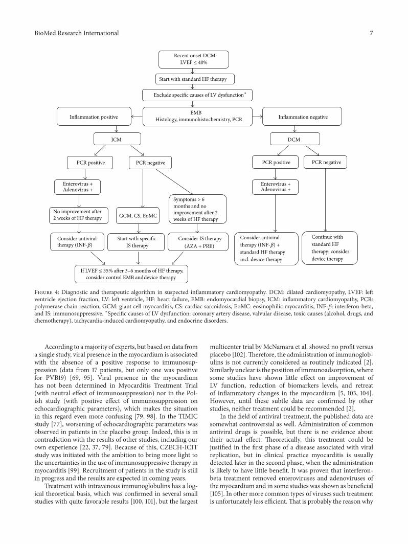
the results of other studies with higher proportion of PVB19were not so optimistic [106]. However, according to Germanauthors, interferon-beta therapy may be at least in the caseof enteroviruses associated with long-term prognostic benefit[29]. In PVB19 infection, telbivudine therapy is currentlytested and we have to wait for the results. For some otherrare agents, such Borrelia burgdorferi, antibiotic treatment isconsidered to be indicated, although the data from placebo-controlled studies are missing and also the results are notunequivocal [7, 8]. Algorithm with the proposal of therapeu-tic decisions based on knowledge of EMB result is shown inFigure 4.
5. Conclusion
The diagnosis of myocarditis and inflammatory cardiomy-opathy remains highly complex and challenging despitethe great expansion in diagnostic methods. Beside carefulanamnestic data and physical examination, a comprehensivediagnostic approach using a range of noninvasive as well asinvasive methods is required, together with highly sophisti-cated laboratory facilities. The most important noninvasivediagnostic method is cardiac magnetic resonance imaging,but endomyocardial biopsy still remains the gold standard.Standard therapy of inflammatory cardiomyopathy is basedon the recommendations for the treatment of heart failureor arrhythmias; specific therapies may be indicated onlywith known results of EMB. Evidence for the therapeuticrecommendation is not entirely convincing, and thereforeindividual assessment of each specific case and experience ofthe attending physician plays an important role in treatmentdecision. It is obvious that without carrying out large multi-center randomized prospective trial our therapeutic decisionswill fall short of the requirements of evidence basedmedicine.A considerable effort is still ahead to reach comparable levelof knowledge in the field of myocarditis and inflammatorycardiomyopathy to other areas of cardiology, where we haveboth clear and proven diagnostic criteria and also clear androbust data-based therapeutic recommendations.
Competing Interests
The authors declare that there is no conflict of interestsregarding the publication of this paper.
Acknowledgments
This study was supported by grant IGAMZ CR 14087-3/2013and the European Regional Development Fund, ProjectFNUSA ICRC (no. CZ.1.05/1.1.00/02.0123).
References
[1] P. Richardson, R. W. McKenna, M. Bristow et al., “Report ofthe 1995 World Health Organization/International Society andFederation of Cardiology Task Force on the definition andclassification of cardiomyopathies,” Circulation, vol. 93, no. 5,pp. 841–842, 1996.
[2] A. L. P. Caforio, S. Pankuweit, E. Arbustini et al., “Current stateof knowledge on aetiology, diagnosis,management, and therapyof myocarditis: a position statement of the European Societyof Cardiology Working Group on Myocardial and PericardialDiseases,” European Heart Journal, vol. 34, no. 33, pp. 2636–2648, 2013.
[3] G. Hufnagel, S. Pankuweit, A. Richter, U. Schonian, and B.Maisch, “The European Study of Epidemiology and Treatmentof Cardiac Inflammatory Diseases (ESETCID): first epidemio-logical results,” Herz, vol. 25, no. 3, pp. 279–285, 2000.
[4] J. P. Breinholt, M. Moulik, W. J. Dreyer et al., “Viral epidemio-logic shift in inflammatory heart disease: the increasing involve-ment of parvovirus B19 in the myocardium of pediatric cardiactransplant patients,” Journal of Heart and Lung Transplantation,vol. 29, no. 7, pp. 739–746, 2010.
[5] B. Maisch and S. Pankuweit, “Current treatment options in(peri)myocarditis and inflammatory cardiomyopathy,” Herz,vol. 37, no. 6, pp. 644–656, 2012.
[6] L. H. Malik, G. D. Singh, and E. A. Amsterdam, “The epidemi-ology, clinical manifestations, and management of chagas heartdisease,” Clinical Cardiology, vol. 38, no. 9, pp. 565–569, 2015.
[7] T. Palecek, P. Kuchynka, D. Hulinska et al., “Presence ofBorrelia burgdorferi in endomyocardial biopsies in patientswith new-onset unexplained dilated cardiomyopathy,” MedicalMicrobiology and Immunology, vol. 199, no. 2, pp. 139–143, 2010.
[8] M. Kubanek, M. Sramko, D. Berenova et al., “Detection ofBorrelia burgdorferi sensu lato in endomyocardial biopsy speci-mens in individuals with recent-onset dilated cardiomyopathy,”European Journal of Heart Failure, vol. 14, no. 6, pp. 588–596,2012.
[9] L. T. Cooper Jr., “Myocarditis,” The New England Journal ofMedicine, vol. 360, no. 15, pp. 1526–1538, 2009.
[10] I. Kindermann, C. Barth, F. Mahfoud et al., “Update onmyocarditis,” Journal of the American College of Cardiology, vol.59, no. 9, pp. 779–792, 2012.
[11] R.Dennert, H. J. Crijns, and S.Heymans, “Acute viralmyocardi-tis,” EuropeanHeart Journal, vol. 29, no. 17, pp. 2073–2082, 2008.
[12] M. Noutsias, M. Pauschinger, W.-C. Poller, H.-P. Schultheiss,and U. Kuhl, “Current insights into the pathogenesis, diagnosisand therapy of inflammatory cardiomyopathy,” Heart FailureMonitor, vol. 3, no. 4, pp. 127–135, 2003.
[13] A. L. P. Caforio, S. Bottaro, and S. Iliceto, “Dilated cardiomyopa-thy (DCM) andmyocarditis: classification, clinical and autoim-mune features,” Applied Cardiopulmonary Pathophysiology, vol.16, no. 1, pp. 82–95, 2012.
[14] C. Wehlou and J. R. Delanghe, “Detection of antibodies incardiac autoimmunity,” Clinica Chimica Acta, vol. 408, no. 1-2,pp. 114–122, 2009.
[15] A. L. P. Caforio, N. J. Mahon, F. Tona, andW. J. McKenna, “Cir-culating cardiac autoantibodies in dilated cardiomyopathy andmyocarditis: pathogenetic and clinical significance,” EuropeanJournal of Heart Failure, vol. 4, no. 4, pp. 411–417, 2002.
[16] N. R. Rose, “Myocarditis: infection versus autoimmunity,”Journal of Clinical Immunology, vol. 29, no. 6, pp. 730–737, 2009.
[17] H.-P. Schultheiss, U. Khl, and L. T. Cooper, “Themanagement ofmyocarditis,” European Heart Journal, vol. 32, no. 21, pp. 2616–2625, 2011.
[18] A. D’Ambrosio, G. Patti, A. Manzoli et al., “The fate of acutemyocarditis between spontaneous improvement and evolutionto dilated cardiomyopathy: a review,” Heart, vol. 85, no. 5, pp.499–504, 2001.
BioMed Research International 9
[19] A. L. P. Caforio, R.Marcolongo, C. Basso, and S. Iliceto, “Clinicalpresentation and diagnosis of myocarditis,” Heart, vol. 101, no.16, pp. 1332–1344, 2015.
[20] J. W. Mason, J. B. O’Connell, A. Herskowitz et al., “A clinicaltrial of immunosuppressive therapy for myocarditis,” The NewEngland Journal of Medicine, vol. 333, no. 5, pp. 269–275, 1995.
[21] U. Kuhl,M. Pauschinger,M. Noutsias et al., “High prevalence ofviral genomes and multiple viral infections in the myocardiumof adults with ‘idiopathic’ left ventricular dysfunction,” Circula-tion, vol. 111, no. 7, pp. 887–893, 2005.
[22] J. Krejcı, H. Poloczkova, P. Hude et al., “Impact of inflammatoryinfiltration and viral genome presence in myocardium on thechanges of echocardiographic parameters,” Cor et Vasa, vol. 55,no. 4, pp. e333–e340, 2013.
[23] U. Kuhl, “Antiviral treatment of myocarditis and acute dilatedcardiomyopathy,”Heart Failure Clinics, vol. 1, no. 3, pp. 467–474,2005.
[24] F. Kuethe, J. Lindner, K. Matschke et al., “Prevalence of par-vovirus B19 and human bocavirus DNA in the heart of patientswith no evidence of dilated cardiomyopathy or myocarditis,”Clinical Infectious Diseases, vol. 49, no. 11, pp. 1660–1666, 2009.
[25] F. Escher, S. Modrow, T. Sabi et al., “Parvovirus B19 profiles inpatients presenting with acute myocarditis and chronic dilatedcardiomyopathy,” Medical Science Monitor, vol. 14, no. 12, pp.CR589–CR597, 2008.
[26] S. A. Koepsell, D. R. Anderson, and S. J. Radio, “Parvovirus B19is a bystander in adult myocarditis,” Cardiovascular Pathology,vol. 21, no. 6, pp. 476–481, 2012.
[27] G. C. Stewart, J. Lopez-Molina, R. V. S. R. K. Gottumukkala etal., “Myocardial Parvovirus B19 persistence: lack of associationwith clinicopathologic Phenotype in adults with heart failure,”Circulation: Heart Failure, vol. 4, no. 1, pp. 71–78, 2011.
[28] A. L. P. Caforio, F. Calabrese, A. Angelini et al., “A prospectivestudy of biopsy-proven myocarditis: prognostic relevance ofclinical and aetiopathogenetic features at diagnosis,” EuropeanHeart Journal, vol. 28, no. 11, pp. 1326–1333, 2007.
[29] U. Kuhl, D. Lassner, J. von Schlippenbach, W. Poller, and H.-P.Schultheiss, “Interferon-beta improves survival in enterovirus-associated cardiomyopathy,” Journal of the American College ofCardiology, vol. 60, no. 14, pp. 1295–1296, 2012.
[30] I. Kindermann, M. Kindermann, R. Kandolf et al., “Predictorsof outcome in patients with suspectedmyocarditis,”Circulation,vol. 118, no. 6, pp. 639–648, 2008.
[31] C.-T. Bock, K. Klingel, andR. Kandolf, “Human parvovirus B19-associated myocarditis,” New England Journal of Medicine, vol.362, no. 13, pp. 1248–1249, 2010.
[32] S. Pankuweit and K. Klingel, “Viral myocarditis: from experi-mentalmodels tomolecular diagnosis in patients,”Heart FailureReviews, vol. 18, no. 6, pp. 683–702, 2013.
[33] C. T. Bock, A. Duchting, and F. Utta, “Molecular phenotypesof human parvovirus B19 in patients with myokarditis,” WorldJournal of Cardiology, vol. 6, pp. 183–195, 2014.
[34] D. Lassner, M. Rohde, C. S. Siegismund et al., “Myocarditis—personalized medicine by expanded endomyocardial biopsydiagnostics,” World Journal of Cardiovascular Diseases, vol. 4,no. 6, pp. 325–340, 2014.
[35] D. Fairweather, L. T. Cooper Jr., and L. A. Blauwet, “Sex andgender differences in myocarditis and dilated cardiomyopathy,”Current Problems in Cardiology, vol. 38, no. 1, pp. 7–46, 2013.
[36] R. E. McCarthy III, J. P. Boehmer, R. H. Hruban et al., “Long-term outcome of fulminant myocarditis as compared with
acute (nonfulminant) myocarditis,”TheNew England Journal ofMedicine, vol. 342, no. 10, pp. 690–695, 2000.
[37] J. Krejci, P. Hude, H. Poloczkova et al., “Correlations of thechanges in bioptic findings with echocardiographic, clinical andlaboratory parameters in patients with inflammatory cardiomy-opathy,” Heart and Vessels, vol. 31, pp. 416–426, 2016.
[38] C. Tschope, C.-T. Bock, M. Kasner et al., “High prevalence ofcardiac parvovirus B19 infection in patients with isolated leftventricular diastolic dysfunction,” Circulation, vol. 111, no. 7, pp.879–886, 2005.
[39] H. Baccouche, H. Mahrholdt, G. Meinhardt et al., “Diagnosticsynergy of non-invasive cardiovascularmagnetic resonance andinvasive endomyocardial biopsy in troponin-positive patientswithout coronary artery disease,” European Heart Journal, vol.30, no. 23, pp. 2869–2879, 2009.
[40] J. Krejci, P. Hude, L. Spinarova et al., “The variable clinicalcourse of peripartum cardiomyopathy,” Biomedical Papers, vol.158, no. 1, pp. 92–97, 2014.
[41] B. D. Bultmann, K. Klingel, M. Nabauer, D. Wallwiener, and R.Kandolf, “High prevalence of viral genomes and inflammationin peripartum cardiomyopathy,” American Journal of Obstetricsand Gynecology, vol. 193, no. 2, pp. 363–365, 2005.
[42] M. G. Midei, S. H. DeMent, A. M. Feldman, G. M. Hutchins,and K. L. Baughman, “Peripartum myocarditis and cardiomy-opathy,” Circulation, vol. 81, no. 3, pp. 922–928, 1990.
[43] K. Sliwa, D. Hilfiker-Kleiner, M. C. Petrie et al., “Currentstate of knowledge on aetiology, diagnosis, management, andtherapy of peripartum cardiomyopathy: a position statementfrom the Heart Failure Association of the European Society ofCardiology Working Group on peripartum cardiomyopathy,”European Journal of Heart Failure, vol. 12, no. 8, pp. 767–778,2010.
[44] L. A. Blauwet and L. T. Cooper, “Diagnosis and managementof peripartum cardiomyopathy,”Heart, vol. 97, no. 23, pp. 1970–1981, 2011.
[45] M. B. Gentry, J. K. Dias, A. Luis, R. Patel, J. Thornton, andG. L. Reed, “African-American women have a higher riskfor developing Peripartum cardiomyopathy,” Journal of theAmerican College of Cardiology, vol. 55, no. 7, pp. 654–659, 2010.
[46] L. T. Cooper, P. J. Mather, J. D. Alexis et al., “Myocardial recov-ery in peripartum cardiomyopathy: prospective comparisonwith recent onset cardiomyopathy in men and nonperipartumwomen,” Journal of Cardiac Failure, vol. 18, no. 1, pp. 28–33, 2012.
[47] A.M. Amos,W. A. Jaber, and S. D. Russell, “Improved outcomesin peripartum cardiomyopathy with contemporary,” AmericanHeart Journal, vol. 152, no. 3, pp. 509–513, 2006.
[48] J. G. Safirstein, A. S. Ro, S. Grandhi, L. Wang, J. D. Fett, and C.Staniloae, “Predictors of left ventricular recovery in a cohort ofperipartum cardiomyopathy patients recruited via the internet,”International Journal of Cardiology, vol. 154, no. 1, pp. 27–31,2012.
[49] J. Krejci, H. Poloczkova, and P. Nemec, “Current therapeuticconcepts in peripartum cardiomyopathy,” Current Pharmaceu-tical Design, vol. 21, no. 4, pp. 507–514, 2015.
[50] M. G. Friedrich, U. Sechtem, J. Schulz-Menger et al., “Car-diovascular magnetic resonance in myocarditis: a JACC WhitePaper,” Journal of the American College of Cardiology, vol. 53, no.17, pp. 1475–1487, 2009.
[51] M. A. G.M. Olimulder, J. van Es, andM. A. Galjee, “The impor-tance of cardiac MRI as a diagnostic tool in viral myocarditis-induced cardiomyopathy,”NetherlandsHeart Journal, vol. 17, no.12, pp. 481–486, 2009.
10 BioMed Research International
[52] O. Bruder, A. Wagner, M. Lombardi et al., “European Car-diovascular Magnetic Resonance (EuroCMR) registry—multinational results from 57 centers in 15 countries,” Journal ofCardiovascular Magnetic Resonance, vol. 15, article 9, 2013.
[53] P. Lurz, I. Eitel, J. Adam et al., “Diagnostic performance ofCMR imaging compared with EMB in patients with suspectedmyocarditis,” JACC Cardiovascular Imaging, vol. 5, no. 5, pp.513–524, 2012.
[54] P. Kuchynka, T. Palecek, E. Nemecek, M. Fikrle, and A. Linhart,“New therapeutic aspects on inflammatory cardiomyopathy,”Current Pharmaceutical Design, vol. 21, no. 4, pp. 459–465, 2015.
[55] S. Grun, J. Schumm, S. Greulich et al., “Long-term follow-up of biopsy-proven viral myocarditis: predictors of mortalityand incomplete recovery,” Journal of the American College ofCardiology, vol. 59, no. 18, pp. 1604–1615, 2012.
[56] M. Francone, C. Chimenti, N. Galea et al., “CMR sensitivityvaries with clinical presentation and extent of cell necrosis inbiopsy-proven acute myocarditis,” JACC Cardiovascular Imag-ing, vol. 7, no. 3, pp. 254–263, 2014.
[57] B. Lauer, C. Niederau, U. Kuhl et al., “Cardiac troponin T inpatients with clinically suspected myocarditis,” Journal of theAmerican College of Cardiology, vol. 30, no. 5, pp. 1354–1359,1997.
[58] C. Ukena, M. Kindermann, F. Mahfoud et al., “Diagnostic andprognostic validity of different biomarkers in patients withsuspectedmyocarditis,”Clinical Research in Cardiology, vol. 103,no. 9, pp. 743–751, 2014.
[59] A. Frustaci, C. Chimenti, F. Calabrese, M. Pieroni, G. Thiene,and A. Maseri, “Immunosuppressive therapy for active lym-phocytic myocarditis. Virological and immunologic profile ofresponders versus nonresponders,” Circulation, vol. 107, no. 6,pp. 857–866, 2003.
[60] F. Mahfoud, B. Grtner, M. Kindermann et al., “Virus serologyin patients with suspected myocarditis: utility or futility?”European Heart Journal, vol. 32, no. 7, pp. 897–903, 2011.
[61] B. Heidecker, M. M. Kittleson, E. K. Kasper et al., “Transcrip-tomic biomarkers for the accurate diagnosis of myocarditis,”Circulation, vol. 123, no. 11, pp. 1174–1184, 2011.
[62] L. T. Cooper Jr., O. K. Onuma, S. Sagar et al., “Genomic andproteomic analysis of myocarditis and dilated cardiomyopathy,”Heart Failure Clinics, vol. 6, no. 1, pp. 75–85, 2010.
[63] M. F. Corsten, A. Papageorgiou,W. Verhesen et al., “MicroRNAprofiling identifies MicroRNA-155 as an adverse mediator ofcardiac injury and dysfunction during Acute Viral MyoCardi-tis,” Circulation Research, vol. 111, no. 4, pp. 415–425, 2012.
[64] J.-L. Bao and L. Lin, “MiR-155 and miR-148a reduce car-diac injury by inhibiting NF-𝜅B pathway during acute viralmyocarditis,” European Review forMedical and PharmacologicalSciences, vol. 18, no. 16, pp. 2349–2356, 2014.
[65] U. Kuhl, M. Rohde, D. Lassner, U.M. Gross, F. Escher, andH.-P.Schultheiss, “miRNAas activitymarkers in ParvoB19 associatedheart disease,” Herz, vol. 37, no. 6, pp. 637–643, 2012.
[66] U.Kuhl, D. Lassner, A.Dorner et al., “A distinct subgroup of car-diomyopathy patients characterized by transcriptionally activecardiotropic erythrovirus and altered cardiac gene expression,”Basic Research in Cardiology, vol. 108, article 372, 2013.
[67] F. Escher, D. Lassner, U. Kuhl et al., “Analysis of endomyocardialbiopsies in suspected myocarditis—diagnostic value of left ver-sus right ventricularbiopsy,” International Journal of Cardiology,vol. 177, no. 1, pp. 76–78, 2014.
[68] A. Yilmaz, I. Kindermann,M. Kindermann et al., “Comparativeevaluation of left and right ventricular endomyocardial biopsy:differences in complication rate and diagnostic performance,”Circulation, vol. 122, no. 9, pp. 900–909, 2010.
[69] C. Chimenti and A. Frustaci, “Contribution and risks ofleft ventricular endomyocardial biopsy in patients with car-diomyopathies: a retrospective study over a 28-year period,”Circulation, vol. 128, no. 14, pp. 1531–1541, 2013.
[70] H. T. Aretz, M. E. Billingham, W. D. Edwards et al., “Myocardi-tis. A histopathologic definition and classification,”The Ameri-can Journal of Cardiovascular Pathology, vol. 1, no. 1, pp. 3–14,1987.
[71] K. L. Baughman, “Diagnosis of myocarditis: death of Dallascriteria,” Circulation, vol. 113, no. 4, pp. 593–595, 2006.
[72] U. Kuhl, M. Noutsias, B. Seeberg, and H.-P. Schultheiss,“Immunohistological evidence for a chronic intramyocardialinflammatory process in dilated cardiomyopathy,” Heart, vol.75, no. 3, pp. 295–300, 1996.
[73] L. T. Cooper, K. L. Baughman, A. M. Feldman et al., “The roleof endomyocardial biopsy in themanagement of cardiovasculardisease: a scientific statement from the American Heart Associ-ation, the American College of Cardiology, and the EuropeanSociety of Cardiology,” Circulation, vol. 116, no. 19, pp. 2216–2233, 2007.
[74] A. M. From, J. J. Maleszewski, and C. S. Rihal, “Current statusof endomyocardial biopsy,”Mayo Clinic Proceedings, vol. 86, no.11, pp. 1095–1102, 2011.
[75] G.Thiene, P. Bruneval, J. Veinot, and O. Leone, “Diagnostic useof the endomyocardial biopsy: a consensus statement,”VirchowsArchiv, vol. 463, no. 1, pp. 1–5, 2013.
[76] B. Maisch, I. Portig, A. Ristic, G. Hufnagel, and S. Pankuweit,“Definition of inflammatory cardiomyopathy (myocarditis): onthe way to consensus: a status report,” Herz, vol. 25, no. 3, pp.200–209, 2000.
[77] A. Frustaci, M. A. Russo, and C. Chimenti, “Randomizedstudy on the efficacy of immunosuppressive therapy in patientswith virus-negative inflammatory cardiomyopathy: the TIMICstudy,” European Heart Journal, vol. 30, no. 16, pp. 1995–2002,2009.
[78] F. Escher, U. Kuhl, D. Lassner et al., “Presence of perforinin endomyocardial biopsies of patients with inflammatorycardiomyopathy predicts poor outcome,” European Journal ofHeart Failure, vol. 16, no. 10, pp. 1066–1072, 2014.
[79] R. Wojnicz, E. Nowalany-Kozielska, C. Wojciechowska et al.,“Randomized, placebo- controlled study for immunosuppres-sive treatment of inflammatory dilated cardiomyopathy. Two-year follow-up results,” Circulation, vol. 104, no. 1, pp. 39–45,2001.
[80] L. H. Chow, S. J. Radio, T. D. Sears, and B. M. Mcmanus,“Insensitivity of right ventricular endomyocardial biopsy in thediagnosis of myocarditis,” Journal of the American College ofCardiology, vol. 14, no. 4, pp. 915–920, 1989.
[81] A. J. Hauck, D. L. Kearney, and W. D. Edwards, “Evaluationof postmortem endomyocardial biopsy specimens from 38patients with lymphocytic myocarditis: implications for role ofsampling error,” Mayo Clinic Proceedings, vol. 64, no. 10, pp.1235–1245, 1989.
[82] J. J. V. McMurray, S. Adamopoulos, S. D. Anker et al., “ESCGuidelines for the diagnosis and treatment of acute and chronicheart failure 2012: the task force for the diagnosis and treatmentof acute and chronic heart failure 2012 of the European Society
BioMed Research International 11
of Cardiology. Developed in collaboration with the HeartFailure Association (HFA) of the ESC,” European Heart Journal,vol. 33, no. 14, pp. 1787–1847, 2012.
[83] C. W. Yancy, M. Jessup, B. Bozkurt et al., “2013 ACCF/AHAguideline for the management of heart failure: a report of theAmerican College of Cardiology Foundation/American HeartAssociation Task Force on practice guidelines,” Circulation, vol.128, no. 16, pp. e240–e327, 2013.
[84] T. J. Bahk, M. D. Daniels, J. S. Leon, K. Wang, and D.M. Engman, “Comparison of angiotensin converting enzymeinhibition and angiotensin II receptor blockade for the preven-tion of experimental autoimmune myocarditis,” InternationalJournal of Cardiology, vol. 125, no. 1, pp. 85–93, 2008.
[85] S. Saegusa, Y. Fei, T. Takahashi et al., “Oral administration ofcandesartan improves the survival of mice with viral myocardi-tis through modification of cardiac adiponectin expression,”Cardiovascular Drugs and Therapy, vol. 21, no. 3, pp. 155–160,2007.
[86] Z. Yuan, K. Shioji, Y. Kihara, H. Takenaka, Y. Onozawa, andC. Kishimoto, “Cardioprotective effects of carvedilol on acuteautoimmune myocarditis: anti-inflammatory effects associatedwith antioxidant property,” American Journal of Physiology—Heart and Circulatory Physiology, vol. 286, no. 1, pp. H83–H90,2004.
[87] M. Pauschinger, S. Rutschow, K. Chandrasekharan et al.,“Carvedilol improves left ventricular function in murinecoxsackievirus-induced acute myocarditis: association withreduced myocardial interleukin-1𝛽 andMMP-8 expression anda modulated immune response,” European Journal of HeartFailure, vol. 7, no. 4, pp. 444–452, 2005.
[88] J. Xiao, M. Shimada, W. Liu, D. Hu, and A. Matsumori,“Anti-inflammatory effects of eplerenone on viral myocarditis,”European Journal of Heart Failure, vol. 11, no. 4, pp. 349–353,2009.
[89] L. T. Cooper Jr., G. J. Berry, and R. Shabetai, “Idiopathicgiant-cell myocarditis-natural history and treatment,”The NewEngland Journal of Medicine, vol. 336, no. 26, pp. 1860–1866,1997.
[90] L. T. Cooper Jr., J. M. Hare, H. D. Tazelaar et al., “Usefulnessof immunosuppression for giant cell myocarditis,” AmericanJournal of Cardiology, vol. 102, no. 11, pp. 1535–1539, 2008.
[91] R. Kandolin, J. Lehtonen, K. Salmenkivi, A. Raisanen-Sokolowski, J. Lommi, and M. Kupari, “Diagnosis, treatment,and outcome of giant-cell myocarditis in the era of combinedimmunosuppression,” Circulation: Heart Failure, vol. 6, no. 1,pp. 15–22, 2013.
[92] S. Kawano, J. Kato, N. Kawano et al., “Clinical features andoutcomes of eosinophilic myocarditis patients treated withprednisolone at a single institution over a 27-year period,”Internal Medicine, vol. 50, no. 9, pp. 975–981, 2011.
[93] T. Yanagisawa, T. Inomata, I. Watanabe et al., “Clinical signif-icance of corticosteroid therapy for eosinophilic myocarditis,”International Heart Journal, vol. 52, no. 2, pp. 110–113, 2011.
[94] L. A. Blauwet and L. T. Cooper, “Idiopathic giant cell myocardi-tis and cardiac sarcoidosis,”Heart Failure Reviews, vol. 18, no. 6,pp. 733–746, 2013.
[95] A. Frustaci and C. Chimenti, “Immunosuppressive therapy inmyocarditis,” Circulation Journal, vol. 79, no. 1, pp. 4–7, 2014.
[96] C. Stanton, F. Mookadam, S. Cha et al., “Greater symptomduration predicts response to immunomodulatory therapy indilated cardiomyopathy,” International Journal of Cardiology,vol. 128, no. 1, pp. 38–41, 2008.
[97] C. Lu, F. Qui, Y. Yan, T. Liu, J. Li, and H. Chen, “Immunosup-pressive treatment for myocarditis: a meta-analysis of random-ized controlled trials,” Journal of Cardiovascular Medicine, 2014.
[98] J. W. Mason, J. B. O’Connell, A. Herskowitz et al., “A clinicaltrial of immunosuppressive therapy for myocarditis,” The NewEngland Journal of Medicine, vol. 333, no. 5, pp. 269–275, 1995.
[99] T. Palecek, J. Krejci, L. Pecen et al., “Czech InflammatoryCardiomyopathy Immunosuppression Trial (CZECH-ICIT):randomized, multicentric study comparing the effect of tworegimens of combined immunosuppressive therapy in the treat-ment of inflammatory cardiomyopathy: the aims and design ofthe trial,” Cor et Vasa, vol. 55, no. 6, pp. e475–e478, 2013.
[100] L. Gullestad, H. Aass, J. G. Fjeld et al., “Immunomodulatingtherapy with intravenous immunoglobulin in patients withchronic heart failure,” Circulation, vol. 103, no. 2, pp. 220–225,2001.
[101] R. Dennert, S. Velthuis, S. Schalla et al., “Intravenous immunog-lobulin therapy for patients with idiopathic cardiomyopathyand endomyocardial biopsy-proven high PVB19 viral load,”Antiviral Therapy, vol. 15, no. 2, pp. 193–201, 2010.
[102] D.M.McNamara, R. Holubkov, R. C. Starling et al., “Controlledtrial of intravenous immune globulin in recent-onset dilatedcardiomyopathy,” Circulation, vol. 103, no. 18, pp. 2254–2259,2001.
[103] A. Staudt, A. Hummel, J. Ruppert et al., “Immunoadsorptionin dilated cardiomyopathy: 6-month results from a randomizedstudy,”American Heart Journal, vol. 152, no. 4, pp. 712.e1–712.e6,2006.
[104] S. B. Felix, A. Staudt, and W. V. Dorffer, “Hemodynamiceffects of immunoadsorption and subsequent immunoglobulinsubstitution in dilated cardiomyopathy: three-month resultsfrom a randomized study,” Journal of the American College ofCardiology, vol. 35, no. 6, pp. 1590–1598, 2000.
[105] U. Kuhl, M. Pauschinger, P. L. Schwimmbeck et al., “Interferon-𝛽 treatment eliminates cardiotropic viruses and improves leftventricular function in patients with myocardial persistence ofviral genomes and left ventricular dysfunction,”Circulation, vol.107, no. 22, pp. 2793–2798, 2003.
[106] O. Zimmermann, C. Rodewald, M. Radermacher et al., “Inter-feron 𝛽-1b therapy in chronic viral dilated cardiomyopathy—isthere a role for specific therapy?” Journal of Cardiac Failure, vol.16, no. 4, pp. 348–356, 2010.