Page 1
REVIEW ARTICLE
Role of glutamate metabolism in bacterial responsestowards acid and other stressesC. Feehily and K.A.G. Karatzas
Bacterial Stress Response Group, Department of Microbiology, School of Natural Sciences, National University of Ireland, Galway, Ireland
Keywords
acid resistance, glutamate decarboxylase,
glutamate metabolism, Listeria
monocytogenes.
Correspondence
Kimon-Andreas G. Karatzas, School of Food
Biosciences, University of Reading, Reading
RG6 6AD, UK. E-mail: [email protected] .
uk
2012/0437: received 8 March 2012, revised
15 August 2012 and accepted 16 August
2012
doi:10.1111/j.1365-2672.2012.05434.x
Summary
Glutamate plays a central role in a wide range of metabolic processes in
bacterial cells. This review focuses on the involvement of glutamate in bacterial
stress responses. In particular, it reviews the role of glutamate metabolism in
response against acid stress and other stresses. The glutamate decarboxylase
(GAD) system has been implicated in acid tolerance in several bacterial genera.
This system facilitates intracellular pH homoeostasis by consuming protons in
a decarboxylation reaction that produces c-aminobutyrate (GABA) from
glutamate. An antiporter system is usually present to couple the uptake of
glutamate to the efflux of GABA. Recent insights into the functioning of this
system will be discussed. Finally, the intracellular fate of GABA will also be
discussed. Many bacteria are capable of metabolizing GABA to succinate via
the GABA shunt pathway. The role and regulation of this pathway will be
addressed in the review.
Introduction
Glutamate is an important molecule for all living organ-
isms, which plays a role in various metabolic processes. It
is a nonessential amino acid involved in protein synthesis
and other fundamental processes such as glycolysis, glu-
coneogenesis and the citric acid cycle (Berg et al. 2007).
It is also a key metabolite because it serves to link nitro-
gen and carbon metabolism (Berg et al. 2007). Catabo-
lism of glutamate occurs mainly by the action of either
glutamate dehydrogenase or glutamate decarboxylase
(GAD) (Berg et al. 2007). The first enzyme, among other
roles, is important for the assimilation of ammonia to
amino acids, while the second is important for resistance
mainly against acid but also other stresses (Berg et al.
2007). As glutamate metabolism in bacterial stress
responses is at the core of this review, it will focus in the
decarboxylation of glutamate to c-aminobutyrate (GABA)
and the subsequent catabolism of GABA through the
GABA shunt.
The GAD is an enzyme that catalyses the decarboxyl-
ation of glutamate to GABA (O’Byrne and Karatzas
2008). The decarboxylation of glutamate is the first step
of the GABA shunt pathway that leads to the production
of succinate by the sequential actions of GABA-amino-
transferase (GABA-AT) and succinic semialdehyde dehy-
drogenase (SSDH; Zhu et al. 2010). The GAD enzyme is
present in a wide variety of organisms from all kingdoms
of life. In mammals, glutamate is the main excitatory and
GABA the main inhibitory neurotransmitter (Petroff
2002). The conversion of the one to the other by GAD
plays an important role in various brain functions.
However, in plants and micro-organisms, the GAD sys-
tem appears to play other roles (Maras et al. 1992). In
plants, the GAD system is expressed in response to a
variety of stress conditions such as temperature shock,
hypoxia or increasing levels of Ca2+ (Shelp et al. 1999).
In micro-organisms, the system seems to be related
mainly with resistance against acidic conditions, although
a role involving other stresses has been seen also. The
GAD system has been described as the most important
mechanism of acid resistance in the highly acid tolerant
bacterium Escherichia coli (Capitani et al. 2003; Foster
2004). Acid resistance would allow various foodborne
pathogens or spoilage bacteria to grow on acidic foods.
Furthermore, this property is also a virulence or probiotic
factor, as it allows pathogens or probiotic organisms,
respectively, to pass through the extremely acidic
Journal of Applied Microbiology 114, 11--24 © 2012 The Society for Applied Microbiology 11
Journal of Applied Microbiology ISSN 1364-5072
Page 2
conditions of the stomach barrier. However, a balance
between acid resistance and other functions is essential as
increased acid and stress resistance have been shown to
impair colonization or virulence (Karatzas et al. 2005,
2007, 2008). The decarboxylation of glutamate consumes
a proton, and therefore, micro-organisms take advantage
of this property to remove protons from the intracellular
milieu under acidic conditions (O’Byrne and Karatzas
2008). It operates in a cycle that starts with the import of
an extracellular glutamate molecule (Glte) in exchange for
an intracellular GABA (GABAi) through the glutamate/
GABA antiporter (O’Byrne and Karatzas 2008). These
antiporters are usually a part of the GAD system in most
micro-organisms (Table 1). Subsequently, the imported
glutamate is decarboxylated by the GAD, with the incor-
poration of a proton in the a-carbon of this molecule in
the place of the carboxyl group to form GABA (Karatzas
et al. 2012; Fig. 1). The bond is highly stable and it can-
not be subjected to ionization, preventing the release of
the proton in the intracellular milieu. Subsequently, the
GABA molecule that carries the removed proton is either
exported by the antiporter as extracellular GABA
(GABAe) or remains inside the cell (GABAi) as has been
shown previously (Karatzas et al. 2010). The current
model for the removal of protons by the GAD system has
been debated, however, and other hypotheses have been
put forward, which are analysed here. In bacteria, the
GAD system is usually accompanied with one or two glu-
tamate/GABA antiporters (Table 1). The antiporter
increases the availability of glutamate to the GAD
enzymes increasing the capacity of the system to remove
protons and, therefore, the efficiency of the system.
The by-product of the GAD system is GABA, which is
the substrate of the GABA shunt pathway. The GABA
shunt pathway was first described during the study of
guinea pig brain cells in 1970 (Balazs et al. 1970). This
pathway involves the catabolism of GABA derived from
the GAD system to succinate via a two step enzymatic
process (Fig. 2). In the first step, a GABA-AT catalyses
the reversible conversion of GABA to succinic semialde-
hyde (SSA), where the amino group of GABA is donated
to an a-ketoglutarate molecule with the subsequent pro-
duction of glutamate (Zhu et al. 2010). The second step
is catalysed by an SSA dehydrogenase and converts the
SSA to succinate (Zhu et al. 2010). Despite over forty
years of study, the role of the GABA shunt pathway in
bacteria has not been fully elucidated, although involve-
ment in glutamate metabolism, anaplerosis and oxidative
stress response has been suggested (de Carvalho et al.
2011). The pathway has been shown to play an important
role in mammalian cells, including a mechanism for the
recovery of four of the five carbons lost from the TCA
cycle, although there is a discrepancy in the estimation of
metabolic activity of this cycle. Cells lose approximately
8% of the energy they would normally generate by pas-
sage through the alternative TCA cycle reactions (Waage-
petersen et al. 1999). The GABA shunt pathway may also
be a way to generate energy from glutamate without the
production of ammonia as the enzymes of the pathway
are pyridoxal phosphate dependent (Waagepetersen et al.
1999).
The glutamate decarboxylase system
The GAD system is present in various bacterial species
(Table 1), and it has been shown to play a role in their
acid resistance (Cotter et al. 2001; Capitani et al.
2003; Su et al. 2011). With the recent developments in
Table 1 The variability in the architecture of the bacterial glutamate decarboxylase (GAD) system
Species Decarboxylases Antiporters Reference
Escherichia coli GadA, GadB GadC Smith et al. (1992)
Shigella flexneri GadA, GadB GadC Waterman and Small (2003a)
Mycobacterium tuberculosis GadA Cole et al. (1998), Cotter et al. (2001)
Listeria monocytogenes GadD1, GadD2, GadD3 GadT1, GadT2 Cotter et al. (2005), Karatzas et al. (2012)
L. monocytogenes ser. 4 and various strains GadD2, GadD3 GadT2 Cotter et al. (2005), Karatzas et al. (2012)
Lactobacillus reuteri GadB GadC1, GadC2 Su et al. (2011)
Lactococcus lactis GadB GadC Su et al. (2011)
Lactobacillus plantarum GadB GadC Su et al. (2011)
Clostridium perfringens GadC
Methanocaldococcus jannaschii* GadB N/A Kezmarsky et al. (2005)
Brucella abortus* GadB GadC Roop et al. (2003)
Bifidobacterium dentium GadB GadC Ventura et al. (2009)
Bifidobacteria (other species than Bif. dentium) Not present Not present Ventura et al. (2009)
N/A represents not available information regarding the presence of the gene in the organism.
*Presence of GAD based on bioinformatic evidence.
12 Journal of Applied Microbiology 114, 11--24 © 2012 The Society for Applied Microbiology
Role of glutamate metabolism in bacterial stress responses C. Feehily and K.-A.G. Karatzas
Page 3
genomics, and the increasing availability of genomes of
various organisms, it became clear that the GAD system
seems to be present even in archaea like Methanocaldococ-
cus jannaschii (Kezmarsky et al. 2005). Despite this
wealth of information, it has not been possible to associ-
ate the presence of the GAD system with a specific kind
of micro-organisms or an environmental niche. It has
been suggested that the GAD system is important for sur-
vival in the low pH of the stomach, although Salmonella
sp. (Foster 2004) and Campylobacter sp. (unpublished
data) that are responsible for most of the cases of food-
related diseases around the world (WHO 2012) do not
possess a GAD system.
Furthermore, the architecture of the GAD system
seems to be subject to high variability between species as
some possess one, two or even three decarboxylases
accompanied with none, one or two antiporters
(Table 1). The case of Mycobacterium tuberculosis is quite
interesting as it seems to possess a GAD that is not
accompanied by an antiporter (Cole et al. 1998; Cotter
et al. 2001). In addition, in other cases, there is variability
within the same species. For example, Listeria monocytog-
enes normally possesses three decarboxylases (GadD1, D2,
D3) and two antiporters (GadT1, T2), but serotype 4 and
a few other strains do not have the GadD1 and GadT1
(Cotter et al. 2005; Karatzas et al. 2012). More interest-
ingly, Bifidobacterium dentium possesses a GAD system,
in contrast to all other Bifidobacteria spp. that do not
seem to possess one, according to the genomes of this
species that have been deciphered (Ventura et al. 2009).
The GAD enzyme, the heart of the GAD system, is a
pyridoxal 5′-phosphate (PLP)-dependent enzyme with an
acidic optimum pH (3�8–4�6) which forms a hexamer as
demonstrated by its crystal structure (Capitani et al.
2003). At neutral pH, the enzyme (GadB) is localized
exclusively in the cytoplasm, while at acidic conditions, it
is recruited to the membrane where it is able to work
synergistically with the glutamate/GABA antiporters.
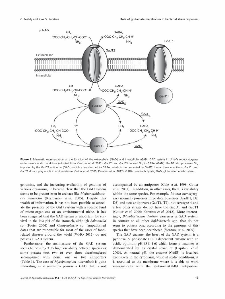
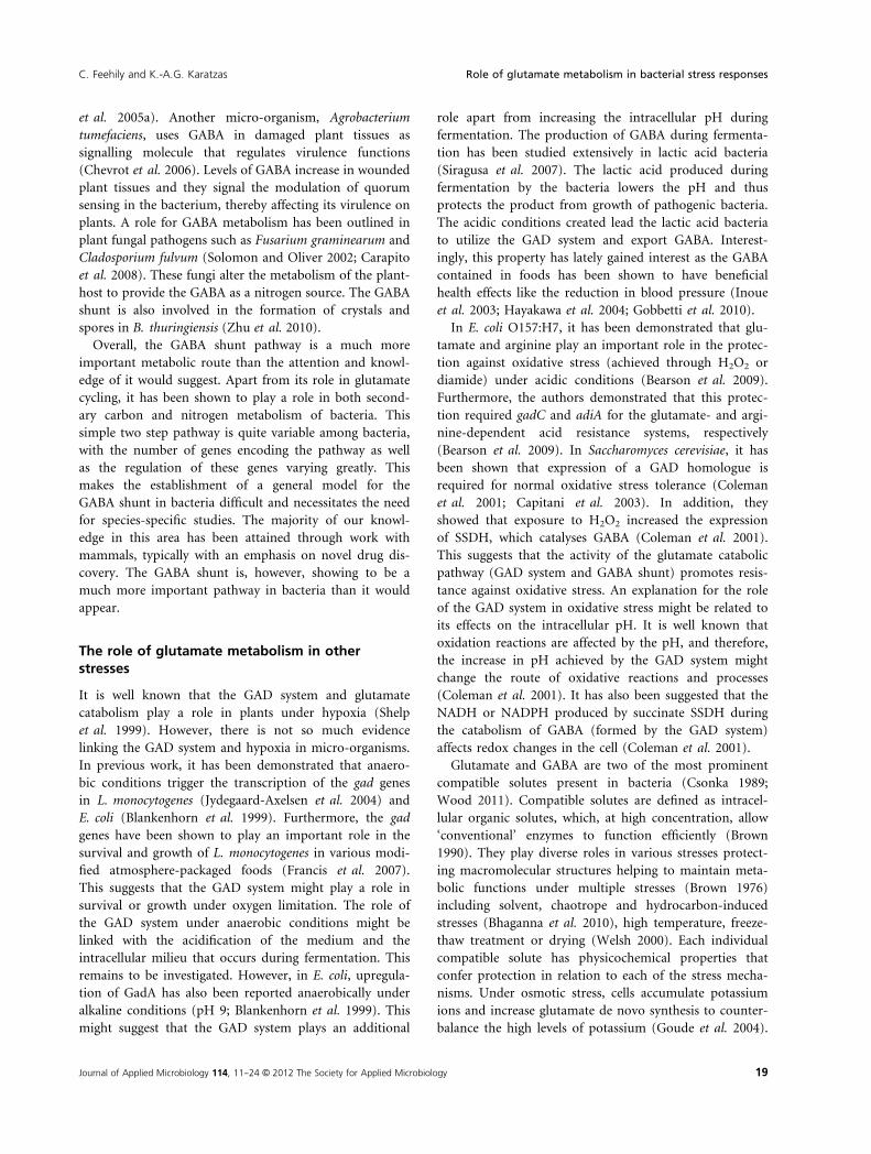
Figure 1 Schematic representation of the function of the extracellular (GADi) and intracellular (GADi) GAD system in Listeria monocytogenes
under severe acidic conditions (adopted from Karatzas et al. 2012). GadD2 and GadD3 convert Glti to GABAi (GADi). GadD2 also processes Glteimported by the GadT2 antiporter (GADe) which is transformed to GABA, which is then exported by GadT2. Under these conditions, GadD1 and
GadT1 do not play a role in acid resistance (Cotter et al. 2005; Karatzas et al. 2012). GABA, c-aminobutyrate; GAD, glutamate decarboxylase.
Journal of Applied Microbiology 114, 11--24 © 2012 The Society for Applied Microbiology 13
C. Feehily and K.-A.G. Karatzas Role of glutamate metabolism in bacterial stress responses
Page 4
Residues 13–57 of GadB act as hexamerization arms,
while residues 3–15 assume different conformations in
the neutral- and low-pH forms, playing a significant role
in the acid pH-driven association of GadB to the mem-
brane (Capitani et al. 2003). However, there is evidence
showing that a GAD from L. monocytogenes (probably
GadD3) deviates from this model, processing solely pools
of Glti and being unable to act synergistically with the
antiporters (Karatzas et al. 2012).
The function of the glutamate decarboxylase system
In various publications, it has been mentioned that the
conversion of glutamate to GABA is beneficial for the cell
because GABA is less acidic than glutamate (Tramonti
et al. 2002; Lei et al. 2011) due to the fact that the pI of
the former is close to 7�0 (neutral), while that of the lat-
ter is approximately 3�1 (acidic). However, how could
this affect their buffering capacity? The glutamate and
GABA have similar pKa values for the side chain carboxyl
group (pKa = 4�0) and the a-amino group (pKa = 10;
Foster 2004). The only difference lies in the fact that
glutamate possesses an a-carboxyl group that has a pKa
of 2�1. This suggests that a possible difference in the abil-
ity of these two compounds to remove protons would
occur in a pHe <3 that is lethal for most bacteria. In
addition, pHi values would never reach to such low
levels. Furthermore, in contrast to GABA, glutamate
incorporates protons at 2 < pH < 3 because of its addi-
tional a-carboxyl group suggesting that Glte is more ben-
eficial than equimolar levels of GABAe. This is
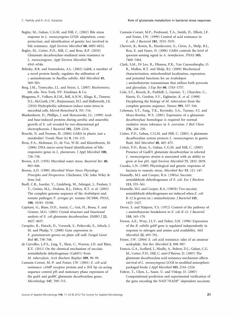
exemplified by a simple experiment where we constructed
the titration curves of 100 mmol l�1 aqueous solutions of
monosodium glutamate and GABA for 10% HCl w/w
(Fig. 3). It is clear that up to pH 3�0, both solutions have
the same buffering capacity. But below pH 3�0, the gluta-
mate solution has a higher buffering capacity as it
requires more than triple the amount of HCl required
compared to the GABA solution, to reduce the pH from
3�0 to 2�0 (Fig. 3). This suggests that it is more beneficial
for the cells not to convert glutamate to GABA. However,
it is evident that this conversion is beneficial because it
occurs in a wide range of pH values and it incorporates
one proton from the intracellular milieu in the molecule,
in the place of the a-carboxyl group (Fig. 1).
The futile proton hypothesis
Until now, there is little experimental work demonstrat-
ing the functionality of the commonly accepted model
for GAD system function and the fate of the protons. In
contrast, the futile proton hypothesis has been proposed
by various investigators in the field (Castanie-Cornet
et al. 1999; Storz and Hengge 2000; Tucker et al. 2002;
Waterman and Small 2003b), which proposes that if the
pKa of glutamate and GABA are taken in account, the
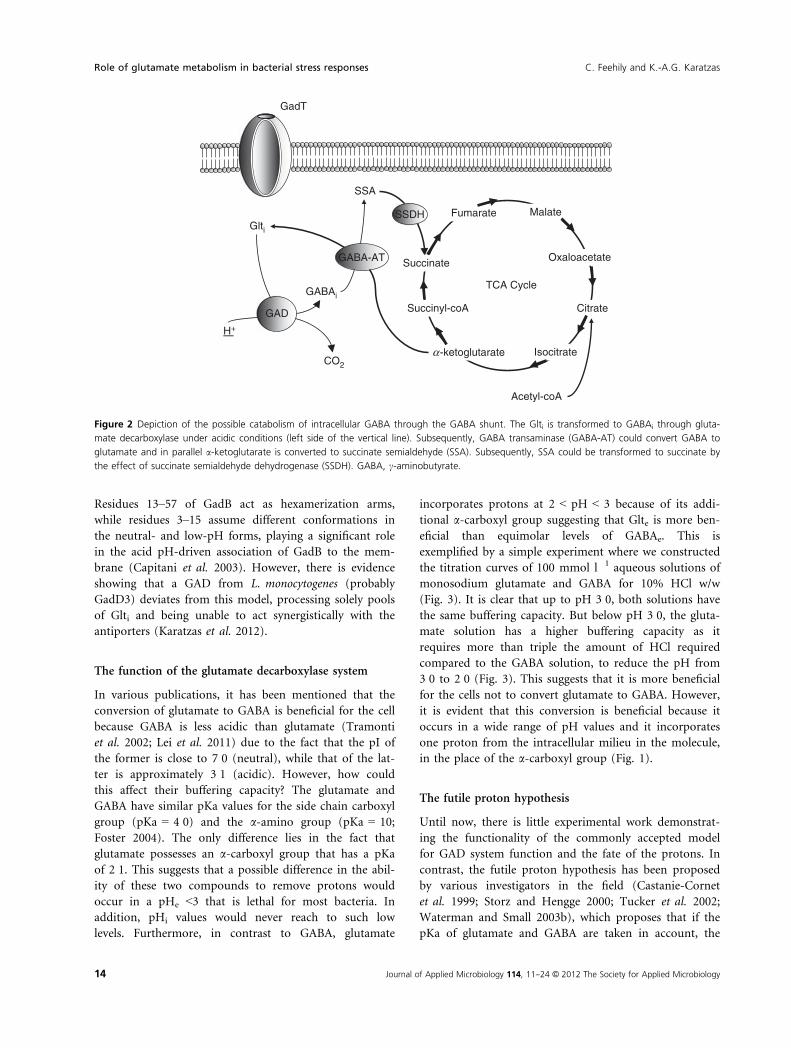
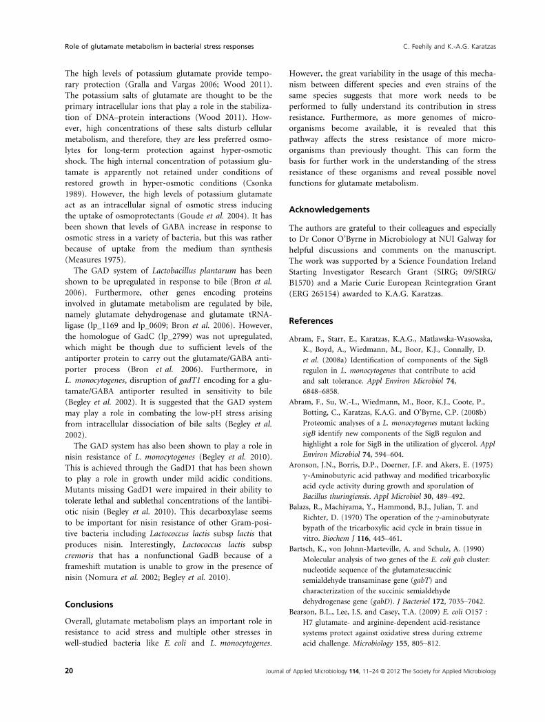
Figure 2 Depiction of the possible catabolism of intracellular GABA through the GABA shunt. The Glti is transformed to GABAi through gluta-
mate decarboxylase under acidic conditions (left side of the vertical line). Subsequently, GABA transaminase (GABA-AT) could convert GABA to
glutamate and in parallel a-ketoglutarate is converted to succinate semialdehyde (SSA). Subsequently, SSA could be transformed to succinate by
the effect of succinate semialdehyde dehydrogenase (SSDH). GABA, c-aminobutyrate.
14 Journal of Applied Microbiology 114, 11--24 © 2012 The Society for Applied Microbiology
Role of glutamate metabolism in bacterial stress responses C. Feehily and K.-A.G. Karatzas
Page 5
GAD system is unable to remove intracellular protons
under severe acid stress and the acid resistance might be
through an unknown mechanism. A careful investigation
into the pKa of glutamate, GABA and the range of pH
values intracellularly and extracellularly leads us in two
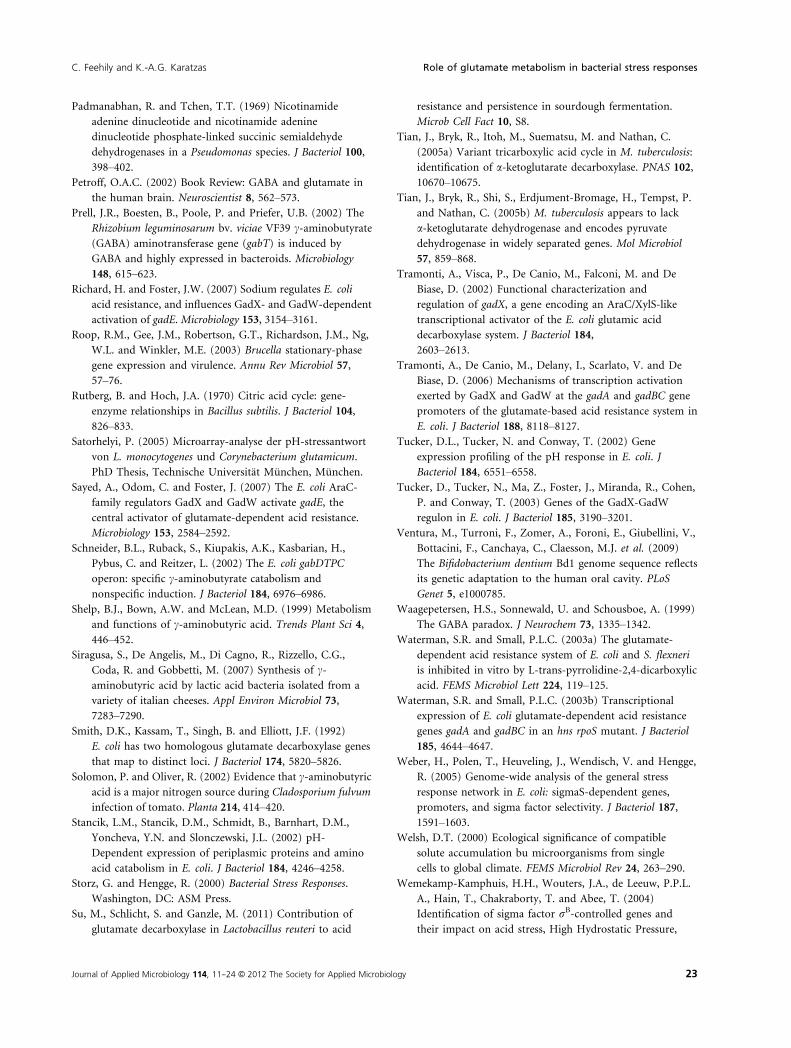
different possibilities that are demonstrated in Fig. 4. The
first scenario (A) refers to a mild pH stress where the
extracellular pH is >4�5. In this case, Glte is mainly
deprotonated and as such is imported in the cell, where
the decarboxylation of glutamate removes one intracellu-
lar proton (H+*). Subsequently, the GABA carries out
this proton, and thus, there is a benefit for the cell. How-
ever, in the second scenario (B) where the pH is very low
(e.g. 3�5), the chain carboxyl group of glutamate is
mainly protonated. This means that the antiporter
imports the glutamate in a protonated state, and once
intracellularly, this proton (H+) is released in the intracel-
lular milieu that counteracts the benefit from the proton
consumed by the decarboxylation process. The latter sce-
nario supports the futile proton hypothesis. However, the
GAD system has an obvious increase in acid resistance
probably through an unknown mechanism. It has been
suggested that this mechanism is a pathway for endoge-
nous glutamate generation or regeneration coupled with
the GAD system (Castanie-Cornet et al. 1999; Tucker
et al. 2002). The intracellularly generated glutamate might
not introduce any protons in the intracellular milieu, and
therefore, its decarboxylation will remove protons with a
net gain for the cell. A possible candidate pathway that
could generate intracellular glutamate (Glti) might
involve a glutaminase that converts glutamine to gluta-
mate (Castanie-Cornet et al. 1999; Tucker et al. 2002;
Waterman and Small 2003b). Glutamine’s only carboxyl
group has a pKa of 2, and it would be deprotonated
under normal acidic conditions (pH >2). Therefore, itsimport would not introduce any protons in the intracel-
lular milieu, and the glutamate decarboxylation would
result in a net loss of protons and a benefit for the cell.
Another alternative pathway could be the GABA shunt
that leads to the regeneration of intracellular glutamate
by the action of GABA-AT (Fig. 2).
The intracellular glutamate decarboxylase system (GADi)
All of the above suggest the conversion of intracellular
glutamate to GABA by the GAD system. Such a process
would also remove intracellular protons from the cell
under acidic conditions. Some workers have previously
demonstrated the presence of GABAi in E. coli (Castanie-
Cornet et al. 1999), Shigella flexneri (Waterman and
Small 2003b) and L. monocytogenes (Karatzas et al. 2010).
However, the significance of this finding has been over-
looked. Until now, all studies on the GAD system assume
that the exported GABAe represents the total GAD activ-
ity. The antiporter works in such a way that, for every
molecule of glutamate that is imported, one molecule of
GABA is exported. Therefore, the GAD system through
the utilization of Glte should not be able to affect the lev-
els of Glti and GABAi. This suggests that GABAi derives
directly through the decarboxylation of Glti under acidic
conditions and that the GAD system utilizes Glti (Fig. 1).
This is supported by data showing that under acidic con-
ditions, mechanisms that increase the levels of Glti are
upregulated. In Streptococcus mutans, it has been demon-
strated that the import of glutamate through the
GlnQHMP transporter increases acid resistance (Krastel
et al. 2010), while in L. monocytogenes, the most highly
acid-induced genes are coding for a putative glutamate
synthase (lmo1734) and a putative ABC transporter of
glutamine (lmo1738 and lmo1740) a glutamate precursor
(Satorhelyi 2005). Because the utilization of the Glte and
Glti are two independent processes, we have previously
proposed the division of the GAD system in extracellular
(GADe) and intracellular (GADi; Karatzas et al. 2012). At
first glance, GADi does not seem to be efficient because
of the limited availability of the Glti substrate compared
to the vast levels of Glte. However, GADi plays a signifi-
cant role in the acid resistance of L. monocytogenes
removing in an average between strains, a quarter of the
protons removed by the overall GAD system (Karatzas
et al. 2012). Furthermore, EGD-e, the most studied strain
of L. monocytogenes, appears to have a defective GADe
unable to export GABA, although it has an active GADi,
Figure 3 Titration curves of 100 mmol l�1 aqueous solutions of
monosodium glutamate (MSG) and c-aminobutyrate (GABA) for a
solution of 10% HCl w/w. (▲) 100 mmol l�1 GABA and
(□) 100 mmol l�1 MSG.
Journal of Applied Microbiology 114, 11--24 © 2012 The Society for Applied Microbiology 15
C. Feehily and K.-A.G. Karatzas Role of glutamate metabolism in bacterial stress responses
Page 6
accumulating GABAi under acidic conditions (Karatzas
et al. 2012). The effect of GADi in acid resistance might
also explain the function of the GAD system in
Myco. tuberculosis that possesses a GadA but no
glutamate/GABA antiporters (Cole et al. 1998; Cotter
et al. 2001). The important role of the GADi plays in
acid resistance remains to be elucidated in other micro-
organisms.
Regulation of the glutamate decarboxylase system
The regulation of the GAD system is complex and it has
been studied extensively in E. coli. In this organism, RpoS
activates gadY, whose small-RNA (sRNA) product stabi-
lizes gadX (Sayed et al. 2007). This increases the GadX
levels, which together with GadW binds and activates the
gadA and gadBC promoters in vitro (Tramonti et al.
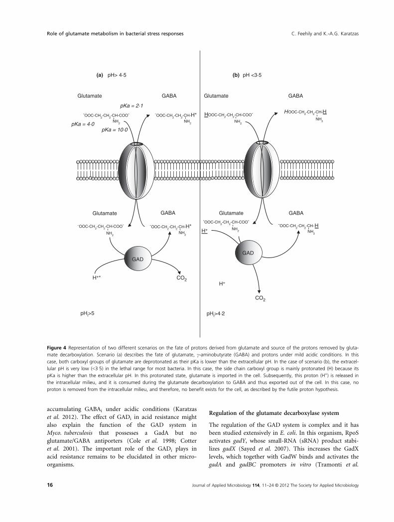
Figure 4 Representation of two different scenarios on the fate of protons derived from glutamate and source of the protons removed by gluta-
mate decarboxylation. Scenario (a) describes the fate of glutamate, c-aminobutyrate (GABA) and protons under mild acidic conditions. In this
case, both carboxyl groups of glutamate are deprotonated as their pKa is lower than the extracellular pH. In the case of scenario (b), the extracel-
lular pH is very low (<3�5) in the lethal range for most bacteria. In this case, the side chain carboxyl group is mainly protonated (H) because its
pKa is higher than the extracellular pH. In this protonated state, glutamate is imported in the cell. Subsequently, this proton (H+) is released in
the intracellular milieu, and it is consumed during the glutamate decarboxylation to GABA and thus exported out of the cell. In this case, no
proton is removed from the intracellular milieu, and therefore, no benefit exists for the cell, as described by the futile proton hypothesis.
16 Journal of Applied Microbiology 114, 11--24 © 2012 The Society for Applied Microbiology
Role of glutamate metabolism in bacterial stress responses C. Feehily and K.-A.G. Karatzas
Page 7
2006). However, in vivo, the latter process requires gadE
(Gong et al. 2004), which is activated by GadX and
GadW (Sayed et al. 2007), and in turn, activates gadA
and gadBC (Tucker et al. 2003; Hommais et al. 2004;
Weber et al. 2005). Similarly to the architecture of the
GAD system, its regulation is subject to variation. In
L. monocytogenes, all regulatory elements from E. coli
(gadE, gadY, gadX, gadW) have not been identified, while
upregulation of gadT2D2 and gadD3 but not gadD1T1 is
SigB-dependent and takes place during entrance in the
stationary phase (Cotter et al. 2001; Wemekamp-
Kamphuis et al. 2004; O’Byrne and Karatzas 2008). How-
ever, exposure of stationary phase cells of L. monocytoge-
nes 10403S to acidic conditions upregulates gadD1T1 and
gadD3, but not gadT2D2 operon. However, this is pecu-
liar as this operon plays an important role under severe
acidic conditions (Karatzas et al. 2010).
Furthermore, various compounds and conditions seem
to activate the GAD system. Apart from acidic condi-
tions, it has been demonstrated that sodium (Richard
and Foster 2007) and polyamines (Jung and Kim 2003)
lead to the upregulation of the GAD system in E. coli. In
both E. coli (Blankenhorn et al. 1999) and L. monocytoge-
nes (Jydegaard-Axelsen et al. 2004), hypoxia has been
shown to upregulate the GAD system. Interestingly, the
GAD system functionality is variable between different
bacteria and probably depends on the environmental
niches they are present. For example, although the GAD
system of E. coli is functional in minimal media (Casta-
nie-Cornet and Foster 2001), in L. monocytogenes, the
system is nonfunctional despite the presence of glutamate
(Karatzas et al. 2010). Variability exists also between
strains of the same species as L. monocytogenes LO28 uti-
lizes the GAD system in rich media like TSBY, but not in
BHI although the 10403S strain utilizes the GAD system
in both media efficiently (Karatzas et al. 2012).
The c-aminobutyrate shunt pathway
The conversion of glutamate to GABA by GAD is the
first step of the GABA shunt pathway. However, in bacte-
ria where most of the GABA is exported by the GAD
antiporter, the GABA shunt deals with the steps that
follow the generation of GABA. The accumulation of
intracellular GABA under acidic conditions suggests that
its removal should involve a catabolic pathway. This
could be assumed as the glutamate/GABA antiporters are
only able to remove quantities of GABA that are equimo-
lar to those of glutamate they import, leaving the excess
in the cell. Therefore, the first enzyme in the GABA shunt
is a GABA-AT (Zhu et al. 2010). This involves the revers-
ible conversion of GABA to SSA, where the amino group
of GABA is donated to an a-ketoglutarate molecule. The
by-product of this reaction is glutamate (Fig. 2). It has
been shown that plants can use pyruvate and glyoxylate
as alternative amino group acceptors (Bouche and
Fromm 2004; Clark et al. 2009); however, bacteria appear
to only have a-ketoglutarate-dependent GABA-AT activ-
ity, with the exception of some Rhizobium spp. (Prell
et al. 2002). The sole reliance on a-ketoglutarate as the
receiving oxoacid is also the case for GABA-ATs present
in mammals (Shelp et al. 1999). The importance of this
enzyme in bacteria is often highlighted by the presence of
more than one gene coding a functional GABA-AT. Stud-
ies with E. coli and Pseudomonas syringae have identified
the possession of up to three genes encoding functional
GABA-ATs (Buell et al. 2003; Kurihara et al. 2010). In
E. coli, it appears that induction of GabT, the primary
GABA-AT, occurs at pH 9�0 (Stancik et al. 2002) suggest-
ing that this pathway may be more active at alkaline pH
conditions.
The second enzyme in the pathway is a SSDH (Zhu
et al. 2010). This irreversible step involves the oxidation
of SSA to succinate with the formation of CO2. Similarly
to GABA-AT, bacteria can possess up to three SSDH
encoding genes (Buell et al. 2003). Each SSDH can pos-
sess different characteristics and cofactor specificities. For
example, in E. coli, there are currently two described
SSDH enzymes, GabD and YneI; the former is dependent
on NADP+, and YneI preferentially utilizes NAD+
(Donnelly and Cooper 1981b; Fuhrer et al. 2007). YneI
can, however, use NADP+, but the activity utilizing this
cofactor is only 15% (Donnelly and Cooper 1981a). The
presence of this varying NAD(P)+ dependency has also
been well described in several species of Pseudomonas (Ja-
koby and Scott 1959; Padmanabhan and Tchen 1969), as
well as both NADP+- and NAD+-dependent SSDH
activity shown in Bacillus thuringiensis (Zhu et al. 2010).
Partially purified SSDH from plants shows an optimal
alkaline pH of 9 (Shelp et al. 1999). This correlates well
with the optimal pH for the GABA-AT if the two
enzymes are to work together in a metabolic pathway.
Metabolism through the GABA shunt is an important
source of nitrogen for bacteria. Arginine, ornithine, agm-
atine and putrescine are all used as nitrogen sources by
E. coli (Schneider et al. 2002). Arginine, ornithine and
agmatine are first converted to putrescine, and the path-
way subsequently converts putrescine to GABA that is
then catabolized via the GABA shunt. In the absence of
either the gabT or gabD genes, GABA-AT and SSDH
activity remain. This activity is induced through the use
of putrescine as a sole nitrogen source (Kurihara et al.
2010). Furthermore, puuE, a gene forming an operon
with other members of the putrescine metabolic pathway,
was shown to act as a secondary GABA-AT (Fuhrer et al.
2007). It is interesting to note that none of the other
Journal of Applied Microbiology 114, 11--24 © 2012 The Society for Applied Microbiology 17
C. Feehily and K.-A.G. Karatzas Role of glutamate metabolism in bacterial stress responses
Page 8
genes in the puu gene cluster encode an SSDH; however,
YneI, which has been predicted as a secondary SSDH
(Kurihara et al. 2010), is induced along with PuuE by
putrescine (Zaboura and Halpern 1978). Despite this evi-
dence for the role of the GABA shunt in nitrogen metab-
olism, E. coli cannot use GABA as a sole source of
nitrogen (Belitsky and Sonenshein 2002). The reasons for
this are unclear, as E. coli does possess a transporter
capable of transporting GABA. Bacillus subtilis has also
been shown to utilize GABA; however, unlike E. coli,
B. subtilis cannot catabolize putrescine or generate GABA
through GAD because of the absence of any GAD encod-
ing genes (Dover and Halpern 1972). Therefore, it
imports extracellular GABA via a GABA permease gapP
(Ferson et al. 1996). This pathway can serve as the sole
nitrogen source for B. subtilis. Prell et al. (2002) sug-
gested that the pathway may be used in nitrogen meta-
bolism for bacteroides, in particular Rhizobium
leguminosarum. Some atmospheric nitrogen that is fixed
to ammonium during its symbiotic relationship with
legume nodules is retained by the bacteria. Results show
that GabT is specifically induced by the bacteroid in the
nodule state, indicating the enzyme may be involved in
catabolizing the glutamate derived from ammonium
(Prell et al. 2002).
The regulation of the GABA shunt pathway is a subject
that little is known about. Much of the initial work inves-
tigating this pathway was performed in E. coli. It was
observed that the pathway is regulated by a form of
catabolite repression (Dover and Halpern 1972). Further
work has since localized the genes to a region called the
gab cluster, which contains the four genes gabT, gabD,
gabP and gabC (Metzer et al. 1979; Bartsch et al. 1990).
Transcriptional control of these genes is complex and
involves the csiD-ygaF-gabDTP regulon. This operon is
reported to be controlled by the three promoters csiDp,
gabDp1 and gabDp2 (Metzner et al. 2004). The first two
promoters are rS-dependent, while the third is likely to
be induced under limited nitrogen availability by Nac/r70
(Metzner et al. 2004). More recent work with B. thuringi-
ensis (Zhu et al. 2010) identified the role of a sigma fac-
tor, r54, in controlling the GABA shunt. This regulates a
transcriptional activator, GabR, which itself is also auto-
regulated. The complete mechanism for regulation has
not yet been elucidated in these bacteria; however, the
gab cluster in B. thuringiensis is a good example of how
difficult is to make comparisons and generalizations on
GABA shunt regulation, even between species of the same
genus. Both gabD and gabT form a GABA-inducible
operon in B. subtilis (Belitsky and Sonenshein 2002);
however, these two genes are individually regulated in
B. thuringiensis by GabR (Zhu et al. 2010). The involve-
ment of ATP as a noncompetitive inhibitor of Arabidopsis
thaliana SSDH increases the ways by which the GABA
shunt in bacteria could be regulated. This implies that
the energy status of the cell itself could be involved in the
regulation of the pathway (de Carvalho et al. 2011).
While much of the focus of the GABA shunt research
has been on its role in carbon and nitrogen metabolism,
there is ground to suggest that it plays a much broader
role in bacteria and has a significant impact in terms of
both survival under environmental stresses and towards
their ability to cause disease. As discussed earlier, the
GABA shunt can operate as an alternative pathway to
certain steps of the TCA cycle, providing succinate. Many
bacteria including L. monocytogenes, Myco. tuberculosis
and B. thuringiensis lack a complete set of genes required
for the TCA cycle (Glaser et al. 2001; Tian et al. 2005a,
b). In fact it has been demonstrated that the SSDHs of
Myco. tuberculosis along with an a-ketoglutarate decar-
boxylase can lead to the generation of succinate in a
pathway which is required for normal growth of the bac-
teria (Metzner et al. 2004). While Myco. tuberculosis does
not use the first stage of the GABA shunt, it does indicate
that shunt-derived succinate could be of beneficial value
to bacteria. Furthermore, the TCA cycle has been pro-
posed as an important process in the formation of spores
in Bacillus species (Rutberg and Hoch 1970); however,
Aronson et al. (1975) describe that the GABA shunt can
successfully complement the loss of a-ketoglutaratedehydrogenase in this process (Tian et al. 2005a).
As the gab gene cluster in E. coli is under the control
the multistress response factor rS, it is thought that the
GABA shunt may play a role in stress response (Metzner
et al. 2004). The mechanism of its involvement is
unclear; however, it has been suggested that the control
of the GABA shunt may affect the glutamate levels that
are seen to increase in response to osmotic stress
(Metzner et al. 2004). This may well be the case as the
first step in the pathway generates glutamate. Further-
more, a putative SSDH in L. monocytogenes, namely
Lmo0913, has been shown to be induced in response to
NaCl (Abram et al. 2008b) that would be required to
remove the toxic SSA accumulated by the generation of
glutamate in this manner. The GABA shunt may also play
a role in acid tolerance. The antiport-independent accumu-
lation of GABA in response to acid by L. monocytogenes
(Karatzas et al. 2010) would necessitate the catabolism of
this metabolite. In fact deletion of lmo0913 in this bacte-
rium results in an acid-sensitive strain (Abram et al.
2008a), highlighting the potential link between acid stress
survival and the GABA shunt.
A further, yet understudied role for the pathway has
been seen to link GABA metabolism and virulence. A
mutation in GABA-AT has been reported to reduce bac-
terial virulence in the plant pathogen Ps. syringae (Tian
18 Journal of Applied Microbiology 114, 11--24 © 2012 The Society for Applied Microbiology
Role of glutamate metabolism in bacterial stress responses C. Feehily and K.-A.G. Karatzas
Page 9
et al. 2005a). Another micro-organism, Agrobacterium
tumefaciens, uses GABA in damaged plant tissues as
signalling molecule that regulates virulence functions
(Chevrot et al. 2006). Levels of GABA increase in wounded
plant tissues and they signal the modulation of quorum
sensing in the bacterium, thereby affecting its virulence on
plants. A role for GABA metabolism has been outlined in
plant fungal pathogens such as Fusarium graminearum and
Cladosporium fulvum (Solomon and Oliver 2002; Carapito
et al. 2008). These fungi alter the metabolism of the plant-
host to provide the GABA as a nitrogen source. The GABA
shunt is also involved in the formation of crystals and
spores in B. thuringiensis (Zhu et al. 2010).
Overall, the GABA shunt pathway is a much more
important metabolic route than the attention and knowl-
edge of it would suggest. Apart from its role in glutamate
cycling, it has been shown to play a role in both second-
ary carbon and nitrogen metabolism of bacteria. This
simple two step pathway is quite variable among bacteria,
with the number of genes encoding the pathway as well
as the regulation of these genes varying greatly. This
makes the establishment of a general model for the
GABA shunt in bacteria difficult and necessitates the need
for species-specific studies. The majority of our knowl-
edge in this area has been attained through work with
mammals, typically with an emphasis on novel drug dis-
covery. The GABA shunt is, however, showing to be a
much more important pathway in bacteria than it would
appear.
The role of glutamate metabolism in otherstresses
It is well known that the GAD system and glutamate
catabolism play a role in plants under hypoxia (Shelp
et al. 1999). However, there is not so much evidence
linking the GAD system and hypoxia in micro-organisms.
In previous work, it has been demonstrated that anaero-
bic conditions trigger the transcription of the gad genes
in L. monocytogenes (Jydegaard-Axelsen et al. 2004) and
E. coli (Blankenhorn et al. 1999). Furthermore, the gad
genes have been shown to play an important role in the
survival and growth of L. monocytogenes in various modi-
fied atmosphere-packaged foods (Francis et al. 2007).
This suggests that the GAD system might play a role in
survival or growth under oxygen limitation. The role of
the GAD system under anaerobic conditions might be
linked with the acidification of the medium and the
intracellular milieu that occurs during fermentation. This
remains to be investigated. However, in E. coli, upregula-
tion of GadA has also been reported anaerobically under
alkaline conditions (pH 9; Blankenhorn et al. 1999). This
might suggest that the GAD system plays an additional
role apart from increasing the intracellular pH during
fermentation. The production of GABA during fermenta-
tion has been studied extensively in lactic acid bacteria
(Siragusa et al. 2007). The lactic acid produced during
fermentation by the bacteria lowers the pH and thus
protects the product from growth of pathogenic bacteria.
The acidic conditions created lead the lactic acid bacteria
to utilize the GAD system and export GABA. Interest-
ingly, this property has lately gained interest as the GABA
contained in foods has been shown to have beneficial
health effects like the reduction in blood pressure (Inoue
et al. 2003; Hayakawa et al. 2004; Gobbetti et al. 2010).
In E. coli O157:H7, it has been demonstrated that glu-
tamate and arginine play an important role in the protec-
tion against oxidative stress (achieved through H2O2 or
diamide) under acidic conditions (Bearson et al. 2009).
Furthermore, the authors demonstrated that this protec-
tion required gadC and adiA for the glutamate- and argi-
nine-dependent acid resistance systems, respectively
(Bearson et al. 2009). In Saccharomyces cerevisiae, it has
been shown that expression of a GAD homologue is
required for normal oxidative stress tolerance (Coleman
et al. 2001; Capitani et al. 2003). In addition, they
showed that exposure to H2O2 increased the expression
of SSDH, which catalyses GABA (Coleman et al. 2001).
This suggests that the activity of the glutamate catabolic
pathway (GAD system and GABA shunt) promotes resis-
tance against oxidative stress. An explanation for the role
of the GAD system in oxidative stress might be related to
its effects on the intracellular pH. It is well known that
oxidation reactions are affected by the pH, and therefore,
the increase in pH achieved by the GAD system might
change the route of oxidative reactions and processes
(Coleman et al. 2001). It has also been suggested that the
NADH or NADPH produced by succinate SSDH during
the catabolism of GABA (formed by the GAD system)
affects redox changes in the cell (Coleman et al. 2001).
Glutamate and GABA are two of the most prominent
compatible solutes present in bacteria (Csonka 1989;
Wood 2011). Compatible solutes are defined as intracel-
lular organic solutes, which, at high concentration, allow
‘conventional’ enzymes to function efficiently (Brown
1990). They play diverse roles in various stresses protect-
ing macromolecular structures helping to maintain meta-
bolic functions under multiple stresses (Brown 1976)
including solvent, chaotrope and hydrocarbon-induced
stresses (Bhaganna et al. 2010), high temperature, freeze-
thaw treatment or drying (Welsh 2000). Each individual
compatible solute has physicochemical properties that
confer protection in relation to each of the stress mecha-
nisms. Under osmotic stress, cells accumulate potassium
ions and increase glutamate de novo synthesis to counter-
balance the high levels of potassium (Goude et al. 2004).
Journal of Applied Microbiology 114, 11--24 © 2012 The Society for Applied Microbiology 19
C. Feehily and K.-A.G. Karatzas Role of glutamate metabolism in bacterial stress responses
Page 10
The high levels of potassium glutamate provide tempo-
rary protection (Gralla and Vargas 2006; Wood 2011).
The potassium salts of glutamate are thought to be the
primary intracellular ions that play a role in the stabiliza-
tion of DNA–protein interactions (Wood 2011). How-
ever, high concentrations of these salts disturb cellular
metabolism, and therefore, they are less preferred osmo-
lytes for long-term protection against hyper-osmotic
shock. The high internal concentration of potassium glu-
tamate is apparently not retained under conditions of
restored growth in hyper-osmotic conditions (Csonka
1989). However, the high levels of potassium glutamate
act as an intracellular signal of osmotic stress inducing
the uptake of osmoprotectants (Goude et al. 2004). It has
been shown that levels of GABA increase in response to
osmotic stress in a variety of bacteria, but this was rather
because of uptake from the medium than synthesis
(Measures 1975).
The GAD system of Lactobacillus plantarum has been
shown to be upregulated in response to bile (Bron et al.
2006). Furthermore, other genes encoding proteins
involved in glutamate metabolism are regulated by bile,
namely glutamate dehydrogenase and glutamate tRNA-
ligase (lp_1169 and lp_0609; Bron et al. 2006). However,
the homologue of GadC (lp_2799) was not upregulated,
which might be though due to sufficient levels of the
antiporter protein to carry out the glutamate/GABA anti-
porter process (Bron et al. 2006). Furthermore, in
L. monocytogenes, disruption of gadT1 encoding for a glu-
tamate/GABA antiporter resulted in sensitivity to bile
(Begley et al. 2002). It is suggested that the GAD system
may play a role in combating the low-pH stress arising
from intracellular dissociation of bile salts (Begley et al.
2002).
The GAD system has also been shown to play a role in
nisin resistance of L. monocytogenes (Begley et al. 2010).
This is achieved through the GadD1 that has been shown
to play a role in growth under mild acidic conditions.
Mutants missing GadD1 were impaired in their ability to
tolerate lethal and sublethal concentrations of the lantibi-
otic nisin (Begley et al. 2010). This decarboxylase seems
to be important for nisin resistance of other Gram-posi-
tive bacteria including Lactococcus lactis subsp lactis that
produces nisin. Interestingly, Lactococcus lactis subsp
cremoris that has a nonfunctional GadB because of a
frameshift mutation is unable to grow in the presence of
nisin (Nomura et al. 2002; Begley et al. 2010).
Conclusions
Overall, glutamate metabolism plays an important role in
resistance to acid stress and multiple other stresses in
well-studied bacteria like E. coli and L. monocytogenes.
However, the great variability in the usage of this mecha-
nism between different species and even strains of the
same species suggests that more work needs to be
performed to fully understand its contribution in stress
resistance. Furthermore, as more genomes of micro-
organisms become available, it is revealed that this
pathway affects the stress resistance of more micro-
organisms than previously thought. This can form the
basis for further work in the understanding of the stress
resistance of these organisms and reveal possible novel
functions for glutamate metabolism.
Acknowledgements
The authors are grateful to their colleagues and especially
to Dr Conor O’Byrne in Microbiology at NUI Galway for
helpful discussions and comments on the manuscript.
The work was supported by a Science Foundation Ireland
Starting Investigator Research Grant (SIRG; 09/SIRG/
B1570) and a Marie Curie European Reintegration Grant
(ERG 265154) awarded to K.A.G. Karatzas.
References
Abram, F., Starr, E., Karatzas, K.A.G., Matlawska-Wasowska,
K., Boyd, A., Wiedmann, M., Boor, K.J., Connally, D.
et al. (2008a) Identification of components of the SigB
regulon in L. monocytogenes that contribute to acid
and salt tolerance. Appl Environ Microbiol 74,
6848–6858.
Abram, F., Su, W.-L., Wiedmann, M., Boor, K.J., Coote, P.,
Botting, C., Karatzas, K.A.G. and O’Byrne, C.P. (2008b)
Proteomic analyses of a L. monocytogenes mutant lacking
sigB identify new components of the SigB regulon and
highlight a role for SigB in the utilization of glycerol. Appl
Environ Microbiol 74, 594–604.
Aronson, J.N., Borris, D.P., Doerner, J.F. and Akers, E. (1975)
γ-Aminobutyric acid pathway and modified tricarboxylic
acid cycle activity during growth and sporulation of
Bacillus thuringiensis. Appl Microbiol 30, 489–492.
Balazs, R., Machiyama, Y., Hammond, B.J., Julian, T. and
Richter, D. (1970) The operation of the c-aminobutyrate
bypath of the tricarboxylic acid cycle in brain tissue in
vitro. Biochem J 116, 445–461.
Bartsch, K., von Johnn-Marteville, A. and Schulz, A. (1990)
Molecular analysis of two genes of the E. coli gab cluster:
nucleotide sequence of the glutamate:succinic
semialdehyde transaminase gene (gabT) and
characterization of the succinic semialdehyde
dehydrogenase gene (gabD). J Bacteriol 172, 7035–7042.
Bearson, B.L., Lee, I.S. and Casey, T.A. (2009) E. coli O157 :
H7 glutamate- and arginine-dependent acid-resistance
systems protect against oxidative stress during extreme
acid challenge. Microbiology 155, 805–812.
20 Journal of Applied Microbiology 114, 11--24 © 2012 The Society for Applied Microbiology
Role of glutamate metabolism in bacterial stress responses C. Feehily and K.-A.G. Karatzas
Page 11
Begley, M., Gahan, C.G.M. and Hill, C. (2002) Bile stress
response in L. monocytogenes LO28: adaptation, cross-
protection, and identification of genetic loci involved in
bile resistance. Appl Environ Microbiol 68, 6005–6012.
Begley, M., Cotter, P.D., Hill, C. and Ross, R.P. (2010)
Glutamate decarboxylase-mediated nisin resistance in
L. monocytogenes. Appl Environ Microbiol 76,
6541–6546.
Belitsky, B.R. and Sonenshein, A.L. (2002) GabR, a member of
a novel protein family, regulates the utilization of
c-aminobutyrate in Bacillus subtilis. Mol Microbiol 45,
569–583.
Berg, J.M., Tymoczko, J.L. and Stryer, L. (2007) Biochemistry,
6th edn. New York, NY: Friedman & Co.
Bhaganna, P., Volkers, R.J.M., Bell, A.N.W., Kluge, K., Timson,
D.J., McGrath, J.W., Ruijssenaars, H.J. and Hallsworth, J.E.
(2010) Hydrophobic substances induce water stress in
microbial cells.Microb Biotechnol 3, 701–716.
Blankenhorn, D., Phillips, J. and Slonczewski, J.L. (1999) Acid-
and base-induced proteins during aerobic and anaerobic
growth of E. coli revealed by two-dimensional gel
electrophoresis. J Bacteriol 181, 2209–2216.
Bouche, N. and Fromm, H. (2004) GABA in plants: just a
metabolite? Trends Plant Sci 9, 110–115.
Bron, P.A., Molenaar, D., de Vos, W.M. and Kleerebezem, M.
(2006) DNA micro-array-based identification of bile-
responsive genes in L. plantarum. J Appl Microbiol 100,
728–738.
Brown, A.D. (1976) Microbial water stress. Bacteriol Rev 40,
803–846.
Brown, A.D. (1990) Microbial Water Stress Physiology.
Principles and Perspectives. Chichester, UK: John Wiley &
Sons Ltd.
Buell, C.R., Joardar, V., Lindeberg, M., Selengut, J., Paulsen, I.
T., Gwinn, M.L., Dodson, R.J., Deboy, R.T. et al. (2003)
The complete genome sequence of the Arabidopsis and
tomato pathogen P. syringae pv. tomato DC3000. PNAS,
100, 10181–10186.
Capitani, G., Biase, D.D., Aurizi, C., Gut, H., Bossa, F. and
Grutter, M.G. (2003) Crystal structure and functional
analysis of E. coli glutamate decarboxylase. EMBO J 22,
4027–4037.
Carapito, R., Hatsch, D., Vorwerk, S., Petkovski, E., Jeltsch, J.
M. and Phalip, V. (2008) Gene expression in
F. graminearum grown on plant cell wall. Fungal Genet
Biol 45, 738–748.
de Carvalho, L.P.S., Ling, Y., Shen, C., Warren, J.D. and Rhee,
K.Y. (2011) On the chemical mechanism of succinic
semialdehyde dehydrogenase (GabD1) from
M. tuberculosis. Arch Biochem Biophys 509, 90–99.
Castanie-Cornet, M.-P. and Foster, J.W. (2001) E. coli acid
resistance: cAMP receptor protein and a 20A bp cis-acting
sequence control pH and stationary phase expression of
the gadA and gadBC glutamate decarboxylase genes.
Microbiology 147, 709–715.
Castanie-Cornet, M.P., Penfound, T.A., Smith, D., Elliott, J.F.
and Foster, J.W. (1999) Control of acid resistance in
E. coli. J Bacteriol 181, 3525–3535.
Chevrot, R., Rosen, R., Haudecoeur, E., Cirou, A., Shelp, B.J.,
Ron, E. and Faure, D. (2006) GABA controls the level of
quorum-sensing signal in A. tumefaciens. PNAS 103,
7460–7464.
Clark, S.M., Di Leo, R., Dhanoa, P.K., Van Cauwenberghe, O.
R., Mullen, R.T. and Shelp, B.J. (2009) Biochemical
characterization, mitochondrial localization, expression,
and potential functions for an Arabidopsis
c-aminobutyrate transaminase that utilizes both pyruvate
and glyoxylate. J Exp Bot 60, 1743–1757.
Cole, S.T., Brosch, R., Parkhill, J., Garnier, T., Churcher, C.,
Harris, D., Gordon, S.V., Eiglmeier, K. et al. (1998)
Deciphering the biology of M. tuberculosis from the
complete genome sequence. Nature 393, 537–544.
Coleman, S.T., Fang, T.K., Rovinsky, S.A., Turano, F.J. and
Moye-Rowley, W.S. (2001) Expression of a glutamate
decarboxylase homologue is required for normal
oxidative stress tolerance in S. cerevisiae. J Biol Chem
276, 244–250.
Cotter, P.D., Gahan, C.G.M. and Hill, C. (2001) A glutamate
decarboxylase system protects L. monocytogenes in gastric
fluid. Mol Microbiol 40, 465–475.
Cotter, P.D., Ryan, S., Gahan, C.G.M. and Hill, C. (2005)
Presence of GadD1 glutamate decarboxylase in selected
L. monocytogenes strains is associated with an ability to
grow at low pH. Appl Environ Microbiol 71, 2832–2839.
Csonka, L.N. (1989) Physiological and genetic responses of
bacteria to osmotic stress. Microbiol Rev 53, 121–147.
Donnelly, M.I. and Cooper, R.A. (1981a) Succinic
semialdehyde dehydrogenases of E. coli. Eur J Biochem
113, 555–561.
Donnelly, M.I. and Cooper, R.A. (1981b) Two succinic
semialdehyde dehydrogenases are induced when E. coli
K-12 Is grown on c-aminobutyrate. J Bacteriol 145,
1425–1427.
Dover, S. and Halpern, Y.S. (1972) Control of the pathway of
c-aminobutyrate breakdown in E. coli K-12. J Bacteriol
110, 165–170.
Ferson, A.E., Wray, J.L.V. and Fisher, S.H. (1996) Expression
of the B. subtilis gabP gene is regulated independently in
response to nitrogen and amino acid availability. Mol
Microbiol 22, 693–701.
Foster, J.W. (2004) E. coli acid resistance: tales of an amateur
acidophile. Nat Rev Microbiol 2, 898–907.
Francis, G.A., Scollard, J., Meally, A., Bolton, D.J., Gahan, C.G.
M., Cotter, P.D., Hill, C. and O’Beirne, D. (2007) The
glutamate decarboxylase acid resistance mechanism affects
survival of L. monocytogenes LO28 in modified atmosphere-
packaged foods. J Appl Microbiol 103, 2316–2324.
Fuhrer, T., Chen, L., Sauer, U. and Vitkup, D. (2007)
Computational prediction and experimental verification of
the gene encoding the NAD+/NADP+-dependent succinate
Journal of Applied Microbiology 114, 11--24 © 2012 The Society for Applied Microbiology 21
C. Feehily and K.-A.G. Karatzas Role of glutamate metabolism in bacterial stress responses
Page 12
semialdehyde dehydrogenase in E. coli. J Bacteriol 189,
8073–8078.
Glaser, P., Frangeul, L., Buchrieser, C., Rusniok, C., Amend,
A., Baquero, F., Berche, P., Bloecker, H. et al. (2001)
Comparative genomics of Listeria species. Science 294,
849–852.
Gobbetti, M., Cagno, R.D. and De Angelis, M. (2010)
Functional microorganisms for functional food quality.
Crit Rev Food Sci Nutr 50, 716–727.
Gong, S., Ma, Z. and Foster, J.W. (2004) The Era-like GTPase
TrmE conditionally activates gadE and glutamate-dependent
acid resistance in E. coli.Mol Microbiol 54, 948–961.
Goude, R., Renaud, S., Bonnassie, S., Bernard, T. and Blanco,
C. (2004) Glutamine, glutamate, and a-glucosylglycerateare the major osmotic solutes accumulated by Erwinia
chrysanthemi strain 3937. Appl Environ Microbiol 70,
6535–6541.
Gralla, J.D. and Vargas, D.R. (2006) Potassium glutamate as a
transcriptional inhibitor during bacterial osmoregulation.
EMBO J 25, 1515–1521.
Hayakawa, K., Kimura, M., Kasaha, K., Matsumoto, K.,
Sansawa, H. and Yamori, Y. (2004) Effect of a
c-aminobutyric acid-enriched dairy product on the blood
pressure of spontaneously hypertensive and normotensive
Wistar-Kyoto rats. Br J Nutr 92, 411–417.
Hommais, F., Krin, E., Coppee, J., Lacroix, C., Yeramian, E.,
Danchin, A. and Bertin, P. (2004) GadE (YhiE): a novel
activator involved in the response to acid environment in
E. coli. Microbiology 150, 61–72.
Inoue, K., Shirai, T., Ochiai, H., Kasao, M., Hayakawa, K.,
Kimura, M. and Sansawa, H. (2003) Blood-pressure-
lowering effect of a novel fermented milk containing
c-aminobutyric acid (GABA) in mild hypertensives. Eur J
Clin Nutr 57, 490–495.
Jakoby, W.B. and Scott, E.M. (1959) Aldehyde oxidation. III.
Succinic semialdehyde dehydrogenase. J Biol Chem 234,
937–940.
Jung, I.L. and Kim, I.G. (2003) Polyamines and glutamate
decarboxylase-based acid resistance in E. coli. J Biol Chem
278, 22846–22852.
Jydegaard-Axelsen, A.M., Hoiby, P.E., Holmstrom, K., Russell,
N. and Knochel, S. (2004) CO2 – and anaerobiosis-
induced changes in physiology and gene expression of
different L. monocytogenes strains. Appl Environ Microbiol
70, 4111–4117.
Karatzas, K.A.G., Valdramidis, V.P. and Wells-Bennik, M.H.J.
(2005) Contingency locus in ctsR of L. monocytogenes
Scott A: a strategy for occurrence of abundant
piezotolerant isolates within clonal populations. Appl
Environ Microbiol 71, 8390–8396.
Karatzas, K.A.G., Zervos, A., Tassou, C.C., Mallidis, C.G. and
Humphrey, T.J. (2007) Piezotolerant small-colony variants
with increased thermotolerance, antibiotic susceptibility,
and low invasiveness in a clonal Staphylococcus aureus
population. Appl Environ Microbiol 73, 1873–1881.
Karatzas, K.A.G., Hocking, P.M., Jørgensen, F., Mattick, K.,
Leach, S. and Humphrey, T.J. (2008) Effects of repeated
cycles of acid challenge and growth on the phenotype
and virulence of S. enterica. J Appl Microbiol 105,
1640–1648.
Karatzas, K.A.G., Brennan, O., Heavin, S., Morrissey, J. and
O’Byrne, C.P. (2010) Intracellular accumulation of high
levels of c-aminobutyrate by L. monocytogenes 10403S in
response to low pH: uncoupling of c-aminobutyrate
synthesis from efflux in a chemically defined medium.
Appl Environ Microbiol 76, 3529–3537.
Karatzas, K.A.G., Suur, L. and O’Byrne, C.P. (2012)
Characterisation of the intracellular-glutamate
decarboxylase system (GADi): analysis of its function,
transcription and role in the acid resistance of various
strains of L. monocytogenes. Appl Environ Microbiol 78,
3571–3579.
Kezmarsky, N.D., Xu, H., Graham, D.E. and White, R.H.
(2005) Identification and characterization of an L-tyrosine
decarboxylase in Methanocaldococcus jannaschii. Biochim
Biophys Acta 1722, 175–182.
Krastel, K., Senadheera, D.B., Mair, R., Downey, J.S.,
Goodman, S.D. and Cvitkovitch, D.G. (2010)
Characterization of a glutamate transporter operon,
glnQHMP, in Streptococcus mutans and its role in acid
tolerance. J Bacteriol 192, 984–993.
Kurihara, S., Kato, K., Asada, K., Kumagai, H. and Suzuki, H.
(2010) A putrescine-inducible pathway comprising PuuE-
YneI in which c-aminobutyrate is degraded into succinate
in E. coli K-12. J Bacteriol 192, 4582–4591.
Lei, G.-S., Syu, W.-J., Liang, P.-H., Chak, K.-F., Hu, W. and
Hu, S.-T. (2011) Repression of btuB gene transcription in
E. coli by the GadX protein. BMC Microbiol 11, 33.
Maras, B., Sweeney, G., Barra, D., Bossa, F. and John, R.A.
(1992) The amino acid sequence of glutamate
decarboxylase from E. coli. Eur J Biochem 204, 93–98.
Measures, J.C. (1975) Role of amino acids in osmoregulation
of non-halophilic bacteria. Nature 257, 398–400.
Metzer, E., Levitz, R. and Halpern, Y.S. (1979) Isolation and
properties of E. coli K-12 mutants impaired in the
utilization of c-aminobutyrate. J Bacteriol 137,
1111–1118.
Metzner, M., Germer, J. and Hengge, R. (2004) Multiple stress
signal integration in the regulation of the complex
rS-dependent csiD-ygaF-gabDTP operon in E. coli. Mol
Microbiol 51, 799–811.
Nomura, M., Kobayashi, M. and Okamoto, T. (2002) Rapid
PCR-based method which can determine both phenotype
and genotype of Lactococcus lactis subspecies. Appl Environ
Microbiol 68, 2209–2213.
O’Byrne, C.P. and Karatzas, K.A.G. (2008) The role of SigB in
the stress adaptations of L. monocytogenes: overlaps
between stress adaptation and virulence. In Adv Appl
Microbiol eds Allen, S., Laskin, I. and Geoffrey, M.G. 65,
pp. 115–140. New York: Academic Press.
22 Journal of Applied Microbiology 114, 11--24 © 2012 The Society for Applied Microbiology
Role of glutamate metabolism in bacterial stress responses C. Feehily and K.-A.G. Karatzas
Page 13
Padmanabhan, R. and Tchen, T.T. (1969) Nicotinamide
adenine dinucleotide and nicotinamide adenine
dinucleotide phosphate-linked succinic semialdehyde
dehydrogenases in a Pseudomonas species. J Bacteriol 100,
398–402.
Petroff, O.A.C. (2002) Book Review: GABA and glutamate in
the human brain. Neuroscientist 8, 562–573.
Prell, J.R., Boesten, B., Poole, P. and Priefer, U.B. (2002) The
Rhizobium leguminosarum bv. viciae VF39 c-aminobutyrate
(GABA) aminotransferase gene (gabT) is induced by
GABA and highly expressed in bacteroids. Microbiology
148, 615–623.
Richard, H. and Foster, J.W. (2007) Sodium regulates E. coli
acid resistance, and influences GadX- and GadW-dependent
activation of gadE. Microbiology 153, 3154–3161.
Roop, R.M., Gee, J.M., Robertson, G.T., Richardson, J.M., Ng,
W.L. and Winkler, M.E. (2003) Brucella stationary-phase
gene expression and virulence. Annu Rev Microbiol 57,
57–76.
Rutberg, B. and Hoch, J.A. (1970) Citric acid cycle: gene-
enzyme relationships in Bacillus subtilis. J Bacteriol 104,
826–833.
Satorhelyi, P. (2005) Microarray-analyse der pH-stressantwort
von L. monocytogenes und Corynebacterium glutamicum.
PhD Thesis, Technische Universitat Munchen, Munchen.
Sayed, A., Odom, C. and Foster, J. (2007) The E. coli AraC-
family regulators GadX and GadW activate gadE, the
central activator of glutamate-dependent acid resistance.
Microbiology 153, 2584–2592.
Schneider, B.L., Ruback, S., Kiupakis, A.K., Kasbarian, H.,
Pybus, C. and Reitzer, L. (2002) The E. coli gabDTPC
operon: specific c-aminobutyrate catabolism and
nonspecific induction. J Bacteriol 184, 6976–6986.
Shelp, B.J., Bown, A.W. and McLean, M.D. (1999) Metabolism
and functions of c-aminobutyric acid. Trends Plant Sci 4,
446–452.
Siragusa, S., De Angelis, M., Di Cagno, R., Rizzello, C.G.,
Coda, R. and Gobbetti, M. (2007) Synthesis of c-aminobutyric acid by lactic acid bacteria isolated from a
variety of italian cheeses. Appl Environ Microbiol 73,
7283–7290.
Smith, D.K., Kassam, T., Singh, B. and Elliott, J.F. (1992)
E. coli has two homologous glutamate decarboxylase genes
that map to distinct loci. J Bacteriol 174, 5820–5826.
Solomon, P. and Oliver, R. (2002) Evidence that c-aminobutyric
acid is a major nitrogen source during Cladosporium fulvum
infection of tomato. Planta 214, 414–420.
Stancik, L.M., Stancik, D.M., Schmidt, B., Barnhart, D.M.,
Yoncheva, Y.N. and Slonczewski, J.L. (2002) pH-
Dependent expression of periplasmic proteins and amino
acid catabolism in E. coli. J Bacteriol 184, 4246–4258.
Storz, G. and Hengge, R. (2000) Bacterial Stress Responses.
Washington, DC: ASM Press.
Su, M., Schlicht, S. and Ganzle, M. (2011) Contribution of
glutamate decarboxylase in Lactobacillus reuteri to acid
resistance and persistence in sourdough fermentation.
Microb Cell Fact 10, S8.
Tian, J., Bryk, R., Itoh, M., Suematsu, M. and Nathan, C.
(2005a) Variant tricarboxylic acid cycle in M. tuberculosis:
identification of a-ketoglutarate decarboxylase. PNAS 102,
10670–10675.
Tian, J., Bryk, R., Shi, S., Erdjument-Bromage, H., Tempst, P.
and Nathan, C. (2005b) M. tuberculosis appears to lack
a-ketoglutarate dehydrogenase and encodes pyruvate
dehydrogenase in widely separated genes. Mol Microbiol
57, 859–868.
Tramonti, A., Visca, P., De Canio, M., Falconi, M. and De
Biase, D. (2002) Functional characterization and
regulation of gadX, a gene encoding an AraC/XylS-like
transcriptional activator of the E. coli glutamic acid
decarboxylase system. J Bacteriol 184,
2603–2613.
Tramonti, A., De Canio, M., Delany, I., Scarlato, V. and De
Biase, D. (2006) Mechanisms of transcription activation
exerted by GadX and GadW at the gadA and gadBC gene
promoters of the glutamate-based acid resistance system in
E. coli. J Bacteriol 188, 8118–8127.
Tucker, D.L., Tucker, N. and Conway, T. (2002) Gene
expression profiling of the pH response in E. coli. J
Bacteriol 184, 6551–6558.
Tucker, D., Tucker, N., Ma, Z., Foster, J., Miranda, R., Cohen,
P. and Conway, T. (2003) Genes of the GadX-GadW
regulon in E. coli. J Bacteriol 185, 3190–3201.
Ventura, M., Turroni, F., Zomer, A., Foroni, E., Giubellini, V.,
Bottacini, F., Canchaya, C., Claesson, M.J. et al. (2009)
The Bifidobacterium dentium Bd1 genome sequence reflects
its genetic adaptation to the human oral cavity. PLoS
Genet 5, e1000785.
Waagepetersen, H.S., Sonnewald, U. and Schousboe, A. (1999)
The GABA paradox. J Neurochem 73, 1335–1342.
Waterman, S.R. and Small, P.L.C. (2003a) The glutamate-
dependent acid resistance system of E. coli and S. flexneri
is inhibited in vitro by L-trans-pyrrolidine-2,4-dicarboxylic
acid. FEMS Microbiol Lett 224, 119–125.
Waterman, S.R. and Small, P.L.C. (2003b) Transcriptional
expression of E. coli glutamate-dependent acid resistance
genes gadA and gadBC in an hns rpoS mutant. J Bacteriol
185, 4644–4647.
Weber, H., Polen, T., Heuveling, J., Wendisch, V. and Hengge,
R. (2005) Genome-wide analysis of the general stress
response network in E. coli: sigmaS-dependent genes,
promoters, and sigma factor selectivity. J Bacteriol 187,
1591–1603.
Welsh, D.T. (2000) Ecological significance of compatible
solute accumulation bu microorganisms from single
cells to global climate. FEMS Microbiol Rev 24, 263–290.
Wemekamp-Kamphuis, H.H., Wouters, J.A., de Leeuw, P.P.L.
A., Hain, T., Chakraborty, T. and Abee, T. (2004)
Identification of sigma factor rB-controlled genes and
their impact on acid stress, High Hydrostatic Pressure,
Journal of Applied Microbiology 114, 11--24 © 2012 The Society for Applied Microbiology 23
C. Feehily and K.-A.G. Karatzas Role of glutamate metabolism in bacterial stress responses
Page 14
and freeze survival in L. monocytogenes EGD-e. Appl
Environ Microbiol 70, 3457–3466.
WHO Campylobacter (2012) Fact sheet No 255 – October
2011. [Online] Available at: http://www.who.int/
mediacentre/factsheets/fs255/en/index.html. [Accessed 08
July 2012].
Wood, J.M. (2011) Bacterial osmoregulation: a paradigm for
the study of cellular homeostasis. Annu Rev Microbiol 65,
215–238.
Zaboura, M. and Halpern, Y.S. (1978) Regulation of
c-aminobutyric acid degradation in E. coli by
nitrogen metabolism enzymes. J Bacteriol 133,
447–451.
Zhu, L., Peng, Q., Song, F., Jiang, Y., Sun, C., Zhang, J. and
Huang, D. (2010) Structure and regulation of the gab gene
cluster, involved in the c-aminobutyric acid shunt, are
controlled by a r54 factor in Bacillus thuringiensis.
J Bacteriol 192, 346–355.
24 Journal of Applied Microbiology 114, 11--24 © 2012 The Society for Applied Microbiology
Role of glutamate metabolism in bacterial stress responses C. Feehily and K.-A.G. Karatzas