The Septum Pellucidum: Normal and Abnormal Mohammad Sarwar 1
From the radiographic perspective, the septum pellucidum is usually relegated to an ignominious position of providing information on the presence and size of a mass lesion by the degree of its dislocation from the midline. However, one must recognize that this thin structure forms an important link in the limbic system and therefore has great clinical importance. Now that MR imaging provides exquisite depiction of the CNS anatomy, it is appropriate to have a thorough knowledge of the anatomic, embryologic, pathologic, and clinical perspectives of the septum pellucidum. This review is intended to provide state-of-the-art information on all aspects of the septum pellucidum. The MR sequences illustrated in this review are T1-weighted, 400-800/20-30 (TR/TE), intermediateweighted , 1500, 2000/20,25 , and T2-weighted, 2000/80; all images were obtained at 1.5 T.
Anatomy
The septum pellucidum is a thin translucent (pellucidum = transparent) plate of two laminae that extends from the anterior part of the body, the genu, and the rostrum of the corpus callosum to the superior surface of the fornix (Figs. 1-3). Its anterior to posterior extent therefore is from the lamina terminalis to the splenium of the corpus callosum. It bridges the midline gap between the corpus callosum above and the fornix below (Figs. 4 and 5). Its width varies from 1 .5 to 3.0 mm [1] . It contains glial cells (Fig. 6), some scattered neurons [2], fiber bundles, and veins that connect with the choroid plexus veins . It is lined with ependyma on the ventricular side. There is no midline septum pellucidum, as the bodies of the lateral ventricles diverge posterior to the interventricular
foramina. The diverging medial wall of the body of each lateral ventricle is instead formed by the ependyma-lined alvei of hippocampi . These alvei form a linkage between the medial margins of the fornices and the inferior surface of the body of the corpus callosum.
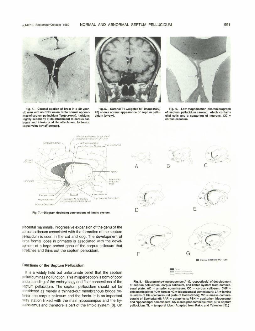
The septum pellucidum serves as an important relay station; its most anatomically and functionally important fiber connections are with the hippocampus and the hypothalamus and not with the primary olfactory structures (Fig . 7). It is linked with the hippocampus by precommissural fornix fibers, with preoptic and hypothalamic nuclei by the phylogenetically regressive median forebrain bundle of Broca that also relates the limbic with the olfactory system, with the amygdaloid nuclei by way of the striae terminalis , with the habenula and colliculi by way of the striae medullaris, and with the midbrain by its connection with the anterior fornix . It has a connection with the infracallosal precommissural primordium hippocampi through the paraterminal body.
Embryologic Considerations
The development of the septum pellucidum occurs from the primitive lamina terminalis (the "lamina reuniens of His" of the older literature) or the commissural plate, which forms the anterior wall of the telencephalic cavity. This development is intricately linked with that of the corpus callosum and the other two forebrain commissures, namely, the anterior and the hippocampal. This development begins by about 1 0-12 weeks of gestation, and the adult form is reached concurrently with that of the corpus callosum by 17 weeks of gestational age [3].
Received October 4, 1988; revision requested November 18, 1988; revision received December 20, 1988; accepted January 5, 1989. ' Department of Radiology, University of Illinois College of Medicine at Chicago, 1740 W. Taylor St., Chicago, IL 60612. Address reprint requests toM. Sarwar.
Fig. 3.-Sagittal and parasagittal perspective of limbic system shows relationship of septum pellucidum to neighboring structures.
The lamina terminalis is an unpaired rostral midline membrane that results from the closure of the anterior neuropore and fusion of the massive lateral plates (4]. This area marks the rostral wall of the prosencephalon. On the side of the lamina terminalis, the symmetric prosencephalic vesicle evaginates to later differentiate into cerebral hemispheres. The prosencephalon subdivides into telencephalon and diencephalon. The delimitation between the two parts is revealed on the external surface of the budding cerebral vesicle by telencephalic-diencephalic sulcus (sulcus hemisphericus). This sulcus has two parts: a dorsal medial part that corresponds to the velum interpositum and a basal medial portion that corresponds to a thickened part of the lamina terminalis , called the commissural plate. It is from this commissural plate that the corpus callosum, the anterior commissure, and the hippocampal commissure develop (Fig. 8). Because of the enormous growth of the corpus callosum in an arched dorsocaudal direction, the commissural plate between the anterior commissure, the hippocampal commissure, and the corpus callosum is progressively stretched and thinned to assume a membranous partition , called the septum pellucidum, between the lateral ventricles. The septum pellucidum therefore is a
Fig. 1.-Horizontal section of brain shows normal appearance of septum pellucidum (arrow).
Fig. 2.-Normal septum pellucidum (arrow) as seen on axial intermediate-weighted MR image (2000/20/ one excitation).
link between the corpus callosum and the fornix (5). It contains some neuronal elements and does not occur in lower animals.
From the embryologic considerations described above, it becomes apparent that the CSF space between the mesial walls of the cerebral hemispheres is the CSF contained in the interhemispheric fissure (6). Rakic and Yakovlev [6] consider the cavum septi pellucidi (CSP) to be a dorsally open pocket that is open at birth and in the neonate, the infant, and the young child . Later it is sealed by the rostrum of the corpus callosum (Figs. 9 and 1 0). The space is bordered above by the corpus callosum, behind by the fornix and the hippocampal commissure (psalterium), and anteriorly by the anterior commissure and rostrum of the corpus callosum; it is delimited from the interhemispheric fissure below by a thick membrane. The pocket remains open in rodents, carnivores, and rhesus monkeys.
Evolution of the Septum Pellucidum
A knowledge of the evolution of the septum pellucidum is helpful in understanding the CNS dysmorphology that occurs in cases of developmental anomalies of the septum pellucidum since there is a known tendency [8] for CNS malformations based on genetic defects to recapitulate phylogeny. In this regard, it is worth noting that the septum pellucidum is a progressive structure. As evolution proceeded, it became higher than wide; the reverse is true in lower primates. This is in contradistinction to the olfactory system, which is a regressive structure in the human.
The evolution of the septum pellucidum is inextricably tied to that of the corpus callosum. In the lamprey, the primordia of the cerebral hemispheres, manifested by the presence of bilateral vesicular outpouchings of the forebrain , are united by the lamina terminalis , which then acts as a pathway for the fibers of the anterior commissure, one of the earliest commissures to evolve phylogenetically (9) . The lamina terminalis delineates the anterior end of the brain in the lamprey. It is differentiated in all vertebrates. It is large and subdivided in most fishes [2). Concomitant with evolution of the corpus callosum, the septum pellucidum begins to be recognized in
AJNR:1 0, September/October 1989 NORMAL AND ABNORMAL SEPTUM PELLUCIDUM 991
' · I '
I
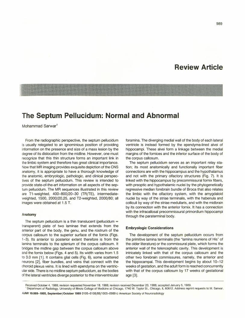
Fig. 4.-Coronal section of brain in a 50-yearold man with no CNS lesion. Note normal appearance of septum pellucidum (large arrow). It widens slightly superiorly at its attachment to corpus callosum and inferiorly at its attachment to fornix. Septal veins (small arrows).
Fig. 5.-Coronal T1-weighted MR image (600/ 20) shows normal appearance of septum pellucidum (arrow).
Fig. 6.-Low-magnification photomicrograph of septum pellucidum (arrow), which contains glial cells and a scattering of neurons. CC = corpus callosum.
L\11~111::; ,IIIQSl/tn
'.
Hypotl1alamus
Mamn111/ary oody
~-fedk11 and larerallongtrudmal smae and mdus1um gnseum
Antenor Nucleus -..__ lnrralammar Nuclei ___r- of Thalamus
Branches to neocorte>. and parahtppocampal gyrus
~·
Fig. 7.-Diagram depicting connections of limbic system.
placental mammals. Progressive expansion of the genu of the corpus callosum associated with the formation of the septum pellucidum is seen in the cat and dog. The development of large frontal lobes in primates is associated with the development of a large arched genu of the corpus callosum that stretches and thins out the septum pellucidum.
Functions of the Septum Pellucidum
It is a widely held but unfortunate belief that the septum pellucidum has no function . This misperception is born of poor understanding of the embryology and fiber connections of the septum pellucidum. The septum pellucidum should not be considered as merely a thinned-out membranous bridge between the corpus callosum and the fornix . It is an important relay station linked with the main hippocampus and the hypothalamus and therefore is part of the limbic system [8] . On
A
0
cc
SP--
-\ MC
>C
F
FO
~\ ~'" MC "\
AC CHP
B
C·?l"PSH
Tl
G
- Fom• M.l 5<1 (;QmmiO:.WidhS
r-) P••motO,Jtn H.apoc.-.mpo
E
FO-{ ~ (/$> J
HCJ ~ CHP AC c
FO X,
c ,,
MC
•c-:?;'-~f ~f
/
--"~' 1 -~- PSH cc,~r
,y/"'~~·r' ' SA
SPA .CJ ' /' '> •
AC ~' TL
m Dale A. Charletta MD 1988
Fig. 8.-Diagram showing sequence (A-G, respectively) of development of septum pellucidum, corpus callosum, and limbic system from commissural plate. AC = anterior commissure; CC = corpus callosum; CHP = chiasmatic plate; FO = fornix; HC = hippocampal commissure; LR = lamina reuniens of His (commissural plate of Hochstetler); MC = massa commissuralis of Zuckerkandl; PAR = paraphysis; PSH = psalterium hippocampi and hippocampal commissure; SA = area praecommissuralis; SP = septum pellucidum; TL = temporal lobe. (Adapted from Rakic and Yakovlev [5] .)
992 SARWAR AJNR :10, September/October 1989
the basis of its connections (2] , the septum pellucidum may be regarded as a correlative center relaying visceral information through hypothalamic autonomic system to the hippocampus, amygdala, habenula, and brainstem reticular formation. It therefore partakes in consciousness and sleep and in emotional response to the environment (Figs. 11 and 12) (1 0-13]. In this regard , it is part of the circuit that subserves the mental processes of self-maintenance, food finding, sexuality, autonomic-vegetative adaptation modes for homeostasis, fight and flight, and species maintenance [4] . Because of the important part played by the hippocampus in memory, and because important connections occur between the septum pellucidum and the hippocampus through the limbic system (14, 15], learning disabilities (Fig. 13) and mental retardation (Fig. 14) can be expected when abnormalities occur in the septum pellucidum. The neuropsychiatric abnormalities can be subtle and are usually elusive on conventional clinical and neurologic assessments unless unraveled by administering a sophisticated battery of neuropsychiatric tests to the patient. Such neuropsychiatric testing should be used routinely in patients who have lesions of the septum pellucidum, to un-
A
.__ _ ___. ll1ppocr~mpal Pru1]ord1um
Septal Area
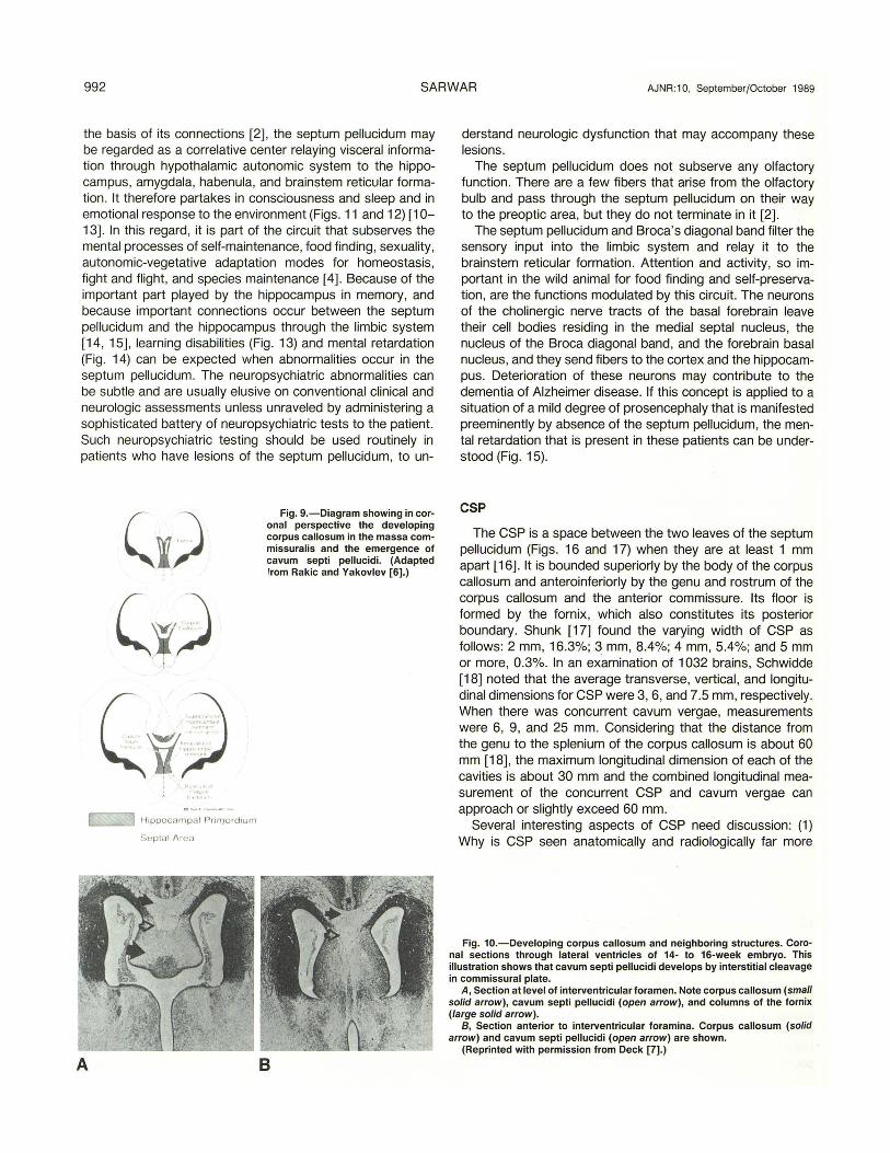
Fig. 9.-Diagram showing in coronal perspective the developing corpus callosum in the massa commissuralis and the emergence of cavum septi pellucidi. (Adapted from Rakic and Yakovlev [6].)
B
derstand neurologic dysfunction that may accompany these lesions.
The septum pellucidum does not subserve any olfactory function . There are a few fibers that arise from the olfactory bulb and pass through the septum pellucidum on their way to the preoptic area, but they do not terminate in it (2].
The septum pellucidum and Broca's diagonal band filter the sensory input into the limbic system and relay it to the brainstem reticular formation . Attention and activity, so important in the wild animal for food finding and self-preservation , are the functions modulated by this circuit. The neurons of the cholinergic nerve tracts of the basal forebrain leave their cell bodies residing in the medial septal nucleus, the nucleus of the Broca diagonal band, and the forebrain basal nucleus, and they send fibers to the cortex and the hippocampus. Deterioration of these neurons may contribute to the dementia of Alzheimer disease. If this concept is applied to a situation of a mild degree of prosencephaly that is manifested preeminently by absence of the septum pellucidum, the mental retardation that is present in these patients can be understood (Fig . 15).
CSP
The CSP is a space between the two leaves of the septum pellucidum (Figs. 16 and 17) when they are at least 1 mm apart (16]. It is bounded superiorly by the body of the corpus callosum and anteroinferiorly by the genu and rostrum of the corpus callosum and the anterior commissure. Its floor is formed by the fornix, which also constitutes its posterior boundary. Shunk [17] found the varying width of CSP as follows: 2 mm, 16.3%; 3 mm, 8.4%; 4 mm, 5.4%; and 5 mm or more, 0.3%. In an examination of 1032 brains, Schwidde (18] noted that the average transverse, vertical, and longitudinal dimensions for CSP were 3, 6, and 7.5 mm, respectively. When there was concurrent cavum vergae, measurements were 6, 9, and 25 mm. Considering that the distance from the genu to the splenium of the corpus callosum is about 60 mm (18], the maximum longitudinal dimension of each of the cavities is about 30 mm and the combined longitudinal measurement of the concurrent CSP and cavum vergae can approach or slightly exceed 60 mm .
Several interesting aspects of CSP need discussion: (1) Why is CSP seen anatomically and radiologically far more
Fig. 10.-Developing corpus callosum and neighboring structures. Coronal sections through lateral ventricles of 14- to 16-week embryo. This illustration shows that cavum septi pellucidi develops by interstitial cleavage in commissural plate.
A, Section at level of interventricular foramen. Note corpus callosum (small solid arrow), cavum septi pellucidi (open arrow), and columns of the fornix (large solid arrow).
B, Section anterior to interventricular foramina. Corpus callosum (solid arrow) and cavum septi pellucidi (open arrow) are shown.
(Reprinted with permission from Deck [7].)
AJNR:10, September/October 1989 NORMAL AND ABNORMAL SEPTUM PELLUCIDUM 993
A Fig. 11.-Partly calcified and predominantly
thrombosed venous angioma in septum pellucidum and neighboring structures.
A and 8, Noncontrast (A) and contrast-enhanced (8) CT scans. lsodense lesion is causing abnormal thickening of septum pellucidum (short arrows) as seen on noncontrast scan. Lesion shows enhancement on postcontrast scan. Septal vein (long arrows) shows abnormal enlargement. On slices at lower level (not shown), calcification was observed in middle of lesion.
C-E, First and second echoes obtained at 2000/20 (C) and 2000/80 (D), respectively, and axial T2-weighted (2000/80) MR image (E) 10 mm higher. Areas of hyperintensity visualized on both intermediate-weighted and T2-weighted images represent disintegrated elements of RBCs, mainly methemoglobin. Lesion is capped by hemosiderin rim (short straight arrows). Abnormally large septal vein (long straight arrows) and large internal cerebral vein (curved arrows) are D shown. Venous radicles that are part of venous angioma can also be seen, particularly in D.
F, Sagittal T1-weighted MR image (600/20). Partly thrombosed lesion can be seen at anterior part of septum pellucidum (short solid arrow) and neighboring structures. Fornix also is involved (open arrow). Prominent internal cerebral vein (long straight arrow) can be seen.
G, Lateral film of left carotid angiogram by digital technique. Note typical appearance of venous angioma (short arrow) that drains through prominent septal vein into internal cerebral vein (long arrow). In arterial phase (not shown), no arterial contribution to this lesion was demonstrated.
This 45-year-old man had had blackouts for several years that would last for several minutes and were followed by aggressive behavior and rage tantrums. Also, he had had amnesia during recent events. In his words, he "loses time." It is suggested that the lesion of the septum pellucidum and neighboring structures had involved the fornix and therefore led to limbic system F dysfunction accounting for the patient's altera-tion of behavior and blackouts, which could be considered seizures.
often in the neonate and infant than in the adult? (2) Why is it that it is seen so frequently at autopsy in the adult brain , yet it is so uncommonly observed in the neuroimaging studies of the adult brain? (3) What is its significance when seen at any age? (4) What is the source of fluid within it?
E
G
From autopsy studies, it has been shown that a macroscopically visible CSP is present in 1 00% of fetuses and premature infants [19, 20]. Its prevalence diminishes sharply after term such that its prevalence is 85% within 1 month, 45% within 2 months, and 15% at 3-6 months [16]. The
994 SARWAR AJNR:1 0, September/October 1989
A B
c D
prevalence of CSP in the reported autopsy studies has shown great variation. Shaw and Alvord [16] studied 37 4 brains of patients who could be considered free of neurologic disease. They ignored in their analysis a small triangular cavity (Fig. 18) that is so frequently encountered at the anterior and superior end of the septum pellucidum. They reported the prevalence of CSP from 6 months to 16 years as 12%. Schwidde [18] studied 1"032 brains from humans of many different ages and reported the prevalence of CSP at 20.34%. Schwidde did not specifically mention whether the patients whose brains were studied had neurologic deficits. Shunk [17] studied 307 brains of patients in whom some type of neurologic disturbances had caused their deaths. This, plus the fact that Shunk included all sizes of CSP including the small triangular clefts seen anteriorly that were ignored by Shaw and Alvord [16] , accounts for his high reported prevalence of CSP of 60.2%. I believe that, considering the method and case selection criteria used by Shaw and Alvord [16], Schwidde [18] , and Shunk [17] , a prevalence of CSP of 12-20% could be considered reasonable in the adult. CSP has been seen on neurosonography in 61 % of premature neonates and in 50% of the full-term neonates [19]. Nakano et al. [21] determined the prevalence of CSP on CT in 1 050
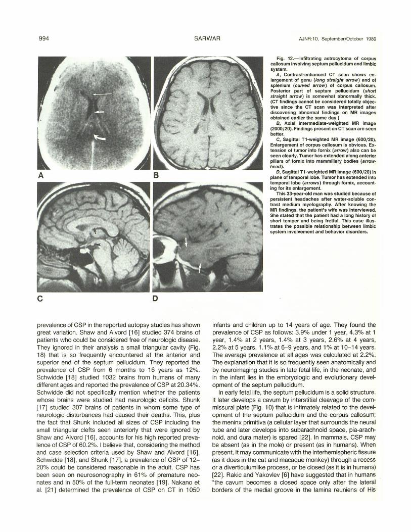
Fig. 12.-lnfiltrating astrocytoma of corpus callosum involving septum pellucidum and limbic system.
A, Contrast-enhanced CT scan shows enlargement of genu (long straight arrow) and of splenium (curved arrow) of corpus callosum. Posterior part of septum pellucidum (short straight arrow) is somewhat abnormally thick. (CT findings cannot be considered totally objective since the CT scan was interpreted after discovering abnormal findings on MR images obtained earlier the same day.)
8, Axial intermediate-weighted MR image (2000/20). Findings present on CT scan are seen better.
C, Sagittal T1-weighted MR image (600/20). Enlargement of corpus callosum is obvious. Extension of tumor into fornix (arrow) also can be seen clearly. Tumor has extended along anterior pillars of fornix into mammillary bodies (arrowhead).
D, Sagittal T1-weighted MR image (600/20) in plane of temporal lobe. Tumor has extended into temporal lobe (arrows) through fornix, accounting for its enlargement.
This 33-year-old man was studied because of persistent headaches after water-soluble contrast medium myelography. After knowing the MR findings, the patient's wife was interviewed. She stated that the patient had a long history of short temper and being fretful. This case illustrates the possible relationship between limbic system involvement and behavior disorders.
infants and children up to 14 years of age. They found the prevalence of CSP as follows: 3.9% under 1 year, 4.3% at 1 year, 1.4% at 2 years, 1.4% at 3 years, 2.6% at 4 years, 2.2% at 5 years, 1.1% at 6-9 years, and 1% at 1 0-14 years. The average prevalence at all ages was calculated at 2.2%. The explanation that it is so frequently seen anatomically and by neuroimaging studies in late fetal life, in the neonate, and in the infant lies in the embryologic and evolutionary development of the septum pellucidum.
In early fetal life, the septum pellucidum is a solid structure. It later develops a cavum by interstitial cleavage of the commissural plate (Fig. 1 0) that is intimately related to the development of the septum pellucidum and the corpus callosum; the meninx primitiva (a cellular layer that surrounds the neural tube and later develops into subarachnoid space, pia-arachnoid , and dura mater) is spared [22]. In mammals, CSP may be absent (as in the mole) or present (as in humans). When present, it may communicate with the interhemispheric fissure (as it does in the cat and macaque monkey) through a recess or a diverticulumlike process, or be closed (as it is in humans) [22]. Rakic and Yakovlev [6] have suggested that in humans "the cavum becomes a closed space only after the lateral borders of the medial groove in the lamina reuniens of His
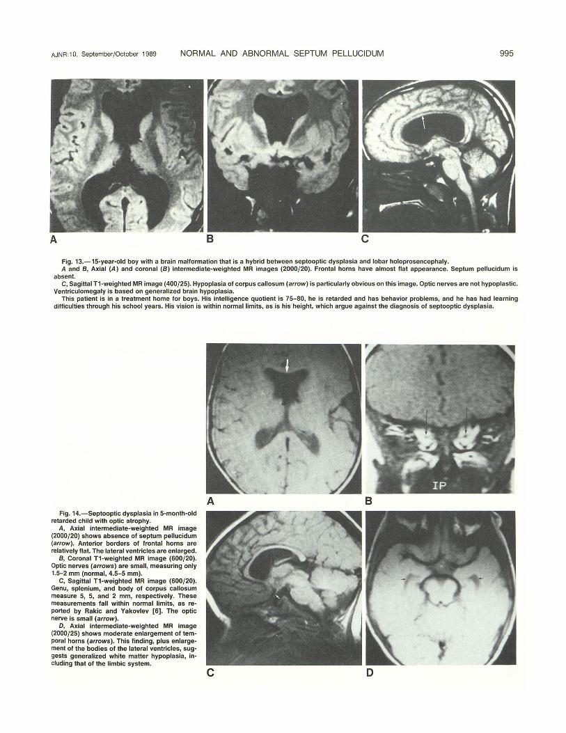
AJNR:1 0, September/October 1989 NORMAL AND ABNORMAL SEPTUM PELLUCIDUM 995
A 8 c Fig. 13.-15-year-old boy with a brain malformation that is a hybrid between septooptic dysplasia and lobar holoprosencephaly. A and 8 , Axial (A) and coronal (8) intermediate-weighted MR images (2000/20). Frontal horns have almost flat appearance. Septum pellucidum is
absent. C, Sagittal T1-weighted MR image (400/25). Hypoplasia of corpus callosum (arrow) is particularly obvious on this image. Optic nerves are not hypoplastic.
Ventriculomegaly is based on generalized brain hypoplasia. This patient is in a treatment home for boys. His intelligence quotient is 75-80, he is retarded and has behavior problems, and he has had learning
difficulties through his school years. His vision is within normal limits, as is his height, which argue against the diagnosis of septooptic dysplasia.
Fig. 14.-Septooptic dysplasia in 5-month-old retarded child with optic atrophy.
A, Axial intermediate-weighted MR image (2000/20) shows absence of septum pellucidum (arrow). Anterior borders of frontal horns are relatively flat. The lateral ventricles are enlarged.
8, Coronal T1-weighted MR image (600/20). Optic nerves (arrows) are small , measuring only 1.5-2 mm (normal, 4.5- 5 mm).
C, Sagittal T1-weighted MR image (600/20). Genu, splenium, and body of corpus callosum measure 5, 5, and 2 mm, respectively. These measurements fall within normal limits, as reported by Rakic and Yakovlev [6]. The optic nerve is small (arrow).
D, Axial intermediate-weighted MR image (2000/25) shows moderate enlargement of temporal horns (arrows). This finding, plus enlargement of the bodies of the lateral ventricles, suggests generalized white matter hypoplasia, including that of the limbic system.
A 8
c D
996 SARWAR AJNR:1 0, September{October 1989
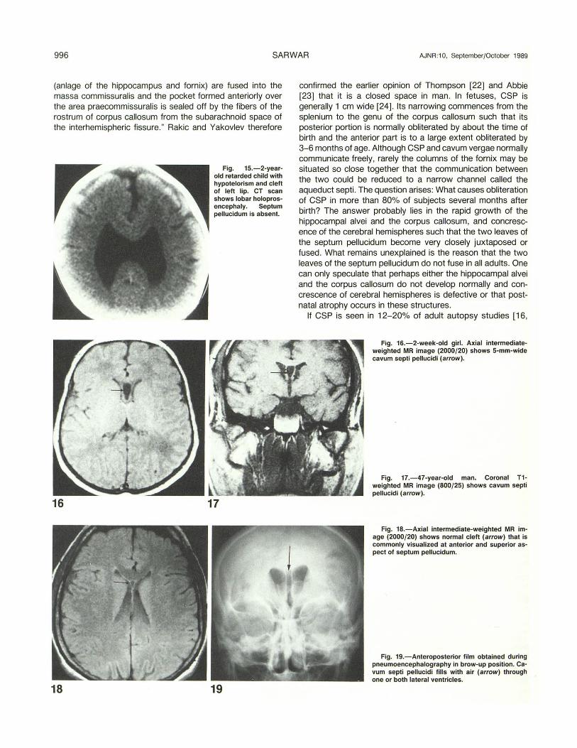
(anlage of the hippocampus and fornix) are fused into the massa commissuralis and the pocket formed anteriorly over the area praecommissuralis is sealed off by the fibers of the rostrum of corpus callosum from the subarachnoid space of the interhemispheric fissure ." Rakic and Yakovlev therefore
18
Fig. 15.-2-year-old retarded child with hypotelorism and cleft of left lip. CT scan shows lobar holoprosencephaly. Septum pellucidum is absent.
17
19
confirmed the earlier opinion of Thompson [22] and Abbie [23] that it is a closed space in man. In fetuses, CSP is generally 1 em wide [24]. Its narrowing commences from the splenium to the genu of the corpus callosum such that its posterior portion is normally obliterated by about the time of birth and the anterior part is to a large extent obliterated by 3-6 months of age. Although CSP and cavum vergae normally communicate freely , rarely the columns of the fornix may be situated so close together that the communication between the two could be reduced to a narrow channel called the aqueduct septi . The question arises: What causes obliteration of CSP in more than 80% of subjects several months after birth? The answer probably lies in the rapid growth of the hippocampal alvei and the corpus callosum, and concrescence of the cerebral hemispheres such that the two leaves of the septum pellucidum become very closely juxtaposed or fused. What remains unexplained is the reason that the two leaves of the septum pellucidum do not fuse in all adults. One can only speculate that perhaps either the hippocampal alvei and the corpus callosum do not develop normally and concrescence of cerebral hemispheres is defective or that postnatal atrophy occurs in these structures.
If CSP is seen in 12-20% of adult autopsy studies [16,
Fig. 18.-Axial intermediate-weighted MR image (2000{20) shows normal cleft (arrow) that is commonly visualized at anterior and superior aspect of septum pellucidum.
Fig. 19.-Anteroposterior film obtained during pneumoencephalography in brow-up position. Cavum septi pellucidi fills with air (arrow) through one or both lateral ventricles.
AJNR :10, SeptemberfOctober 1989 NORMAL AND ABNORMAL SEPTUM PELLUCIDUM 997
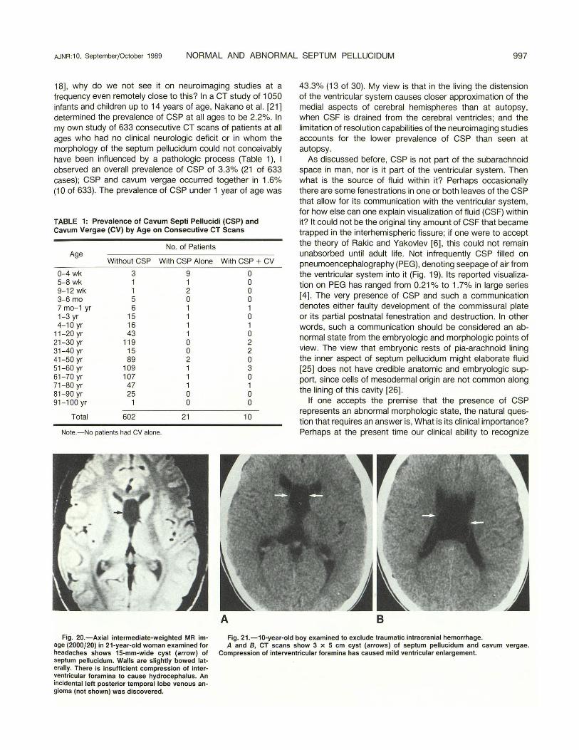
18], why do we not see it on neuroimaging studies at a frequency even remotely close to this? In a CT study of 1 050 infants and children up to 14 years of age, Nakano et al. [21] determined the prevalence of CSP at all ages to be 2.2%. In my own study of 633 consecutive CT scans of patients at all ages who had no cl inical neurologic deficit or in whom the morphology of the septum pellucidum could not conceivably have been influenced by a pathologic process (Table 1 ), I observed an overall prevalence of CSP of 3.3% (21 of 633 cases) ; CSP and cavum vergae occurred together in 1.6% (1 0 of 633). The prevalence of CSP under 1 year of age was
TABLE 1: Prevalence of Cavum Septi Pellucidi (CSP) and Cavum Vergae (CV) by Age on Consecutive CT Scans
No. of Patients Age
Without CSP With CSP Alone With CSP + CV
0-4 wk 3 9 0 5-8 wk 1 1 0 9-12 wk 1 2 0 3-6 mo 5 0 0 7 mo-1 yr 6 1 1 1-3 yr 15 1 0 4-10 yr 16 1 1
11-20 yr 43 1 0 21-30 yr 119 0 2 31-40 yr 15 0 2 41-50 yr 89 2 0 51-60 yr 109 1 3 61-70yr 107 1 0 71-80 yr 47 1 1 81-90 yr 25 0 0 91-100 yr 1 0 0
Total 602 21 10
Note.-No patients had CV alone.
A
43.3% (13 of 30). My view is that in the living the distension of the ventricular system causes closer approximation of the medial aspects of cerebral hemispheres than at autopsy, when CSF is drained from the cerebral ventricles; and the limitation of resolution capabilities of the neuroimaging studies accounts for the lower prevalence of CSP than seen at autopsy.
As discussed before, CSP is not part of the subarachnoid space in man, nor is it part of the ventricular system. Then what is the source of fluid within it? Perhaps occasionally there are some fenestrations in one or both leaves of the CSP that allow for its communication with the ventricular system, for how else can one explain visualization of fluid (CSF) within it? It could not be the original tiny amount of CSF that became trapped in the interhemispheric fissure; if one were to accept the theory of Rakic and Yakovlev [6], this could not remain unabsorbed until adult life. Not infrequently CSP filled on pneumoencephalography (PEG), denoting seepage of air from the ventricular system into it (Fig . 19). Its reported visualization on PEG has ranged from 0.21 % to 1.7% in large series [4]. The very presence of CSP and such a communication denotes either faulty development of the commissural plate or its partial postnatal fenestration and destruction. In other words, such a communication should be considered an abnormal state from the embryologic and morphologic points of view. The view that embryonic rests of pia-arachnoid lining the inner aspect of septum pellucidum might elaborate fluid [25] does not have credible anatomic and embryologic support , since cells of mesodermal origin are not common along the lining of this cavity [26].
If one accepts the premise that the presence of CSP represents an abnormal morphologic state, the natural question that requires an answer is, What is its clinical importance? Perhaps at the present time our clinical ability to recognize
8
Fig. 20. - Axial intermediate-weighted MR image (2000/20) in 21-year-old woman examined for headaches shows 15-mm-wide cyst (arrow) of septum pellucidum. Walls are slightly bowed laterally. There is insufficient compression of interventricular foramina to cause hydrocephalus. An incidental left posterior temporal lobe venous angioma (not shown) was discovered.
Fig. 21.-10-year-old boy examined to exclude traumatic intracranial hemorrhage. A and B, CT scans show 3 x 5 em cyst (arrows) of septum pellucidum and cavum vergae.
Compression of interventricular foramina has caused mild ventricular enlargement.
998 SARWAR AJNR:1 0, September/October 1989
such a neurologic deficit, which in this situation would imply limbic system dysfunction, remains at best crude.
Cyst of the Septum Pellucidum
The distinction between a large CSP and a cyst of the septum pellucidum remains at best nebulous. No readily available or generally accepted definition exists. I define the cyst as a fluid-containing structure between the lateral ventricles , whose walls exhibit lateral bowing instead of being parallel and are 1 0 mm apart or greater (Fig. 20). This arbitrary definition is clinically important since a cyst larger than this may be expected to cause narrowing of the interventricular foramina and lead to hydrocephalus (Fig. 21 ), or to cause neurologic impairment simply by compression of the neighboring neural structures. No symptoms may be expected if cyst walls are 5 mm or less apart . Large cysts cause splaying of the choroid plexi of the lateral ventricles, which assume a somewhat parallel course instead of their anterior parts converging toward the interventricular foramina. Such symptomatic cysts are distinctly rare [27]. Cowley et al. [27] reviewed the literature and found a total of 21 cases up to 1979, including one of their own. In this regard it is especially interesting to note that in their 1936 report Dyke and Davidoff [28] found only one such lesion in 5000 PEG studies.
TABLE 2: Changes in the Septum Pellucidum in Boxers and Control Subjects*
Group
Boxers (n = 13) Controls (n = 500)
· From [36].
Cavum Septi Pellucidi
12 (92) 142 (28)
No.(%)
Fenestration of Leaves
10 (77) 15 (3)
A
Mean Width (mm)
5.2 1.6
Since the report by Cowley et al. [27] in 1979, an additional four cases have been reported in the English language literature [29-32]. The cysts may or may not communicate with the ventricular system. The ones that do not communicate are more likely to be symptomatic and are treated surgically by drainage or by establishing communication with the ventricular system. Spontaneous collapse of cysts of the septum pellucidum has been reported [17].
CSP and Chronic Encephalopathy in Boxers
Boxing is a violent sport designed to selectively cause injury to the face and the cranium and its most vulnerable contents, the brain . In addition to subdural and parenchymal hemorrhages, the midline structures, which include the corpus callosum, the septum pellucidum, and the fornix, are particularly susceptible to injury as the brain undergoes repeated distortions and displacements in all imaginable planes. Chronic brain injury in the living has been revealed by CT as corticocerebral atrophy [33-35] and an increased prevalence of CSP [34]. In this regard, the most elegant work was reported by Corsellis et al. [36] in 1973. They had access to 15 retired boxers. Two were excluded from study because the intraventricular hemorrhage had been deemed to have caused artifactual tearing or separation of the leaves of the septum pellucidum. The findings in the remaining 13 boxers were compared with those in the brains of 500 normal control subjects 21-100 years old (200 men and 300 women) (Table 2). CSP was observed in 12 (92%) of 13 brains in the boxers, compared with a prevalence of 28% (142 of 500) in the control population. The main width of the CSP in boxers was 5.17 mm compared with 1.6 mm in the control group. The septal leaves were fenestrated in 77% of the boxers, compared with only 3% of control subjects. The reason that septal leaves are so prone to fenestration is that they are relatively thin at their superior attachment to the corpus callosum and some-
8
Fig. 22.-47-year-old man with history of Fig. 23.-Severe hydrocephalus in two patients causes fraying and fenestration (A, arrow) and trauma. Coronal section of brain shows tears (ar· total destruction (B) of septum pellucidum. rows) of septum pellucidum. (Courtesy of C. A. Hailer, Chicago.)
AJNR:1 0, September/October 1989 NORMAL AND ABNORMAL SEPTUM PELLUCIDUM 999
what thicker at their inferior attachment to the fornix. Repeated bouts and rotational injury lead to stretching , tearing (Fig. 22), and fenestration , especially of the thinner and fragile parts superiorly. It is not uncommon for the fornix to become totally severed from the corpus callosum [36]. Because of the loss of neural tissue in the corpus callosum, fornix, and neighboring structures, there is atrophic ventriculomegaly that further stretches these leaves, accentuating fenestration. Part of the explanation of CSP in such cases may lie in moving apart the closely apposed septal leaves. The fenestrations allow ventricular CSF to accumulate with CSP. Such an occurrence cannot be expected in obstructive ventriculomegaly, when CSP is either compressed if present or not seen as such because there is rupture of its leaflets (Fig. 23). Whereas similar septum pellucidum injury can be present in other kinds of trauma [37 , 38], no such vulnerability exists in Alzheimer disease, Pick disease, Huntington chorea, or senile dementia [36].
Dementia pugilistica [39] is a term that is applied to the dementing process seen in boxers. Whereas the generalized cerebral cortical atrophy that is seen in these individuals could account for dementia, of particular interest is the injury to the medial temporal lobes that form part of the limbic system.
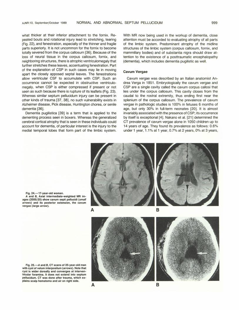
Fig. 24.-17-year-old woman. A and 8 , Axial intermediate-weighted MR im
ages (2000/20) show cavum septi pellucidi (small arrows) and its posterior extension, the cavum vergae (large arrow).
Fig. 25.-A and 8 , CT scans of 25-year-old man with cyst of velum interpositum (arrows). Note that cyst is wider dorsally and converges at interventricula r foramina. It does not extend into septum pellucidum. CT was done after trauma, which explains scalp hematoma and air on right side.
A
A
With MR now being used in the workup of dementia, close attention must be accorded to evaluating atrophy of all parts of the limbic system. Predominant atrophy of the midline structures of the limbic system (corpus callosum, fornix , and mammillary bodies) and of substantia nigra should draw attention to the existence of a posttraumatic encephalopathy (dementia), which includes dementia pugilistic as well.
Cavum Vergae
Cavum vergae was described by an Italian anatomist Andrea Verga in 1851 . Embryologically the cavum vergae and CSP are a single cavity called the cavum corpus callosi that lies under the corpus callosum. This cavity closes from the caudal to the rostral extremity, thus ending first near the splenium of the corpus callosum. The prevalence of cavum vergae in pathologic studies is 1 00% in fetuses 6 months of age, but only 30% in full-term neonates [20]. It is almost invariably associated with the presence of CSP; its occurrence by itself is exceptional [ 4] . Nakano et al. [21 ] determined the CT prevalence of cavum vergae alone in 1 050 children up to 14 years of age. They found its prevalence as follows: 0.6% under 1 year, 1.1% at 1 year, 0.7% at 2 years, 0% at 3 years,
B
B
1000 SARWAR AJNR:1 0, September/October 1989
0.9% at 4 years , 0% at 5 years, 0% at 6-9 years, and 0% at 10-14 years of age. The average prevalence at all ages was determined to be 0.4%. In none of the 1032 brains studied by Schwidde [18] was cavum vergae found alone. The concurrence rate of CSP and cavum vergae has been reported as 3% [21] and 11 % [18]. The following structures constitute the boundaries of the cavum vergae: anteriorly, the anterior limbs of the fornix ; superiorly, the body of the corpus callosum; posteriorly, the splenium of the corpus callosum; and inferiorly, the psalterium and hippocampal commissure, the fibers of which bridge the space between the diverging posterior pillars of the fornix and rest on the tela choroidea. On axial CT and MR, the cavum vergae appears as a posterior continuation of the CSP. Their junction is marked by beginning expansion of the cavum vergae such that its dorsal terminus assumes a broad base (Fig . 24). A mild constriction at their juncture may be noted. It can be differentiated from the cavum or cyst of velum interpositum by the following characteristics of the cavum or cyst: it lies under the fornix , converges at the interventricular foramina, and does not extend into the septum pellucidum (Fig. 25).
Expected Neurologic Dysfunction from an Embryologic Perspective in the Presence of CSP and Cavum Vergae
One might ask, if CSP is present in all fetuses and in premature neonates and its prevalence rapidly diminishes after birth to about 15% at 6 months, then which of the following two considerations should be considered a normal occurrence: (1) its absence in the adult or (2) its presence? Obviously if more than 80% of the adult population do not have CSP, its absence cannot be considered abnormal. The presence of CSP in the adult population may then be considered an aberration of a normal developmental process of fusion of the leaflets of the septum pellucidum and resorption of the fluid trapped between them. If one then accepts CSP as dysmorphology of the septum pellucidum, the natural question is whether there is a corresponding neurologic dysfunction . The search for an answer to this question especially interested German neuroscientists between 1935 and 1965 [4] . The differing points of view and controversies ignited dazzling literary skirmishes. In 1938, Wilder (cited in [4]) described a "septum pellucidum syndrome" in association with CSP; its features included mental disturbances, disordered speech , and seizures. Considering that the septal area by itself traditionally is not considered epileptogenic, the described association between epilepsy and CSP could perhaps be a manifestation of a concomitant limbic system dysmorphology, a facet of this anomaly that has not been explored and perhaps deserves attention . This aspect of investigation becomes especially interesting when one considers that in the presence of CSP, epileptic seizures have been described in 31-55% of cases and psychosis , dementia, and personality changes in about 15% [4), not to mention other symptoms like headache, giddiness, signs of increased intracranial pressure, and even hemiparesis.
In most reports that have dealt with CSP, cavum vergae has been lumped together with CSP. Very little information is
available on the prevalence of cavum vergae alone and the attendant neurologic importance. Since (1) CSP and cavum vergae are a single cavity in the fetus and (2) the process of closure begins from behind and extends rostrally, it becomes understandable that if cavum vergae were not to close, the CSP also would not. But what is to be made of the situation of cavum vergae alone? It could be surmised that the process of closure was defective such that the cavum vergae was ignored. But that would be too simplistic an explanation. I submit that persistence of cavum vergae alone is perhaps a reflection of an abnormal neural organizational phenomenon that denotes impaired development of the fornix , psalterium, and splenium of the corpus callosum. In other words, cavum vergae could be considered a participant in the spectrum of midline developmental defects. This aspect was investigated by Miller et al. [ 40). They studied 1 0 patients with CT, aged 1 day to 9 years, who had cavum vergae alone or in association with CSP. Eight of 10 had concurrent CSP and cavum vergae. Of these 1 0 patients, five had delayed development, four had macrocephaly, two had learning disabilities, two had abnormal EEGs, and one had Apert syndrome (some patients had more than one abnormal feature). They encountered no cases of cavum vergae in 50 children in whom CT was performed for head trauma. Nakano et al. [21] reported a concurrence rate of CSP and cavum vergae at all ages at 3% in a population of 1 050 infants and children aged 1 to 14 years. This prevalence was higher than those of CSP and cavum vergae occurring alone of 2.2% and 0.4%, respectively. All of the patients in their study had neurologic deficits, which were classified as follows: (1) convulsive disorders in 312 (average age, 2.4 years); (2) developmental delay in 468 (average age, 4.1 years); and (3) various neurologic syndromes in 270 (average age, 2.2 years). Their study did not include any controls. They stated that no statistically significant relationship could be established between the prevalence of CSP and cavum vergae occurring alone or concurrently and the neurologic deficit.
Absence of the Septum Pellucidum
Absence of the septum pellucidum almost always signifies substantial neurologic disease. Its absence (agenesis) can be part of a developmental brain malformation, or it can be acquired. In the latter situation it is invariably a sequela of long-standing substantial hydrocephalus. The increased intracranial pressure that follows severe hydrocephalus initially frays and fenestrates the leaves of the septum pellucidum [ 41] before they finally disintegrate, allowing the two lateral ventricles to form a freely communicating single cavity (Fig. 23B). I am not aware of any published work that has correlated the relation between intraventricular pressure and the occurrence of fenestration and rupture of the leaves of the septum pellucidum.
In the developmental category, absence of the septum pellucidum can be considered part of a continuum of the spectrum of prosencephaly. Prosencephaly implies maldevelopment of the forebrain structures, which include the corpus callosum and the septum pellucidum. In this regard,
AJNR :10, September/October 1989 NORMAL AND ABNORMAL SEPTUM PELLUCIDUM 1001
septooptic dysplasia (originally described by deMorsier [42] in 1956) is preeminent. This condition is characterized by (1) a variable degree of hypoplasia of the optic pathways, with the optic chiasm often assuming a vertical orientation instead of the normally encountered oblique configuration, with or without the presence of an "optic ventricle" close to the optic chiasm; (2) absence of the septum pellucidum; and (3) hypothalamic-pituitary dysfunction manifested by deficiency in growth hormone and hypothyroidism. deMorsier suggested that this syndrome resulted from an agenesis of the embryonic neuroglial anlage that bridges the origin of the corpus callosum and the anterior commissure. He described the corpus callosum as being thin and the bodies of the fornices as being fused in this disorder. Considering that the septum pellucidum forms a part of the limbic system and that the embryologic origins of the septum pellucidum and of the corpus callosum are so closely entwined, it should come as no surprise if one encounters hypoplasia of the corpus callosum (Fig. 13) [ 43] and of the hippocampus in cases of septooptic dysplasia. This finding is generally not recognized because most of the cases of septooptic dysplasia that were reported during the CT era had one major deficiency: axial CT provides a poor anatomic depiction of the corpus callosum and of the hippo-
TABLE 3: Causes of the Absence of the Septum Pellucidum
Developmental (congenital) Holoprosencephaly Septooptic dysplasia Dysgenesis of the corpus callosum Apert syndrome (acrocephalosyndactyly) Brain hypoplasia Cortical dysmorphogenesis (gray-matter heterotopia) Schizencephaly (including Gruner syndrome) Lissencephaly Congenital hydrocephalus Chiari malformation type II
Acquired Significant and long-standing hydrocephalus Leptomeningitis Trauma Porencephaly
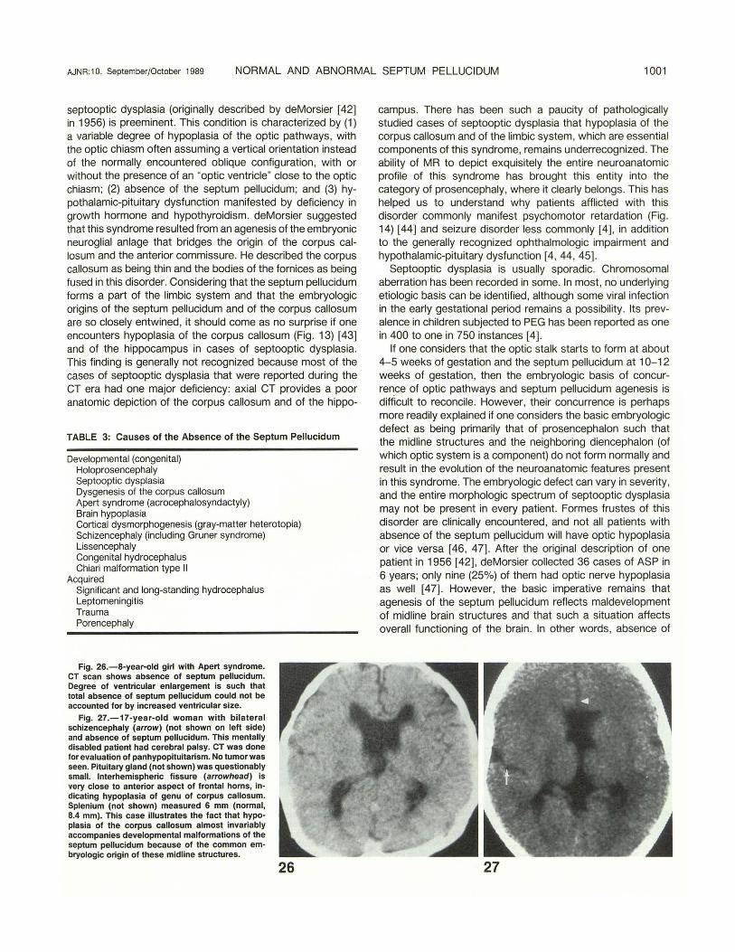
Fig. 26.-8-year-old girl with Apert syndrome. CT scan shows absence of septum pellucidum. Degree of ventricular enlargement is such that total absence of septum pellucidum could not be accounted for by increased ventricular size.
Fig. 27.-17-year-old woman with bilateral schizencephaly (arrow) (not shown on left side) and absence of septum pellucidum. This mentally disabled patient had cerebral palsy. CT was done lor evaluation of panhypopituitarism. No tumor was seen. Pituitary gland (not shown) was questionably small. Interhemispheric fissure (arrowhead) is very close to anterior aspect of frontal horns, indicating hypoplasia of genu of corpus callosum. Splenium (not shown) measured 6 mm (normal, 8.4 mm). This case illustrates the fact that hypoplasia of the corpus callosum almost invariably accompanies developmental malformations of the septum pellucidum because of the common embryologic origin of these midline structures.
26
campus. There has been such a paucity of pathologically studied cases of septooptic dysplasia that hypoplasia of the corpus callosum and of the limbic system, which are essential components of this syndrome, remains underrecognized. The ability of MR to depict exquisitely the entire neuroanatomic profile of this syndrome has brought this entity into the category of prosencephaly, where it clearly belongs. This has helped us to understand why patients afflicted with this disorder commonly manifest psychomotor retardation (Fig . 14) [44) and seizure disorder less commonly [4) , in addition to the generally recognized ophthalmologic impairment and hypothalamic-pituitary dysfunction [4 , 44, 45).
Septooptic dysplasia is usually sporadic. Chromosomal aberration has been recorded in some. In most, no underlying etiologic basis can be identified, although some viral infection in the early gestational period remains a possibility. Its prevalence in children subjected to PEG has been reported as one in 400 to one in 750 instances [4].
If one considers that the optic stalk starts to form at about 4-5 weeks of gestation and the septum pellucidum at 1 0-12 weeks of gestation, then the embryologic basis of concurrence of optic pathways and septum pellucidum agenesis is difficult to reconcile . However, their concurrence is perhaps more readily explained if one considers the basic embryologic defect as being primarily that of prosencephalon such that the midline structures and the neighboring diencephalon (of which optic system is a component) do not form normally and result in the evolution of the neuroanatomic features present in this syndrome. The embryologic defect can vary in severity, and the entire morphologic spectrum of septooptic dysplasia may not be present in every patient. Formes frustes of this disorder are clinically encountered, and not all patients with absence of the septum pellucidum will have optic hypoplasia or vice versa [46, 47). After the original description of one patient in 1956 [ 42], deMorsier collected 36 cases of ASP in 6 years; only nine (25%) of them had optic nerve hypoplasia as well [47). However, the basic imperative remains that agenesis of the septum pellucidum reflects maldevelopment of midline brain structures and that such a situation affects overall functioning of the brain . In other words, absence of
27
1002 SARWAR AJNR:1 0, September/October 1989
the septum pellucidum on a developmental basis invariably connotes neurologic impairment that varies in severity. In this regard it is well to quote Friede [41 ], the great pediatric neuropathologist: "Occasional observations of complete absence of the septum pellucidum without cerebral lesions are curiosities without known clinical significance. " In a personal review of 633 consecutive CT scans at all ages, only eight cases had absence of the septum pellucidum. Of these eight, the cause was severe hydrocephalus in seven ; only one patient with schizencephaly had absence of the septum pellucidum.
In addition to long-standing substantial hydrocephalus, absence of the septum pellucidum (Table 3) is routinely present in holoprosencephaly (Fig. 15) [41] . It may be part of generalized brain hypoplasia [ 48] and gray-matter heterotopia [49]. It may also be considered an integral component of Apert syndrome (acrocephalosyndactyly) (Fig. 26) [4, 50]. It
A B
can be observed as a sequela of neonatal leptomeningitis and neonatal brain trauma if the septum pellucidum is destroyed. It can be part of porencephaly, whether acquired or developmental (schizencephaly) (Fig. 27) [51 , 52] . It is noted in at least 20% of cases of Chiari type II malformation [53] , very likely as a consequence of hydrocephalus.
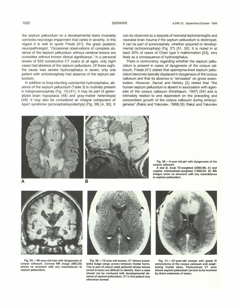
There is controversy regarding whether the septum pellucidum is present in cases of dysgenesis of the corpus callosum. Friede [41] stated that ependyma-lined septum pellucidum becomes laterally displaced in dysgenesis of the corpus callosum and that its absence is "simulated" on gross examination. However, Sarnat and Netsky [2] stated that "the human septum pellucidum is absent in association with agenesis of the corpus callosum (Kirshbaum, 1947) [54] and is intimately related to and dependent on the preceding and concomitant growth of the corpus callosum during embryogenesis" (Rakic and Yakovlev, 1968) [6]. Rakic and Yakovlev
Fig. 28.-3-year-old girl with dysgenesis of the corpus callosum.
A and 8 , Axial T2-weighted (2000/80, A) and sagittal intermediate-weighted (1500/25, B) MR images s.how no structure with any resemblance to septum pellucidum.
Fig. 29.- 49-year-old man with dysgenesis of corpus callosum. Coronal MR image (800/25) shows no structure with any resemblance to septum pellucidum.
Fig. 30.-73-year-old woman. CT shows knucklelike bulge (large arrow) between frontal horns. This is part of cavum septi pellucidi whose leaves (small arrows) are difficult to identify. Such a case should not be confused with developmental absence of septum pellucidum. CT in this patient was otherwise normal.
Fig. 31 .- 47-year-old woman with grade Ill astrocytoma of the corpus callosum and neighboring frontal lobes. Postcontrast CT scan shows septum pellucidum (arrow) to be involved by direct extension of tumor.
AJNR:1 0, September/October 1989 NORMAL AND ABNORMAL SEPTUM PELLUCIDUM 1003
[6] stated that "there is no septum pellucidum and no cavum with the corpus callosum. " Be that as it may, from the neuroimaging perspective there is nothing on CT and MR that looks like the septum pellucidum in dysgenesis of the corpus callosum (Figs. 28 and 29).
From the neuroimaging point of view, it is only in severe and long-standing hydrocephalus that the septum pellucidum ruptures such that the appearance of an absent septum pellucidum emerges. If no septum pellucidum is recognized in a case in which there is only slight ventriculomegaly. then such a situation almost invariably denotes a developmental cause of its absence. Because an absent septum pellucidum can be considered part of the spectrum of holoprosencephaly. one should look additionally for midline facial dysmorphism and hypotelorism, findings that would further reinforce the developmental basis of an absent septum pellucidum. Also, there should be no signs of increased intracranial pressure and the head should not be abnormally large. Occasionally the two leaves of CSP may be too thin to be imaged adequately on CT. In such cases it is most likely that CSP can mimic absent septum pellucidum. However, such confusion should not occur, for in almost all cases of CSP there is a knucklelike protrusion of a CSF-containing space in the midline of what appears to be a monoventricle (Fig. 30). In the rare instance in which the midline protrusion is not present, a structure termed the "pseudomonoventricle" (55] can appear. This should not be confused with semilobar or lobar holoprosencephaly since in these conditions the anterior and superior borders of the frontal horns are flat on axial and coronal images, respectively, and the normal narrowing of the
frontal horns at their lateral margins transforms into a variable degree of bulbous configuration .
Miscellaneous Lesions of the Septum Pellucidum
Neoplasms
Neoplasms that primarily originate from the septum pellucidum are extremely rare. Generally the septum pellucidum is involved from direct extension of tumors that arise from the neighboring structures, principally the corpus callosum. These tumors are gliomas of varying histologic types- astrocytomas, oligodendrogliomas, mixed astrocytomas-oligodendrogliomas, subependymomas, and glioblastomas [56] . The infiltrative tumors, including gliomatosis cerebri , that involve the septum pellucidum and the corpus callosum can be difficult to diagnose on axial CT. However, if the septum pellucidum has irregular and shaggy borders and its absolute thickness is more than 3 mm, an infiltrating tumor may be suspected. This suspicion can be further strengthened if the thickness of the genu of the corpus callosum and of the splenium of the corpus callosum (measured from the pericallosal cistern to the genu and the splenium of the corpus callosum, respectively) exceeds 1 0 mm. This observation is based on adult average measurements of 7.93 and 8.37 mm for the genu and splenium of the corpus callosum, respectively , as reported by Rakic and Yakovlev [6]. Corpus callosum lesions that secondarily involve the septum pellucidum are best assessed by MR. since it provides the sagittal perspective of the corpus callosum that is so important in evaluating its
Fig. 32.-lntraventricular cysticercosis in 21-year-old woman with headaches. Clinical and other CT and MR findings (not shown) suggested neurocysticercosis. Axial T1-weighted MR image shows shift of septum pellucidum to the left (arrow). On T2-weighted images (not shown) this cyst had the same signal characteristics as CSF. Absence of other leaf of septum pellucidum excludes large cavum septi pellucidi or cyst of septum pellucidum.
Fig. 33.-58-year-old man with large old infarct in left cerebral hemisphere. CT scan shows absence of shift of septum pellucidum in malacic process. Even in the presence of extensive neural tissue volume loss there is no ipsilateral shift of septum pellucidum (arrow) .
Fig. 34.- Thinning of septum pellucidum as a consequence of severe ischemic-hypoxic encephalopathy in an 8-month-old boy born at 32 weeks of gestation weighing 1456 g. He suffered severe neonatal ischemic-hypoxic insult. He had presumed germinal matrix hemorrhage. Coronal T1-weighted MR image (500/20) shows considerable enlargement of ventricular system due to atrophy. There is thinning of corpus callosum and of septum pellucidum. White-matter radiations are thin, indicating their underdevelopment as a consequence of ischemic-hypoxic encephalopathy. Hyperintense signal seen along ventricular system is caused by methemoglobin.
1004 SARWAR AJNR :10, September/October 1989
morphology (Fig . 12). Russell and Naidich [57] described thickening and contrast enhancement of the septum pellucidum as an important and reliable, but not pathognomonic, sign of an intraaxial mass lesion (Fig . 31 ). Midline lipomas usually constitute a component of the spectrum of developmental malformations of the corpus callosum [5]. A lipoma confined to the septum pellucidum was described recently [58].
Angiomatous Malformations
Angiomatous lesions confined exclusively to the septum pellucidum, to my knowledge, have not been described. The septum pellucidum may appear thicker andjor show linear contrast enhancement if the septal veins that drain into the internal cerebral veins partake in the venous drainage of an angiomatous lesion that involves the neighboring deeper structures of the brain . These angiomatous lesions are usually arteriovenous malformations. I recently encountered a patient who had a venous angioma that predominantly involved the septum pellucidum and in whom MR had shown evidence of previous hemorrhage (Fig. 11 ).
Calcification
Calcified lipomas of the corpus callosum could involve the septum pellucidum. In 1966, Wesenberg et al. [59] described calcification in the roof of CSP in two cases of lissencephaly. They suggested that it was present in the paraphysis, a structure that is located in the septum pellucidum but that involutes and disappears late in the third month of fetal life. They considered the calcification as part of the developmental arrest of brain growth in lissencephaly. Calcified intraaxial and extraaxial neoplasms and angiomatous lesions that occur in the structures adjoining the septum pellucidum may also involve it by direct extension.
Displacement and Atrophy of the Septum Pellucidum
Minimal asymmetry of the frontal horns is not uncommon. In a study of 300 patients who had no neurologic deficits, Shapiro et al. [60] observed that the frontal horns were equal in size in 89.7% of cases. The right frontal horn was larger than the left in 7%, and the left was larger than the right in 3.3%. They also noted that the septum pellucidum was shifted 1-2 mm to the side opposite the larger frontal horn. The septum pellucidum normally maintains a straight orientation. A septal tilt, especially when the tilt assumes a curved appearance, in the absence of compression of the frontal horn on the side opposite to the tilt should prompt a careful search for an intraventricular lesion. This search should be especially careful if the frontal horn is not excessively large or it does not contain a lesion with a density higher than that of CSF, both on plain and contrast-enhanced CT, and if it is not of an intensity different from that of CSF on MR. In other words, the frontal horn could harbor a lesion that is isodense or isointense relative to CSF. The lesions that are most likely to present in such fashion include cysticercosis (Fig. 32) and
neuroepithelial cysts. It is a peculiar phenomenon that, unlike mediastinal shift to the side of lung collapse or fibrosis, the septum pellucidum may not shift toward the side of neural tissue volume loss (Fig. 33).
One generally does not think in terms of the septum pellucidum participating in a generalized atrophic process. Notwithstanding its small volume, it does contain fibers that connect it with the limbic system, the cerebral white matter, and the cortex. Therefore, it is not unreasonable to speculate that if carefully sought, septal atrophy that is part of a generalized atrophic process may be recognizable both by neuroimaging (Fig. 34) and at autopsy.
Summary and Conclusions
The common embryologic origin of the septum pellucidum, of the corpus callosum, and of the limbic system explains the anatomic basis of a varying degree of concurrent involvement of these structures in disease processes, both developmental and acquired. Developmental absence of the septum pellucidum portends a more generalized dysplasia that encompasses the corpus callosum and the limbic system. When considered in this context , it is easy to understand the limbic system dysfunction that is engendered by this anomaly. It is rare for an acquired lesion to involve the septum pellucidum per se because of its small size. However, it is common for it to be secondarily involved in almost all types of acquired disease processes. With the exquisite anatomic morphology that MR now provides, one must carefully assess involvement of the septum pellucidum, of the corpus callosum, and of the limbic system in disease processes that occur in the vicinity of these structures, so that more incisive clinical insight into the functions that are subserved by these structures can be obtained.
ACKNOWLEDGMENTS
I thank Dale Charletta for the artwork , Mark Reyes for providing the anatomic material , and Claribel Olichwier for manuscript preparation.
REFERENCES
1. Pendergrass EP, Hodes PJ . Dilatation of the cavum septi pellucidi and cavum vergae. Ann Surg 1935;101 :269-295
2. Sarna! HB, Netsky MG. Evolution of the nervous system. New York: Oxford University, 1974:247-249
3. Loeser JD, Alvord EC. Agenesis of the corpus callosum. Brain 1968; 91 :553-570
4. Bruyn GW. Agenesis septi pellucidi, cavum septi pellucidi, cavum vergae, and cavum veli interpositi. In: Vinken PJ, Bruyn GW, eds. Handbook of clinical neurology, Vol. 30. Congenital malformations of the brain and skull, part I. Amsterdam, The Netherlands: Elsevier/North-Holland Biomedical, 1977: 299-336
5. Atlas SW, Zimmerman RA, Bilanuik L T, et al. Corpus callosum and limbic system: neuroanatomic MR evaluation of developmental anomalies. Radiology 1986;160:355-362
6. Rakic P, Yakovlev Pl. Development of the corpus callosum and cavum septi in man. J Comp Neurol1968;132:45-72
7. Deck MDF. The lateral ventricles. In: Newton TH , Potts DG, eds. Radiology of the skull and brain. Ventricles and cisterns. St. Louis: Mosby, 1978: 3489-3494
AJNR:1 0, September/October 1989 NORMAL AND ABNORMAL SEPTUM PELLUCIDUM 1005
9. Kier EL. The cerebral ventricles. A phylogenetic and ontogenetic study. In: Newton TH , Potts DG, eds. Radiology of the skull and brain. St. Louis: Mosby, 1977:2877-2890
10. Smythies JR. The neurological foundation of psychiatry. Oxford: Blackwell Scientific, 1966
11 . Spiegel EA, Miller HR, Oppenheimer MJ. Forebrain and rage reactions. J Neurophysio/1940;3: 538-548
12. lssacson RL. The limbic system. New York: Plenum, 1974 13. Zeman W, King FA. Tumors of the septum pellucidum and adjacent
structures with abnormal affective behavior: an anterior midline structure syndrome. J Nerv Ment Dis 1958;127:490-502
14. Papez JW. A proposed mechanism of emotion. Arch Neural Psychiatry 1937;38: 725-7 44
15. Maclean PD. Cerebral evolution and emotional processes. Ann NY Acad Sci 1972;193:137-149
16. Shaw CM, Alvord EC. Cava septi pellucidi at vergae: their normal and pathological states. Brain 1969;92:213-223
17. Shunk H. Congenital dilatations of the septi pellucidum. Radiology 1963;81 :610-618
18. Schwidde JT. Incidence of cavum septi pellucidi and cavum vergae in 1 ,032 human brains. Arch Neural Psychiatry 1952;67: 625-633
19. Farrugia S, Babcock DS. The cavum septi pellucidi: its appearance and incidence with cranial ultrasonography in infancy. Radiology 1981 ; 139:147-150
20. Larroche JC, Baudey J. Cavum septi lucidi , cavum vergae, cavum veli interpositi: cavities of the median line. Bioi Neonate 1961;3:193-236
21. Nakano S, Hojo H, Kataoka K, Yamasaki S. Age related incidence of cavum septi pellucidi and cavum vergae on CT scans of pediatric patients. J Comput Assist Tomogr 1981;5:348
22. Thompson IM. On the cavum septi pellucidi. J Anat 1932;67:59-77 23. Abbie AA. The origin of the corpus callosum and the fate of the structures
related to it. J Comp Neuro/1940;70:9-44 24. Lemire RJ , Loeser JD, Leech RW, Alvord EC Jr. Normal and abnormal
development of the human nervous system. Hagerstown, MD: Harper & Row, 1975:265-272
25. Van Wagenen WP, Aird RB. Dilatations of the cavity of the septum pellucidum and cavum vergae: report of cases. Am J Cancer 1934;20 : 539-557
26. Wolf A, Bamford TE. Cavum septi pellucidi and cavum vergae. Bull Neural lnst NY 1935;4: 294-309
27. Cowley AR, Moody OM, Alexander E, Ball MR, Laster OW. Distinctive CT appearance of cyst of the cavum septi pelludici. AJR 1979;133:548-550
28. Dyke CG, Davidoff LM. The pneumoencephalographic diagnosis of tumors of the corpus callosum. Bull Neurollnst NY 1936;4: 602-603
29. Garza-Mercado R. Giant cyst of the septum pellucidum: case report . J Neurosurg 1981;55:646-650
30. Butt W, Havill D, Daneman A, Pape K. Hemorrhage and cyst development in the cavum septi pellucidi and cavum vergae: report of three cases. Pediatr Radio/ 1985; 15: 368-371
31 Aoki N. Cyst of the septum pellucidum presenting as hemiparesis. Childs Nerv Syst 1986;2:326-328
32. Amin BH. Symptomatic cyst of the septum pellucidum. Childs Nerv Syst 1986;2 : 320-322
33. Casson IR , Sham R, Campbell EA, Tarlau M, Didomenico A. Neurological and CT evaluation of knocked-out boxers. J Neural Neurosurg Psychiatry 1982;45: 170-17 4
34. Lampert PW, Hardman JM. Morphological changes in brains of boxers.
JAMA 1984;251 :2676-2679 35 . Ross RJ, Cole M, Thompson JS, Kim KH . Boxers-computed tomography,
EEG, and neurological evaluation. JAMA 1983;249 : 211-213 36. Corsellis JAN, Bruton CJ , Freeman-Browne D. The aftermath of boxing .
Psycho/ Med 1973;3:270-303 37 . Mawdsley C, Ferguson RF. Late sequelae of head injuries. 8th lnt Gong
42 . deMorsier G. Agenesie du septum pellucidum avec malformation du tractus optique: Ia dysplasie septo-optique. Schweiz Arch Neural Psychiatr 1956;77 :267-292
43. Curnes JT, Laster OW, Koubek TO, Moody OM, Ball MR, Witcofski RL. MRI of the corpus callosal syndromes. AJNR 1986;7:617- 622
44. lzenberg N, Rosenblum M, Parks JS. The endocrine spectrum of septooptic dysplasia. Clin Pediatr (Phila) 1986;23 :632-636
46. Wilson OM, Enzmann DR, Hintz RL, Rosenfield G. Computed tomography findings in septo-optic dysplasia: discordance between clinical and radiological findings. Neuroradiology 1984;26:279-283
47. Acers TE. Optic nerve hypoplasia: septo-optic-pituitary dysplasia syndrome. Trans Am Ophthalmol Soc 1981 ;79:425-457
48. Probst FP. The prosencephalies. Morphology, neuroradiological appearances and differential diagnosis. Berlin: Springer-Verlag, 1979 :116- 123
49. Olson LD. Agenesis of the septum pellucidum with gray matter heterotopia. Minn Med 1985;68 :843-845
50. deleon GA, deleon G, Grover WD, Zaeri N, Alburger PD. Agenesis of the corpus callosum and limbic malformation in Apert syndrome (type I acrocephalosyndatyly). Arch Neuro/1987;44:979-982
51 . Barkovich AJ , Chuang SH, Norman D. MR of neuronal migration anomalies. AJNR 1987;8:1009-1017
52. Aicardi J, Goutieres F. The syndrome of absence of the septum pellucidum with parencephalis and other developmental defects. Neuropediatrics 1981 ;12:319-329
53. Gooding CA, Carter A, Hoar RD. New ventriculographic aspects of ArnoldChiari malformation. Radiology 1967;89:626-632
54. Kirschbaum WR. Agenesis of the corpus callosum and associated malformations. J Neuropathol Exp Neuro/1947;6 :78-94
55. Poll-The BT. Pseudomonoventricle due to a malformation of the septum pellucidum. Neuropediatrics 1985;16:39-42
56. Geuna E, Regalia F, Pappada G, Arrigani M. Septum pellucidum oligodendroglioma: case report and review of literature. J Neurosurg Sci 1981 ; 25:49-53
57. Russell EJ, Naidich TP. The enhancing septalfalveal wedge: a septal sign of intraaxial mass. Neuroradiology 1982;23:33-40
58. Wilberger JE Jr, Abla A, Rothfus W. Lipoma of the septum pellucidum: case report . J Comput Assist Tomogr 1987;11 :79-82
59. Wesenberg RL, Juhl JH, Daube JR. Radiological findings in lissencephaly (congenital agyria). Radiology 1966;87 : 436-445
60. Shapiro R, Galloway SJ, Shapiro MC. Minimal asymmetry of the brain: a normal variant. AJR 1986;147:753-756