Hindawi Publishing CorporationThe Scientific World JournalVolume 2013, Article ID 683685, 15 pageshttp://dx.doi.org/10.1155/2013/683685
Review ArticleUltrasound for the Anesthesiologists: Present and Future
Abdullah S. Terkawi,1,2 Dimitrios Karakitsos,3 Mahmoud Elbarbary,4
Michael Blaivas,5 and Marcel E. Durieux1
1 Department of Anesthesiology, University of Virginia Health System, Charlottesville, VA 22903, USA2Department of Anesthesiology, King Fahad Medical City, Riyadh, Saudi Arabia3 Department of Internal Medicine, University of South Carolina, School of Medicine, Columbia, SC, USA4Department of Cardiac Sciences, Critical Care & National and Gulf Center for Evidence Base Health Practice,King Saud bin Abdulaziz University for Health Sciences, Riyadh, Saudi Arabia
5 Department of Emergency Medicine, University of South Carolina, School of Medicine, Columbia, SC, USA
Correspondence should be addressed to Abdullah S. Terkawi; [email protected]
Received 12 August 2013; Accepted 26 September 2013
Academic Editors: B. Bein, A. Kotanidou, and E. O. Martin
Ultrasound is a safe, portable, relatively inexpensive, and easily accessible imaging modality, making it a useful diagnostic andmonitoring tool in medicine. Anesthesiologists encounter a variety of emergent situations and may benefit from the applicationof such a rapid and accurate diagnostic tool in their routine practice. This paper reviews current and potential applications ofultrasound in anesthesiology in order to encourage anesthesiologists to learn and use this useful tool as an adjunct to physicalexamination. Ultrasound-guided peripheral nerve blockade and vascular access represent themost popular ultrasound applicationsin anesthesiology. Ultrasound has recently started to substitute for CT scans and fluoroscopy in many pain treatment procedures.Although the application of airway ultrasound is still limited, it has a promising future. Lung ultrasound is a well-established fieldin point-of-care medicine, and it could have a great impact if utilized in our ORs, as it may help in rapid and accurate diagnosisin many emergent situations. Optic nerve sheath diameter (ONSD) measurement and transcranial color coded duplex (TCCD)are relatively new neuroimaging modalities, which assess intracranial pressure and cerebral blood flow. Gastric ultrasound canbe used for assessment of gastric content and diagnosis of full stomach. Focused transthoracic (TTE) and transesophageal (TEE)echocardiography facilitate the assessment of left and right ventricular function, cardiac valve abnormalities, and volume status aswell as guiding cardiac resuscitation.Thus, there are multiple potential areas where ultrasound can play a significant role in guidingotherwise blind and invasive interventions, diagnosing critical conditions, and assessing for possible anatomic variations that maylead to plan modification. We suggest that ultrasound training should be part of any anesthesiology training program curriculum.
1. Introduction
Anesthesiologists require quick and accurate diagnostic toolsfor the effective management of emergencies. Ultrasound(US) is a safe, easily accessible point-of-care imaging modal-ity that is being increasingly adopted in modern anesthesi-ology practice. As physician-performed ultrasound becomesmore practical and practiced, it is important to assure thatanesthesiologists are aware of the expanding applications ofthis technology and the status of its use. Current and potentialfuture applications of US in anesthesiology are summarizedas follows:
1Electrical wave generated: sound is created when a vibrating source contacts a medium
Electrical energy converted into acoustic energy, using piezoelectric crystals (PEC). The thickness of the PEC determines the characteristic frequency of the transducer, which then determines the depth and focus of the transducer (linear array transducer contains higher concentration of crystals and therefore generates high frequency waves, which helps in visualizing superficial structures)
When waves hit tissue, they are reflected, refracted, scattered, attenuated, or absorbed. The returned waves produce echoes, which are finally converted into electrical signals. Air, bone, and calcified tissues absorb most of the beam, while fluid
Sound travels at different speeds through different media (propagation velocity)
Crystals also receive returned echoes and convert them into electrical energy. The time taken for each echo to return to the transducer is proportional to the travelled distance
The returned electrical signals are reproduced as an image on the monitor. Based on the time and the strength of the returned echoes, a microprocessor assigns each one a position and color in a gray scale
Machine
Transducer∗∗
Tissues
is a good conductor∗ .
Figure 1: Concept of ultrasonography: ∗tissues that allow the beam to pass easily (e.g., containing fluids or blood) create only littleecho (hypoechoic) and appear black on the screen, while tissues that allows less beam to pass (e.g., fat and bone) create stronger echoes(hyperechoic) and thus appear white on the screen; ∗∗linear transducers have a higher frequency (10–15MHz) and are usually used forsuperficial structures; curved transducers have a lower frequency (4–8MHz) and are usually used for deeper structures.
This paper highlights current and potential future appli-cations of ultrasound in the field of anesthesiology. Ourobjective is to increase awareness of the current benefits andlimitations of selected ultrasound applications that may berelevant to anesthesiologists.We aim to encourage practition-ers to acquire appropriate training that will allow them toapply well-established ultrasound techniques in their routinepractice, to assure that they are aware of constraints andlimitations in various settings, and to remain alert for thedevelopment of ultrasound techniques that are a focus ofongoing research.
2. Principles of Ultrasound
Ultrasound (“extrasound”) refers to the use of sound waves(typically from 2 to 15MHz, but in modern probes up to22MHz), which are above the frequency of those soundwaves that can be heard by the human ear (20 to 20,000Hzrange). The concept of ultrasound is explained in Figure 1[1–3].
Several basic and advancedmodes of ultrasound imagingexist, but B-mode, M-mode, and color-Doppler are thosemost commonly used in anesthesiology [3]. B-mode (bright-ness) is themainmode of any ultrasoundmachine. Each grayscale tomographic image in B-mode is composed of pixelswith brightness depending on the intensity of the echo that
is received from the corresponding location in the body.Thismode is used to evaluate and scan organs in real time [4, 5].M-mode (motion) displays themovement of structures alonga single line (axis of the ultrasound beam) chosen by theoperator.M-mode is used for evaluation of heart wall or valvemotion (echocardiography), hemodynamic status (vena cavaanalysis), and documentation of lung sliding or movement ofthe diaphragm [5].
Doppler modes detect frequency shifts that are createdby sound reflections of a moving target (Doppler effect).It uses the change in pitch of the sound waves to provideinformation about blood flow [6]. Three Doppler techniquesare commonly used. (1) Color flow Doppler: this gives animage of the blood vessel that represents the speed anddirection of blood flow through the vessel.The colors (usuallyred and blue) denote flow towards and away from thetransducer, regardless of the vessel’s nature (artery or vein).Power Doppler is a special type of color Doppler. (2) Pulsedwave Doppler (PWD) transmits short pulses of ultrasoundandDoppler signals. It allowsmeasurements of blood velocityfrom a small region, by converting the Doppler sounds into agraph that gives information about the speed and directionof blood flow through the blood vessel. (3) Continuouswave Doppler (CWD) transmits and receives continuousultrasound waves. The region from which Doppler signalsare obtained is determined by the overlap of the transmitted
The Scientific World Journal 3
and received ultrasound beams. It is useful for measuringhigh velocities but with poor ability to localize a flow signalaccurately, since the signal can originate from any point alongthe ultrasound beam.
3. Regional Anesthesia
Ultrasound-guided peripheral nerve blockade is perhaps themost popular ultrasound application used by anesthesiolo-gists. It might be the gold standard for regional anesthesia; itallows anesthesiologists to perform regional anesthesia moreaccurately, and it expands the ability to block smaller nervesand those in more difficult anatomic locations.
Ultrasound-guided peripheral nerve blocks offer thefollowing advantages: direct observation of the nerves andsurrounding structures (e.g., vessels), thus decreasing com-plications (e.g., accidental intraneural or intravascular injec-tion), and direct observation of local anesthetic spread. Themore accurate deposition leads to faster onset and longerduration of block, improves block quality, and allows dosereduction of local anesthetics [7–9]. It has been shown thatwhen peripheral nerves are adequately imaged by ultrasound,the simultaneous use of the nerve stimulator offers no furtheradvantages [7].
In children, ultrasound guidance carries similar advan-tages as for adults and has become more popular recently.However, there is still a shortage of clinical studies comparingthe advantages of ultrasound guidance over traditional tech-niques (nerve stimulation), particularlywith respect to safety;ilioinguinal blocks may be an exception. Further studies arewarranted [10, 11].
4. Neuraxial and Chronic Pain Procedures
Ultrasound has become a commonly used modality in theperformance of chronic pain interventions and has begun tosubstitute for CT scans and fluoroscopy inmany chronic painprocedures. It allows direct visualization of tissue structurewhile allowing real-time guidance of needle placement andmedication administration. The following list summarizessome of current and potential applications of ultrasound inneuraxial and chronic pain procedures:
Ultrasound can aid in neuraxial blocks in two ways: (1)ultrasound-assisted neuraxial technique and (2) real-timeultrasound-guided neuraxial technique. It helps in identifica-tion of landmarks andmidline structures, estimating epiduralspace depth, and facilitating epidural catheter insertion [12].Improvement in efficacy of epidural analgesia and techniquedifficulties are two other advantages of preprocedural ultra-sound [13].
Karmakar et al. [14] in 14 out of 15 patients demonstratedsuccessful use of real-time ultrasound-guided paramedianepidural access with in-plane needle insertion, without inad-vertent dural punctures or complications. Real-time tech-nique requires more expert personnel and a minimum ofthree hands, which may make it unpractical.
Willschke et al. [15] evaluated ultrasound guidance forepidural catheter placement in children below six years,found that ultrasonography is helpful in reducing bonecontacts, faster epidural placement, and offered direct visu-alization of neuraxial structures and the spread of localanesthetic inside the epidural space. Again, it needs highlyskilled hands.
Nerve root blocks under US guidance can be as effectiveas those placed using a fluoroscopy-guided method [16]. USfacilitates identifying critical vessels at unexpected locations,thereby avoiding injury [17]. Transforaminal injection is acommonly used technique in management of spinal radic-ular pain. Ultrasound-guided transforaminal injection canbe accurate and feasible in the preclinical setting, and itcarries an advantage over traditional fluoroscopy or CT scantechnique by avoiding radiation exposure and the ability tobe performed as an outpatient procedure [18].
Ultrasound-guided facet joint block is another applica-tion that provides a minimal invasive procedure, with lesstime consumed, lower expenses, and fewer complications, incomparison with fluoroscopy-guided technique [19, 20].
Ultrasound-guided epidural blood patch allows confir-mation of proper placement of injectate into the epiduralspace [21]. Clendenen et al. [22] presented a case series ofsix patients whowere treatedwith 4-dimensional ultrasound-guided epidural blood patch for symptomatic postlaminec-tomy cerebrospinal fluid leak; all of them had relief of theirheadache.
Ultrasound guidance of intra-articular joint injections(mainly the knee joint) improves needle placement andinjection accuracy in comparison with palpation/anatomiclandmark techniques, which improves patient-reported clin-ical outcomes and cost-effectiveness [23, 24]. Ultrasound-guided interventional procedures for patients with chronicpelvic pain (e.g., pudendal neuralgia, piriformis syndrome,and “border nerve” syndrome) were also reported [25].
5. Vascular Access
Advantages of ultrasound-guided central venous catheteri-zation include identification of the vein, detection of vari-able anatomy and intravascular thrombi, and avoidanceof inadvertent arterial puncture. It is safer and less timeconsuming than the traditional landmark technique [26,
4 The Scientific World Journal
27]. It is of particular benefit when used in patients withunderlying coagulopathy or platelet dysfunction, by reducingthe number of puncture attempts. Ultrasound can also beused for localization of central vein catheters and detectionof postprocedural pneumothorax, as an alternative to chestradiography [28]. Ultrasound-guided vascular access hashelped in various challenging patient positions: in sittingpatients, patient with kyphosis and fixed chin-on-chest defor-mity [29], and in the prone position [30].
Ultrasound arterial cannulation helps in reducing thenumber of attempts, shortening the procedure time, andincreasing the success rate, even in children [26]. A linear orhockey-stick probe can be used. However, it requires trainingto achieve a level of consistent proficiency.
There is a marked reduction in complication rates afterimplementation of US-guided central venous cannulationapproaches. Although some complications still happen, ratesof 4.6% have been reported, comparing with 10.5% whenusing landmark technique, which represents an absoluterisk reduction of 5.9% (95% CI 0.5–11.3%) [31]. Most ofthese complications occur due to inadequate operator’sexperience; “overshooting” the needle to exit the vein orfailing to differentiate between vein and artery. French et al.[32] suggested a new 4-dimensional imaging (real-time3-dimensional imaging) approach, using a matrix arraystransducer, for central venous cannulation, which showspromising results in preventing “overshooting” the needleand provides better visualization of anatomy.
Peripheral vascular access in pediatrics can be verychallenging especially in small, obese, or dehydrated childrenor in those with previously failed venipuncture. Studiesshowed that ultrasound-guided peripheral vascular accessmay improve the success rate of difficult vascular access whenperformed by well-trained physicians [33]. Recently, theHigh-frequency UltraSound in Kids studY (HUSKY) group,suggested that high-frequency (50MHz) micro-ultrasound(HFMU) may allow better visualization for the sub-10mmspace. This could be a valuable tool for difficult vascularaccess in pediatric patients [34].
6. Airway Assessment
Airway ultrasound can visualize and assess the tongue,oropharynx, hypopharynx, epiglottis, larynx, vocal cords,cricothyroid membrane, cricoid cartilage, trachea, and cer-vical esophagus [4, 35, 36]. The posterior pharynx, posteriorcommissure, and posterior wall of the trachea cannot bevisualized due to artifacts that are created by the intraluminalair column [35]. In comparison with computed tomography(CT scan), it has been found that ultrasound can reliablyimage all the structures that are visualized by CT scanand provides almost identical infrahyoid parameter mea-surements and minimal differences in suprahyoid anatomicparameter measurements, as the latter may be affected by theunintentional head extension [37].
Superficial structures can be scanned by linear high-frequency transducer, while deep structures are better visu-alized in sagittal and parasagittal views using the curved low-
frequency transducer [4]. Current and potential applicationsof airway ultrasound are summarized as follows.
(1) prediction of difficult airway;(2) confirmation of proper endotracheal tube placement
and ventilation;(3) evaluation of airway pathologies that may affect the
choice of airwaymanagement (e.g., subglottic heman-giomas and stenosis), or mandate urgent securing ofairway (e.g., Epiglottitis);
(4) prediction of obstructive sleep apnea;(5) prediction of size of endotracheal, endobronchial, and
tracheostomy tubes;(6) airway related nerve blocks;(7) assessing and guidance for proper percutaneous
dilatational tracheostomy (PDT);(8) prediction of successful extubation:
(a) prediction of airway edema;(b) assessment of the diaphragm movement;(c) assessment of vocal cord movements.
Prediction of the difficult airway is a research area of greatinterest, with promising preliminary findings. Adhikari et al.[38] recently have reported that measurements of anteriorsoft neck tissue thickness at the level of the hyoid bone andthyrohyoid membrane can be used to predict difficult laryn-goscopies, even though no significant correlation is foundbetween sonographic measurements and clinical screeningtests. An early study with a smaller sample size by Komatsuet al. [39] measured the distance from the skin to the anterioraspect of the airway at the level of the vocal cords, anteriorto the thyroid cartilage, and failed to show a prediction ofdifficult laryngoscopy in obese patients.Thus, further studiesare still needed in this area.
Confirmation of proper endotracheal tube placementcan be done by two methods, direct and indirect [4, 40].One direct method is the use of a real-time ultrasoundprobe placed transversely on the neck at the level of thesuprasternal notch during intubation to observe whether thetube enters the trachea or esophagus. An indirect methodis by observing bilateral lung sliding with ventilation as theprobe is placed in the midaxillary line. Marciniak et al.[41] describe some characteristic ultrasonographic findingsin the pediatric airway (e.g., shape changes of the glottis asthe tracheal tube passes, enhanced posterior shadowing ofthe trachea, visualization of the vocal cords, and confirma-tion of bilateral lung movements) that could help duringtracheal intubation. Recently, Fiadjoe et al. [42] reportedan ultrasound-guided tracheal intubation in a 14-month-old baby, using a 15MHz linear ultrasound probe at thelevel of the thyrohyoid membrane.They introduced (withoutlaryngoscope) the tracheal tube containing a malleable styletuntil it was visualized by ultrasound at the glottis level andthen further adjusted the position and direction into theglottis until widening of the vocal cords was observed.
The Scientific World Journal 5
AC
VCVC
PC
TCSM
(a) (b) (c)
Figure 2: Vocal cords assessment: SM: strap muscles; TC: thyroid cartilage; AC: anterior commissure; PC: posterior commissure; and VC:vocal cords. (a) Vocal cords are abducted on inspiration, (b) adducted partially during expiration, (c) and are tightly closed when asking thepatient to say “Eeeee.” Linear transducer was placed transversely on the midline of the cricothyroid membrane.
Preliminary results of bedside ultrasonography show thatit is a safe and effective tool to diagnose acute epiglottitis. Intwo recent studies, Hung et al. [43] visualized the “P sign” ina longitudinal view through the thyrohyoid membrane. Ko etal. [44] found a significant difference in the anteroposteriordiameter of the epiglottis in acute epiglottitis patients. Thesefindings can facilitate early and proper airway management.
Diagnosis and prediction of obstructive sleep apnea is achallenge, as many patients come for surgery undiagnosed.Lahav et al. [45] found that tongue base width, measured byultrasound, may influence the severity of obstructive sleepapnea, including the patients’ sensation of choking duringnight. Another important correlation, found by Liu et al. [46],is the lateral parapharyngeal wall thickness. Further studiesare still needed in this area.
Ultrasonography has been used successfully to guide thechoice of the appropriate size of endotracheal tube [4, 47],tracheostomy tube [4, 36], and even double-lumen tube [48,49]. Ultrasound is successfully improving the performance ofairway related nerve blocks [36], including superior laryngealnerve, deep cervical plexus, alveolar nerve, and superficialtrigeminal nerve. Kaur et al. [50] recently published theirpreliminary results in ultrasound-guided superior laryngealnerve block for upper airway anesthesia, using a hockey stick-shaped 8 to 15MHz transducer and concluded that it is afeasible approach.
Percutaneous dilatational tracheostomy (PDT) is a fre-quent procedure in intensive care units [36], with possiblepotential complications, like hemorrhage from local ves-sels and tracheal stenosis, due to a higher placement ofthe tracheostomy. Advantages of ultrasound in this settinginclude [4, 36, 51] identification of possible vessels in thefield and localization of the midline and the tracheal ringsfor optimal intercartilaginous space selection, to avoid anypossible laryngotracheal stenosis. The distance from the skinto the surface of the trachea can also be measured in orderto estimate the required length of the puncture cannula.Another approach for using real-time ultrasonic guidance
with visualization of the needle has been reported [52] andappears to be feasible, accurate, and safe. Selecting the goodcandidate seems to be themain advantage of ultrasound PDT.
Prediction of successful extubation is another challenge,especially in long-term intubated patients and in those whohave a high risk of airway edema and vocal cord injuries(e.g., after thyroid surgery). A pilot study by Ding et al.[53] reported a useful method for predicting postextubationstridor. They found that the air-column width during cuffdeflation at the level of the cricothyroid membrane is apotential predictor of postextubation stridor that reflectslaryngeal edema. Jiang et al. [54] found that the cranio-caudaldisplacement of the liver and spleen with a cut-off valueof 1.1 cm during spontaneous breathing trials, measured byultrasonography, is a good predictor for extubation outcome.
Laryngeal ultrasound (Figure 2) to assess vocal fold paral-ysis in children has been suggested as a useful adjunct toendoscopy in diagnosis of vocal cord palsy [55]. Shaath et al.[56] assessed the accuracy of US in detection the vocal cordmobility in children after cardiac surgery in comparison withstandard fiber-optic laryngoscopy and reported a sensitivityof 100% and specificity of 80% in 10 patients with persistentsignificant upper airway obstruction. A recent case report[57] shows a successful detection of recurrent laryngeal nervepalsy in the immediate postoperative period after thyroidsurgery. Although endoscopy is still considered the goldstandard for diagnosis of vocal cord palsy, the noninvasivenature and portabilitymake ultrasound a good screening toolpre- and postthyroidectomy.
7. Lung Ultrasound
In a number of emergency situations, hypoxia will requireurgent and appropriate diagnosis for its management. Pneu-mothorax, pulmonary edema, pulmonary embolism, andARDS are situations where ultrasound can be an importanttool for diagnosis (as shown in the following list). Lichten-stein et al. [58] introduced a quick and accurate ultrasound
6 The Scientific World Journal
protocol (BLUE protocol) for a rapid diagnosis and differen-tiating the cause of acute respiratory failure in critical caresettings. We believe that a similar protocol could possiblybe applied to our anesthetized patients. Lung ultrasound hasa higher diagnostic yield than chest X-ray for most of theaforementioned conditions [59]; it is easier to carry out andless time consuming. However, it has some limitations whenused in patients with subcutaneous emphysema, pleuralcalcifications, and in the obese [5]. Current and potentialapplications of lung ultrasound are as follows.
(1) diagnosis of pneumothorax;(2) diagnosis of interstitial syndrome;(3) diagnosis and differentiation of underlying cause of
Pleural effusion, and selecting the optimal puncturesite for pleurocentesis;
(4) diagnosis of pulmonary consolidation and pneumo-nia;
(5) diagnosis of atelectasis(6) diagnosis of pulmonary edema and differentiate it
from acute respiratory distress syndrome (ARDS);(7) diagnosis of pulmonary embolism;(8) monitoring of lung disease (severity, progress, and
response to therapy);(9) optimizing mechanical ventilation.
A high frequency (7.5 to 10MHz) transducer is anappropriate choice for detecting pleural line abnormalities,while lower frequency (3.5MHz) convex and microconvextransducers can be used to diagnose pleural effusions andlung parenchymal abnormalities. [5]. B- and M-mode maybe used during lung ultrasound scanning and the producedsonographic images are a virtual interplay of two elements:air and fluid. Lung ultrasound interprets mainly the presenceor absence of various artifacts since air is an acoustic barrier.
7.1. Normal Lung Aeration Patterns Reflect Specific Sono-graphic Signs [5] (Figures 3(a) and 3(b))
(i) “Lung sliding” signs are sliding of visceral and parietallayers of pleura with respiration.
(ii) Seashore sign is a complex picture of parallel linessignifying the static thoracic wall and sandy “gran-ulous” pattern, which reflect the normal pulmonaryparenchyma.
(iii) A-lines are a basic artifact of normally aerated lung.
7.2. Pathological Lung Signs and Patterns Include the following[5]
(i) B-lines represent discrete laser-like vertical hypere-choic lines that arise from the pleural line and extendto the bottom of the screen.These lines are consistentwith interlobular pulmonary edema and can be foundin both ARDS and cardiogenic pulmonary edema.
(ii) Dynamic and static air bronchograms which consistof hyperechoic punctiform elements within the lungparenchyma can be used to diagnose consolidationand atelectasis, respectively.
(iii) Lung pulse is an early and dynamic diagnostic signof complete atelectasis, in which US perceives thevibrations of heart activity, along with the absence oflung sliding [60].
The International Liaison Committee on Lung Ultrasound(ILC-LUS) has recommended the following signs for thedetection of various lung abnormalities [59].
(i) Pneumothorax (Figures 3(c) and 3(d)). Absence of lungsliding, presence of lung point(s), absence of B-lines, andabsence of lung pulse. Lung ultrasound rules out the diagno-sis of pneumothorax more accurately than a supine anteriorchest X-ray (evidence level A).
(ii) Interstitial Syndrome (Figures 3(e) and 3(f)). Presence of aB-profile consisting of more than 3 B-lines on a longitudinalscanning plane. Interstitial syndrome includes pulmonaryedema, interstitial lung disorders and ARDS (evidence levelB). [59, 61].
(iii) Lung Consolidation. Sonographic signs are a subpleu-ral echo-poor region or one with tissue-like echotexture.Lung ultrasound can differentiate between consolidation ofpulmonary embolism, pneumonia, and atelectasis (evidencelevel A).
(iv) Pleural Effusion.Ahypoechoic or anechoic space betweensonoanatomical boundaries (i.e., chest wall, the diaphragmand subdiaphragmatic organs). Lung ultrasound is moreaccurate than chest X-ray (evidence level A).
(v) Monitoring Interstitial Syndrome. The number of B-lines is directly proportional to the severity of pulmonarycongestion. This could be used as a monitoring parameterof severity and response to therapy (evidence level A). Pul-monary edema can be diagnosed, quantified, and monitoredby detection of B-lines [62].
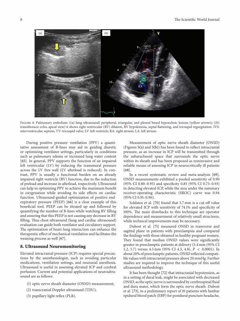
Pulmonary embolism (PE) (Figure 4), “mainly periph-eral” can be diagnosed sonographically by the recognition ofa peripheral, triangular, and pleural based hypoechoic lesion[5]. Mathis et al. [63], in a multicenter study that involves 352patients, defined diagnostic criteria as (1) PE confirmed: twoor more typical triangular or rounded pleural-based lesions;(2) PE probable: one typical lesion with pleural effusion; (3)PE possible: small (<5mm) subpleural lesions or a singlepleural effusion only.The sensitivity was 74%, specificity 95%,positive predictive value 95%, negative predictive value 75%,and accuracy 84%.
Laursen et al. [64] have studied the utility of lung ultra-sound in near-drowning victims. Lung ultrasound showedmultiple B-lines on the anterior and lateral surfaces of bothlungs, consistent with pulmonary edema.These findings mayencourage anesthesiologists to consider lung ultrasound fordiagnosing aspiration pneumonia during anesthesia.
The Scientific World Journal 7
(a) (b)
(c) (d)
(e) (f)
Figure 3: Normal lung ultrasound: (a) 2D “red arrows” point to the pleura, where the normal “sliding sign” should be seen, while the “yellowarrows” represent the A-lines that are normal reverberation from the pleura. (b) M-mode shows the “seashore sign.” Pneumothorax: (c) 2D;absence of lung sliding, (d) M-mode; “stratosphere sign” or “barcode sign,” lung point may also be seen during inspiration and representsthe border between pneumothorax and normal pleura. Cardiac pulmonary edema: (e) homogeneous distribution of B-lines (yellow arrows),normal sliding, and no spared areas. Acute respiratory distress syndrome (ARDS): (f) “patchy” distribution of B-lines, reduced/abolishedsliding, spared areas, and peripheral consolidations.
8 The Scientific World Journal
RA LA
LVTV
IVS
(a) (b)
Figure 4: Pulmonary embolism: ((a) lung ultrasound) peripheral, triangular, and pleural based hypoechoic lesions (yellow arrows); ((b)transthoracic echo, apical view) it shows right ventricular (RV) dilation, RV hypokinesia, septal flattening, and tricuspid regurgitation. IVS:interventricular septum; TV: tricuspid valve; LV: left ventricle; RA: right atrium; LA: left atrium.
During positive pressure ventilation (PPV) a quanti-tative assessment of B-lines may aid in guiding diureticor optimizing ventilator settings, particularly in conditionssuch as pulmonary edema or increased lung water content[65]. In general, PPV supports the function of an impairedleft ventricular (LV) by reducing the transmural pressureacross the LV free wall (LV afterload is reduced). In con-trast, PPV is usually a functional burden on an alreadyimpaired right ventricle (RV) function, due to the reductionof preload and increase in afterload, respectively. Ultrasoundcan help in optimizing PPV to achieve the maximum benefitin oxygenation while avoiding its side effects on cardiacfunction. Ultrasound-guided optimization of positive end-expiratory pressure (PEEP) [66] is a clear example of thisbeneficial tool. PEEP can be titrated up and followed byquantifying the number of B-lines while watching RV fillingand assuring that this PEEP is not causing any decrease in RVfilling. Thus chest ultrasound (lung and cardiac ultrasound)evaluation can guide both ventilator and circulatory support.The optimization of heart-lung interaction can enhance thetherapeutic effect of mechanical ventilation and facilitates theweaning process as well [67].
8. Ultrasound Neuromonitoring
Elevated intracranial pressure (ICP) requires special precau-tions by the anesthesiologist, such as avoiding particularmedications, ventilation settings, and neuraxial anesthesia.Ultrasound is useful in assessing elevated ICP and cerebralperfusion. Current and potential applications of neuroultra-sound are as follows:
Measurement of optic nerve sheath diameter (ONSD)(Figures 5(a) and 5(b)) has been found to reflect intracranialpressure, as an increase in ICP will be transmitted throughthe subarachnoid space that surrounds the optic nervewithin its sheath and has been proposed as noninvasive andreliable means of assessing ICP in neurocritically ill patients[68].
In a recent systematic review and meta-analysis [69],ONSD measurements exhibited a pooled sensitivity of 0.90(95% CI 0.80–0.95) and specificity 0.85 (95% CI 0.73–0.93)in detecting elevated ICP, while the area under the summaryreceiver-operating characteristic (SROC) curve was 0.94(95% CI 0.91–0.96).
Soldatos et al. [70] found that 5.7mm is a cut-off valuefor elevated ICP with sensitivity of 74.1% and specificity of100%. The main drawbacks to this technique are operatordependence and measurement of relatively small structures,while technical improvements may be necessary.
Dubost et al. [71] measured ONSD in transverse andsagittal plane in patients with preeclampsia and comparedthe findings with those obtained in healthy pregnant women.They found that median ONSD values were significantlygreater in preeclamptic patients at delivery (5.4mm (95% CI5.2, 5.7) versus 4.5mm (95% CI 4.3, 4.8), 𝑃 < 0.0001). Inabout 20%of preeclamptic patients,ONSD reflected compati-ble valueswith intracranial pressure above 20mmHg. Furtherstudies are required to improve the technique of this usefulultrasound methodology.
It has been thought [72] that intracranial hypotension, asin a setting of dural leak, might be associated with decreasedONSD, as the optic nerve is surrounded by cerebrospinal fluidand dura mater, which form the optic nerve sheath. Dubostet al. [73], in a preliminary report of 10 patients with lumbarepidural blood patch (EBP) for postdural puncture headache,
The Scientific World Journal 9
ONSD
(a)
ONSD
(b)
(c) (d)
Figure 5: Optic nerve sheath: (a) normal diameter and (b) large diameter that represents increase intracranial pressure. ONSD: optic nervesheath diameter. It is usually measured 3mm behind the retina. Ultrasound pupillary light reflex: (c) diameter of the pupil before shininglight to the contralateral eye and (d) the pupil constricted after shining the light.
indeed found that successful EBP was followed by ONSDenlargement.
Ultrasound assessment of the pupillary light reflex (PLR)was initially developed for the U.S. Space Program (NASA)and is not standardized for clinical use. However, the methodcan be used even when visual access to the pupil is impos-sible, and interpreting its results is straightforward [74].Consensual pupillary light reflex is elicited with contralateraltransillumination through the eyelids with both eyes closed(Figures 5(c) and 5(d)). The pupillary light reflex ultrasoundtest can be conducted with a linear array probe at the highestavailable frequency (e.g., 12–15MHz), using the coronal pri-mary view,whileM-modemeasurements are used tomeasurethe constriction velocity of the PLR [75]. This method mightbe used as pupillometry as well.
Transcranial color coded duplex (TCCD) (Figure 6) isan accurate, real-time, noninvasive (permits bedside exam-ination), and inexpensive tool used for the study of the
intracranial circulation and the diagnosis of nonthrombosedaneurysms, largely due to its ability to reveal flow phenomena[76]. The main limitation of TCCD is the few availableultrasonic windows, which can limit the area of insonationof the cerebral arteries including their proximal branchingand lower spatial resolution and can obstruct transtemporalinsonation [77]. TCCD has advantages over transcranialDoppler (TCD) by showing the images of the intracranialanatomy and arteries throughout duplex B-mode, while stillhaving the capacity to measure velocities using Doppler. Inother words, different from TCD technology, TCCD shootsmultiple ultrasound beams to expose a larger brain areaat dual emitting frequencies, one for gray scale imagingand one for Doppler imaging. Thus this tool can illustratearterial position on color flow imaging as well as on B-modeultrasonography [78]. TCD and TCCD measured velocitiesare comparable using zero angle correction, resulting inmoreaccurate measurement of flow velocities and allowing for
10 The Scientific World Journal
(a)
End diastolic velocity (EDV)
Early diastolic hump
Dicrotic shoulder
Catacrotic shoulder
Systolic peak velocity (SPV)
S D
(b)
Figure 6: Transcranial color coded duplex (TCCD): (a) middle cerebral artery (MCA) color Doppler and (b) MCA pulsed wave Doppler. S:systole, D: diastole.
superior precision in order to define intracranial arterialnarrowing. TCCD can be used for monitoring of cerebralblood flow alterations which follow traumatic brain injuryand in patients with sickle cell anemia. It also can be usedin the detection of patent foramen ovale and in the diagnosisof cerebral circulatory arrest which is a component of braindeath [79].
9. Gastric Ultrasound
A full stomach may lead to aspiration pneumonia and subse-quent morbidities. Anesthesiologists may encounter patientswith unknown prandial status, and even fasting “sufficient”time cannot guarantee an empty stomach in many cases (e.g.,in the elderly or in patients with gastroparesis). Ultrasoundcan help in this setting, and the perioperative evaluationof bowel motility is also feasible by means of sonography.Current and potential applications of Gastric ultrasound areas follows:
(1) assessment of gastric content and diagnosis of fullstomach;
(2) confirmation of gastric tube placement.
Bouvet et al. [80], measured the antral cross-sectionalarea (CSA) in 180 patients after intubation and analyzed therelationship between antral CSA and the volume of gastriccontents. The cut-off value of antral CSA of 340mm2 forthe diagnosis of “at risk” stomach was associated with asensitivity of 91% and a specificity of 71%. The area underthe receiver operating characteristic (ROC) curve for thediagnosis of “at-risk” stomach was 90%. (“At risk” stomachwas defined as the presence of solid particles and/or gastricfluid volume more than 0.8mL/kg.) These findings showthat antral CSA volume assessment can be important inminimizing the risk of pulmonary aspiration of gastriccontents. Perlas et al. [81] performed gastric sonography in86 patients before anesthetic induction, and patients were
classified using a 3-point grading system; grade 0 (emptyantrum); grade 1 (minimal fluid volume detected only in theright lateral decubitus position (16 +/− 36mL, within normalranges expected for fasted patients); and grade 2 (antrumclearly distended with fluid visible in both supine and lateralpositions (180 +/− 83mL, beyond previously reported “safe”limits). One patient with a grade 2 antrum had an episode ofa significant regurgitation of gastric contents on emergencefrom anesthesia. They concluded that this grading systemcould be a promising “biomarker” to assess perioperativeaspiration risk. Perlas et al. [82], in another work, validated amathematicalmodel for quantitativeUS assessment of gastricvolume. Arzola et al. [83] found that anesthesiologists willachieve a 95% success rate in bedside qualitative ultrasoundassessment after performing approximately 33 examinations,with appropriate training and supervision.
Confirmation of a gastric tube placement is also possibleusing ultrasound [84], which might replace the conventionalradiography method unless sonography is inconclusive.
10. Focused Transthoracic Echo (TTE)
Focus assessed transthoracic echo (FATE) was introducedby Jensen et al. [85] for cardiopulmonary monitoring in theintensive care unit. This approach basically involves fourstandardized acoustic views for cardiopulmonary screeningand monitoring (Figure 7). Recent studies show a greatimpact of FATE in preoperative assessment [86, 87] when itis performed by anesthesiologists. Dennis and Stenson [88],recently presented a case that shows how anesthesiologists,using basic TTE skills, can diagnose and save a patient withpostpartum hemorrhage by using a rapid obstetric screeningechocardiography approach [89]. Learning the basic skillsto perform focused transthoracic echo allows assessing theglobal function of the heart and diagnosing certain patholo-gies (e.g., pulmonary embolism). Cowie [90] presented their3-year experience of focused cardiovascular ultrasound in
The Scientific World Journal 11
LV
RV
LVOTAV
MV
(a)
LV
LARA
RV
(b)
LV
Liver
(c)
AVTV
RV
RAMV
(d)
Figure 7: Basic transthoracic echo views: (a) left parasternal long axis, (b) apical, (c) subcostal, and (d) left parasternal short axis; aortic valve“Mercedes sign,” mitral valve “fish mouth sign,” and papillary muscles (two arrows), respectively, from left to right. RV: right ventricle, LV:left ventricle, LVOT: left ventricular outlet, RA: right atrium, LA: left atrium, AV: aortic valve, MV: mitral valve, and TV: tricuspid valve.
the perioperative period, which shows that focused car-diovascular ultrasound performed by anesthesiologists inthe perioperative period accurately detects major cardiacpathology and significantly alters perioperativemanagement.Neelankavil et al. [91] suggested a simulation method to trainanesthesiologists in basic transthoracic echocardiographyskills. Tanzola et al. [92] have suggested that implementationof a focused bedside TTE curriculum within anesthesiaresidency training is feasible, quantifiable, and effective forincreasing anesthesia residents’ TTE knowledge.
Recent studies show that preoperative excess testing andconsultation are common, adding to the cost of care withoutnoticeably improving patient outcome. These findings mustencourage anesthesiologists to play an effective role in thepreoperative assessment field by implementing clinicallyinnovative approaches and developing training curricula aswell as performing research [93]. Therefore, we stronglyencourage the use of a focused protocol for perioperativeassessment and incorporation of a training curriculum inresidency training.
11. Technological Advances
New technologies have greatly improved the image quality,diagnostic abilities, and size of theUSmachine.These includeadvances in transducers, scanning schemes, three- and four-dimensional visualization, contrast agents (microbubbles),strain imaging, and others [94].
The matrix array is a new transducer with improvedresolution; it has a lens that is placed in front of thepiezoelectric element to allow a mechanical focusing in theY- andZ-planes. Four-dimensional ultrasound provides real-time 3D images (the 4th “D” is time) and currently is used forfetal imaging, where it provides remarkable images. It mayhave potential applications in our field. Endobronchial andendoscopic ultrasounds are two other new modalities withgreat and potential implications [95].
Recently, the use of three-dimensional high resolutionultrasound was reported [96] for nerve blockade, providingbetter visualization of local anesthetic spread and cathetertip location. Small and portable ultrasound systems have
12 The Scientific World Journal
become increasingly available, even a mobile ultrasound-guided peripheral nerve block has been developed [97]. TheSonixGPS needle guidance system (Ultrasonix, Richmond,BC, Canada) is a GPS technology with a new needle trackingsystem, using sensors in both the needle and transducer toobtain a real-time image of needle shaft and tip positionrelated to the ultrasound beam that is based on the needletrajectory. This can be used for vascular access and nerveblocks. Recently, it has been used for real-time thoracicparavertebral block [98] and spinal anesthesia [99] in pilotstudies.
12. Conclusion
Ultrasound is a unique tool which provides the anesthesi-ologist with diagnostic and monitoring capabilities enablingoptimization of perioperative management. Indeed, ultra-sound has an important role in problem-based managementof various anesthesiology emergencies such as hypoxia,hypotension, dyspnea, and cardiopulmonary arrest. Finally,procedural ultrasound applications in the field of anesthesi-ology are numerous and improve the quality of care.
We believe that ultrasound can be the third eye of theanesthesiologist that helps in the performance of previouslyblind procedures and allows discovery ofmany hidden spacesto uncover their mysteries. Anesthesiologists, in the nearfuture, may need to carry a portable ultrasound around theirneck instead of a stethoscope.
Conflict of Interests
The authors declare no conflict of interests.
Acknowledgment
The authors are thankful to Dr. Rayan S. Terkawi, an internmedical doctor at King Fahad Medical City, Riyadh, SaudiArabia, who designed (Figure 1) in this paper.
References
[1] J. P. Kline, “Ultrasound guidance in anesthesia,” AANA Journal,vol. 79, no. 3, pp. 209–217, 2011.
[2] C. R. Falyar, “Ultrasound in anesthesia: applying scientificprinciples to clinical practice,” AANA Journal, vol. 78, no. 4, pp.332–340, 2010.
[3] A. Hatfield and A. Bodenham, “Ultrasound: an emerging rolein anaesthesia and intensive care,”British Journal of Anaesthesia,vol. 83, no. 5, pp. 789–800, 1999.
[4] M. S. Kristensen, “Ultrasonography in the management of theairway,” Acta Anaesthesiologica Scandinavica, vol. 55, no. 10, pp.1155–1173, 2011.
[5] K. Stefanidis, S. Dimopoulos, and S. Nanas, “Basic principlesand current applications of lung ultrasonography in the inten-sive care unit,” Respirology, vol. 16, no. 2, pp. 249–256, 2011.
[6] S. Kaddoura, Echo Made Easy, Elsevier, 2009.[7] Z. J. Koscielniak-Nielsen, “Ultrasound-guided peripheral nerve
blocks: what are the benefits?” Acta Anaesthesiologica Scandi-navica, vol. 52, no. 6, pp. 727–737, 2008.
[8] J. Griffin and B. Nicholls, “Ultrasound in regional anaesthesia,”Anaesthesia, vol. 65, supplement 1, pp. 1–12, 2010.
[9] S. S. Liu, J. Ngeow, and R. S. John, “Evidence basis forultrasound-guided block characteristics: onset, quality, andduration,” Regional Anesthesia and Pain Medicine, vol. 35, no.2, pp. S26–S35, 2010.
[10] G. Ivani and V. Mossetti, “Pediatric regional anesthesia,” Min-erva Anestesiologica, vol. 75, no. 10, pp. 577–583, 2009.
[11] K. Rubin, D. Sullivan, and S. Sadhasivam, “Are peripheral andneuraxial blocks with ultrasound guidance more effective andsafe in children?” Paediatric Anaesthesia, vol. 19, no. 2, pp. 92–96, 2009.
[12] A. Perlas, “Evidence for the use of ultrasound in neuraxialblocks,” Regional Anesthesia and Pain Medicine, vol. 35, no. 2,pp. S43–S46, 2010.
[13] K. J. Chin and A. Perlas, “Ultrasonography of the lumbar spinefor neuraxial and lumbar plexus blocks,” Current Opinion inAnaesthesiology, vol. 24, no. 5, pp. 567–572, 2011.
[14] M. K. Karmakar, X. Li, A. M.-H. Ho, W. H. Kwok, and P.T. Chui, “Real-time ultrasound-guided paramedian epiduralaccess: evaluation of a novel in-plane technique,” British Journalof Anaesthesia, vol. 102, no. 6, pp. 845–854, 2009.
[15] H. Willschke, P. Marhofer, A. Basenberg et al., “Epiduralcatheter placement in children: comparing a novel approachusing ultrasound guidance and a standard loss-of-resistancetechnique,” British Journal of Anaesthesia, vol. 97, no. 2, pp. 200–207, 2006.
[16] H. Jee, J. H. Lee, J. Kim, K. D. Park, W. Y. Lee, and Y.Park, “Ultrasound-guided selective nerve root block versusfluoroscopy-guided transforaminal block for the treatment ofradicular pain in the lower cervical spine: a randomized,blinded, controlled study,” Skeletal Radiology, vol. 42, no. 1, pp.69–78, 2012.
[17] S. N. Narouze, A. Vydyanathan, L. Kapural, D. I. Sessler,and N. Mekhail, “Ultrasound-guided cervical selective nerveroot block A fluoroscopy-controlled feasibility study,” RegionalAnesthesia and Pain Medicine, vol. 34, no. 4, pp. 343–348, 2009.
[18] M. Gofeld, S. J. Bristow, S. C. Chiu, C. K. McQueen, and L.Bollag, “Ultrasound-guided lumbar transforaminal injections:feasibility and validation study,” Spine, vol. 37, no. 9, pp. 808–812, 2012.
[19] D. H. Ha, D. M. Shim, T. K. Kim, Y. M. Kim, and S.S. Choi, “Comparison of ultrasonography- and fluoroscopy-guided facet joint block in the lumbar spine,” Asian SpineJournal, vol. 4, pp. 15–22, 2010.
[20] H. Jung, S. Jeon, S. Ahn, M. Kim, and Y. Choi, “The validationof ultrasound-guided lumbar facet nerve blocks as confirmedby fluoroscopy,” Asian Spine Journal, vol. 6, pp. 163–167, 2012.
[21] I. Khayata, J. Lance Lichtor, and P. Amelin, “Ultrasound-guidedepidural blood patch,” Anesthesiology, vol. 114, no. 6, Article ID1453, 2011.
[22] S. R. Clendenen, S. Pirris, C. B. Robards, B. Leone, and E. W.Nottmeier, “Symptomatic postlaminectomy cerebrospinal fluidleak treated with 4-dimensional ultrasound-guided epiduralblood patch,” Journal of Neurosurgical Anesthesiology, vol. 24,pp. 222–225, 2012.
[23] D. J. Berkoff, L. E. Miller, and J. E. Block, “Clinical utilityof ultrasound guidance for intra-articular knee injections: areview,” Clinical Interventions in Aging, vol. 7, pp. 89–95, 2012.
The Scientific World Journal 13
[24] C. A. Gilliland, L. D. Salazar, and J. R. Borchers, “Ultra-sound versus anatomic guidance for intra-articular and peri-articular injection: a systematic review,” The Physician andSportsmedicine, vol. 39, no. 3, pp. 121–131, 2011.
[25] P. W. H. Peng and P. S. Tumber, “Ultrasound-guided inter-ventional procedures for patients with chronic pelvic pain:a description of techniques and review of literature,” PainPhysician, vol. 11, no. 2, pp. 215–224, 2008.
[26] A. Kumar and A. Chuan, “Ultrasound guided vascular access:efficacy and safety,” Best Practice and Research, vol. 23, no. 3, pp.299–311, 2009.
[27] M. Lamperti, A. R. Bodenham,M. Pittiruti et al., “Internationalevidence-based recommendations on ultrasound-guided vas-cular access,” IntensiveCareMedicine, vol. 38, pp. 1105–1117, 2012.
[28] A. Vezzani, C. Brusasco, S. Palermo, C. Launo, M. Mergoni,and F. Corradi, “Ultrasound localization of central vein catheterand detection of postprocedural pneumothorax: an alternativeto chest radiography,” Critical Care Medicine, vol. 38, no. 2, pp.533–538, 2010.
[29] D. Castillo, D. S. McEwen, L. Young, and J. Kirkpatrick, “Mi-cropuncture needles combined with ultrasound guidance forunusual central venous cannulation: desperate times call fordesperate measures: a new trick for old anesthesiologists,”Anesthesia and Analgesia, vol. 114, no. 3, pp. 634–637, 2012.
[30] K. Sofi and S. Arab, “Ultrasound-guided central venouscatheterization in prone position,” Saudi Journal of Anaesthesia,vol. 4, pp. 28–30, 2010.
[31] T. J. Wigmore, J. F. Smythe, M. B. Hacking, R. Raobaikady,and N. S. MacCallum, “Effect of the implementation of NICEguidelines for ultrasound guidance on the complication ratesassociated with central venous catheter placement in patientspresenting for routine surgery in a tertiary referral centre,”British Journal of Anaesthesia, vol. 99, no. 5, pp. 662–665, 2007.
[32] J. L. H. French, N. J. Raine-Fenning, J. G. Hardman, and N. M.Bedforth, “Pitfalls of ultrasound guided vascular access: the useof three/four-dimensional ultrasound,” Anaesthesia, vol. 63, no.8, pp. 806–813, 2008.
[33] E. Oakley and A.-M. Wong, “Ultrasound-assisted peripheralvascular access in a paediatric ED: paediatric emergencymedicine,” Emergency Medicine Australasia, vol. 22, no. 2, pp.166–170, 2010.
[34] G. J. Latham, M. L. Veneracion, D. C. Joffe, A. T. Bosenberg,S. H. Flack, and D. K. Low, “High-frequency micro-ultrasoundfor vascular access in young children: a feasibility study by theHigh-frequency UltraSound in Kids studY (HUSKY) group,”Paediatric Anaesthesia, vol. 23, no. 6, pp. 529–535, 2013.
[35] M. Singh, K. J. Chin, V. W. S. Chan, D. T. Wong, G. A.Prasad, and E. Yu, “Use of sonography for airway assessment:an observational study,” Journal of Ultrasound in Medicine, vol.29, no. 1, pp. 79–85, 2010.
[36] J. S. Green and B. C. H. Tsui, “Applications of ultrasonographyin ENT: airway assessment and nerve blockade,” AnesthesiologyClinics, vol. 28, no. 3, pp. 541–553, 2010.
[37] A. Prasad, E. Yu, D. T. Wong, R. Karkhanis, P. Gullane, and V.W. S. Chan, “Comparison of sonography and computed tomog-raphy as imaging tools for assessment of airway structures,”Journal of Ultrasound in Medicine, vol. 30, no. 7, pp. 965–972,2011.
[38] S. Adhikari, W. Zeger, C. Schmier et al., “Pilot study to deter-mine the utility of point-of-care ultrasound in the assessment ofdifficult laryngoscopy,” Academic Emergency Medicine, vol. 18,no. 7, pp. 754–758, 2011.
[39] R. Komatsu, P. Sengupta, A. Wadhwa et al., “Ultrasound quan-tification of anterior soft tissue thickness fails to predict difficultlaryngoscopy in obese patients,”Anaesthesia and Intensive Care,vol. 35, no. 1, pp. 32–37, 2007.
[40] P. Rudraraju and L. A. Eisen, “Confirmation of endotrachealtube position: a narrative review,” Journal of Intensive CareMedicine, vol. 24, no. 5, pp. 283–292, 2009.
[41] B. Marciniak, P. Fayoux, A. Hebrard, R. Krivosic-Horber,T. Engelhardt, and B. Bissonnette, “Airway management inchildren: ultrasonography assessment of tracheal intubation inreal time?”Anesthesia andAnalgesia, vol. 108, no. 2, pp. 461–465,2009.
[42] J. E. Fiadjoe, P. Stricker, H. Gurnaney et al., “Ultrasound-guidedtracheal intubation: a novel intubation technique,” Anesthesiol-ogy, vol. 117, pp. 1389–1391, 2012.
[43] T.-Y. Hung, S. Li, P.-S. Chen et al., “Bedside ultrasonographyas a safe and effective tool to diagnose acute epiglottitis,” TheAmerican Journal of Emergency Medicine, vol. 29, no. 3, pp.351.e1–353.e1, 2011.
[44] D. R. Ko, Y. E. Chung, I. Park et al., “Use of bedside sonographyfor diagnosing acute epiglottitis in the emergency department,”Journal of Ultrasound in Medicine, vol. 31, no. 1, pp. 19–22, 2012.
[45] Y. Lahav, E. Rosenzweig, Z. Heyman, J. Doljansky, A. Green,and Y. Dagan, “Tongue base ultrasound: a diagnostic toolfor predicting obstructive sleep apnea,” Annals of Otology,Rhinology and Laryngology, vol. 118, no. 3, pp. 179–184, 2009.
[46] K.-H. Liu,W. C.W. Chu, K.-W. To et al., “Sonographicmeasure-ment of lateral parapharyngeal wall thickness in patients withobstructive sleep apnea,” Sleep, vol. 30, no. 11, pp. 1503–1508,2007.
[47] A. Sustic, “Role of ultrasound in the airway management ofcritically ill patients,” Critical Care Medicine, vol. 35, no. 5, pp.S173–S177, 2007.
[48] J. B. Brodsky, A. Macario, and J. B. D. Mark, “Tracheal diameterpredicts double-lumen tube size: a method for selecting leftdouble-lumen tubes,” Anesthesia and Analgesia, vol. 82, no. 4,pp. 861–864, 1996.
[49] J. B. Brodsky, K. Malott, M. Angst, B. G. Fitzmaurice, S. P. Kee,and L. Logan, “The relationship between tracheal width and leftbronchial width: implications for left-sided double-lumen tubeselection,” Journal of Cardiothoracic and Vascular Anesthesia,vol. 15, no. 2, pp. 216–217, 2001.
[50] B. Kaur, R. Tang, A. Sawka, C. Krebs, and H. Vaghadia,“A method for ultrasonographic visualization and injectionof the superior laryngeal nerve: volunteer study and cadaversimulation,” Anesthesia and Analgesia, vol. 115, no. 5, pp. 1242–1245, 2012.
[51] A. Hatfield and A. Bodenham, “Portable ultrasonic scanningof the anterior neck before percutaneous dilatational tra-cheostomy,” Anaesthesia, vol. 54, no. 7, pp. 660–663, 1999.
[52] V. Rajajee, J. J. Fletcher, L. R. Rochlen, and T. L. Jacobs,“Real-time ultrasound-guided percutaneous dilatational tra-cheostomy: a feasibility study,”Critical Care, vol. 15, no. 1, articleR67, 2011.
[53] L.-W.Ding,H.-C.Wang,H.-D.Wu, C.-J. Chang, and P.-C. Yang,“Laryngeal ultrasound: a useful method in predicting post-extubation stridor: a pilot study,” European Respiratory Journal,vol. 27, no. 2, pp. 384–389, 2006.
[54] J.-R. Jiang, T.-H. Tsai, J.-S. Jerng, C.-J. Yu, H.-D. Wu, and P.-C.Yang, “Ultrasonographic evaluation of liver/spleen movementsand extubation outcome,” Chest, vol. 126, no. 1, pp. 179–185,2004.
14 The Scientific World Journal
[55] A. Vats, G. A.Worley, R. De Bruyn, H. Porter, D. M. Albert, andC.M.Bailey, “Laryngeal ultrasound to assess vocal fold paralysisin children,” Journal of Laryngology and Otology, vol. 118, no. 6,pp. 429–431, 2004.
[56] G. A. Shaath, A. Jijeh, A. Alkurdi, S. Ismail, M. Elbarbary, andM. S. Kabbani, “Ultrasonography assessment of vocal cordsmobility in children after cardiac surgery,” Journal of the SaudiHeart Association, vol. 24, pp. 187–190, 2012.
[57] P. Kundra, K. Kumar, V. Allampalli, R. Anathkrishnan, S.Gopalakrishnan, and S. Elangovan, “Use of ultrasound to assesssuperior and recurrent laryngeal nerve function immediatelyafter thyroid surgery,” Anaesthesia, vol. 67, no. 3, pp. 301–302,2012.
[58] D. A. Lichtenstein and G. A. Meziere, “Relevance of lungultrasound in the diagnosis of acute respiratory failure: theBLUE protocol,” Chest, vol. 134, no. 1, pp. 117–125, 2008.
[59] G. Volpicelli, M. Elbarbary, M. Blaivas et al., “Internationalevidence-based recommendations for point-of-care lung ultra-sound,” Intensive Care Medicine, vol. 38, pp. 577–591, 2012.
[60] D. A. Lichtenstein, N. Lascols, S. Prin, and G. Meziere, “The“lung pulse”: an early ultrasound sign of complete atelectasis,”Intensive Care Medicine, vol. 29, no. 12, pp. 2187–2192, 2003.
[61] R. Copetti, G. Soldati, and P. Copetti, “Chest sonography:a useful tool to differentiate acute cardiogenic pulmonaryedema from acute respiratory distress syndrome,” Cardiovascu-lar Ultrasound, vol. 6, article 16, 2008.
[62] G. Volpicelli, L. A. Melniker, L. Cardinale, A. Lamorte, and M.F. Frascisco, “Lung ultrasound in diagnosing and monitoringpulmonary interstitial fluid,” La Radiologia Medica, vol. 118, no.2, pp. 196–205, 2012.
[63] G. Mathis, W. Blank, A. Reissig et al., “Thoracic ultrasoundfor diagnosing pulmonary embolism: a prospective multicenterstudy of 352 patients,” Chest, vol. 128, no. 3, pp. 1531–1538, 2005.
[64] C. B. Laursen, J. R. Davidsen, and P. H. Madsen, “Utility of lungultrasound in near-drowning victims,” BMJ Case Reports, vol.2012, 2012.
[65] R. Copetti, L. Cattarossi, F. Macagno,M. Violino, and R. Furlan,“Lung ultrasound in respiratory distress syndrome: a useful toolfor early diagnosis,” Neonatology, vol. 94, no. 1, pp. 52–59, 2008.
[66] T. Luecke, F. Corradi, and P. Pelosi, “Lung imaging for titrationof mechanical ventilation,” Current Opinion in Anaesthesiology,vol. 25, no. 2, pp. 131–140, 2012.
[67] N. Xirouchaki, E. Magkanas, K. Vaporidi et al., “Lung ultra-sound in critically ill patients: comparison with bedside chestradiography,” Intensive Care Medicine, vol. 37, no. 9, pp. 1488–1493, 2011.
[68] R. Moretti and B. Pizzi, “Ultrasonography of the optic nerve inneurocritically ill patients,” Acta Anaesthesiologica Scandinav-ica, vol. 55, no. 6, pp. 644–652, 2011.
[69] J. Dubourg, E. Javouhey, T. Geeraerts, M. Messerer, and B.Kassai, “Ultrasonography of optic nerve sheath diameter fordetection of raised intracranial pressure: a systematic reviewand meta-analysis,” Intensive Care Medicine, vol. 37, no. 7, pp.1059–1068, 2011.
[70] T. Soldatos, D. Karakitsos, K. Chatzimichail, M. Papathanasiou,A. Gouliamos, and A. Karabinis, “Optic nerve sonography inthe diagnostic evaluation of adult brain injury,” Critical Care,vol. 12, no. 3, article R67, 2008.
[71] C. Dubost, A. Le Gouez, V. Jouffroy et al., “Optic nerve sheathdiameter used as ultrasonographic assessment of the incidenceof raised intracranial pressure in preeclampsia: a pilot study,”Anesthesiology, vol. 116, pp. 1066–1071, 2012.
[72] M. Rollins and P. Flood, “Imaging intracranial pressure: anintroduction to ultrasonography of the optic nerve sheath,”Anesthesiology, vol. 116, pp. 983–984, 2012.
[73] C. Dubost, A. Le Gouez, P. J. Zetlaoui, D. Benhamou, F. J.Mercier, and T. Geeraerts, “Increase in optic nerve sheath diam-eter induced by epidural blood patch: a preliminary report,”British Journal of Anaesthesia, vol. 107, no. 4, pp. 627–630, 2011.
[74] L. Chiao, S. Sharipov, A. E. Sargsyan et al., “Ocular examinationfor trauma; clinical ultrasound aboard the International SpaceStation,” Journal of Trauma, vol. 58, no. 5, pp. 885–889, 2005.
[75] A. E. Sargsyan, D. R. Hamilton, S. L. Melton, D. Amponsah,N. E. Marshall, and S. A. Dulchavsky, “Ultrasonic evaluation ofpupillary light reflex,” Critical Ultrasound Journal, vol. 1, no. 2,pp. 53–57, 2009.
[76] M. Nedelmann, E. Stolz, T. Gerriets et al., “Consensus recom-mendations for transcranial color-coded duplex sonography forthe assessment of intracranial arteries in clinical trials on acutestroke,” Stroke, vol. 40, no. 10, pp. 3238–3244, 2009.
[77] F. A. Rasulo, E. De Peri, and A. Lavinio, “Transcranial Dopplerultrasonography in intensive care,” European Journal of Anaes-thesiology, vol. 25, no. 42, pp. 167–173, 2008.
[78] E. Bor-Seng-Shu, R. Hirsch, M. J. Teixeira, A. F. De Andrade,and R. Marino Jr., “Cerebral hemodynamic changes gauged bytranscranial Doppler ultrasonography in patients with post-traumatic brain swelling treated by surgical decompression,”Journal of Neurosurgery, vol. 104, no. 1, pp. 93–100, 2006.
[79] M. A. Topcuoglu, “Transcranial Doppler ultrasound in neu-rovascular diseases: diagnostic and therapeutic aspects,” Journalof Neurochemistry, vol. 123, supplement 2, pp. 39–51, 2012.
[80] L. Bouvet, J.-X.Mazoit, D. Chassard, B. Allaouchiche, E. Boselli,andD. Benhamou, “Clinical assessment of the ultrasonographicmeasurement of antral area for estimating preoperative gastriccontent and volume,” Anesthesiology, vol. 114, no. 5, pp. 1086–1092, 2011.
[81] A. Perlas, L. Davis, M. Khan, N. Mitsakakis, and V. W. S. Chan,“Gastric sonography in the fasted surgical patient: a prospectivedescriptive study,” Anesthesia and Analgesia, vol. 113, no. 1, pp.93–97, 2011.
[82] A. Perlas, N. Mitsakakis, L. Liu et al., “Validation of a mathe-matical model for ultrasound assessment of gastric volume bygastroscopic examination,” Anesthesia and Analgesia, vol. 116,pp. 357–363, 2013.
[83] C. Arzola, J. C. Carvalho, J. Cubillos, X. Y. Ye, and A. Perlas,“Anesthesiologists’ learning curves for bedside qualitative ultra-sound assessment of gastric content: a cohort study,” CanadianJournal of Anaesthesia, vol. 60, no. 8, pp. 771–779, 2013.
[84] C. Vigneau, J.-L. Baudel, B. Guidet, G.Offenstadt, and E.Maury,“Sonography as an alternative to radiography for nasogastricfeeding tube location,” Intensive Care Medicine, vol. 31, no. 11,pp. 1570–1572, 2005.
[85] M. B. Jensen, E. Sloth, K. M. Larsen, and M. B. Schmidt,“Transthoracic echocardiography for cardiopulmonary moni-toring in intensive care,” European Journal of Anaesthesiology,vol. 21, no. 9, pp. 700–707, 2004.
[86] D. J. Canty, C. F. Royse, D. Kilpatrick, L. Bowman, and A. G.Royse, “The impact of focused transthoracic echocardiographyin the pre-operative clinic,” Anaesthesia, vol. 67, pp. 618–625,2012.
[87] D. J. Canty, C. F. Royse, D. Kilpatrick, D. L. Williams, and A.G. Royse, “The impact of pre-operative focused transthoracicechocardiography in emergency non-cardiac surgery patients
The Scientific World Journal 15
with known or risk of cardiac disease,” Anaesthesia, vol. 67, pp.714–720, 2012.
[88] A. Dennis and A. Stenson, “Echo rounds: the use of transtho-racic echocardiography in postpartumhypotension,”Anesthesiaand Analgesia, vol. 115, pp. 1033–1037, 2012.
[89] A. T. Dennis, “Transthoracic echocardiography in obstetricanaesthesia and obstetric critical illness,” International Journalof Obstetric Anesthesia, vol. 20, no. 2, pp. 160–168, 2011.
[90] B. Cowie, “Three years’ experience of focused cardiovascularultrasound in the peri-operative period,” Anaesthesia, vol. 66,no. 4, pp. 268–273, 2011.
[91] J. Neelankavil, K. Howard-Quijano, T. C. Hsieh et al.,“Transthoracic echocardiography simulation is an efficientmethod to train anesthesiologists in basic transthoracicechocardiography skills,” Anesthesia and Analgesia, vol. 115, pp.1042–1051, 2012.
[92] R. C. Tanzola, S. Walsh, W. M. Hopman, D. Sydor, R. Arellano,and R. V. Allard, “Brief report: focused transthoracic echocar-diography training in a cohort of Canadian anesthesiologyresidents: a pilot study,” Canadian Journal of Anesthesia, vol. 60,pp. 32–37, 2013.
[93] M. F. Newman, J. P. Mathew, and S. Aronson, “The evolutionof anesthesiology and perioperative medicine,” Anesthesiology,vol. 118, pp. 1005–1007, 2013.
[94] P. N. T.Wells, H.-D. Liang, and T. P. Young, “Ultrasonic imagingtechnologies in perspective,” Journal ofMedical Engineering andTechnology, vol. 35, no. 6-7, pp. 289–299, 2011.
[95] R. T. O’Brien and S. P. Holmes, “Recent advances in ultrasoundtechnology,” Clinical Techniques in Small Animal Practice, vol.22, no. 3, pp. 93–103, 2007.
[96] O. Choquet and X. Capdevila, “Case report: three-dimensionalhigh-resolution ultrasound-guided nerve blocks: a newpanoramic vision of local anesthetic spread and perineuralcatheter tip location,” Anesthesia and Analgesia, vol. 116, pp.1176–1181, 2013.
[97] C. L. Jeng, T. M. Torrillo, M. R. Anderson, R. S. Morrison,K. H. Todd, and M. A. Rosenblatt, “Development of a mobileultrasound-guided peripheral nerve block and catheter service,”Journal of Ultrasound in Medicine, vol. 30, no. 8, pp. 1139–1144,2011.
[98] B. Kaur, H. Vaghadia, R. Tang, and A. Sawka, “Real-timethoracic paravertebral block using an ultrasound-guided posi-tioning system,” British Journal of Anaesthesia, vol. 110, pp. 852–853, 2013.
[99] S. W.Wong, A. U. Niazi, K. J. Chin, and V. W. Chan, “Real-timeultrasound-guided spinal anesthesia using the SonixGPS(R)needle tracking system: a case report,” Canadian Journal ofAnesthesia, vol. 60, pp. 50–53, 2013.