Biochimica et Biophysica Acta 1768 (2007) 794–807www.elsevier.com/locate/bbamem
Abbreviations: !2AR, beta 2 adrenoceptor; GPCR, G protein coupled receptor; TM, transmembrane! Amino acid numbering: The position of !2AR residues are followed by the Ballesteros general number [1] in superscript, in the form X.YY, where X refers to theTM segment and YY to the position relative to the most highly conserved amino acid in the TM segment, which is assigned an arbitrary position of 50.
G protein coupled receptors (GPCRs) represent the largestfamily of membrane proteins in the human genome and therichest source of targets for the pharmaceutical industry. Therehas been remarkable progress in the field of GPCR biologyduring the past two decades. Notable milestones include thecloning of the first GPCR genes, and the sequencing of thehuman genome revealing the size of the GPCR family and thenumber of orphan GPCRs. Moreover, there is a growingappreciation that GPCR regulation and signaling is much morecomplex than originally envisioned, and includes signalingthrough G protein independent pathways [2–4]. Consequently,it has been proposed that the term GPCR be abandoned in favorof 7 transmembrane or 7TM receptors.
In spite of the remarkable advances in the biology andpharmacology of GPCRs, progress in the area of proteinstructure has been more limited. To date, the only high-resolution structures of a GPCR have been for bovinerhodopsin. In this manuscript I will briefly review thegroundbreaking structural work on rhodopsin and discuss thechallenges in obtaining high-resolution structures of otherGPCRs. I will also discuss what we know about the structuralchanges associated with receptor activation.
2. GPCR structure
2.1. Common structural features of GPCRs
G protein coupled receptors (GPCRs) represent the singlelargest class of membrane proteins in the human genome. Arecent and detailed analysis of the human genome reveals over800 unique GPCRs, of which approximately 460 are predictedto be olfactory receptors [5]. Based on sequence similaritywithin the 7 TM segments, these receptors can be clustered into5 families: the rhodopsin family (701 members), the adhesionfamily (24 members), the frizzled/taste family (24 members),the glutamate family (15 members), and the secretin family (15members) [5]. The physiologic function of a large fraction ofthese 800 GPCRs is unknown; these receptors are referred to asorphan GPCRs. However, deorphanization of non-olfactoryGPCRs is an ongoing process [6], as they are a promising groupof targets for the pharmaceutical industry. Therefore, the actualnumber of orphan GPCRs continues to decline.
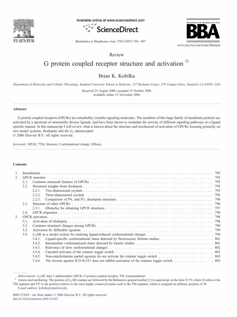
GPCRs share a common structural signature of sevenhydrophobic transmembrane (TM) segments, with an extra-cellular amino terminus and an intracellular carboxyl terminus(Fig. 1). GPCRs share the greatest homology within the TMsegments. The most variable structures among the family ofGPCRs are the carboxyl terminus, the intracellular loop
spanning TM5 and TM6, and the amino terminus. The greatestdiversity is observed in the amino terminus. This sequence isrelatively short (10–50 amino acids) for monoamine andpeptide receptors, and much larger (350–600 amino acids) forglycoprotein hormone receptors, and the glutamate familyreceptors. The largest amino terminal domains are observed inthe adhesion family receptors.
This structural and functional similarity of GPCRs stands incontrast to the structural diversity of the natural GPCR ligands[7]. These range from subatomic particles (a photon), to ions(H+ and Ca++), to small organic molecules, to peptides andproteins. The location of the ligand binding domains for manyGPCRs has been determined [7]. While many small organicagonists bind within the TM segments, peptide hormones andproteins often bind to the amino terminus and extracellularsequences joining the TM domains. However, size of the ligandalone cannot be used to predict the location of the binding site:for instance, glycoprotein hormones, glutamate, and Ca2+ allactivate their respective receptors by binding to relatively largeamino terminal domains [7,8]. It is interesting to note that it hasbeen possible to identify small molecular weight allostericmodulators that bind within the TM domains [9–14] for manyGPCRs that bind their native agonists on the extracellular loopsor the amino terminus.
2.2. Structural insights from rhodopsin
Our understanding of GPCR structure is based largely on thehigh-resolution structures of the inactive state of rhodopsin.Rhodopsin is better suited for structural studies than most otherGPCRs because it is possible to obtain large quantities of highlyenriched protein from bovine retina. Rhodopsin is also aremarkably stable GPCR, retaining function under conditionsthat denature many other GPCRs.
2.2.1. Two-dimensional crystalsThe first structures of rhodopsin came from cryoelectron
microscopy of two-dimensional crystals of bovine rhodopsinfrom Gebhard Schertler's group [15–19]. While the resolutionof these structures was limited (ranging from 5 to 9 Å), theyprovided the first picture of the orientation of the TM segmentsin a lipid environment. These structures, together with thesubsequent computational work from Baldwin [20] providedthe template from which most molecular models for otherGPCRs were generated.
2.2.2. Three-dimensional crystalsMore recently, three dimensional crystals structures of
rhodopsin have been obtained by several groups [21–26].Three-dimensional crystals of bovine rhodopsin have been
795B.K. Kobilka / Biochimica et Biophysica Acta 1768 (2007) 794–807
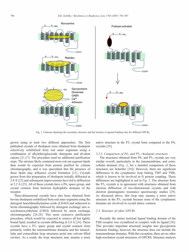
grown using at least two different approaches. The firstpublished crystals of rhodopsin were obtained from rhodopsinselectively solubilized from rod outer segments using acombination of alkyl(thio)glucoside detergents and divalentcations [21,27]. The procedure used no additional purificationsteps. The mixture likely contained more rod out segment lipidsthan would be expected from protein purified by columnchromatography, and it was speculated that the presence ofthese lipids may influence crystal formation [21]. Crystalsgrown from this preparation of rhodopsin initially diffracted at2.8 Å [22] and subsequent improvements have led to diffractionat 2.2 Å [25]. All of these crystals have a P41 space group, andcrystal contacts form between hydrophilic domains of thereceptor.
Three-dimensional crystals have also been obtained frombovine rhodopsin solubilized from rod outer segments using thedetergent lauryldimethylamine-oxide (LDAO) and subjected tolectin chromatography followed by detergent exchange into n-octyltetraoxyethylene (C8E4) followed by anion exchangechromatography [26,28]. This more extensive purificationprocedure, which would be expected to remove all but tightlybound lipid, resulted in crystals diffracting at 2.6 Å [26]. Thesecrystals have a P31 space group and crystal contacts formprimarily within the transmembrane domains and the intracel-lular and extracellular loop structures point into solvent filledcavities. As a result, the loop structures may assume a more
native structure in the P31 crystal form compared to the P41crystals [29].
2.2.3. Comparison of P41 and P31 rhodopsin structuresThe structures obtained from P41 and P31 crystals are very
similar overall, particularly in the transmembrane, and extra-cellular domains (Fig. 2, for a detailed comparison of thesestructures see Schertler [29]). However, there are significantdifferences in the cytoplasmic loop linking TM5 and TM6,which is known to be involved in G protein coupling. Thesedifferences are highlighted in red in Fig. 2. The structure fromthe P31 crystals is in agreement with structures obtained fromelectron diffraction of two-dimensional crystals and withelectron paramagnetic resonance spectroscopy studies [29].As discussed above, this loop may assume a more nativestructure in the P31 crystals because none of the cytoplasmicdomains are involved in crystal lattice contacts.
2.3. Structure of other GPCRs
Recently the amino terminal ligand binding domain of theFSH receptor was crystallized in complex with its ligand [30].This provides important structural insights into glycoproteinhormone binding; however, the structure does not include thetransmembrane domains. With this exception, there are no otherhigh-resolution crystal structures of GPCRS. Structure analysis
Fig. 1. Cartoons depicting the secondary structure and the location of agonist binding sites for different GPCRs.
of other GPCRs has largely been limited to the use of site-directed mutagenesis and cysteine scanning mutagenesis [31] todetect receptor–ligand interactions, and the use of engineeredmetal ion binding sites to probe intramolecular interactions [32].While these approaches provide low-resolution structuralinformation, this information can be used to verify and improvethe accuracy of homology models based on rhodopsin.Ballesteros and Javitch found that structural insights obtainedfrom mutagenesis data and substituted cysteine accessibilitystudies on monoamine receptors were consistent with the high-resolution structure of rhodopsin, suggesting that rhodopsinserves as a good template for homology modeling [33].However, rhodopsin might not be a good template for modelsof more distantly related family rhodopsin family members suchas the cholecystokinin CCK1 receptor [34].
2.3.1. Obstacles for obtaining GPCR structuresThe major obstacles to obtaining structures of other GPCRs
include protein production and purification, and protein stabilityand homogeneity. In terms of production, it is now possible togenerate sufficient quantities (tens of milligrams) of severalGPCRs for crystal screening using bacterial, yeast, insect cell,
and mammalian cell expression systems [35–40]. The avail-ability of robotic systems for preparing setups of 100 nlvolumes (or smaller) has enabled large parameter screens withrelatively small amounts of protein. As such, protein productionis no longer the major limitation for crystallography efforts.
Perhaps a greater problem is the stability of purified GPCRsin detergents compatible with crystallography. For example, the!2 adrenoceptor (!2AR) and many other GPCRs are not stablein the detergents used to obtain rhodopsin crystals; and thus farrhodopsin crystals have not been obtained in dodecylmaltoside,a detergent in which the !2AR is relatively stable. GPCRs tendto be more stable in non-ionic detergents with relatively longalkyl chains. These detergents may form larger micelles thatprevent the formation of crystal contacts [41]. Another problemis the potential for both structural and conformational hetero-geneity in GPCRs. By structural heterogeneity I meanheterogeneity in posttranslational modifications such as glyco-sylation, phosphorylation and palmitoylation. These sources ofheterogeneity can often be eliminated by site directedmutagenesis of the protein, or enzymatic removal of sugarsand phosphates. This source of heterogeneity is minimized if theGPCR can be expressed in bacteria.
Fig. 2. A comparison of the rhodopsin structures determined from the P41 [25] and P31 [26] crystal forms. The loop connecting TM5 and TM6 (shown in red) is themost divergent sequence.
797B.K. Kobilka / Biochimica et Biophysica Acta 1768 (2007) 794–807
The greatest obstacle to the formation of crystals may beconformational heterogeneity due to the inherent flexibility ofGPCRs. This flexibility may be functionally important,enabling structural changes associated with agonist bindingand activation of a membrane bound receptor; however, thisproperty may lead to the existence of different protein con-formations and to denaturation, particularly in detergentsolutions. There is evidence that the conformational hetero-geneity due to relative movements of the TM segments can bereduced by specific ligands [42]. Other sources of conforma-tional heterogeneity are the amino and carboxyl termini, and theloops connecting the TM segments. These sequences are oftenlonger than the homologous segments in rhodopsin and may beunstructured. In some cases the problem of conformationalheterogeneity can be mitigated by structural modifications;however, identifying these poorly structured domains can bechallenging. Moreover, removing hydrophilic sequences mayeliminate potential crystal lattice sites.
While the challenges facing GPCR structural biologists areformidable, I believe structures will begin to appear within thenext several years. The problems outlined above are solvable.This may require a highly focused effort on a specific target,where structural modifications and/or associated proteins(antibodies, arrestins, G proteins) are used to facilitate crystalformation. Another approach is to screen a large number ofGCPRs to identify those with the best characteristics for struc-tural studies. This approach has been used by a Europeanconsortium known as the Membrane Protein Network (MEP-NET, http://www.mepnet.org/) consisting of academic andindustrial groups. This program has made significant progressin identifying the best GPCR candidates for crystallographytrials.
2.4. GPCR oligomers
There is a growing body of evidence that GPCRs exist asdimers (or oligomers) and that these dimers may be importantfor G protein activation for at least some GPCR families. Thistopic has been addressed in several excellent reviews [43–46]and is the subject of another review in this series. Therefore,GPCR dimers will be only briefly discussed here. Dimerizationis clearly an important mechanism of receptor activation for theglutamate family of GPCRs [8,47], where ligand-inducedchanges in the dimer interface of the amino terminal ligandbinding domain has been demonstrated by crystallography[48,49]. However, the role of dimerization in the activation ofrhodopsin family members is less clear. For instance, recentcryoelectron microscopy images suggest that rhodopsin mayexist as homodimers in rod outer segment membranes [50]. Inaddition, neutron scattering studies provide evidence that apentameric complex forms when purified leukotriene B(4) isreconstituted with purified Gi, suggesting that a receptorhomodimer is needed to complex with a heterotrimeric Gprotein [51]. Nevertheless, it remains to be seen if a receptordimer is required for G protein activation. The effect of agonistbinding on the formation or disruption of dimers is notconsistent among the rhodopsin family members that have
been examined [43]. Moreover, ligands interact with individualreceptor monomers, and there is currently no evidence thatligands span the interface between receptor dimers. If changesin dimerization occur, it is likely a secondary consequence ofligand-induced changes in the arrangement of the TM segments.Evidence in support of this comes from biophysical studies onleukotriene B(4) homodimers demonstrating that ligand bindingto one protomer leads to conformational changes in its partner[52]. While dimers may be important for G protein activation, itis essential to understand the agonist-induced structural changesthat occur in the context of individual GPCR monomers.
3. GPCR activation
3.1. Activation of rhodopsin
The currently available three-dimensional, high-resolutionstructures of rhodopsin correspond to an inactive form of thereceptor. The most detailed information about structuralchanges associated with activation of a GPCR comes fromlower-resolution biophysical studies on rhodopsin. Rhodopsinstructure and what is known about its light-induced conforma-tional changes have been the subject of several excellentreviews [29,53–56], and some of the main points will be brieflydiscussed here. Electron paramagnetic resonance spectroscopy(EPR) studies provide evidence that photoactivation ofrhodopsin involves a rotation and tilting of TM6 relative toTM3 [57]. Further support for motion of TM6 during rhodopsinactivation was provided by chemical reactivity measurementsand fluorescence spectroscopy [58], as well as by ultravioletabsorbance spectroscopy [59] and by zinc crosslinking ofhistidines [60]. Light-induced conformational changes havealso been observed in the cytoplasmic domain spanning TM1and TM2, and the cytoplasmic end of TM7 [61–63].
Obtaining a high-resolution structure of metarhodopsin II,the active form of rhodopsin has been hindered by the instabilityof rhodopsin crystals following light activation. However, therehas been some progress towards this goal. Shertler's groupsucceeded in obtaining two dimensional crystals and a lowresolution map of metarhodopsin I [64,29]. Metarhodopsin I isan intermediate in the process of rhodopsin activation thatoccurs after photoisomerization of 11-Cisretinal, but beforestructural changes required for transducin activation (metarho-dopsin II). Electron diffraction studies reveal that the formationof metarhodopsin I is not accompanied by the large rigid-bodymovements in TM segments shown to be involved in rhodopsinactivation [57]. However, a more subtle change, consisting inthe rearrangement in the conformation of the Trp residue of thehighly conserved CWxxP motif in TM6, has been detected inthis intermediate. Thus, it seems that there is no gradualtransformation of the inactive protein into the active form, butthe activation is initiated through small scale changes in theconformation of some key residues, which will presumablytrigger the larger conformational changes related to thesubsequent stages of the activation process [29]. Recently,conditions for growing more light stable three-dimensionalcrystals of rhodopsin have been identified [65], suggesting that
a high-resolution structure of metarhodopsin II may be availablein the near future.
3.2. Common structural changes among GPCRs
In spite of the remarkable diversity of ligands and ligandbinding domains in the family of GPCRs, there is alsoconsiderable evidence for a common mechanism of activation.When comparing sequences, GPCRs are most similar at thecytoplasmic ends of the transmembrane segments adjacent tothe second and third cytoplasmic domains, the regions known tointeract with cytoplasmic G proteins [66]. Members of the largefamily of GPCRs transduce signals by activating one or moremembers of the relatively small family of highly homologousheterotrimeric G proteins. For example, the follicle stimulatinghormone (FSH) receptor is activated by a large glycoproteinhormone that binds to the amino terminus while the !2AR isactivated by adrenaline (approximately the size of a singleamino acid) that binds to the TM segments; yet both of thesereceptors activate the same G protein (Gs), indicating that thestructural changes in the cytoplasmic domains of these tworeceptors must be very similar. Moreover, many GPCRs exhibitpromiscuous coupling to more than one G protein. For example,rhodopsin preferentially couples to transducin while the !2ARpreferentially couples to Gs; however, both are capable ofactivating Gi [67].
Additional evidence that GPCRs undergo similar conforma-tional changes within TM segments and cytoplasmic domainscomes from biophysical and biochemical studies. Fluorescencespectroscopic studies of !2AR labeled with florescent probesdemonstrate movement in both TM3 and TM6 upon activation[68]. More recent studies of !2AR labeled with fluorescentprobes at the cytoplasmic end of TM6 provide evidence thatagonists induce a rotation or tilting movement of thecytoplasmic end of TM6 similar to that observed in rhodopsin[69,70]. A key structural change involving the disruption of anionic interaction between the highly conserved D(E)RYsequence at the cytoplasmic end of TM3 and an acidic residueat the cytoplasmic end of TM6 is observed upon activation ofboth rhodopsin and the !2AR [57,71] Additional support formovement of TM3 and TM6 in the !2AR comes from zinccrosslinking studies [72,73], and chemical reactivity measure-ments in constitutively active !2AR mutants [74,75]. Cysteinecrosslinking studies on the M3 muscarinic receptor provideevidence for the movement of the cytoplasmic ends of TM5 andTM6 toward each other upon agonist activation [76–78].
3.3. Activation by diffusable agonists
While the structural changes associated with activation maybe similar for rhodopsin and other GPCRs, the mechanism bywhich these changes are brought about is quite different. Ininactive rhodopsin, there is virtually no activity towards the Gprotein transducin. Absorption of a photon of light convertscovalently bound 11-Cisretinal (an inverse agonist) to all transretinal (a full agonist) within femptoseconds. Rhodopsin thenrapidly undergoes a series of conformational changes that have
been characterized spectroscopically (rhodopsin>bathorhodop-sin> lumirhodopsin>metarhodopsin I>metarhodopsin II). Thestructural changes associated with the formation of metarho-dopsin II, the active form of rhodopsin, are observed withinmicroseconds of photoactivation [79]. Photoisomerization ofbound retinal is extremely efficient in using the energy of thecaptured photon to induce protein structure changes. It isinteresting that free trans-retinal is not a very effective agonistfor opsin (the unliganded form of rhodopsin) [80,81], producingonly about 14% of the response observed for light-activatedrhodopsin[80].
For the vast majority of other GPCRs, activation occurswhen an agonist diffuses into an unliganded receptor. In manycases the unliganded receptor has some basal (constitutive)activity towards a G protein. The term “efficacy” is used todescribe the effect of a ligand on the functional properties of thereceptor [For a more complete discussion of efficacy refer to[82]]. Agonists are defined as ligands that fully activate thereceptor. Partial agonists induce submaximal activation of the Gprotein even at saturating concentrations. Inverse agonistsinhibit basal activity. Antagonists have no effect on basalactivity, but competitively block access of other ligands. Theefficacy of a given drug may vary depending on the signalingpathway being examined [83].
A number of kinetic models have been developed to explainGPCR activation using information derived from indirectmeasures of receptor conformation, such as ligand bindingaffinity and the activation of G proteins or effector enzymes[84–87]. The simplest of them, the two-state model proposesthat a receptor exists primarily in two states, the inactive state(R) and the active state (R*). In the absence of ligands, the levelof basal receptor activity is determined by the equilibriumbetween R and R*. The efficacy of ligands reflects their abilityto alter the equilibrium between these two states. Full agonistsbind to and stabilize R*, while inverse agonists bind to andstabilize R. Partial agonists have some affinity for both R andR* and are therefore less effective in shifting the equilibriumtowards R*.
The two-state model can describe much of the functionalbehavior of GPCRs and explain the spectrum of responses toligands of different efficacy in simple experimental systemsconsisting of one receptor and one G protein. However, there isa growing body of experimental evidence for the existence ofmultiple conformational states [summarized in [83]]. Withinthis framework, each ligand may induce or stabilize a uniqueconformational state that can be distinguished by the activity ofthat state towards different signaling molecules (G proteins,kinases, arrestins).
3.4. !2AR as a model system for studying ligand-inducedconformational changes
With the exception of rhodopsin, the !2AR is perhaps themost extensively characterized member of the GPCR family.The !2AR is a good model system for studying agonist bindingbecause much is known about the sites of interactions betweencatecholamine ligands and the receptor (Fig. 3). Moreover,
799B.K. Kobilka / Biochimica et Biophysica Acta 1768 (2007) 794–807
there is a rich source of structurally similar ligands having aspectrum of efficacies ranging from inverse agonists to fullagonists (Fig. 3B).
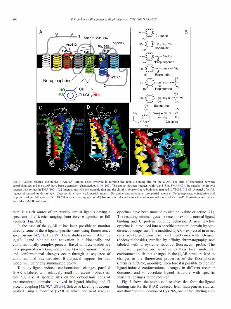
In the case of the !2AR it has been possible to monitordirectly some of these ligand-specific states using fluorescencespectroscopy [42,70,71,88,89]. These studies reveal that for the!2AR ligand binding and activation is a kinetically andconformationally complex process. Based on these studies wehave proposed a working model (Fig. 4) where agonist bindingand conformational changes occur through a sequence ofconformational intermediates. Biophysical support for thismodel will be briefly summarized below.
To study ligand induced conformational changes, purified!2AR is labeled with relatively small fluorescent probes (lessthat 500 Da) at specific sites on the cytoplasmic ends oftransmembrane domains involved in ligand binding and Gprotein coupling [42,70,71,88,89]. Selective labeling is accom-plished using a modified !2AR in which the most reactive
cysteines have been mutated to alanine, valine or serine [71].The resulting minimal cysteine receptor exhibits normal ligandbinding and G protein coupling behavior. A new reactivecysteine is introduced into a specific structural domain by site-directed mutagenesis. The modified !2AR is expressed in insectcells, solubilized from insect cell membranes with detergent(dodecylmaltoside), purified by affinity chromatography, andlabeled with a cysteine reactive fluorescent probe. Thefluorescent probes are sensitive to their local molecularenvironment such that changes in the !2AR structure lead tochanges in the fluorescent properties of the fluorophores(intensity, lifetime, mobility). Therefore it is possible to monitorligand-induced conformational changes in different receptordomains, and to correlate ligand structure with specificstructural changes in the receptor.
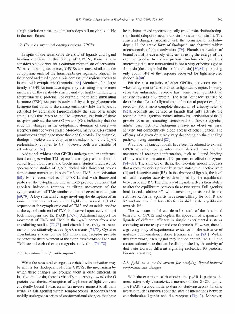
Fig. 3 shows the amino acid residues that form the ligandbinding site for the !2AR deduced from mutagenesis studies,and illustrates the location of Cys 265, one of the labeling sites
Fig. 3. Agonist binding site in the !2AR. (A) Amino acids involved in forming the agonist binding site for the !2AR. The sites of interaction betweencatecholamines and the !2AR have been extensively characterized [100–102]. The amine nitrogen interacts with Asp 113 in TM3 [103], the catechol hydroxylsinteract with serines in TM5 [100–102]. Interactions with the aromatic ring and the chiral !-hydroxyl have both been mapped to TM6 [101]. (B) A panel of !2ARligands discussed in this review. Catechol is a very weak partial agonist. Dopamine and salbutamol are partial agonists. Norepinephrine, epinephrine andisoproterenol are full agonists. ICI118,551 is an inverse agonist. (C–E) Isoproterenol docked into a three-dimensional model of the !2AR. Illustrations were madewith MacPyMOL software.
we have used for our spectroscopy studies. An environmentallysensitive fluorophore covalently bound to Cys265 is wellpositioned to detect agonist-induced conformational changesrelevant to G protein activation. Based on homology withrhodopsin, Cys265 is located in the third intracellular loop (IC3)at the cytoplasmic end of the TM6. Mutagenesis studies haveshown this region of IC3 to be important for G protein coupling[90,91]. Moreover, TM6, along with TM3 and TM5 containamino acids that form the agonist binding site.
3.4.1. Ligand-specific conformational states detected byfluorescence lifetime studies
We examined ligand-dependent changes in fluorescencelifetime of purified !2AR labeled at Cys265 with fluoresceinmaleimide in an effort to identify the existence of agonist-specific conformational states [42]. Fluorescence lifetimeanalysis can detect discrete conformational states in a popula-tion of molecules, while fluorescence intensity measurementsreflect the weighted average of one or more discrete states.Fluorescence lifetime, ", refers to the average time that afluorophore that has absorbed a photon remains in the excitedstate before returning to the ground state. The lifetime offluorescein (nanoseconds) is much faster than the predicted off-rate of the agonists we examined (#s–ms), and much shorter
than the half-life of conformational states of bacteriorhodopsin(#s)[92], rhodopsin (ms) [79,93] or of ion channels (#s–ms)[94]. Therefore, lifetime analysis of fluorescein bound toCys265 is well suited to capture even short-lived, agonist-induced conformational states. We observed that the full agonistisoproterenol induced a conformation that was distinct from theconformations induced by the partial agonists salbutamol anddobutamine [42]. These studies also revealed the existence of anintermediate state in equilibrium with the active state for bothfull agonists and partial agonists. In contrast, the neutralantagonist appeared to stabilize a state that was indistinguish-able from the unliganded receptor.
3.4.2. Intermediate conformational states detected by kineticstudies
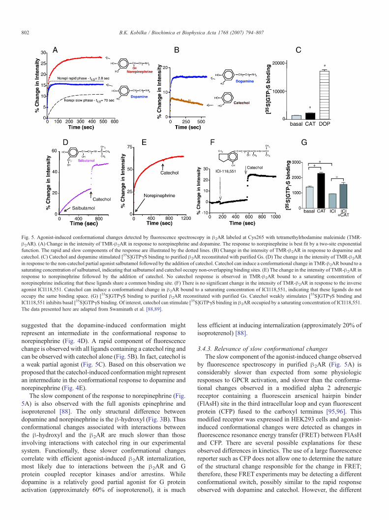
Agonist-induced conformational changes in purified !2ARlead to an increase in the fluorescence intensity of tetramethylr-hodamine bound to Cys265 [88,89]. The increase in fluorescenceintensity as a function of time following activation by the agonistnorepinephrine is best fit with a two component exponentialfunction [88] (Fig. 5A). In contrast, the response to dopamine (apartial agonist) is adequately fit by a one component exponentialfunction. Of interest, the rapid component of norepinephrine isvery similar to the response to dopamine (Fig. 5A), which
Fig. 4. Sequential Binding Model. (A) Arrangement of the TM domains of the !2AR as viewed from the extracellular surface. The agonist binding domains are shownin red (TM3), green (TM5) and blue (TM6). (B) Cartoon representing structural components of norepinephrine. (C–E) In the absence of ligand, the receptor (R) isconformationally flexible. Conformational state R1 is stabilized by interactions between TMs 5 and 6 and the catechol ring. The transition to state R2 occurs when Asp113 in TM3 binds the amine nitrogen. The transitions from R to R2 are rapid. The slow transition from R2 to R3 involves interactions between the chiral !-hydroxyl andAsn293 on TM6. Adapted from Swaminath et al. [88].
801B.K. Kobilka / Biochimica et Biophysica Acta 1768 (2007) 794–807
suggested that the dopamine-induced conformation mightrepresent an intermediate in the conformational response tonorepinephrine (Fig. 4D). A rapid component of fluorescencechange is observed with all ligands containing a catechol ring andcan be observed with catechol alone (Fig. 5B). In fact, catechol isa weak partial agonist (Fig. 5C). Based on this observation weproposed that the catechol-induced conformationmight representan intermediate in the conformational response to dopamine andnorepinephrine (Fig. 4E).
The slow component of the response to norepinephrine (Fig.5A) is also observed with the full agonists epinephrine andisoproterenol [88]. The only structural difference betweendopamine and norepinephrine is the !-hydroxyl (Fig. 3B). Thusconformational changes associated with interactions betweenthe !-hydroxyl and the !2AR are much slower than thoseinvolving interactions with catechol ring in our experimentalsystem. Functionally, these slower conformational changescorrelate with efficient agonist-induced !2AR internalization,most likely due to interactions between the !2AR and Gprotein coupled receptor kinases and/or arrestins. Whiledopamine is a relatively good partial agonist for G proteinactivation (approximately 60% of isoproterenol), it is much
less efficient at inducing internalization (approximately 20% ofisoproterenol) [88].
3.4.3. Relevance of slow conformational changesThe slow component of the agonist-induced change observed
by fluorescence spectroscopy in purified !2AR (Fig. 5A) isconsiderably slower than expected from some physiologicresponses to GPCR activation, and slower than the conforma-tional changes observed in a modified alpha 2 adrenergicreceptor containing a fluorescein arsenical hairpin binder(FlAsH) site in the third intracellular loop and cyan fluorescentprotein (CFP) fused to the carboxyl terminus [95,96]. Thismodified receptor was expressed in HEK293 cells and agonist-induced conformational changes were detected as changes influorescence resonance energy transfer (FRET) between FlAsHand CFP. There are several possible explanations for theseobserved differences in kinetics. The use of a large fluorescencereporter such as CFP does not allow one to determine the natureof the structural change responsible for the change in FRET;therefore, these FRET experiments may be detecting a differentconformational switch, possibly similar to the rapid responseobserved with dopamine and catechol. However, the different
Fig. 5. Agonist-induced conformational changes detected by fluorescence spectroscopy in !2AR labeled at Cys265 with tetramethylrhodamine maleimide (TMR-!2AR). (A) Change in the intensity of TMR-!2AR in response to norepinephrine and dopamine. The response to norepinephrine is best fit by a two-site exponentialfunction. The rapid and slow components of the response are illustrated by the dotted lines. (B) Change in the intensity of TMR-!2AR in response to dopamine andcatechol. (C) Catechol and dopamine stimulated [35S]GTP$S binding to purified !2AR reconstituted with purified Gs. (D) The change in the intensity of TMR-!2ARin response to the non-catechol partial agonist salbutamol followed by the addition of catechol. Catechol can induce a conformational change in TMR-!2AR bound to asaturating concentration of salbutamol, indicating that salbutamol and catechol occupy non-overlapping binding sites. (E) The change in the intensity of TMR-!2AR inresponse to norepinephrine followed by the addition of catechol. No catechol response is observed in TMR-!2AR bound to a saturating concentration ofnorepinephrine indicating that these ligands share a common binding site. (F) There is no significant change in the intensity of TMR-!2AR in response to the inverseagonist ICI118,551. Catechol can induce a conformational change in !2AR bound to a saturating concentration of ICI118,551, indicating that these ligands do notoccupy the same binding space. (G) [35S]GTP$S binding to purified !2AR reconstituted with purified Gs. Catechol weakly stimulates [35S]GTP$S binding andICI118,551 inhibits basal [35S]GTP$S binding. Of interest, catechol can stimulate [35S]GTP$S binding in !2AR occupied by a saturating concentration of ICI118,551.The data presented here are adapted from Swaminath et al. [88,89].
rates observed could also be due to the fact that the !2ARexperiments use purified receptor in the absence of G protein,while the FRET experiments were performed on receptorexpressed in cell membranes that contain G proteins. It isknown that receptors form complexes with G proteins in theplasma membrane (precoupling), and that these complexes havea higher affinity for agonists than do receptors alone. Otherfactors that might contribute to differences between experimentsusing purified receptor and those on receptors in cells could bethe influence of the pH and salt gradients across the plasmamembrane in living cells, as well as the asymmetry of plasmamembrane lipids. Nevertheless, while the slow component of theconformational response to agonists may be attributable to theuse of purified receptor protein, we believe the structuralchanges are physiologically relevant because they can be linkedto specific interactions between the ligand and the receptor (e.g.,the !-hydroxyl of catecholamine agonists interacting withAsn293 in TM6, Fig. 3), and they correlate with specificfunctional behaviors stimulated by the agonists in !2ARexpressed in cells (e.g., agonist-induced internalization [88]).
3.4.4. Catechol activates of the rotamer toggle switchBased on what is known about the binding site for the
catechol ring of catecholamines (Fig. 3), we proposed that therapid component of the conformational response associatedwith catecholamine and catechol binding involves changes inthe rotameric position of aromatic amino acids surrounding thehighly conserved proline Pro2886.50 in TM6 (Fig. 3C–E). Thisconformational change, known as a rotamer toggle switch, hasbeen proposed to be involved in the activation of amine andopsin receptor families [97]. Upon binding, the aromaticcatechol ring of catecholamines would interact directly withthe aromatic residues of the rotamer toggle switch, Trp2866.48
and Phe2906.52. Molecular dynamics simulations suggestthat rotamer configurations of Cys2856.47, Trp2866.48 andPhe2906.52, the residues that comprise the rotamer toggleswitch, are coupled and modulate the bend angle of TM6around the highly conserved proline kink at Pro2886.50, leadingto the movement of the cytoplasmic end of TM6 upon activation[97]. This movement could be detected by tetramethylrhoda-mine bound to Cys265 at the cytoplasmic end of TM6 (Fig. 5B).
3.4.5. Non-catecholamine partial agonists do not activate therotamer toggle switch
In the same experimental system, activation of tetramethylr-hodamine-labeled !2AR by salbutamol, a noncatechol partialagonist, produced only a slow monophasic increase influorescence intensity [89] (Fig. 5C). Catechol could induce afurther increase in fluorescence in !2AR bound to saturatingconcentrations of salbutamol, but not in !2AR bound tonorepinephrine (Fig. 5C and D). This suggests that the aromaticring of salbutamol does not occupy the same binding space ascatechol and does not activate the rotamer toggle switch [89].Thus, the active state induced by salbutamol would be differentfrom that induced by catecholamine agonists [89]. This is inagreement with fluorescent lifetime experiments discussedabove [42].
3.4.6. The inverse agonist ICI118,551 does not inhibitactivation of the rotamer toggle switch
The neutral antagonist alprenolol and the inverse agonistsICI118,551 and timolol do not produce a detectable change influorescence in !2AR labeled with tetramethylrhodamine onCys 265 [89], although they do inhibit the response to agonistsand partial agonists with one exception. In tetramethylrhoda-mine-labeled !2AR bound to saturating concentrations of theseantagonists there is no effect on the response to catechol. This isshown in Fig. 5F for ICI118,551. The fluorescence response tocatechol in !2AR bound to ICI118,551 is associated with afunctional response in a G protein activation assay (Fig. 5G).This suggests that ICI118,551 does not occupy the catecholbinding pocket and does not prevent activation of the rotamertoggle switch by catechol.
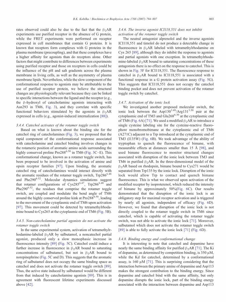
3.4.7. Activation of the ionic lockWe investigated another proposed molecular switch, the
ionic lock between the Asp1303.49/Arg1313.50 pair at thecytoplasmic end of TM3 and Glu2686.30 at the cytoplasmic endof TM6 (Fig. 6A) [71]. We used a modified !2AR to introduce asingle cysteine labeling site for the cysteine-reactive fluoro-phore monobromobimane at the cytoplasmic end of TM6(A271C) adjacent to a Trp introduced at the cytoplasmic end ofTM3 (I135W) (Fig. 6B). We took advantage of the ability oftryptophan to quench the fluorescence of bimane, withmeasurable effects at distances smaller than 15 Å [98], andused bimane fluorescence to monitor structural changesassociated with disruption of the ionic lock between TM3 andTM6 in purified !2AR. In the three-dimensional model of the!2AR based on rhodopsin, bimane bound to Cys271 would beseparated from Trp135 by the ionic lock. Disruption of the ioniclock would allow Trp to contact and quench bimanefluorescence. This is what we observed upon activation of themodified receptor by isoproterenol, which reduced the intensityof bimane by approximately 50%(Fig. 6C). Our resultsdemonstrated that the disruption of the ionic lock is anobligatory step for maximal receptor activation and is triggeredby nearly all agonists, independent of efficacy (Fig. 6D).However, we found that disruption of the ionic lock is notdirectly coupled to the rotamer toggle switch in TM6 sincecatechol, which is capable of activating the rotamer toggleswitch, was not able to activate the ionic lock [71]. Moreover,salbutamol which does not activate the rotamer toggle switch[89] is able to fully activate the ionic lock [71] (Fig. 6D).
3.4.8. Binding energy and conformational changeIt is interesting to note that catechol and dopamine have
nearly the same binding affinity for purified !2AR [71]. The Kiof dopamine, as determined by competition binding, is 350 #M,while the Kd for catechol, determined by a conformationalassay, is 160 #M [71]. This is surprising considering that theinteraction between the primary amine of dopamine and Asp113makes the strongest contribution to the binding energy. Sincedopamine and catechol bind with the same affinity, but onlydopamine disrupts the ionic lock, part of the binding energyassociated with the interaction between dopamine and Asp113
803B.K. Kobilka / Biochimica et Biophysica Acta 1768 (2007) 794–807
may be used to offset by the energetic cost of breaking the ioniclock.
4. Conclusions
Based on these fluorescence studies we proposed a modelwhere agonists stabilize partially or fully active states by usingdifferent chemical groups to activate different combinations of
molecular switches, which are not necessarily interdependent.In the unliganded inactive state of a GPCR, the arrangement ofTM segments is stabilized by non-covalent interactionsbetween side chains. Structurally distinct ligands are able tobreak different combinations of the basal state stabilizinginteractions either directly by binding to amino acids that areinvolved in these intramolecular interactions, or indirectly bystabilizing new intramolecular interactions. These ligand-
Fig. 6. Fluorescence spectroscopy to monitor disruption of the ionic lock in the !2AR. (A) Model of TM3 (red) and TM6 (blue) from the !2AR depicting the aminoacids that comprise the ionic lock at the cytoplasmic end of these TM segments. (B) Close up view of the ionic lock and the modifications made to monitorconformational changes in this region. Alanine 271 was mutated to cysteine (C271) and isoleucine 135 was mutated to tryptophan (W135). C271 was labeled withmonobromobimane in purified !2AR. Upon activation, W135 moves closer to bimane on C271 and quenches fluorescence. (C) Emission spectrum of bimane on C271before and after activation by the agonist isoproterenol. (D) Effect of different ligands on disruption of the ionic lock as determined by bimane fluorescence. The partialagonists dopamine and salbutamol are as effective at disrupting the ionic lock as the full agonists norepinephrine and isoproterenol. Only catechol has no effect on theionic lock. These data are adapted from Yao et al. [71].
specific conformational changes may be responsible fordifferential activation of the signaling cascades of the receptor.The affinity of a particular ligand will then be dependent on theenergy costs and gains associated with each disrupted andcreated interaction, while its efficacy will be dependent on theability to trigger the switches associated with activation. Thesemolecular switches are normally activated by agonist binding,but will also be revealed in constitutively active mutants, wheresingle point mutations in virtually any structural domain canlead to elevated basal activity[99]. A better understanding of theprocess by which ligands bind and modify GPCR structure mayultimately help in the design of more selective drugs with theappropriate efficacy for the desired physiologic function.
References
[1] J.A. Ballesteros, H. Weinstein, Integrated methods for the construction ofthree-dimensional models and computational probing of structure–function relations in G protein coupled receptors, Methods Neurosci.25 (1995) 366–428.
[2] R.J. Lefkowitz, S.K. Shenoy, Transduction of receptor signals by beta-arrestins, Science 308 (5721) (2005) 512–517.
[3] L.M. Luttrell, R.J. Lefkowitz, The role of beta-arrestins in the terminationand transduction of G-protein-coupled receptor signals, J. Cell. Sci. 115(Pt. 3) (2002) 455–465.
[4] M. Azzi, P.G. Charest, S. Angers, G. Rousseau, T. Kohout, M. Bouvier,G. Pineyro, Beta-arrestin-mediated activation of MAPK by inverseagonists reveals distinct active conformations for G protein-coupledreceptors, Proc. Natl. Acad. Sci. U. S. A. 100 (20) (2003) 11406–11411.
[5] R. Fredriksson, M.C. Lagerstrom, L.G. Lundin, H.B. Schioth, TheG-protein-coupled receptors in the human genome form five mainfamilies. Phylogenetic analysis, paralogon groups, and fingerprints,Mol. Pharmacol. 63 (6) (2003) 1256–1272.
[6] A.D. Howard, G. McAllister, S.D. Feighner, Q. Liu, R.P. Nargund, L.H.Van der Ploeg, A.A. Patchett, Orphan G-protein-coupled receptors andnatural ligand discovery, Trends Pharmacol. Sci. 22 (3) (2001) 132–140.
[7] T.H. Ji, M. Grossmann, I. Ji, G protein-coupled receptors. I. Diversityof receptor–ligand interactions, J. Biol. Chem. 273 (28) (1998)17299–17302.
[8] J.P. Pin, T. Galvez, L. Prezeau, Evolution, structure, and activationmechanism of family 3/C G-protein-coupled receptors, Pharmacol. Ther.98 (3) (2003) 325–354.
[9] F. Knoflach, V. Mutel, S. Jolidon, J.N. Kew, P. Malherbe, E. Vieira, J.Wichmann, J.A. Kemp, Positive allosteric modulators of metabotropicglutamate 1 receptor: characterization, mechanism of action, and bindingsite, Proc. Natl. Acad. Sci. U. S. A. 98 (23) (2001) 13402–13407.
[10] F.Y. Carroll, A. Stolle, P.M. Beart, A. Voerste, I. Brabet, F. Mauler, C.Joly, H. Antonicek, J. Bockaert, T. Muller, J.P. Pin, L. Prezeau, BAY36-7620: a potent non-competitive mGlu1 receptor antagonist with inverseagonist activity, Mol. Pharmacol. 59 (5) (2001) 965–973.
[11] A. Pagano, D. Ruegg, S. Litschig, N. Stoehr, C. Stierlin, M. Heinrich, P.Floersheim, L. Prezeau, F. Carroll, J.P. Pin, A. Cambria, I. Vranesic, P.J.Flor, F. Gasparini, R. Kuhn, The non-competitive antagonists 2-methyl-6-(phenylethynyl)pyridine and 7-hydroxyiminocyclopropan[b]chromen-1a-carboxylic acid ethyl ester interact with overlapping binding pocketsin the transmembrane region of group I metabotropic glutamate receptors,J. Biol. Chem. 275 (43) (2000) 33750–33758.
[12] S. Litschig, F. Gasparini, D. Rueegg, N. Stoehr, P.J. Flor, I. Vranesic, L.Prezeau, J.P. Pin, C. Thomsen, R. Kuhn, CPCCOEt, a noncompetitivemetabotropic glutamate receptor 1 antagonist, inhibits receptor signalingwithout affecting glutamate binding, Mol. Pharmacol. 55 (3) (1999)453–461.
[13] K. Ray, J. Tisdale, R.H. Dodd, P. Dauban, M. Ruat, J.K. Northup,Calindol, a positive allosteric modulator of the human Ca(2+) receptor,activates an extracellular ligand-binding domain-deleted rhodopsin-like
seven-transmembrane structure in the absence of Ca(2+), J. Biol. Chem.280 (44) (2005) 37013–37020.
[14] K. Ray, J. Northup, Evidence for distinct cation and calcimimeticcompound (NPS 568) recognition domains in the transmembrane regionsof the humanCa2+ receptor, J. Biol. Chem. 277 (21) (2002) 18908–18913.
[15] A. Krebs, P.C. Edwards, C. Villa, J. Li, G.F. Schertler, The three-dimensional structure of bovine rhodopsin determined by electroncryomicroscopy, J. Biol. Chem. (2003) 50217–50225.
[16] A. Krebs, C. Villa, P.C. Edwards, G.F. Schertler, Characterisation of animproved two-dimensional p22121 crystal from bovine rhodopsin, J. Mol.Biol. 282 (5) (1998) 991–1003.
of rhodopsin transmembrane alpha-helices, Nature 389 (6647) (1997)203–206.
[19] G.F. Schertler, C. Villa, R. Henderson, Projection structure of rhodopsin,Nature 362 (6422) (1993) 770–772.
[20] J.M. Baldwin, G.F. Schertler, V.M. Unger, An alpha-carbon template forthe transmembrane helices in the rhodopsin family of G-protein-coupledreceptors, J. Mol. Biol. 272 (1) (1997) 144–164.
[21] T. Okada, I. Le Trong, B.A. Fox, C.A. Behnke, R.E. Stenkamp, K.Palczewski, X-Ray diffraction analysis of three-dimensional crystals ofbovine rhodopsin obtained from mixed micelles, J. Struct. Biol. 130 (1)(2000) 73–80.
[22] K. Palczewski, T. Kumasaka, T. Hori, C.A. Behnke, H. Motoshima, B.A.Fox, I. Le Trong, D.C. Teller, T. Okada, R.E. Stenkamp, M. Yamamoto,M. Miyano, Crystal structure of rhodopsin: a G protein-coupled receptor[see comments], Science 289 (5480) (2000) 739–745.
[23] T. Okada, Y. Fujiyoshi, M. Silow, J. Navarro, E.M. Landau, Y. Shichida,Functional role of internal water molecules in rhodopsin revealed byX-ray crystallography, Proc. Natl. Acad. Sci. U. S. A. 99 (9) (2002)5982–5987.
[24] D.C. Teller, T. Okada, C.A. Behnke, K. Palczewski, R.E. Stenkamp,Advances in determination of a high-resolution three-dimensionalstructure of rhodopsin, a model of G-protein-coupled receptors(GPCRs), Biochemistry 40 (26) (2001) 7761–7772.
[25] T. Okada, M. Sugihara, A.N. Bondar, M. Elstner, P. Entel, V. Buss, Theretinal conformation and its environment in rhodopsin in light of a new2.2 A crystal structure, J. Mol. Biol. 342 (2) (2004) 571–583.
[26] J. Li, P.C. Edwards, M. Burghammer, C. Villa, G.F. Schertler, Structure ofbovine rhodopsin in a trigonal crystal form, J. Mol. Biol. 343 (5) (2004)1409–1438.
[27] T. Okada, K. Takeda, T. Kouyama, Highly selective separation ofrhodopsin from bovine rod outer segment membranes using combinationof divalent cation and alkyl(thio)glucoside, Photochem. Photobiol. 67 (5)(1998) 495–499.
[28] P.C. Edwards, J. Li, M. Burghammer, J.H. McDowell, C. Villa, P.A.Hargrave, G.F. Schertler, Crystals of native and modified bovinerhodopsins and their heavy atom derivatives, J. Mol. Biol. 343 (5)(2004) 1439–1450.
[29] G.F. Schertler, Structure of rhodopsin and the metarhodopsin Iphotointermediate, Curr. Opin. Struct. Biol. 15 (4) (2005) 408–415.
[30] Q.R. Fan, W.A. Hendrickson, Structure of human follicle-stimulatinghormone in complex with its receptor, Nature 433 (7023) (2005)269–277.
[31] L. Shi, J.A. Javitch, The binding site of aminergic G protein-coupledreceptors: the transmembrane segments and second extracellular loop,Annu. Rev. Pharmacol. Toxicol. 42 (2002) 437–467.
[32] C.E. Elling, K. Thirstrup, S.M. Nielsen, S.A. Hjorth, T.W. Schwartz,Metal-ion sites as structural and functional probes of helix–helixinteractions in 7TM receptors, Ann. N. Y. Acad. Sci. 814 (1997) 142–151.
[33] J.A. Ballesteros, L. Shi, J.A. Javitch, Structural mimicry in G protein-coupled receptors: implications of the high-resolution structure ofrhodopsin for structure–function analysis of rhodopsin-like receptors,Mol. Pharmacol. 60 (1) (2001) 1–19.
[34] E. Archer, B. Maigret, C. Escrieut, L. Pradayrol, D. Fourmy, Rhodopsincrystal: new template yielding realistic models of G-protein-coupledreceptors? Trends Pharmacol. Sci. 24 (1) (2003) 36–40.
805B.K. Kobilka / Biochimica et Biophysica Acta 1768 (2007) 794–807
[35] C.G. Tate, R. Grisshammer, Heterologous expression of G-protein-coupled receptors, Trends Biotechnol. 14 (11) (1996) 426–430.
[36] D. Massotte, G protein-coupled receptor overexpression with thebaculovirus-insect cell system: a tool for structural and functionalstudies, Biochim. Biophys. Acta. 1610 (1) (2003) 77–89.
[37] V. Sarramegna, F. Talmont, P. Demange, A. Milon, Heterologousexpression of G-protein-coupled receptors: comparison of expressionsystems fron the standpoint of large-scale production and purification,Cell. Mol. Life Sci. 60 (8) (2003) 1529–1546.
[38] R. Grisshammer, P. Averbeck, A.K. Sohal, Improved purification of a ratneurotensin receptor expressed in Escherichia coli, Biochem. Soc. Trans.27 (6) (1999) 899–903.
[39] H.M. Weiss, R. Grisshammer, Purification and characterization of thehuman adenosine A(2a) receptor functionally expressed in Escherichiacoli, Eur. J. Biochem. 269 (1) (2002) 82–92.
[40] J.L. Baneres, A. Martin, P. Hullot, J.P. Girard, J.C. Rossi, J. Parello,Structure-based analysis of GPCR function: conformational adaptation ofboth agonist and receptor upon leukotriene B4 binding to recombinantBLT1, J. Mol. Biol. 329 (4) (2003) 801–814.
[41] C. Ostermeier, H. Michel, Crystallization of membrane proteins, Curr.Opin. Struct. Biol. 7 (1997) 697–701.
[42] P. Ghanouni, Z. Gryczynski, J.J. Steenhuis, T.W. Lee, D.L. Farrens, J.R.Lakowicz, B.K. Kobilka, Functionally different agonists induce distinctconformations in the G protein coupling domain of the beta 2 adrenergicreceptor, J. Biol. Chem. 276 (27) (2001) 24433–24436.
[43] S. Angers, A. Salahpour, M. Bouvier, Dimerization: an emerging conceptfor G protein-coupled receptor ontogeny and function, Annu. Rev.Pharmacol. Toxicol. 42 (2002) 409–435.
[44] L.A. Devi, Heterodimerization of G-protein-coupled receptors: pharma-cology, signaling and trafficking, Trends Pharmacol. Sci. 22 (10) (2001)532–537.
[45] S. Bulenger, S. Marullo, M. Bouvier, Emerging role of homo- andheterodimerization in G-protein-coupled receptor biosynthesis andmaturation, Trends Pharmacol. Sci. 26 (3) (2005) 131–137.
[46] J.A. Javitch, The ants go marching two by two: oligomeric structure ofG-protein-coupled receptors, Mol. Pharmacol. 66 (5) (2004) 1077–1082.
[47] J.P. Pin, J. Kniazeff, C. Goudet, A.S. Bessis, J. Liu, T. Galvez, F. Acher, P.Rondard, L. Prezeau, The activation mechanism of class-C G-proteincoupled receptors, Biol. Cell 96 (5) (2004) 335–342.
[48] N. Kunishima, Y. Shimada, Y. Tsuji, T. Sato, M. Yamamoto, T.Kumasaka, S. Nakanishi, H. Jingami, K. Morikawa, Structural basis ofglutamate recognition by a dimeric metabotropic glutamate receptor,Nature 407 (6807) (2000) 971–977.
[49] D. Tsuchiya, N. Kunishima, N. Kamiya, H. Jingami, K. Morikawa,Structural views of the ligand-binding cores of a metabotropic glutamatereceptor complexed with an antagonist and both glutamate and Gd3+,Proc. Natl. Acad. Sci. U. S. A. 99 (5) (2002) 2660–2665.
[50] Y. Liang, D. Fotiadis, S. Filipek, D.A. Saperstein, K. Palczewski, A.Engel, Organization of the G protein-coupled receptors rhodopsin andopsin in native membranes, J. Biol. Chem. 278 (24) (2003) 21655–21662.
[51] J.L. Baneres, J. Parello, Structure-based analysis of GPCR function:evidence for a novel pentameric assembly between the dimericleukotriene B4 receptor BLT1 and the G-protein, J. Mol. Biol. 329 (4)(2003) 815–829.
[52] D. Mesnier, J.L. Baneres, Cooperative conformational changes in aG-protein-coupled receptor dimer, the leukotriene B(4) receptor BLT1,J. Biol. Chem. 279 (48) (2004) 49664–49670.
[53] T.P. Sakmar, Structure of rhodopsin and the superfamily of seven-helicalreceptors: the same and not the same, Curr. Opin. Cell Biol. 14 (2) (2002)189–195.
[54] T.P. Sakmar, R.R. Franke, H.G. Khorana, The role of the retinylideneSchiff base counterion in rhodopsin in determining wavelengthabsorbance and Schiff base pKa, Proc. Natl. Acad. Sci. U. S. A. 88 (8)(1991) 3079–3083.
[55] K.D. Ridge, N.G. Abdulaev, M. Sousa, K. Palczewski, Phototransduc-tion: crystal clear, Trends Biochem. Sci. 28 (9) (2003) 479–487.
[56] W.L. Hubbell, C. Altenbach, C.M. Hubbell, H.G. Khorana, Rhodopsinstructure, dynamics, and activation: a perspective from crystallography,
site-directed spin labeling, sulfhydryl reactivity, and disulfide cross-linking, Adv. Protein Chem. 63 (2003) 243–290.
[57] D.L. Farrens, C. Altenbach, K. Yang, W.L. Hubbell, H.G. Khorana,Requirement of rigid-body motion of transmembrane helices for lightactivation of rhodopsin, Science 274 (5288) (1996) 768–770.
[58] T.D. Dunham, D.L. Farrens, Conformational changes in rhodopsin.Movement of helix f detected by site-specific chemical labeling andfluorescence spectroscopy, J. Biol. Chem. 274 (3) (1999) 1683–1690.
[59] S.W. Lin, T.P. Sakmar, Specific tryptophan UV-absorbance changes areprobes of the transition of rhodopsin to its active state, Biochemistry 35(34) (1996) 11149–11159.
[60] S.P. Sheikh, T.A. Zvyaga, O. Lichtarge, T.P. Sakmar, H.R. Bourne,Rhodopsin activation blocked by metal-ion-binding sites linkingtransmembrane helices C and F, Nature 383 (6598) (1996) 347–350.
[61] C. Altenbach, J. Klein-Seetharaman, K. Cai, H.G. Khorana, W.L.Hubbell, Structure and function in rhodopsin: mapping light-dependentchanges in distance between residue 316 in helix 8 and residues in thesequence 60–75, covering the cytoplasmic end of helices TM1 andTM2 and their connection loop CL1, Biochemistry 40 (51) (2001)15493–15500.
[62] C. Altenbach, J. Klein-Seetharaman, J. Hwa, H.G. Khorana, W.L.Hubbell, Structural features and light-dependent changes in the sequence59–75 connecting helices I and II in rhodopsin: a site-directed spin-labeling study, Biochemistry 38 (25) (1999) 7945–7949.
[63] C. Altenbach, K. Cai, H.G. Khorana, W.L. Hubbell, Structural featuresand light-dependent changes in the sequence 306–322 extending fromhelix VII to the palmitoylation sites in rhodopsin: a site-directed spin-labeling study, Biochemistry 38 (25) (1999) 7931–7937.
[64] J.J. Ruprecht, T. Mielke, R. Vogel, C. Villa, G.F. Schertler, Electroncrystallography reveals the structure of metarhodopsin I, EMBO J. 23(18) (2004) 3609–3620.
[65] D. Salom, I. Le Trong, E. Pohl, J.A. Ballesteros, R.E. Stenkamp, K.Palczewski, D.T. Lodowski, Improvements in G protein-coupled receptorpurification yield light stable rhodopsin crystals, J. Struct. Biol. (2006)497–504.
[66] T. Mirzadegan, G. Benko, S. Filipek, K. Palczewski, Sequence analysesof G-protein-coupled receptors: similarities to rhodopsin, Biochemistry42 (10) (2003) 2759–2767.
[67] R.A. Cerione, C. Staniszewski, J.L. Benovic, R.J. Lefkowitz, M.G.Caron, P. Gierschik, R. Somers, A.M. Spiegel, J. Codina, L. Birnbaumer,Specificity of the functional interactions of the b-adrenergic receptor andrhodopsin with guanine nucleotide regulatory proteins reconstituted inphospholipid vesicles, J. Biol. Chem. 260 (3) (1985) 1493–1500.
[68] U. Gether, S. Lin, P. Ghanouni, J.A. Ballesteros, H. Weinstein, B.K.Kobilka, Agonists induce conformational changes in transmembranedomains III and VI of the beta2 adrenoceptor, EMBO J. 16 (22) (1997)6737–6747.
[69] A.D. Jensen, F. Guarnieri, S.G. Rasmussen, F. Asmar, J.A. Ballesteros, U.Gether, Agonist-induced conformational changes at the cytoplasmic sideof TM 6 in the {beta}2 adrenergic receptor mapped by site-selectivefluorescent labeling, J. Biol. Chem. (2000) 9279–9290 (p. Record assupplied by publisher).
[70] P. Ghanouni, J.J. Steenhuis, D.L. Farrens, B.K. Kobilka, Agonist-inducedconformational changes in the G-protein-coupling domain of the beta 2adrenergic receptor, Proc. Natl. Acad. Sci. U. S. A. 98 (11) (2001)5997–6002.
[71] X. Yao, C. Parnot, X. Deupi, V.R. Ratnala, G. Swaminath, D. Farrens, B.Kobilka, Coupling ligand structure to specific conformational switches inthe beta(2)-adrenoceptor, Nat. Chem. Biol. (2006) 417–422.
[72] S.P. Sheikh, J.P. Vilardarga, T.J. Baranski, O. Lichtarge, T. Iiri, E.C.Meng, R.A. Nissenson, H.R. Bourne, Similar structures and sharedswitch mechanisms of the beta2-adrenoceptor and the parathyroidhormone receptor. Zn(II) bridges between helices III and VI blockactivation, J. Biol. Chem. 274 (24) (1999) 17033–17041.
[73] C.E. Elling, T.M. Frimurer, L.O. Gerlach, R. Jorgensen, B. Holst, T.W.Schwartz, Metal ion site engineering indicates a global toggle switchmodel for seven-transmembrane receptor activation, J. Biol. Chem. 281(25) (2006) 17337–17346.
[74] J.A. Javitch, D. Fu, G. Liapakis, J. Chen, Constitutive activation of thebeta2 adrenergic receptor alters the orientation of its sixth membrane-spanning segment, J. Biol. Chem. 272 (30) (1997) 18546–18549.
[75] S.G. Rasmussen, A.D. Jensen, G. Liapakis, P. Ghanouni, J.A. Javitch, U.Gether, Mutation of a highly conserved aspartic acid in the beta2adrenergic receptor: constitutive activation, structural instability, andconformational rearrangement of transmembrane segment 6, Mol.Pharmacol. 56 (1) (1999) 175–184.
[76] S.D. Ward, F.F. Hamdan, L.M. Bloodworth, J. Wess, ConformationalChanges That Occur during M3 Muscarinic Acetylcholine ReceptorActivation Probed by the Use of an in Situ Disulfide Cross- linkingStrategy, J. Biol. Chem. 277 (3) (2002) 2247–2257.
[77] S.D. Ward, F.F. Hamdan, L.M. Bloodworth, N.A. Siddiqui, J.H. Li, J.Wess, Use of an in situ disulfide cross-linking strategy to study thedynamic properties of the cytoplasmic end of transmembrane domain VIof the M3 muscarinic acetylcholine receptor, Biochemistry 45 (3) (2006)676–685.
[78] S.J. Han, F.F. Hamdan, S.K. Kim, K.A. Jacobson, L.M. Bloodworth, B.Li, J. Wess, Identification of an agonist-induced conformational changeoccurring adjacent to the ligand-binding pocket of the M(3) muscarinicacetylcholine receptor, J. Biol. Chem. 280 (41) (2005) 34849–34858.
[79] Z.T. Farahbakhsh, K. Hideg, W.L. Hubbell, Photoactivated conforma-tional changes in rhodopsin: a time-resolved spin label study, Science 262(5138) (1993) 1416–1419.
[80] M. Han, S.W. Lin, M. Minkova, S.O. Smith, T.P. Sakmar, Functionalinteraction of transmembrane helices 3 and 6 in rhodopsin. Replacementof phenylalanine 261 by alanine causes reversion of phenotype of aglycine 121 replacement mutant, J. Biol. Chem. 271 (50) (1996)32337–32342.
[81] S. Jager, K. Palczewski, K.P. Hofmann, Opsin/all-trans-retinal complexactivates transducin by different mechanisms than photolyzed rhodopsin,Biochemistry 35 (9) (1996) 2901–2908.
[82] T. Kenakin, Efficacy at G-protein-coupled receptors, Nat. Rev., DrugDiscov. 1 (2) (2002) 103–110.
[83] T. Kenakin, Ligand-selective receptor conformations revisited: thepromise and the problem, Trends Pharmacol. Sci. 24 (7) (2003) 346–354.
[84] R.J. Lefkowitz, S. Cotecchia, P. Samama, T. Costa, Constitutive activityof receptors coupled to guanine nucleotide regulatory proteins, TrendsPharmacol. Sci. 14 (8) (1993) 303–307.
[85] P. Leff, The two-state model of receptor activation [see comments],Trends Pharmacol. Sci. 16 (3) (1995) 89–97.
[86] J.M. Weiss, P.H. Morgan, M.W. Lutz, T.P. Kenakin, The cubic ternarycomplex receptor-occupancy model. III. resurrecting efficacy, J. Theor.Biol. 181 (4) (1996) 381–397.
[87] T. Kenakin, Inverse, protean, and ligand-selective agonism: matters ofreceptor conformation, FASEB J. 15 (3) (2001) 598–611.
[88] G. Swaminath, Y. Xiang, T.W. Lee, J. Steenhuis, C. Parnot, B.K. Kobilka,Sequential Binding of Agonists to the {beta}2 Adrenoceptor: kineticevidence for intermediate conformational states, J. Biol. Chem. 279 (1)(2004) 686–691.
[89] G. Swaminath, X. Deupi, T.W. Lee, W. Zhu, F.S. Thian, T.S. Kobilka, B.Kobilka, Probing the beta2 Adrenoceptor Binding Site with Catechol
Reveals Differences in Binding and Activation by Agonists and PartialAgonists, J. Biol. Chem. 280 (23) (2005) 22165–22171.
[90] B.F. O'Dowd, M. Hnatowich, J.W. Regan, W.M. Leader, M.G. Caron,R.J. Lefkowitz, Site-directed mutagenesis of the cytoplasmic domainsof the human b2-adrenergic receptor. Localization of regions involvedin G protein- receptor coupling, J. Biol. Chem. 263 (31) (1988)15985–15992.
[91] S.B. Liggett, M.G. Caron, R.J. Lefkowitz, M. Hnatowich, Coupling of amutated form of the human beta 2-adrenergic receptor to Gi and Gs.Requirement for multiple cytoplasmic domains in the coupling process,J. Biol. Chem. 266 (8) (1991) 4816–4821.
[92] S. Subramaniam, R. Henderson, Molecular mechanism of vectorialproton translocation by bacteriorhodopsin [see comments], Nature 406(6796) (2000) 653–657.
[93] S. Arnis, K. Fahmy, K.P. Hofmann, T.P. Sakmar, A conserved carboxylicacid group mediates light-dependent proton uptake and signaling byrhodopsin, J. Biol. Chem. 269 (39) (1994) 23879–23881.
[94] T. Hoshi, W.N. Zagotta, R.W. Aldrich, Shaker potassium channel gating.I: transitions near the open state, J. Gen. Physiol. 103 (2) (1994) 249–278.
[95] J.P. Vilardaga, R. Steinmeyer, G.S. Harms, M.J. Lohse, Molecular basisof inverse agonism in a G protein-coupled receptor, Nat. Chem. Biol. 1(1) (2005) 25–28.
[96] C. Hoffmann, G. Gaietta, M. Bunemann, S.R. Adams, S. Oberdorff-Maass, B. Behr, J.P. Vilardaga, R.Y. Tsien, M.H. Ellisman, M.J. Lohse, AFlAsH-based FRET approach to determine G protein-coupled receptoractivation in living cells, Nat. Methods 2 (3) (2005) 171–176.
[97] L. Shi, G. Liapakis, R. Xu, F. Guarnieri, J.A. Ballesteros, J.A. Javitch,Beta2 adrenergic receptor activation. Modulation of the proline kink intransmembrane 6 by a rotamer toggle switch, J. Biol. Chem. 277 (43)(2002) 40989–40996.
[98] S.E. Mansoor, H.S. McHaourab, D.L. Farrens, Mapping proximity withinproteins using fluorescence spectroscopy. A study of T4 lysozymeshowing that tryptophan residues quench bimane fluorescence, Biochem-istry 41 (8) (2002) 2475–2484.
[99] C. Parnot, S. Miserey-Lenkei, S. Bardin, P. Corvol, E. Clauser, Lessonsfrom constitutively active mutants of G protein-coupled receptors, TrendsEndocrinol. Metab. 13 (8) (2002) 336–343.
[100] C. Strader, M. Candelore, W. Hill, I. Sigal, R. Dixon, Identification of twoserine residues involved in agonist activation of the !-adrenergicreceptor, J. Biol. Chem. 264 (1989) 13572–13578.
[101] K. Wieland, H.M. Zuurmond, C. Krasel, A.P. Ijzerman, M.J. Lohse,Involvement of Asn-293 in stereospecific agonist recognition and inactivation of the beta 2-adrenergic receptor, Proc. Natl. Acad. Sci. U. S. A.93 (17) (1996) 9276–9281.
[102] G. Liapakis, J.A. Ballesteros, S. Papachristou, W.C. Chan, X. Chen, J.A.Javitch, The forgotten serine. A critical role for Ser-2035.42 in ligansbinding to and activation of the beta 2-adrenergic receptor, J. Biol. Chem.275 (48) (2000) 37779–37788.
[103] C.D. Strader, M.R. Candelore, W.S. Hill, R.A. Dixon, I.S. Sigal, A singleamino acid substitution in the beta-adrenergic receptor promotes partialagonist activity from antagonists, J. Biol. Chem. 264 (28) (1989)16470–16477.
807B.K. Kobilka / Biochimica et Biophysica Acta 1768 (2007) 794–807