review I Degradation of steroids in the h I. A. Macdonald, V. D. Bokkenheuser, J. Winter, A. M. McLernon,' and E. H. Mosbach Departments of Medicine and Biochemistry, Dalhousie University, Halifax, Nova Scotia, Canada;' Department of Pathology, St. Luke's-Roosevelt Hospital Center, New York, NY 1 0025;3 and Department of Surgery, Beth Israel Medical Center, New York, NY 100034 TABLE OF CONTENTS Introduction Systematic and trivial names Enzyme and steroid nomenclature Classes of steroid in the human gut The enterohepatic circulation Cholesterol Bile acids Neutral, phenolic, and synthetic steroids Experimental approaches in studying the degradation of steroids by human intestinal bacteria Administration of labeled steroids Analysis of steroids in bile, urine, and feces Cholesterol Bile acids Steroid hormones Animal models Metabolism of steroids by mixed fecal flora Studies with crude and purified enzyme and pure cultures in vitro systems in vitro Metabolism of cholesterol Reduction of cholesterol to coprostanol Coprostanone from cholesterol Side chain cleavage of cholesterol Hydrolysis of conjugated bile acids Oxidation of 3a-, 7a-, and 12a-hydroxyl 7-Dehydroxylation Epimerization of the 3a- and 7a-hydroxyl Formation of 5a-(allo) bile acids Other bile acid transformations Metabolism of bile acids groups to ketones groups Formation of C-24 ethyl esters 675 676 677 677 678 678 678 679 680 680 680 680 680 680 680 68 1 68 1 682 682 684 684 684 686 686 686 687 689 689 689 Formation of unsaturated bile acids 689 Hydrolysis of bile acid 3-sulfate 689 Effect of bile acids on intestinal bacteria 690 Metabolism of neutral steroids 690 Hydrolysis of glucuronides and sulfates 690 Dehydroxylation of the 2 1-hydroxyl group 692 Reduction of ring A 692 Epimerization of the Sa-hydroxyl-group 692 Introduction of a double bond conjugated to a keto group 692 Side chain cleavage 693 Reduction of a 20-ketone to an alcohol 693 1 6a-Dehydroxylation 693 Metabolism of phenolic steroids 693 Conclusion 693 INTRODUCTION The intestinal tract of adults contains approximately 1 kg of bacteria, equivalent to 1014 organisms repre- senting at least 400 distinct species (1-3). More than 99% of the organisms are obligate anaerobes. The con- centration of bacteria increases from jejunum through the ileum and constitutes the bulk of the intestinal Abbreviations: CCK, cholecystokinin; GLC, gas-liquid chromatog- raphy; EHC, enterohepatic circulation; Eh, oxidation-reduction po- tential; HSDH, hydroxysteroid dehydrogenase; MS, mass spectros- copy; menadione, vitamin KS = 2-methyl-l,4-naphthoquinone; TLC, thin-layer chromatography; NDC, nuclear dehydrogenating Clostridia; NDH, nuclear dehydrogenase. Sterols are 27 derivatives of cholestane, usually with a 3P-hydroxyl-group. Neutral steroids, in this review, are defined as C-19 and C-21 derivatives of pregnane and androstane. Phenolic steroids are derivatives of estrone in which the A ring is aromatic and hydroxylated. ' Present address: Ethicon Inc., Summerville, NJ 08876. ' I. A. Macdonald. ' V. D. Bokkenheuser, J. Winter, and A. M. McLernon. E. H. Mosbach. Journal of Lipid Research Volume 24, 1983 675 by guest, on November 6, 2017 www.jlr.org Downloaded from
Transcript
review I Degradation of steroids in the h
I. A. Macdonald, V. D. Bokkenheuser, J. Winter, A. M. McLernon,' and E. H. Mosbach Departments of Medicine and Biochemistry, Dalhousie University, Halifax, Nova Scotia, Canada;' Department of Pathology, St. Luke's-Roosevelt Hospital Center, New York, N Y 1 0025;3 and Department of Surgery, Beth Israel Medical Center, New York, NY 100034
TABLE O F CONTENTS
Introduction Systematic and trivial names Enzyme and steroid nomenclature Classes of steroid in the human gut The enterohepatic circulation
Cholesterol Bile acids Neutral, phenolic, and synthetic steroids
Experimental approaches in studying the degradation of steroids by human intestinal bacteria
Administration of labeled steroids Analysis of steroids in bile, urine, and feces
Cholesterol Bile acids Steroid hormones
Animal models Metabolism of steroids by mixed fecal flora
Studies with crude and purified enzyme and pure cultures in vitro
systems in vitro Metabolism of cholesterol
Reduction of cholesterol to coprostanol Coprostanone from cholesterol Side chain cleavage of cholesterol
Hydrolysis of conjugated bile acids Oxidation of 3a-, 7a-, and 12a-hydroxyl
7-Dehydroxylation Epimerization of the 3a- and 7a-hydroxyl
Formation of 5a-(allo) bile acids Other bile acid transformations
Metabolism of bile acids
groups to ketones
groups
Formation of C-24 ethyl esters
675 676 677 677 678 678 678 679
680 680 680 680 680 680 680
68 1
68 1 682 682 684 684 684 686
686 686
687 689 689 689
Formation of unsaturated bile acids 689 Hydrolysis of bile acid 3-sulfate 689
Effect of bile acids on intestinal bacteria 690 Metabolism of neutral steroids 690
Hydrolysis of glucuronides and sulfates 690 Dehydroxylation of the 2 1-hydroxyl group 692 Reduction of ring A 692 Epimerization of the Sa-hydroxyl-group 692 Introduction of a double bond conjugated to
a keto group 692 Side chain cleavage 693 Reduction of a 20-ketone to an alcohol 693 1 6a-Dehydroxylation 693
Metabolism of phenolic steroids 693 Conclusion 693
INTRODUCTION
The intestinal tract of adults contains approximately 1 kg of bacteria, equivalent to 1014 organisms repre- senting at least 400 distinct species (1-3). More than 99% of the organisms are obligate anaerobes. The con- centration of bacteria increases from jejunum through the ileum and constitutes the bulk of the intestinal
Abbreviations: CCK, cholecystokinin; GLC, gas-liquid chromatog- raphy; EHC, enterohepatic circulation; Eh, oxidation-reduction po- tential; HSDH, hydroxysteroid dehydrogenase; MS, mass spectros- copy; menadione, vitamin KS = 2-methyl-l,4-naphthoquinone; TLC, thin-layer chromatography; NDC, nuclear dehydrogenating Clostridia; NDH, nuclear dehydrogenase. Sterols are 27 derivatives of cholestane, usually with a 3P-hydroxyl-group. Neutral steroids, in this review, are defined as C-19 and C-21 derivatives of pregnane and androstane. Phenolic steroids are derivatives of estrone in which the A ring is aromatic and hydroxylated.
' Present address: Ethicon Inc., Summerville, NJ 08876. ' I . A. Macdonald. ' V. D. Bokkenheuser, J. Winter, and A. M. McLernon.
stream in the colon. Thus both dietary and endogenous substances are destined to come intimately in contact with this population in the lower gut. A wide spectrum of microbial species, both anaerobes and facultatives, are capable of transforming the steroids with the result that the physical and biological properties of the latter can undergo drastic changes. In man and in other ver- tebrates these steroidal metabolites may often be ab- sorbed and returned to the liver where further metab- olism can take place. Recycled metabolites are generally re-excreted in the bile but some are delivered to the blood stream for renal excretion. Thus there is a need to understand the nature of these steroid transforma- tions and their biological significance. Such information may be gained through experiments with humans as well as with appropriate animal models, mixed fecal cul- tures, pure bacterial cultures, and crude or purified en- zyme systems. In this report we will review cholesterol, bile acid, and steroid hormone metabolism by the hu- man intestinal flora including experimental approaches, involved organisms, molecular mechanisms, and medi- cal significance of various steroid transformations. The metabolism of plant and marine steroids will not be ad- dressed in this review.
SYSTEMATIC AND TRIVIAL NAMES
C24 and C27 steroids
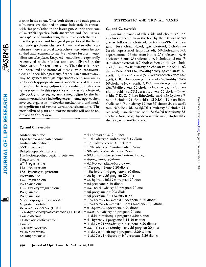
Systematic names of bile acids and cholesterol me- tabolites referred to in the text by their trivial names are as follows: cholesterol, 5-cholesten-3P-01; choles- tanol, 5a-cholestan-3/3-ol; epicholesterol, 5-cholesten- 3a-01; coprostanol (coprosterol), 5P-cholestan-3P-ol; coprostanone, 5P-cholestan-3-one; A4-cholestenone, 4- cholesten-3-one; A5-cholestenone, 5-cholesten-3-one; 7- dehydrocholesterol, 5,7-cholestadien-3P-ol; CA, cholic acid (3a,7a, 12a-trihydroxy-5P-cholan-24-0ic acid); DC, deoxycholic acid (3a, 12a-dihydroxy-5~-cholan-24-oic acid); LC, lithocholic acid (3a-hydroxy-5P-cholan-24-oic acid); CDC, chenodeoxycholic acid (3a,7a-dihydroxy- 5P-cholan-24-oic acid); UDC, ursodeoxycholic acid (3a,7/3-dihydroxy-5P-cholan-24-oic acid); UC, urso- cholic acid (3a,7~,12a-trihydroxy-5P-cholan-24-oic acid); Y-KLC, 7-keto-lithocholic acid (3a-hydroxy-7- oxo-5~-cholan-24-oic acid); 12-KLC, 12-keto-litho- cholic acid (Sa-hydroxy- 12-oxo-5~-cholan-24-oic acid); 0-muricholic acid, 3a,6/3,7P-trihydroxy-5P-cholan-24- oic acid; w-muricholic acid, 3a,Ga,7P-trihydroxy-5P- cholan-24-oic acid; hyodeoxycholic acid, 3a,6a-dihy- droxy-5P-cholan-24-oic acid.
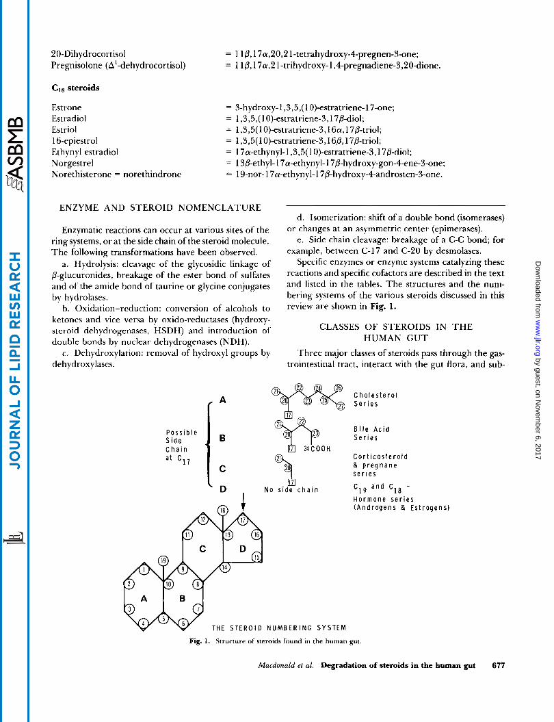
Enzymatic reactions can occur at various sites of the ring systems, o r at the side chain of the steroid molecule. The following transformations have been observed.
a. Hydrolysis: cleavage of the glycosidic linkage of P-glucuronides, breakage of the ester bond of sulfates and of the amide bond of taurine or glycine conjugates by hydrolases.
b. Oxidation-reduction: conversion of alcohols to ketones and vice versa by oxido-reductases (hydroxy- steroid dehydrogenases, HSDH) and introduction of double bonds by nuclear dehydrogenases (NDH).
c . Dehydroxylation: removal of hydroxyl groups by deh ydrox y lases.
P o s s i b l e S i d e C h a i n
a t ' 1 7
a
B
C
D I
d. Isomerization: shift of a double bond (isomerases) o r changes at an asymmetric center (epimerases).
e. Side chain cleavage: breakage of a C-C bond; for example, between C-I7 and C-20 by desmolases.
Specific enzymes or enzyme systems catalyzing these reactions and specific cofactors are described in the text and listed in the tables. The structures and the num- bering systems of the various steroids discussed in this review are shown in Fig. 1.
CLASSES OF STEROIDS IN T H E HUMAN G U T
Three major classes of steroids pass through the gas- trointestinal tract, interact with the gut flora, and sub-
C h o l e s t e r o l
B i l e A c i d S e r i e s
C o r t i c o s t e r o i d & p r e g n a n e s e r i e s
C 1 9 a n d C18 -
H o r m o n e s e r i e s ( A n d r o g e n s & E s t r o g e n s )
@% N o s i d e c h a i n
N U M B E R I N G S Y S T E M
Fig. 1. Structure of steroids found in the human gut.
Macdonald et al. Degradation of steroids in the human gut 677
sequently can undergo microbial transformations. These classes (see Fig. 1) are i) cholesterol originating from the diet, liver, intestinal epithelium, and other tissues; ii) bile acids synthesized from cholesterol in the liver and excreted via the biliary tract, and iii) steroid hormones synthesized from cholesterol in the adrenal cortex and gonads, cleared by the liver, and excreted via the biliary tract.
Roughly, 400- 1000 mg of cholesterol, 100-500 mg of bile acids, and less than 2 mg of steroid hormones pass daily through the colon in a healthy nonpregnant human subject on a “mixed western” diet. Both the cholesterol and the steroid hormones are generally wa- ter-insoluble. In bile they form mixed micelles (poly- molecular aggregates) with the highly soluble bile acid conjugates and other polar lipids such as phospholipids. Bile acids can occur both in the monomeric and micellar forms and are generally ionized. During the passage through the lower gastrointestinal tract, all three types of steroids interact with the intestinal flora. This review highlights the various established transformations and, when known, the properties of the organism(s) and en- zyme system(s) responsible.
THE ENTEROHEPATIC CIRCULATION
Several reviews dealing with different aspects of the enterohepatic circulation (EHC) are available including historic (4) and current (5-8) perspectives. However, for the sake of clarity and completion, a brief summary of the role of the EHC in the metabolism of cholesterol, bile acids, and steroid hormones is outlined below.
Cholesterol
Strictly speaking, cholesterol does not undergo a clearly defined EHC. After absorption, it enters the lym- phatic system in chylomicrons that are partially metab- olized by lipoprotein lipases in the walls of the blood vessels. The ensuing secondary particles, containing the cholesterol, are further metabolized in the liver to li- poproteins.
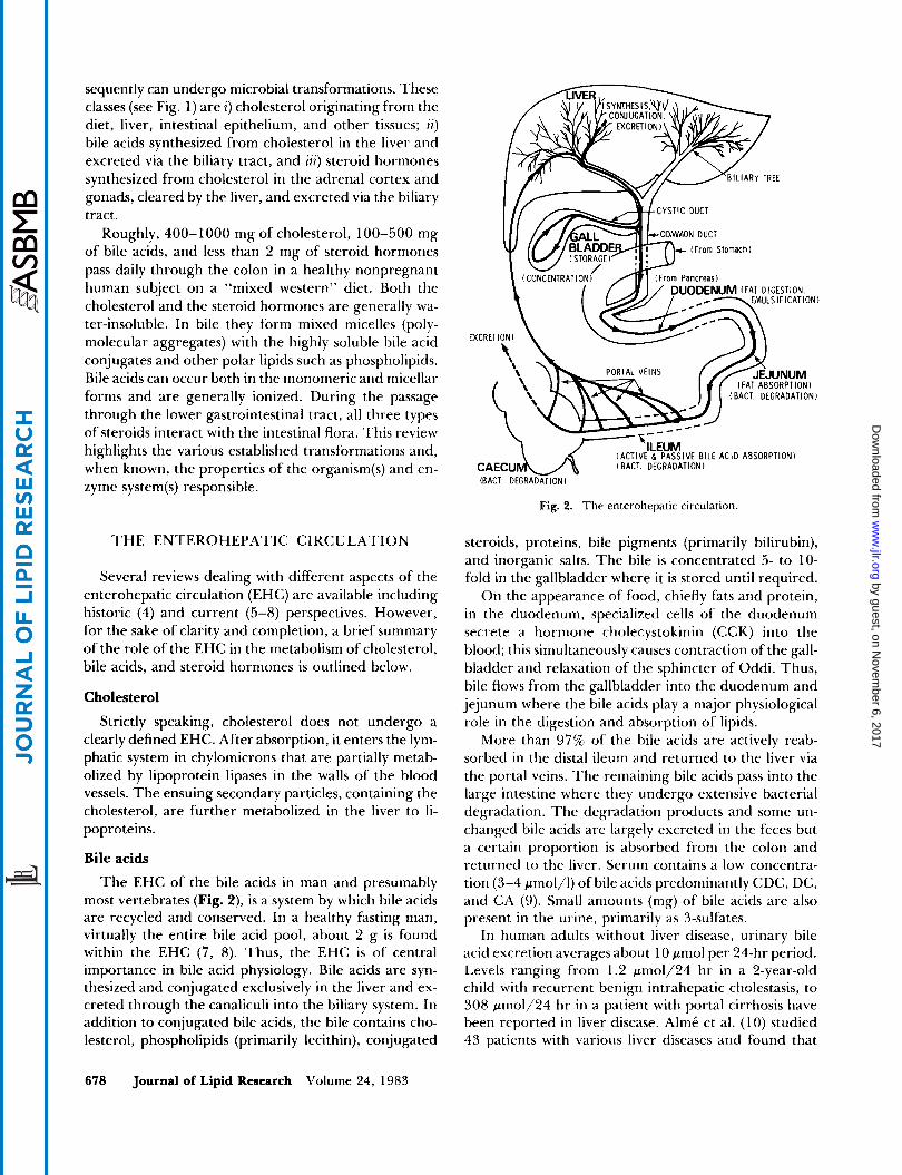
Bile acids
The EHC of the bile acids in man and presumably most vertebrates (Fig. 2), is a system by which bile acids are recycled and conserved. In a healthy fasting man, virtually the entire bile acid pool, about 2 g is found within the EHC (7, 8). Thus, the EHC is of central importance in bile acid physiology. Bile acids are syn- thesized and conjugated exclusively in the liver and ex- creted through the canaliculi into the biliary system. In addition to conjugated bile acids, the bile contains cho- lesterol, phospholipids (primarily lecithin), conjugated
- /BILIARY TREE
S S l V E BILE ACID ABSORPTION1
Fig. 2. T h e enterohepatic circulation
steroids, proteins, bile pigments (primarily bilirubin), and inorganic salts. The bile is concentrated 5- to 10- fold in the gallbladder where it is stored until required.
On the appearance of food, chiefly fats and protein, in the duodenum, specialized cells of the duodenum secrete a hormone cholecystokinin (CCK) into the blood; this simultaneously causes contraction of the gall- bladder and relaxation of the sphincter of Oddi. Thus, bile flows from the gallbladder into the duodenum and jejunum where the bile acids play a major physiological role in the digestion and absorption of lipids.
More than 97%’ of the bile acids are actively reab- sorbed in the distal ileum and returned to the liver via the portal veins. The remaining bile acids pass into the large intestine where they undergo extensive bacterial degradation. The degradation products and some un- changed bile acids are largely excreted in the feces but a certain proportion is absorbed from the colon and returned to the liver. Serum contains a low concentra- tion (3-4 pmol/l) of bile acids predominantly CDC, DC, and CA (9). Small amounts (mg) of bile acids are also present in the urine, primarily as 3-sulfates.
In human adults without liver disease, urinary bile acid excretion averages about 10 pmol per 24-hr period. Levels ranging from 1.2 pmo1/24 hr in a 2-year-old child with recurrent benign intrahepatic cholestasis, to 308 pmo1/24 hr in a patient with portal cirrhosis have been reported in liver disease. AlmC et al. (1 0) studied 43 patients with various liver diseases and found that
CA and CDC accounted for 49-78%, and 3,12-disub- stituted and monohydroxylated bile acids for 8-27% of total urinary bile acids. Bile acids hydroxylated at C-1 or C-6 (e.g., 1, 3, 6, 7, 12; 1, 3, 7, 12 or 3, 6, 7 , 12) constitute 5-15% of the total. The occurrence of rel- atively high proportions of such bile acids in urine clearly distinguishes hepatic disease from the normal state (1 0-1 2). In cirrhotic patients, norcholic acid (3a,7a, 12a-trihydroxy-24-nor-5~-cholanoic acid) was considerably more abundant, accounting for 6% of total bile acid, than in noncirrhotic subjects (average 1%). Otherwise it has not been possible so far to distinguish between different liver diseases by comparing urinary bile acid profiles.
The origin of polyhydroxylated bile acids in urine is not known; they are probably of hepatic origin. We have not encountered hydroxylation in experiments with in- testinal microorganisms.
The quantitatively most significant secondary bile acids occurring in the gut are DC derived from CA, and LC derived from CDC by microbial 7-dehydroxylation. The secondary bile acids, like the primary bile acids, are conjugated with either glycine or taurine during hepatic passage. DC may comprise up to 20-30% of the human bile acid pool, while LC accounts for only 1- 3% of the total. A large proportion of conjugated LC is sulfated at the 3-position in the human liver but this detoxification mechanism appears to be largely unavail- able in other species, such as the baboon (1 3) or rhesus monkey (1 4). The conjugate is poorly absorbed. In the colon, tauro-LC and glyco-LC sulfates are presumably first hydrolyzed to LC sulfate, which is also poorly ab- sorbed, and hydrolyzed further by bacterial sulfatases so that LC is finally excreted mostly in the unconjugated form.
Neutral, phenolic, and synthetic steroids Like bile acids, neutral and phenolic steroids can un-
dergo EHC. Differences in chemical structure such as an extra hydroxyl group, or the shape of the molecule can influence the excretion route of the steroids (15). Otherwise, little is known about the factors determining whether a particular molecule leaves the liver cell via the biliary canaliculi or through the sinusoids of the circulatory system. It is believed that a molecular weight of 300-600 is required for EHC, precisely the range of weights of the conjugated compounds. The steroids are conjugated in the liver with sulfuric or glucuronic acid or both. The type of conjugation of neutral steroids is determined by the chemical structure of the steroid. For example, CI9O2 and CZ1O2 steroids with 3a-hy- droxy-5~-structures are usually conjugated with gluc- uronic acid (ethereal glucuronides) while 3a-hydroxy- 5 a and 3P-hydroxy-A5 steroids generally form sulfates
(esters). The conjugation increases the polarity of the molecule which, at the prevailing pH, leads to ionization and therefore increased solubility. Quantification of biliary steroid hormones is difficult. Measurements in fistula bile are obviously erroneous since the EHC is interrupted. Analyses in intact individuals indicate that males secrete about 13 mg and females 6.5 mg of neu- tral steroids in the bile per 24 hr (16), but less than 2 mg arrives in the colon.
Some of the phenolic steroids are conjugated with both sulfuric and glucuronic acids, e.g., estriol is se- creted in the bile as the 3-sulfate, 16a-glucuronide, while others are conjugated with sulfuric acid only (es- trone) or glucuronic acid only (estradiol) (16-20). It appears that quantification has only been attempted in fistula patients. Aldercreutz and Luukkainen (2 1) found that nonpregnant and pregnant women excrete 16 mg and 3750 mg estriol, respectively, per 24 hr. Estrone and estradiol, both biologically less active than estriol, constitute less than 15% of the biliary phenolic steroids. Aldercreutz and Martin (1 8) noted that the bulk of es- trogens participating in the EHC are hydroxylated at the 15 or 16 position in the liver.
Synthetic steroids also undergo EHC. After absorp- tion these compounds are transported to the liver. Little is known about their immediate fate but it seems safe to assume that the bulk of the progestins escapes ring- A reduction in the liver and enters the circulation via the sinusoids. A small proportion of the molecules is reduced in the liver, despite the protective ethynyl group, conjugated, and excreted in the bile. These mol- ecules are devoid of hormonal activity. Most of the syn- thetic estrogens probably also pass unaltered through the liver, but sooner or later circulating estrogens are taken up by the liver cells, conjugated, and excreted in the bile.
Most of the conjugated biliary steroids, normal or synthetic, are deconjugated in the gut, and reabsorbed before or after bacterial alterations. It is noteworthy that synthetic steroids have a higher rate of fecal ex- cretion than the natural compounds. For example, fol- lowing intravenous administration, 7% of estrone and estradiol (22), but 30% of ethynyl estradiol is excreted in the feces (23). The fecal excretion of norgestrel and norethisterone is even higher (24, 25).
The hepatic uptake and conjugation of steroids ap- pear to be abnormal in certain diseases (17). For ex- ample, in Gilbert’s disease, a genetic disorder associated with glucuronyl transferase deficiency, the hepatic up- take and conjugation of steroids are decreased. In Du- bin-Johnson syndrome (an inherited liver transport de- fect of conjugated bilirubin) the excretion of conjugated anions is reduced. Cholestasis decreases biliary excre- tion of estrogen glucuronides.
Macdonald et al. Degradation of steroids in the human gut 679
EXPERIMENTAL APPROACHES IN STUDYING T H E DEGRADATION
OF STEROIDS BY HUMAN INTESTINAL BACTERIA
Administration of labeled steroids Labeled steroids can be given orally or intravenously.
The label should be situated in the steroid ring or on the side chain (e.g., C24) in a position refractory to chemical or biological removal. The dynamics of EHC of any given steroid can be studied by draining the bile through a fistula or removing bile-rich duodenal sam- ples periodically. The bile is then fractionated to mea- sure the specific activity of the substance in question. Parameters such as pool size, turnover rate, and syn- thesis rate can be measured in the intact animal or hu- man using the isotope dilution principle ( 5 , 6). Alter- nately, bile can be drained and the total product (e.g., bile acid) can be measured. If the label appears in frac- tion(s) other than the administered compound, the new metabolite can be identified and its rate of formation can be calculated. Labeled products or their metabolites leaving the EHC may be detected in urine or feces.
Analysis of steroids in bile, urine and feces Cholesterol. A number of publications have described
the determination of cholesterol in bile by GLC (26), HPLC (27), and enzymic methods (28). The determi- nation of total sterols in feces is difficult because in the gut, cholesterol is partially degraded to coprostanol and coprostanone. These analyses are rendered more com- plex by the presence of the corresponding degradation products of plant sterols, e.g., coprositosterol (24-ethyl- 5P-cholestan-3P-ol) and by high concentrations of pig- mented material. Elaborate and reliable techniques for the determination of fecal neutral sterols by a combi- nation of solvent extraction, TLC, and GLC were de- veloped by Miettinen, Ahrens, and Grundy (29). Small amounts of cholesterol in urine are detectable by GLC (30).
Bile acids. Methods for the analysis of biliary bile acids, widely used at present, employ deproteinization with methanol, and alkaline or enzymatic hydrolysis followed by GLC. Final confirmation of bile acid structures often requires MS. A reliable method for the analysis of total fecal bile acids based on solvent extraction, TLC, and GLC has been published by Grundy, Ahrens, and Miet- tinen (31). Urine contains small amounts of a variety of unusual bile acids. The analytical procedures have been worked out (10, 12) and similar techniques have been used to analyze fecal bile acid composition (32). The above-mentioned techniques, although sophisticated, are not without artifacts such as the destruction of 3- keto bile acids by alkaline hydrolysis of conjugates which
are found in trace amounts in feces (32) and higher amounts in urine (1 0-1 2).
Steroid hormones. Bile contains many different types of steroid hormones ranging from the CIS estrogens with phenolic hydroxyl groups, the C19 androgens, and CP1 progestational hormones to the Cpl corticosteroids with various oxygenated substituents. These classes of steroids exhibit considerable differences in polarity, sol- ubility, and stability which require a variety of special- ized procedures for their isolation and identification (33, 34).
Animal models In order to determine whether or not a given met-
abolic process is carried out by the intestinal flora, the metabolism of labeled steroids can be studied in germ- free animals and compared to the metabolism in con- ventional animals. The appearance of labeled transfor- mation products in the feces or urine of conventional animals and their absence in germ-free animals strongly suggest the involvement of the microbial flora. This principle was first employed in the field of bile acid metabolism (35-37). By monoinfecting germ-free ani- mals with a pure bacterial culture and examining the metabolic products in the feces, the formation of distinct metabolites could be directly associated with a given organism. Similar techniques were applied to the study of steroid hormones. For example, it was shown that 2 1 -dehydroxylated derivatives of certain corticoids re- quired the existence of normal intestinal microflora (1 6, 38). Such observations in animal models have led to the isolation from human fecal flora of microorganisms ca- pable of carrying out a variety of transformations (39, 40). Wostmann and coworkers (41, 42) compared the composition of bile acids in the feces and bile of germ- free rats and conventional animals. Fecal bile acids were analyzed by a modification of the method by Grundy et al. (31). Briefly, this consisted of solvent extraction and separation of the bile acids by two TLC systems followed by GLC analysis; a trace label of ['4C]labeled CA was used as an internal standard. The fecal bile acid composition of germ-free and conventional Wistar rats is summarized in Table 1, adopted from Madsen et al. (4 1). Similarly, Table 2 summarizes the biliary bile acid composition of germ-free dogs, rats, rabbits, and man. Data for the rat and mouse are adapted from Beaver, Wostmann, and Madsen (42), for the rabbit from Hof- mann et al. (43, 44), and for man from Garbutt et al. (45). From the bile acid composition listed in Table 2, it is evident that in the rat, DC, w-muricholic acid (3a,6a,7/3-trihydroxy-5P-cholan-24-oic acid), hyodeox- ycholic acid (3a,6a-dihydroxy-5P-cholan-24-oic acid), and the ketonic bile acids are secondary bile acids (Table 1). Also muricholic acids (3,6,7-trihydroxy-5/3-cholan-
24-oic acids) are found largely in mice and rats but are absent in the other species including humans. Forma- tion of hyodeoxycholic acid appears to be largely by 7- dehydroxylation of w-muricholic acid (42, 43). It is ap- parent that P-muricholic acid (3a,6/3,7P-trihydroxy-5/3- cholan-24-oic acid) may be epimerized to w-muricholic acid by the rat intestinal flora. Confirming this proposal, a strain of Clostridium group 111, which transforms P- muricholic acid to w-muricholic acid, has been isolated from rat feces by Sacquet and coworkers (46); In con- trast to 7-hydroxyl epimerization (See Epimerization of 3a- and 7a-hydroxyl groups.), this reaction results in formation of a Ga-hydroxyl group and is peculiar to the fecal flora of rats and mice. Rabbits, on the other hand, make some allo-CA(b-) as a primary bile acid, (as ob- served in germ-free animals) and in conventional ani- mals this is quantitatively 7-dehydroxylated to allo-DC by the flora (Table 2) (See also below, Formation of 5a- (allo) bile acids.)
Metabolism of steroids by mixed fecal flora and pure cultures in vitro
The bacterial metabolism of steroids may also be studied in vitro. By incubating a steroid precursor with a culture of mixed fecal flora or with a pure bacterial culture it is possible to follow the sequential alterations of the substrate (16). The cultures are sampled at spe- cific intervals and the steroids are extracted, identified, and quantified.
In vitro techniques enable the investigator to identify specific microorganisms and isolate enzymes associated with a given steroid transformation (39, 47). This ap- proach has provided information on metabolic pathways of steroid compounds (39), and microbiologists have used the pertinent enzymes as a basis for bacterial clas- sification (48).
Studies with crude and purified enzyme systems in vitro
Studying enzyme systems in vitro throws some light on the mechanism by which certain bacterial transfor- mations take place in vivo. Example 1 : Ursodeoxycholic acid (UDC) is a fecal bile acid and is also found as a minor biliary bile acid. Yet the 1 Sa-hydroxy analogue, UC, is scarce in both bile and feces. This finding can be explained in terms of a bacterial 7a-HSDH which has a greater affinity (lower K,,! value) for CDC than for CA (49). It is also possible that CDC is a better inducer of the epimerizing enzymes, 7a- and YP-HSDH, than CA (49), or that UC is more rapidly 7-dehydroxylated to give DC than CA. (for details see below, Epimer- ization of 3a- and 7a-hydroxyl groups.)
Example 2: Deoxycholic acid (DC) is the most com- mon secondary bile acid. Yet Stellwag and Hylemon (50) found that the number of organisms capable of synthesizing the specific 7-dehydroxylase is only about 1 04- 1 O6 organisms per g wet feces. Studies on C. leptum
TABLE 2. Percent composition of biliary bile acids in germ-free dogs, rats, mice, rabbits, and humans"
Mouse R d t Rabbit Dog H uiiiaii
Bile Acid GFb CVb GF CV GF (:V G F c V <; F c: V ~ ~~
c A CDC DC allo-CA allo-DC P-Muricholicd H yodeoxyc h o k e
25 1.5 0
N Rc N R 68 0
53 0 3.5
N R N R 38 trace
50 1 0
N R N R 49 0
~ ~ ~~
75 94 0 4 1 0 1.0 0 89
N R 5 0 N R 0 6.4 15 0 0 3 0 0
~
95 84 4.6 3.7 0 12
N R N R N R N R 0 0 0 0
~
Present Present
0 N R N R 0 0
~~
45 35 20 N R N R
0 0
' Adapted from Beaver et al. (42) and Hofmann et al. (43). GF, germ-free; CV, conventional. N R , not reported. P-Muricholic acid, 3a,6a,7~-trihydroxy-5P-cholan-24-oic acid. Hyodeoxycholic acid, 3n,Ga-dihydroxy-5fl-cholan-24-oir acid.
Macdonald et al. Degradation of steroids in the human gut 681
show that the affinity of this enzyme toward the sub- strate is extremely high (K,,, value of the order of lo-' M ) (50, 51) and the reaction occurs on the cell surface so that the substrate need not even enter the cell for transformation.
Example 3: Pregnanolone and 16a-hydroxypregnan- olone are known to undergo EHC. In these metabolites, the side chain normally has the 17P-configuration. Hu- man urine contains both 17a- and 17P-pregnanolone. Studies with pure cultures and isolated enzymes explain the origin of the 17a-isomer (52-54). I t results from the 1 6a-dehydroxylation reaction of 1 Ga-hydroxypreg- nanolone. In the gut, E. lrntuin synthesizes a 16a-de- hydrase which converts the 1 6a-hydroxypregnanolone to the intermediary, A'fi-pregnanolone. This in turn, is metabolized by a specific reductase to the l7a-isomer (54). 17P-Pregnanolone arises from an entirely different source. It is derived either from the ring A reduction of progesterone or from the 2 1 -dehydroxylation of THDOC. In this case, the stereochemistry of the uri- nary steroids discloses the origin of a given metabolite.
METABOLISM OF CHOLESTEROL
Coprostanone and lesser amounts of cholestanol can be detected in normal feces. The latter saturated sterol arises predominantly from the bacterial reduction of cholestenone (55, 56).
The early observations (57, 58) that mixed fecal cul- tures of human or rat readily transform cholesterol to coprostanol led to later attempts to isolate the micro- organism responsible for this transformation. Crowther et al. (59) claimed that this reaction was carried out by strains of common intestinal bacteria; Bijdoharteriuni sp., Clostridium .$I., and Brccteroides spp. of which Bacteroides thetniotcto,iiirro,i produced the largest amount of copros- tanol (59). In contrast, Sadzikowski, Sperry, and Wilkins (60) and Eyssen et al. (38, 61) isolated microorganisms from human and rat feces that reduced A5-3P-hy- droxysteroids to 5P-saturated derivatives. These organ- isms were identified as Eubnctrrium species; according to the authors (38, 60, 61) they should be solely re- sponsible for the conversion of cholesterol to copros- tanol. The discrepancy remains to be resolved.
Two major pathways have been postulated for the conversion of cholesterol to coprostanol. The first in- volves direct reduction of the double bond at C-5. The second pathway involves the initial oxidation of the 3P- OH group and isomerization of the double bond to form the intermediate 4-cholesten-3-one which then undergoes nuclear reduction to 5P-cholestan-3-one and further reduction of the ketone to yield coprostanol. Evidence has been presented for both alternatives. Ro- senfeld and Gallagher (62) incubated [3a-3H]cholesterol with human feces and observed that the resulting co-
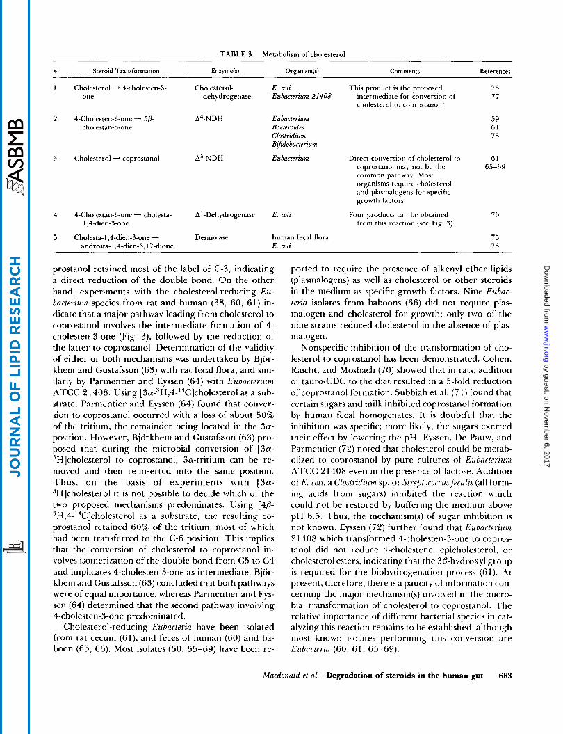
Outlined in Fig. 3 are the major known degradative products of cholesterol. The reactions are presented in Table 3, describing the microorganisms and enzyme systems responsible.
Reduction of cholesterol to coprostanol Depending upon the diet, 50% or more of the total
fecal sterol can be present in the form of coprostanol.
0
4- \ androstene-3.17-dione
Cholesterol
I, 4-androstad iene- 5/3 -cho les tan-3 -one Coprostanol 3, 17- d i o n e
1 Cholesterol - 4-cholesten-3- Cholesterol- E. coli This product is the proposed 76 one dehydrogenase Eubacterium 2 I408 intermediate for conversion of 77
Eubacterium Direct conversion of cholesterol to 61 coprostanol may not be the common pathway. Most organisms require cholesterol and plasmalogens for specific growth factors.
65-69
4 4-Cholestan-3-one - cholesta- A'-Dehydrogenase E. coli 1,4-dien-3-0ne
5 Cholesta-l,4-dien-3-one - Desmolase human fecal flora androsta- 1,4-dien-3,17-dione E. coli
Four products can be obtained 76 from this reaction (see Fig. 3).
75 76
prostanol retained most of the label of C-3, indicating a direct reduction of the double bond. On the other hand, experiments with the cholesterol-reducing Eu- bacterium species from rat and human (38, 60, 61) in- dicate that a major pathway leading from cholesterol to coprostanol involves the intermediate formation of 4- cholesten-3-one (Fig. 3), followed by the reduction of the latter to coprostanol. Determination of the validity of either or both mechanisms was undertaken by Bjor- khem and Gustafsson (63) with rat fecal flora, and sim- ilarly by Parmentier and Eyssen (64) with Eubacterium ATCC 21408. Using [3a-3H,4-14C]cholesterol as a sub- strate, Parmentier and Eyssen (64) found that conver- sion to coprostanol occurred with a loss of about 50% of the tritium, the remainder being located in the 3a- position. However, Bjorkhem and Gustafsson (63) pro- posed that during the microbial conversion of [3a- 3H]cholesterol to coprostanol, 3a-tritium can be re- moved and then re-inserted into the same position. Thus, on the basis of experiments with [3a- 3H]cholesterol it is not possible to decide which of the two proposed mechanisms predominates. Using [40- 3H,4-14C]cholesterol as a substrate, the resulting co- prostanol retained 60% of the tritium, most of which had been transferred to the C-6 position. This implies that the conversion of cholesterol to coprostanol in- volves isomerization of the double bond from C5 to C4 and implicates 4-cholesten-3-one as intermediate. Bjor- khem and Gustafsson (63) concluded that both pathways were of equal importance, whereas Parmentier and Eys- sen (64) determined that the second pathway involving 4-cholesten-3-one predominated.
Cholesterol-reducing Eubacteria have been isolated from rat cecum (61), and feces of human (60) and ba- boon (65, 66). Most isolates (60, 65-69) have been re-
ported to require the presence of alkenyl ether lipids (plasmalogens) as well as cholesterol or other steroids in the medium as specific growth factors. Nine Eubac- teria isolates from baboons (66) did not require plas- malogen and cholesterol for growth; only two of the nine strains reduced cholesterol in the absence of plas- malogen.
Nonspecific inhibition of the transformation of cho- lesterol to coprostanol has been demonstrated. Cohen, Raicht, and Mosbach (70) showed that in rats, addition of tauro-CDC to the diet resulted in a 5-fold reduction of coprostanol formation. Subbiah et al. (7 1) found that certain sugars and milk inhibited coprostanol formation by human fecal homogenates. It is doubtful that the inhibition was specific; more likely, the sugars exerted their effect by lowering the pH. Eyssen, De Pauw, and Parmentier (72) noted that cholesterol could be rnetab- olized to coprostanol by pure cultures of Eubacterzum ATCC 2 1408 even in the presence of lactose. Addition of E . coli, a Clostridium sp. or Streptororcus fecalis (all form- ing acids from sugars) inhibited the reaction which could not be restored by buffering the medium above pH 6.5. Thus, the mechanism(s) of sugar inhibition is not known. Eyssen (72) further found that Eubacterium 2 1408 which transformed 4-cholesten-3-one to copros- tanol did not reduce 4-cholestene, epicholesterol, or cholesterol esters, indicating that the 30-hydroxyl group is required for the biohydrogenation process (61). At present, therefore, there is a paucity of information con- cerning the major mechanism(s) involved in the micro- bial transformation of cholesterol to coprostanol. The relative importance of different bacterial species in cat- alyzing this reaction remains to be established, although most known isolates performing this conversion are Eubucterin (60, 61, 65-69).
Macdonald et al. Degradation of steroids in the human gut 683
Coprostanone from cholesterol Coprostanone is a relatively minor constituent of the
fecal sterol fraction. It is readily reduced by rat cecal contents to coprostano~ (73). In vivo conditions n,ay favor this reaction and the accumulation of coprostanone.
Side chain cleavage of cholesterol Though well documented for fungi and various soil
microorganisms, there is little evidence of a side chain
bacteria (74). The studies summarized below are pre- liminary and ,lnconfirmed data from animal experi- ments.
~ ~ l l ~ ~ i ~ ~ intracecal administration of [ 14C]cho]esterol
chain of cholesterol and CA, nuclear dehydrogenate ring and dehydroxylate at C-7 (76). The meta- bolic products of cholesterol were 4-cholesten-3-one, androsta-4-en-3,17-dione, cholesta-l,4-dien-3-one, and androsta-1,4-dien-3,17-dione. All four metabolites were produced in aerobic incubation over a 14-day period while only the two latter compounds were obtained un- der anaerobic conditions.
The clinical significance of cholesterol and its metab- olites coprostanol and coprostanone in the human in-
shown that the fecal levels of both cholesterol and its two major microbial products are higher in patients with bowel cancer, adenomatous polyps, and ulcerative co- litis compared to age- and sex-matched controls. The
cleavage of the cholesterol molecule by human intestinal testine k still far from clear. ReddY et a'. (78-80) have
to guinea pigs, Goddard and Hill (75) found that 1.7% appeared in the urine as estrogens. In vitro conversion of cholesterol by gut flora to estrone and estradiol has
little of the intracecally administered cholesterol in the
Same researchers (8 l ) have shown that the degree Of degradation Of cholesterol was lower in a population hereditarily predisposed to bowel cancer.
perhaps its metdbolites~ may play a role as co-carcino- also been observed (75). In contrast, the rat excreted Cruse et al. (82,83) have proposed that cholesterol, and
urine. The bulk appeared in the feces, with 6% as LC gen(s) in colon cancer. This hinges
and is0-Lc. The authors concluded that the gut flora of guinea pig is capable of removing the side chain of cholesterol, whereas in the rat only a 3-carbon unit is removed. Both activities were suppressed by adminis- tering antibiotics to the animals, further emphasizing the role of bacteria in the conversion.
A strain of E. coli isolated from a patient with colo- rectal cancer was reported by Owen et al. (76) and Tenneson, Owen, and Mason (77) to cleave the side
on an epidemiological link and remains to be substan- tiated (84).
METABOLISM OF BILE ACIDS
The major known degradative reactions of CA are outlined in Fig. 4. Analogous reactions with the excep- tion of 12a-OH dehydrogenation (reaction 4) can be listed for CDC. Table 4 describes the organisms and
0
H Isocholic
f Ethyl cholic
10
H 7d,l2ol-dihydroxy-3- keio- 5/3 - chol an - 2 4 - oi c oc id
Deoxycholic
Cholic
HO
7-ke to deoxycholic
a' u n fa tu ra 18 d d er i vat i v e
-4 Allocho I ic Ursodeoxycholic unsaturated derivative
S g y m Ei-\ 2 8 b Lky; 8 % p o m u 5 ' Z E O t .=z ya- E < 3 c .S"o a s ? m u 5 L - Z - Z s . ;yr; 5 s-2 u g * '5 3 .- & g % E $ 3 2 z2.s 5 o w . c u u S O i n O W
,Q :c n c - c > c w M.Q .5 ? % ; $ 5 . 8 i $4 2 u - - m gy.5 c * u.5 o z z m 2 2 CESS? m y 2 z r r i g , g 4 5 c : u . % * $ 3 O S L u . ? w ; i . 5 . " ~ 2
a .m 0 i e.:'& $. 9; T 5 . 2 s 2 .- *g; i=" $ $ - e u c .E :=Q C
.2:2k . " Z S . - Q m " 3 ; u ; E s 5 8 o g 5 2 D
$ g+
C
> F G m a - a -
O .s Y
.- u c w u - . s L O Q vi TI
u '5 's .'" .'" u .r! I:
4 Z p . E E E E = . c w O c i l 5
s b b b
a011 2 2
I
c c m m
c . c c c
5 s .- .- 2 2 S S 2 2 2 - 2 - E E e c ! $ $ m m T I T I u u m m w w "
5 5 Z - g 2 8 5 . ' m m
2 2 32 8 3 3
e" "" " 9 599 9 L O W - - m m m m o t. 2 9
rD ,x. "I
" - ?I 11 t I t I ; ; I I-
h I: 2 F.
:a a % a $:a 22 c % ,I. 3: k 2 k 2 d d d b d d b d 2 d d 0 z z z z z z z z ir
a-i
z z w' > > .- .-
a; I p 9 i .p
. p 3 j .$E 3 $ E.3 E E E'E: E $ q u .E 5
E E
2 L 2 8c 5 'E:
2 t e 2 2 % 2 d B . 'fi. 3 8; E.2.22 & A. .s f Z;, & 3
2 2 2 4 2 $ y.4 3: 5 Q
$ 2 m - u
I * X L * u
s z s k 2 r . xz g g Q
% % x %$ % p z % & Z Q'S 2 g U & bz - 4 s Q- u$- - g z 2 8 4 U U 2 c Qz t i a j E tiki rriajrri ticrri t i G t i tirri2 tirr i rqUQQ u ti
s - x x
- z a - X W A n 34=
n e T 3: C P 5 2 2 2 wi q z 2 p
-2 o w = % I W :> 5 z
& g 2 c 2 - 2 4 3 T T ; 2 n n
3 : T m n r - t. t. 2 9 m n n
P 3:
2 r q , v, * .- O r 6 6
c ri z : - 2 4 @i
6 . 2 2 E + 2 +2 .-
O
u w 0 Q Q c c a- u k s s i
c L 2
w a w
W
O m e, QJ 2 W L L a- m .c 2 $ 4 I m
Y I: I: K % 9 9 4 2 2
: e ' ' T L T 7.6 3: T 3: L a a ri
.- L > a
T .-
- b0.S T T T T I
x h 5 , " I
L C n n Q - - m
a W O
0 0
v T m 2 I
m r - - r. t. co 2 m
> O
> m .- L E L g T 's .$ : '5 .E
$ s - $ :+ 0 i T & & 9
a- m W
u
3: c.l .- m d
rD r . m m 0 I 0 4 - n - W - m e e - 2
Macdonald et al. Degradation of steroids in the human gut 685
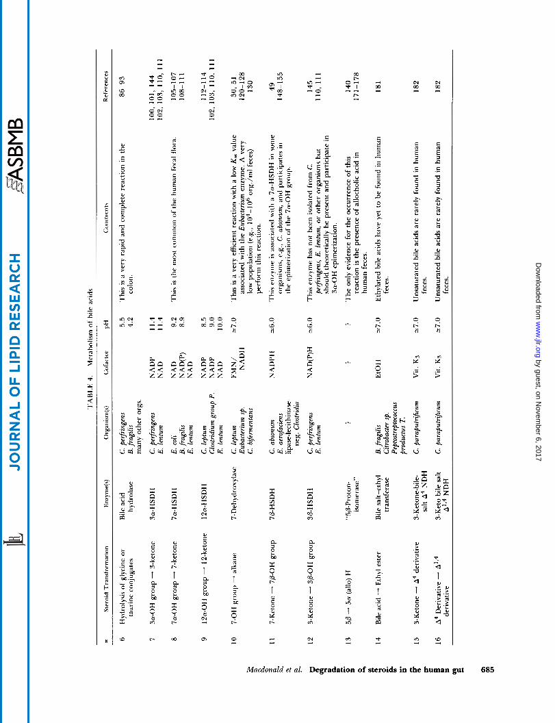
enzyme systems responsible for a given transformation. These reactions have been reviewed by Hayakawa (85), Midtvedt (86), Lewis and Gorbach (87) and Hill (88); important advances are summarized below.
Hydrolysis of conjugated bile acids Bacterial deconjugation by “B. coli” was first de-
scribed in 1934 by Basu and Chakravarty (89). Later Norman and Grub (90) reported deconjugation by a number of Clostridia and Enterococci. It is now known that bile acid hydrolysis may be caused by many intes- tinal organisms including Bacteroides, Clostridium, Eubac- terium, Lactobacillus, and Streptococcus, but it has not been demonstrated in mammalian cells (85-88). Bacterial deconjugation in the gut is so thorough that hydrolysis of both taurine and glycine conjugates goes to comple- tion in the colon (5, 6). The enzyme “bile acid hydro- lase” (cholylglycine hydrolase) from C. perfringens (9 1) and B. fraplis (92) has been purified and characterized. The C. perjiringens and the B. fragilis enzymes are both intracellular and have pH optima around 5.5 and 4.2, respectively (91, 92).
Among deconjugating microorganisms there is con- siderable variation in preference for taurine- versus gly- cine-conjugated bile acids. For example, a bile acid hy- drolase from Peptostreptococcus intermedius displays a spec- ificity for taurine conjugates while the enzyme from S.
f p c d i s and L. brevis will hydrolyze chiefly glycine con- jugates (93). In contrast, the C. pe?jringens enzyme will hydrolyze both forms of conjugates with some prefer- ence for the glycine derivatives (91).
I t must be emphasized that the physical and chemical properties of bile acids change drastically when they are hydrolyzed (92). The solubility decreases, particularly at low pH. Free bile acids are poorer detergents than their conjugates and are far less efficient in forming mixed micelles (94, 95). Bacterial overgrowth in the small intestine can lead to premature deconjugation (95). Thus conjugated ionized forms are not available for fat absorption and digestion leading to fat malab- sorption and steatorrhea (95). Premature deconjugation can also lead to some passive reabsorption of the un- ionized forms of free bile acids, which will not occur under physiological pH with conjugated bile acids.
Oxidation of 3a-, 7a-, and 1 2a-hydroxyl groups to ketones
Ketonic bile acids are not found in normal human bile in appreciable quantities but they may represent a significant fraction of human fecal bile acids. The bile of cholecystectomized patients contains small amounts of keto bile acids (96), presumably because the bile acids in the absence of the gallbladder are exposed more in- tensively to bacterial action. The presence of ketonic
functions reduces the solubility, polarity, and deter- gency of the 5P-cholanoic acids. Ketonic bile acids are produced exclusively by bacterial NAD- or NADP-de- pendent hydroxysteroid dehydrogenases (HSDH). If they are reabsorbed and returned to the liver, the keto- groups are reduced primarily to the a-hydroxyl con- formation by hepatic enzymes, which accounts for the low levels of keto-bile acids normally in bile (96).
Cell-free preparation of 3a-, 7a-, and 12a-HSDH were first reported by Aries and Hill (97) who studied these enzymes in isolates of Clostridia, Bacteroides, Bijido bacterium, and Enterobacter. In the human intestinal flora, the 7a-HSDH was much more abundant than the 3a-HSDH or the 12a-HSDH. More recently, the po- sitional- and stereo-specificity of HSDH associated with specific microorganisms have been characterized. A given organism may elaborate a single enzyme acting solely upon a single portion of the bile acid molecule, while others may produce several HSDHs that can ox- idize hydroxyl groups at positions 3, 7, and 12. Organ- isms known to elaborate these enzymes (98-1 15) are summarized on Table 4 and Fig. 4. The HSDH enzymes usually have different pH optima for oxidation and re- duction; alkaline conditions favor oxidation while pH values below 7 promote reduction (99, 1 15). These en- zymes are intracellular and possess a high degree of positional and stereochemical specificity. However, it is reasonable to assume that the 3a-HSDH acting upon bile acids and neutral steroids is one and the same en- zyme (48, 103). Organisms synthesizing HSDH are thought to obtain metabolic energy by the formation of intracellular reduced nucleotides (NADH, NADPH) by hydride ion transfer (1 05).
7-Dehydroxylation 7-Dehydroxylation of the primary bile acids CA and
CDC is quantitatively the most important transforma- tion giving rise to DC and LC. In man, DC accounts for about 20% of the total biliary bile acids, with CDC and CA making up most of the remainder. I t is known that DC undergoes EHC. Man is incapable of rehydroxylat- ing the 7-position of DC to CA as is known to occur in rats (1 16). In rabbits, 7a-dehydroxylation of CA by an- aerobes primarily concentrated in the cecum is so thor- ough that as much as 95% of the rabbits’ biliary bile acids is, in fact, DC (1 17). In contrast to DC, LC, the product of 7a-dehydroxylation of CDC, is not effi- ciently conserved in the EHC of most animal species including man (1 18) i n whom it accounts for about 2%) of biliary bile acids. I X is poorly absorbed from the ileum because it is present mainly as a 3-sulfate ester. The compound is hydrolyzed i n the colon by bacterial enzyines and the LC formed is mainly adsorbed to bac- terial debris arid excreted in the feces.
7a-Dehydroxylation can readily be demonstrated in mixed fecal cultures of man (99) and laboratory animals (1 19). Pure cultures of anaerobic bacteria synthesizing 7a-dehydroxylase were first isolated by Gustafsson, Midtvedt, and Norman (120) from feces and classified as “Lactobactlli” (1 20). Transformation studies were fur- ther performed on these isolates by Midtvedt (1 2 l) .
Independently, both Bokkenheuser, Hoshita, and Mosbach (122) and Aries and Hill (97) isolated a 7a- dehydroxylating strain of Bacteroides. 7a-Dehydroxylase activity has more recently been demonstrated in cul- tures of C. h$ermeiatans (1 23), C. leptum (50, 5 l) , a Eu- bacterium species (1 24), and a variety of other anaerobes including Clostridzal and non-Clostrzdial isolates from human feces and from sewers (1 25). Attempts to purify the C. leptzim eniyme have been unsuccessful (50, 51). However, the Eubnctertum enzyme is strongly inducible by CA and weakly inducible by CDC (124). I t has been purified 7-fold (124) and studied in some detail by White and co-workers (126, 127). It has a molecular weight of 1 14,000 by gel filtration (1 27) and is activated 4- to 6-fold by NAD. Low concentrations of NADH further enhance activity (1 28) while higher concentra- tions are strongly inhibitory, suggesting that the intra- cellular ratio of NAD/NADH may be a controlling fac- tor for the cellular level of activity. Moreover, kinetics for the NAD saturation curve do not obey the Michaelis- Menton equation in the presence of NADH, (but do so in its absence) suggesting allosteric binding of NADH to the enzyme (128). The same workers (126, 128) showed by two-dimensional electrophoresis that five polypeptides of molecular weights 77,000, 55,000, 55,000,27,000, and 23,500, respectively, are associated with enzyme induction. Purification of the enzyme by HPLC and subsequent electrophoresis showed that all of the above peptides except the one with the lowest molecular weight are associated with enzyme activity. In addition, a substance of even smaller molecular weight is apparently associated with the enzyme in the intact bacterial cell and may act as an a~ t iva to r .~
The purified enzyme 7-dehydroxylated CA, CDC, and UDC but the reaction with the 7P-epimer (UDC) was somewhat slower (1 27). This observation is in agree- ment with findings by Fedorowski et al. (129) that de- hydroxylation of both CDC and UDC occurred in mixed fecal cultures but the reaction with CDC was about 5-fold faster. Presumably, the rates of 7-dehy- droxylation of UDC and CDC in mixed fecal cultures will also depend upon the relative concentrations of the organisms involved, some of which do not react with UDC (1 29). Interestingly, 7-dehydroxylation of CA by
Hylemon, P. B. Private communication.
cultures of Eubacterium sp. can be enhanced by cocul- turing this organism with a variety of species of Bacter- oides (130).
There is some controversy regarding the existence of intestinal microorganisms capable of 7-dehydroxy- lating conjugated bile acids. The organisms studied by White and co-workers (128) did not produce enzymes that reacted with conjugated, methylated, or unsatu- rated bile acids. Other workers have found, largely on the basis of experimental evidence in humans, that con- jugated bile acids can be dehydroxylated prior to hy- drolysis (131, 132). The reason for this discrepancy is not clear, unless other organism(s) yet to be studied in vitro can 7-dehydroxylate conjugated bile acid.
Samuelsson (1 33) proposed that the first step of the 7a-dehydroxylation reaction is the diaxial trans-elimi- nation of water from the 6,7-position of CA, yielding a 6,7-unsaturated intermediate. The second step is the reduction of the intermediate to form DC. So far, it has not been possible to isolate the intermediate but White and co-workers (1 26, 128) have shown that the synthetic unsaturated intermediate, 3a-hydroxy-5P-6-cholen-24- oic acid, was rapidly converted to LC by Eubacterium 7- dehydroxylase ( 1 28) lending support to Samuelsson’s proposal (1 33). No evidence of hydration of the A6-in- termediate to give CDC or UDC was found (1 28), sug- gesting the first step in the process is irreversible. 7- Dehydroxylation may be a source of metabolic energy to the bacterium but the significance of 7a-dehydrox- ylation for the host is not clear. Drasar and Hill (1 15) and Hill (1 34) noted a positive correlation between the proportion of DC in bile and the risk of developing colon cancer. Moreover, LC, the 7-dehydroxylation product of CDC, is a liver toxin for animals (1 35, 136) and possesses comutagenic properties (1 37, 138). On the other hand, Hofmann (1 39) has suggested that 7- dehydroxylation of bile acids may have some benefit for the host, for example the formation of insoluble LC from CDC can result in removal of bile acids from so- lution thus preventing their cathartic effect. It is inter- esting to note that germ-free animals have watery stools that can be restored to normal consistency by feeding a bile acid sequestrant such as cholestyramine (1 39).
Epimerization of 3a- and 7a-hydroxyl groups
The transformation of the 3a- and 7a-hydroxyl groups of primary bile acids to 3P- and 7P-epimers by intestinal microorganisms has been suggested by the presence of 3P- and 7P-epimers occurring in the feces of laboratory animals (140) and UDC in the bile of man (1 4 1) and some species of bears (1 42). Several studies also demonstrate 3 and 7 epimerization by human in- testinal microorganisms in vitro (129, 143, 144). In 1970, Hayakawa (85) proposed that 3a-hydroxyl epi-
Macdonald et al. Degzadation of steroids in the human gut 687
merization occurred by oxidation of the 3a-hydroxyl group to the 3-ketone, then subsequent reduction of the ketone to the 3P-hydroxyl group. An analogous mechanism can be assumed for 7a-hydroxyl epimer- ization. Recently 3a-hydroxyl epimerization of CDC has been shown with C. perfringens (145, 146) and some strains of E. lentum (1 10, 11 l) , implying the presence of a 3a- and 30-HSDH and a 3-keto-intermediate. 3P- HSDH has not been clearly demonstrated in either C. perfringens (1 00) or E. lentum (1 03- 105) although theo- retically this enzyme should be present. The reverse reaction, the transformation of 3P-hydroxyl (iso) bile acid to 3a-hydroxyl bile acid can also be observed in C. perfringens (1 46) and in rat liver (1 47). The hepatic en- zyme appears to be physiologically mor,e significant than the microbial enzyme for the reverse reaction.
In the case of the epimerization of the 7a-hydroxyl group, the number of participating anaerobes in pure culture include C. absonum (49, 148-1 50), lecithinase- lipase-negative Clostridaa (1 5 1, 152), Eubacterium uero- faciens (1 53, 154), and a gram-positive unidentified anaerobe (1 55). Eubacterium aerofuczens and the gram- positive anaerobe possess only the 7P-HSDH and, there- fore, must be co-cultured with an organism synthesizing 7a-HSDH to epimerize the 7a-OH group (1 53-1 55).
Both C. absonum (49, 148) and E. aerofuczens (154) oxidize UDC and UC to the 7-kcto bile acid interme- diate when the Eh (redox potential) of the culture rises to above -100 mV (i.e., the medium is less reduced as a result of oxygen diffusion). This observation suggests that high redox potential lowers the intracellular NADPH/NADP ratio, a controlling factor in the ratio of hydroxy-bile acid/keto bile acid (1 49, 150).
The enzymes 7a- and 7P-HSDH in C. absonum are inducible by CDC and DC. Thus a two-enzyme pathway consisting of (a) 7a-HSDH (oxidative direction) and (b) 7P-HSDH (reductive direction) is, in effect, induced
(Le., CDC 7-KLC UDC). These enzymes are also inducible by 12-KLC and 7-KLC but repressed by the endproduct UDC. (1 49, 150). It is unexpected that DC and 12-KLC, which are not reactants, are both good inducers. Epimerization of bile acids at the 7-position may represent a detoxification process for the bacterium as UDC is less hydrophobic (less toxic) than CDC (1 56). In the human host, however, UDC, although less toxic than CDC, represents only a very small portion of biliary bile acids (1 4 1).
Two groups of investigators have proposed that at least part of the 7-epimerization occurring in mixed fe- cal cultures may proceed directly without a 7-keto in- termediate (129, 143). Evidence for this lies in the fail- ure to detect the 7-keto intermediate (143) and the con- servation of tritium in the transformation of [ 7P-’HH]CDC to [7a-3H]UDC by human fecal bacteria (129). 7-Keto-
688 Journal of Lipid Research Volume 24, 1983
(a) (b)
intermediates may escape detection because their con- version to 7P-OH bile acids is very rapid. Conservation of tritium at the 7-position during epimerization does not exclude the existence of a 7-keto-intermediate since the label could be recycled by bacterial intracellular nucleotides (63). Present evidence therefore favors the theory that 7-OH epimerization takes place via the 7- keto-intermediate. In mammals, epimerization of the 7a-hydroxyl group may result from oxidation of pri- mary bile acid by bacterial 7a-HSDH followed by either hepatic or bacterial reduction of the 7-keto-intermedi- ate to 7P-OH bile acid (8, 35, 157). The hepatic par- ticipation can be illustrated in germ-free animals mon- oinfected with E. coli which has no 7P-HSDH (35, i05).
Evidence for 12a-OH epimerization lies only in one report of the presence of 12P-OH bile acids in human feces (1 58). An organism elaborating 12P-HSDH re- mains to be isolated. Two established 12a-HSDH con- taining organisms have been shown to be devoid of ac- tivity with 12P-OH-containing substrates (1 04).
The clinical significance of in vivo formation of urso- bile acids is somewhat controversial. Fedorowski et al. (1 29) showed that UDC was about five times as resistant as CDC to 7-dehydroxylation in mixed fecal cultures. On the other hand, Bazzoli et al. (159) were unable to detect significant differences in the rates of 7-dehy- droxylation of UDC and CDC whether in fecal cultures or following colonic instillation experiments in human volunteers. Both CDC and UDC are potent gallstone- dissolving agents (160-162) but UDC is thought to be somewhat efficacious (160-162). Since UDC is less hy- drophobic (less toxic) (1 56), it should be the safer agent for gallstone dissolution. However, Barns and Powrie (1 63) showed that UDC and to a small extent CA (but not CDC, DC, or LC) were clastogenic (chromosome breaking) to Chinese hamster ovary cells. In animal models, UDC does not appear to be co-carcinogenic for colon cancer (1 64) as does CDC (1 64, 165). VanTassell, MacDonald, and Wiikins (166) recently showed that a number of bile acids including CA, DC, and CDC (but not LC) stimulate formation of a mutagen when incu- bated with human feces; UDC and UC remain to be tested in their system.
T w o model systems attempting to enhance 7-epi- merization of bile acids at the expense of 7-dehydrox- ylation are described (167, 168). In vitro, Macdonald and Hutchison (1 67) failed to reduce 7-dehydroxylation of CDC or CA in mixed fecal cultures by inoculation of C. absonum into such cultures. Although UDC and UC formation were enhanced, this occurred primarily at the expense of 7-KLC and 7-KDC formation. In vivo, Yahiro, Setoguchi, and Katsuki (168), gave CDC orally to rabbits that had been cecectomized and appendec- tomized. They found a marked enhancement of 7-epi- merization and a depression of 7-dehydroxylation as
reflected by the increased levels of UDC and decreased levels of DC and LC in the bile. This altered microbial metabolism presumably reflects changes both in the flora and the transit time resulting from surgery. Oral administration of CDC to human subjects enhances epimerization of the 7a-OH group in some patients, but in most cases as much as 90% of the bile acid pool be- comes CDC (169, 170).
Formation of 5a-(allo) bile acids
In 1936, allo-DC (termed “lagodeoxycholic acid”) was isolated by Kishi (171) from rabbit bile. Allo-DC has since been found in small amounts in rat (172) and rabbit feces (173); allo-CA occurs in reptile bile as a major constituent (174) and in human feces in minor amounts (140, 175). ,4110-CDC has been identified as a minor constituent in the bile of the giant salamander (176). The origin of such bile acids is not clear. Some of the 5a-cholanoic acid in reptilian bile can probably be formed without bacterial intervention via the ste- reospecific reduction of 7a, 1 2a-dihydroxy-4-cholesten- %one to a 5a-intermediate (1 77). Kallner (1 72) showed that rats could transform intracecally administered “C- labeled DC to allo-DC. He later demonstrated that labeled 12a-hydroxy-3-keto-5/3-cholanoic, 12a-hy- droxy-3-keto-5a-cholanoic, and 1 2a-hydroxy-3-keto- chol-4-en-oic acids similarly administered to rats could all be transformed to both DC and allo-DC (178). AI- though metabolism by the rat intestinal flora is impli- cated here, similar experiments with mixed fecal cul- tures have not been published and organism(s) capable of carrying out this transformation have not been iso- lated.
The significance of allo-bile acids is not known but they are undoubtedly less soluble and probably more toxic than 5P-bile acids. Cholestanol-fed rabbits form gallstones containing a high proportion of the calcium salts of deoxycholyl- and allodeoxycholyl-glycine (1 79). The insoluble allo-salt arises from a combination of he- patic and microbial transformations (44). With choles- tanol feeding, allo-CA acid is formed in the liver of rabbits as a primary bile acid and then transformed by microbial 7-dehydroxylation to allo-DC. Similarly, the small amounts of allo-CA found in human bile (180) may in part, be derived directly from hepatic transfor- mation of cholestanol.
Other bile acid transformations A number of bile acid transformations can be dem-
onstrated either in mixed fecal culture or in pure cul- ture. These include: a ) formation of C24-ethyl esterified bile acids, b) formation of unsaturated bile acids, and c) hydrolysis of a 3-sulfated bile acid. The first t w o re- actions have been illustrated in Table 2 and Fig. 4. The occurrence of these transformations in the human in-
testine have to be established and the medical relevance remains at best conjectural.
Formation of C-24 ethylesters. Bile acid ethyl esters have been shown to form in rat fecal cultures (181) and in cultures of three rat fecal isolates: B. frugdis, Citrobucter. sp., and Peptostreptococcus productus T (1 8 1). To the au- thors’ knowledge these derivatives have not been ob- served in human feces.
Formation of unsaturated bile acids. Hill and coworkers (1 82- 185) have shown that lecithinase-negative organ- isms, primarily C. paraputri,ficum, C. indolis, and C. ter- t ium, contain nuclear dehydrogenases (NDH) which, in the presence of a suitable hydrogen acceptor such a menadione, can introduce double bonds at the 1 , 2 , and 4, 5 positions of 3-keto bile acids (Fig. 4). This reaction, however, requires nonphysiological amounts of mena- dione and a considerably higher Eh value than that found in the colon. Moreover, the bile acid substrate must be first oxidized in the 3-position since nuclear dehydrogenating clostridia (NDC) are lacking 3a-HSDH (186). A second possible mechanism of obtaining an unsaturated bile acid could be via hydrolysis of bile acid 3-sulfates (see below). Until recently, unsaturated bile acids have not been found in the feces or bile of man. Tanida et al. (32) were able to show the presence of a cholenoic acid in the feces of one out of four healthy subjects analyzed in detail. Indirect evidence for the formation of unsaturated bile acids was provided by Larusso, Hoffman, and Hofmann (187) who showed that about 10% of tritium from [2,2’,4,4’-’HH]CDC was removed in human subjects and could be recovered in the urine as 3H20. A much higher percentage of the tritium (20-35%) was removed when this substrate was incubated with feces from the same subjects or with 7 of 24 anaerobic fecal isolates (1 87). Thus it appears that desaturation at the A 1,2 and A 4,5 position of CDC may be catalyzed by the human fecal flora, (presumably by NDC) but this reaction is reversible and saturated bile acid predominates (32). Additionally, [ 1 1,12-’H]- CDC appears to be stable to tritium loss in man (188); but this does not appear to be the case with [ 1 1,12-’HH]- LC which looses tritium very readily (1 88). The stability of 1 1,12-tritiated bile acids in mixed fecal cultures re- mains to be studied.
Bowel cancer patients in the United Kingdom have a significantly higher number of NDC than do age- and sex-matched controls (1 83, 189). Blackwood et al. (190) confirmed this observation and noted a similar rela- tionship with breast cancer patients (as well as obtaining confirmatory data with colon cancer patients). How- ever, this finding may not have general significance as very few NDC could be isolated from populations of Hong Kong (1 9 1).
Hydrolysis of bile acid 3-sulfatr. Kelsey, Muschik, and Sexton (1 92) and Borriello and Owen (193) showed that
Macdonald et al. Degradation of steroids in the human gut 689
3c l -hydroxy-5P- SA-d ihydro- n ' d i h y d r o - Prednisolone tetrohydrocort isol 2 0 7 cort isol cor t i so l
C H 2 0 H I
H
Ilp - hydroxy- ondrostenedione
3,6-hydroxy-5/3- tetrohydrocort isol
Co r t i so l
20B- d i hydrocort isol 2 I- deoxycortisol
Fig. 5. Metabolism of cortisol.
LC-3-sulfate was metabolized by human mixed fecal cultures to LC, 3-keto-LC, iso-LC (3P-OH), and a 5P- cholenoic acid (193). Two organisms from rat feces, Ps. aerugimxa (1 94) and a Clostrididuin strain "S" (1 95) have been found to synthesize a 3-sulfatase. The latter sul- fatase was shown to be specific for equatorial-3 sulfates of CY4- or CY6-bile acids and yields nonsulfated cholan- oate and sulfate ions on hydrolysis (195-197). Such or- ganisms have not yet been recovered from human feces. It is not known whether the elaboration of 3-sulfatase has physiological significance.
Effect of bile acids on intestinal bacteria Bile acids, particularly the dihydroxycholanoic acids
are bacteriostatic against certain intestinal bacteria (1 98, 199). It must be expected therefore, that both com- position and concentration of bile acids in the gut in- fluence the flora selectively.
METABOLISM OF NEUTRAL STEROIDS
The major known catabolic reactions of neutral ste- roids are shown in Fig. 5 and Fig. 6. Table 5 lists the same reactions with the organisms responsible and the enzyme systems involved.
Hydrolysis of glucuronides and sulfates Steroid hormones undergoing EHC are reduced in
the liver in ring A, conjugated with sulfate or glucuro- nide, and excreted in the bile. In the normal human colon these compounds are deconjugated by the action of sulfatases and glucuronidases. Sulfatases, exclusively of bacterial origin (1 8, 200-2 IO) , can hydrolyze steroid sulfates conjugated at the 3a-, 3/3-, 17P-, and 21-hy- droxyl positions, and it is not known if they are active at other positions e.g., 16. Glucuronidases are synthe- sized both in the intestinal wall and by bacteria (201).
16 I6 CY- h ydroxyprogesteron e A progesterone I 70- progesterone
The enzymes are absent from human feces at birth but present in increasing concentrations with age. In the rat, glucuronidase activity is related to the presence of Bactrroides; similar types of Bactrroidrs are also present in human fecal flora (18). The hydrolysis of glucuro- nides or sulfates is very important because bacteria can- not modify the conjugated molecule. For example, E. lriituin is incapable of 2 1-dehydroxylating DOC-2 l-sul- fate, but does so in the presence of sulfatase.'
Dehydroxylation of the 21-hydroxyl group 2 1 -Hydroxycorticoids undergoing EHC lose their 2 1-
hydroxyl group. In contrast, those not undergoing EHC are excreted in the urine with the 21-hydroxyl group intact albeit conjugated. The enzyme responsible for this conversion is exclusively of bacterial origin (39). 2 1- Dehydroxylating bacteria are present in stools of normal subjects regardless of the diet.' The enzyme is synthe- sized by Euhactrrium lentuni (203) and phenotypically similar organisms (48) of which there are about 107/g of human feces (203, 204). Colonization takes place early in life, usually before 1 year of age, and may well be achieved at birth since the organisms have been iso- lated from the vaginal secretion of 27% of pregnant women.' 2 1-Dehydroxylase is a constitutive enzyme with a pH optimum between 6.4 and 6.8. It requires an a-keto1 group at C-20-2 1 ; a hydroxyl group at C-20 protects the molecule against the action of the enzyme (205). The enzyme has no effect on hydroxyl groups at C-1 1 and C-17, and it acts independently of the config- uration of ring A (40). The enzyme has been extracted and partially purified (206). I t is inhibited by both water- soluble and lipophilic metal ion-chelators and is inacti- vated by oxygen in a matter of seconds (47). Interest- ingly, the enzyme functions equally well whether the medium is reduced by mechanical and chemical means or is reduced by co-culturing with a rapidly growing, fermenting organism (203, 206). FMNHP or NAD(P)H and FMN, required as coenzymes, are usually synthe- sized by the bacteria manufacturing the 2 1 -dehydrox- ylase. The molecular weight of the enzyme determined by chromatography on Sepharose 6B is 582,000 (203, 206).
Reduction of ring A
Saturation of ring A in A4-3-keto steroids proceeds in two steps. Frequently, both reactions can be carried out by enzymes secreted by a single microbial strain but others synthesize enzymes catalyzing only one of these reductions (207, 208). Schubert et al. (209) demon- strated that A4-3-keto steroids incubated with intestinal
Bokkenheuser, V. D., and J. Winter. Unpublished observations.
flora were stereospecifically reduced to the Sa-hydroxy- 5P-configuration. Quantitative reduction is performed by C. paruputrlficum while partial reduction may be ac- complished by L. lezchmanzz, Bijidohacteriutn adolascentis, and Peptostreptococcus (204). With C. paraputr$rum, an in vitro substrate concentration of 200 pg/ml or more inhibits the reduction of the 3-keto group. This prin- ciple has been utiliied in the biosynthesis of rare and expensive reference compounds, e.g., 3a-hydroxy, 50- derivatives of 18-hydroxylated corticoids (2 10 ,2 1 1) and 3-keto, 50-derivatives of medroxyprogesterone ace- tate.'
Kinetic studies with C. paraputrlficum (204) showed that the A4-3-keto reduction begins with hydrogenation of the double bond which is then followed by the for- mation of a hydroxyl group at C-3. The structure of rings A and B influences the rate of the reaction. For example, a methyl function at C-6 together with a 6, 7 double bond increases the resistance of the molecule to bacterial reduction (2 12). Moreover, 1,4-dienes and 4,6-dienes are more resistant to C. pnraputrlficuin than 4-mono-ene steroids (2 13, 2 14) and megestrol acetate, the most active oral progestin, owes its resistance to the 6-methyl, 4-6-diene configuration rendering it refrac- tory to both hepatic and bacterial reduction (1 8).
The preservation of the 3-keto-4-ene structures is essential for the hormonal function of the molecule. It follows, therefore, that hepatic or bacteriologic reduc- tion of ring A plays a profound role in the biological activity of these steroid structures.
Epimerization of Sa-hydroxyl group E. lrntum and phenotypically similar organisms pos-
sess a 3a-HSDH responsible for the epimerization of the 3-hydroxyl group from a to P. Incubation of preg- nanolone (30-) with E. lentum strains yields pregnane- dione, 3P- and 30-pregnanolone under mild anaerobic conditions (Eh, -150 mV T 20) (48, 202). The epi- nierization may proceed via oxidation to a 3-keto group, followed by reduction to the 30- or 3P-compound. Al- ternatively, the reaction may involve the formation of an unsaturated intermediate which is rapidly converted to 3a- or 3P-pregnanolone without accumulation of the intermediate. It should be noted that the 3a-HSDH re- quires a substrate with a 3a-OH group and has no effect on substrates with 3P-OH groups.
Introduction of a double bond conjugated to a keto group
Nuclear dehydrogenation may be carried out in vitro by some strains of C. welchii and C. paraputrijcum. The inducible enzyme synthesized by the latter organism requires a hydrogen acceptor such as menadione or
phenazine methosulfate (2 15). The unsaturated com- pound may be observed only within the first 24 hr; thereafter it becomes reduced to the original substrate. It seems unlikely that nuclear dehydrogenation takes place in the gut because of the highly reduced environ- ment (Eh less than -300 mV). (See also Formation of unsaturated bile acids.)
Side chain cleavage
Formation of 17-keto steroids (2 10) from cortisol by slurries of human feces was observed by Wade et al. (2 16), Gustafsson (2 17), and Eriksson and Gustafsson (2 18). Recently, C19 metabolites, 5t-androstane-3,- 1 lf17-triol and 5&androstane-3a, 1 1/3-diol-1 y-one, were isolated from incubation of cortisol with human fecal flora (2 19). The prevalence of converting organisms was approximately 1 06/g of feces. T h e conversion required an Eh below - 130 mV, and an initial pH of 7.0. Pre- liminary investigation indicated that the ability of con- verting organisms to form colonies on solid media was related to the composition of the media and the gaseous environment. Only corticoids with a hydroxyl group at the CI7 position were transformed to CI9 steroids by fecal flora. Recently, two of us6 recovered from human feces a gram-positive obligate anaerobic rod that syn- thesizes desmolase. This organism is yet to be identified. Free-living soil organisms can also remove the side chain of C,, steroids in the absence of the 17-hydroxyl group (52, 207).
Reduction of a 20-ketone to a n alcohol
Bacteroides frugalis, present in normal human fecal flora, converts DOC to a metabolite tentatively identi- fied as 20,2 l-dihydroxy-5&pregnan-3-one (205). This metabolite, resistant to further alteration by fecal bac- teria (204) is formed in yields of 10% when DOC is the substrate; it cannot be produced from such closely re- lated structures as THDOC and pregnanolone (204). These experiments suggest that the 20-HSDH of B. fra- galis requires an intact 3-keto-4-ene structure for its ac- tion.
A different 20-HSDH is synthesized by Bijidobacte- rium adolescentis, a common anaerobe in the intestinal flora of human and rat (220). It is more efficient than the enzyme elaborated by B. fragzlis and acts regardless of an unsaturation in the A ring. T h e enzyme shows a wide substrate specificity and reduces the 20-keto group to a 20@-hydroxyl group. It neither metabolizes, nor is affected by, hydroxyl groups at C3, C11, C17, or C21. Once formed, 20-hydroxyl compounds are resistant to further bacterial metabolism of the side chain. How- ever, existing A4-3-keto groups may be reduced by C. paraputrijicum to entirely refractory compounds, e.g., cortol (220).
1 6a-Dehydroxylation
As with most other bacteria capable of altering ring D, the organisms responsible for the conversion of 16a- hydroxy progesterone to 1 7a-progesterone also belong to the Eubacteria (53, 221). They are present in human feces in a concentration of 105/g. Two strains, #144 and # 146, have been isolated from fecal flora of the rat. Although phenotypically identical, strain # 144 synthes- izes a 1 6a-dehydratase and a AI6 nuclear dehydrogenase while a strain #146 also manufactures a 21-dehydrox- ylase (53, 221).
The transformation of 16a-OH progesterone to 17a- progesterone is a two-step reaction. The first, the re- moval of the hydroxyl group with formation of AI6-
progesterone, is completed within 9- 12 hr of incuba- tion. The second step, reduction of the A'' bond, takes place only after 24 hr of incubation and results in the isomerization of the side chain in the 17 position from /3 to a (54, 222). This mechanism, proposed by Calvin and Lieberman (223) was confirmed by isolation of AI6 derivatives from incubations of strain #144 with 16a- OH progesterone; moreover, 17a-progesterone was formed in cultures supplemented with A"-progesterone (54). Thus, the position of the side chain of urinary metabolites reveals which steroids have been 1 6a-de- hydroxylated. The 1 6a-dehydratase is unusually resis- tant to oxygen as evidenced by the specific enzymatic activity of cell fractions whether incubated aerobically or anaerobically (54).
METABOLISM OF PHENOLIC STEROIDS The metabolism of these compounds was reviewed
in detail by Adlercreutz et al. (1 7), Diczfalusy and Levitz (224), and Taylor (16). Some important transformations are outlined below.
Like other steroids, conjugated estrogens are hydro- lyzed by the intestinal microflora; as expected, admin- istration of antibiotics causes a huge increase in the ex- cretion of conjugates in the feces. Estrone and 15a-hy- droxy-estrone can be reduced in the 17-position. The reduction can take place also in the 16-position con- verting 16-oxo-estradiol to 16-epiestriol. Adlercreutz et al. (225) suggested that the transformation of 16a-hy- droxy-estrone to the 1 Sa-compound takes place in two steps: first, 16a-dehydration due to the intestinal mi- croflora and second, 15a-hydroxylation which can take place in the intestinal wall and the liver.
Experiments with fecal flora or pure bacterial cul- tures are needed to clarify the precise mechanism and site of the transformation.
CONCLUSION We have described a number of microbial transfor-
mations of the three classes of steroids occurring in the
Macdonald et al. Degradation of steroids in the human gut 693
human intestine. These include: hydrolysis, dehydro- genation (hydroxyl a n d nuclear), dehydroxylation, epi- merization, and side chain cleavage reactions all leaving the steroid nucleus intact. In many cases the responsible organism has been isolated and the enzynie(s) has been purified and studied. Areas of paucity in information are nonetheless evident. Isolation of unknown organ- isms, details of the mechanism of action of most of'the described enzymes, and a thorough understanding of the biological significance of the reaction products to the host must await further research. New applications of these enzymes remain in the future.EJ T h e authors wish to acknowledge financial support from the following granting agencies: the National Institute of Health (grants #AM-25324 and HL-24061), the National Cancer In- stitute (grant CA-25763), the Medical Research Council of Canada (grant MA 5075), and the National Cancer Institute of Canada. ,Moriusriipi rrtrir~rd I8 lMn, 1982 nrid r r r rurvcrrl forrii 18 J(iriiior>' I983
REFERENCES
1 .
2.
3.
4.
5.
6.
7.
8.
9.
10.
694
Holdeman, L. V., I. J. Good, and W. E. C;. Moore. 1976. Human fecal flora: a variation in bacterial composition within individuals and a possible effect of emotional stress. AppI. Ermzron. Microbial. 31: 359-375. Hentges, D. J., B. R. Maier, G. C. Burton, M. A. Flynn, and R. T . Tsutakawa. 1977. Effect of a high-beef diet on the fecal bacterial flora of humans. Cnticer Rrs. 37: 568-57 1. Moore, W. E. C., E. P. Cato, and L. V. Holdeman. 1978. Some current concepts in intestinal bacteriology. ,4m. ,/. (.'lit/. S u t r . 31: S33-S42. Hislop, I. G. 1970. T h e absorption and enterohepatic circulation of bile salts: an historical review. Mrd. J . Aust.
Hofinann, A. F. 1979. T h e enterohepatic circulation of bile acids in man. In Bile Acids Clinics in Gastroenter- ology. Vol. 6. G. Paumgartner, editor. Saunders, Phil- adelphia. 3-24. Carey, M. C. 1982. T h e enterohepatic circulation. 1~ T h e Liver, Biology and Pathobiology. I. Arias, H . Pop- per, D. Schachter, and D. A. Shafritz, editors. Raven Press, New York. 429-465. Hofmann, A. F. 1976. T h e enterohepatic circulation of bile acids in man. Adz!. I u / p r ) t . M r d . 21: 501-534. Hofmann, A. F. 1977. T h e enterohepatic circulation of conjugated bile acids in healthy man: quantitative de- scription and function. In Expos& Annuels de Biochimie Medicale (33' serie). Cholesterol Metabolism and Li- polytic Enzymes. J. Polonovsky, editor. Masson Publish- ing USA Inc., New York. 69-86. Ahlberg, J., B. Angelin, I . Bjorkhem, and K. Einarsson. 1977. Individual bile acids in portal venous and systemic blood serum in fasting inan. Gns t ror t i t r ,u lo~ . 73: 1377- 1382. Almk, B., A. Brernmelgaard, J. Sjovall, and P. Thonias- sen. 1977. Analysis of metabolic profiles of bile acids in urine using lipophilic anion exchanger and computer-
1 I . Amuro, Y., T . Endo, K. Higashino, K. Uchida, and Y. Yamamura. 198 1. Urinary and fecal keto bile acids in liver cirrhosis. Clin. Chim. Acta. 114: 137-147.
12. Brenimelgaard, A., and J. Sjovall. 1974. Bile acid pro- files in urine of patients with liver diseases. Eur. J . Cliti.
13. Palmer, R. H., and C. K. McSherry. 1982. Lithocholate metabolism in baboons fed chenodeoxycholate. J . Lnh.
14. Allan, R. N. , J. L. Thistle, A. F. Hofinann, J. A. Carter, and P. Y. S. Yu. 1976. Lithocholate metabolism during chenotherapy for gallstone dissolution. 2. Absorption arid sulphation. Gut. 17: 4 13-4 19.
15. Laatikainen, T . , and R. Vikko. 1970. Identification of' C I 9 O p and C 2 1 0 2 steroids in the mono- and disulphate fractions of human faeces. Eur. J . Biochrtti. 13: 534-538.
16. Taylor, W. 1971. 'The excretion of steroid hormone metabolites in bile and feces. Vitron. Horm. 29: 201-285.
17. Aldercreutz, H., F. Martin, R. Jarvenpaa, and T . Fotsis. 1979. Steroid absorption and enterohepatic recycling.
18. Adlercreutz, H., and F. Martin. 1980. Biliary excretion and intestinal metabolism of progesterone and estrogens in man. J . Strroid Biochrm. 13: 23 1-244.
19. Sandberg, A. A., M." R. Slaunwhite, Jr. and R. Y. Kir- dani. 1970. Metabolic Conjugation and Metabolic Hy- drolysis. W. M. Fishinan, editor. Academic Press, New
20. Levit7, M. , and B. K. Young. 1977. Estrogens in preg- nancy. I'rtcirr~. f fortn. 35: 109-147.
21. Adlercreutz, H., arid T. Luukkainen. 1967. Biochemical and clinical aspects of enterohepatic circulation of es- trogens. A d o . Eric/oc.r.irtol. (Coprnhagrtr) Suppl. 124: 10 1- 140.
22. Sandberg, A. A., and W. R. Slaunwhite, Jr. 1951. Stud- ies on phenolic steroids in human subjects. 11. T h e met- abolic fate and hepatobiliary-enteric circulation of CI4- estrone and C14-estradiol in women. J . C/in. I ) 7 ~ 5 / . 36: 1266-1278.
23. Reed, M. J., K . Fotherby, and S . J. Steele. 1972. Me- tabolism of ethynyloestradiol in man. J . Eridorrittol. 55: 3.5-361.
24. Huempel, M., M. Wendt, G. Jogs, C. Weiss, S. Reitz, and U. Speck. 1977. Intraindividual comparison of phar- macokinetic parameters of d-norgestrel, lynestienol and cyproterone acetate in six women. Controcrptioti. 16: 199-2 15.
25. Smith, R. L. 1974. Biliary excretion and hepatotoxicity of contraceptive steroids. Pharmacological models in contraceptive development. Acta Endorri)iol. Suppl. 185:
26. Grundy, S. M. 1975. Effects of polyunsaturated fiat on lipid nietabolism in patients with hypertriglyceridemia. J . O'l i t i . 1tizlr.V. 55: 269-282.
27. Duncan, I. W., P. H. Culbreth, and C . A. Burtis. 1979. Determination of free, total and esterified cholesterol by high pressure liquid chromatography. J . Chrorn(itogr. 162: 281-292.
28. Deacon, A. C., and P. J. G. Dorsonn. 1979. Enzymic assay of total cholesterol involving chemical or enzymic hydrolysis: a comparison of methods. Glitz. Cl iun i . 25:
29. Miettinen, T. A,, E. H. Ahrens, Jr., and S. M. Grundy. 1965. Quantitative isolation and gas-liquid chromato- graphic analysis of total dietary and fecal neutral ste- roids. J . Lipid Res. 6 41 1-424.
30. Juengst, D., A. Pickel, E. Elsaesser, F. J. Marx, and H. J. Karl. 1979. Urinary cholesterol excretion in man with benign prostatic hyperplasia and carcinoma of the prostate. Cancer. 43: 353-359.
31. Grundy, S. M., E. H. Ahrens, Jr., and T . A. Miettinen. 1965. Quantitative isolation and gas-liquid chromato- graphic analysis of total fecal bile acids. J . Lipid Res. 6:
32. N. Tanida, Y. Hikasa, M. Hosomi, M. Satoni, I. Ooh- ama, and T . Shimoyama. 198 1. Fecal bile acid analysis in healthy Japanese subjects using a lipophilic anion ex- changer, capillary column gas chromatography and mass spectrometry. Gastroenterol Jpn. 16: 363-37 1.
33. Heftmann, E., editor. 1973. Modern Methodsof Steroid Analyses. Academic Press, New York.
34. Karger, B. L., L. R. Snyder, and C. Horvath, editors. 1973. Introduction to Separation Science. John Wiley & Sons.
35. Gustafsson, B. E., A. Norman, and J. Sjovall. 1960. In- fluence of E. coli infection on turnover and metabolism of cholic acid in germ-free rats. Arch. Biochem. Biophys.
36. Norman, A., and S. Bergman. 1960. The action of in- testinal microorganisms on bile acids: bile acids and ste- roids 101. Acto C ~ P V I . Scand. 14: 1781-1789.
37. Gustafsson, B. E., T. Midtvedt, and A. Norman. 1968. Metabolism of cholic acid in germ-free animals after the establishment in the intestinal tract of deconjugating and 7a-dehydroxylating bacteria. Acta Pathol. Microbiol. Scand. 72: 433-443.
38. Eyssen, H., and G. Parmentier. 1977. Biohydrogenation of sterols and fatty acids by the intestinal microflora. Am. J. (:/in, ,\:utr. 27: 1329-1340.
39. Winter, J., and V. D. Bokkenheuser. 1978. 21-Dehy- droxylation of corticoids by anaerobic bacteria isolated from human fecal flora. J . Steroid Biochem. 9: 379-384.
40. Winter, J., V. D. Bokkenheuser, and L. Ponticorvo. 1979. Bacterial metabolism of corticoids with particular reference to the 2 1-dehydroxylation. J. Bioi. Chem. 254:
41. Madsen, D., M. Beaver, L. Chang, E. Bruckner-Kardoss, and B. Wostmann. 1976. Analysis of bile acids in con- ventional and germ-free rats.J. Lipid Res. 17: 107-1 I 1.
42. Beaver, M. H., B. S. Wostmann, and D. C. Madsen. 1978. Bile acids in bile of germ-free and conventional dogs. Prof. Sor. Exp. Bid . Med. 157: 386-389.
43. Hofmann, A. F., E. H. Mosbach, and C. C. Sweeley. 1969. Bile acid composition of bile from germ-free rab- bits. Biochim. Biophys. Acta. 176: 204-207.
44. Hofmann, A. F., V. Bokkenheuser, R. L. Hirsch, and E. H. Mosbach. 1968. Experimental cholelithiasis in the rabbit induced by cholestanol feeding: effect of neo- mycin treatment on bile composition and gallstone for- mation. J . Lipid Res. 9: 244-253.
45. Garbutt, J. T., K. W. Heaton, L. Lack, and M. P. Tyor. 1969. Increased ratio of glycine to taurine conjugated bile salts in patients with ileal disorders. Gastroenterology. 56: 71 1-720.
46. Sacquet, E. C., P. M. Raibaud, C. Mejean, M. J. Riottot, C. Leprince, and P. C. Leglise. 1979. Bacteriol forma-
397-4 10.
91: 83-100.
2626-2629.
tion of w-muricholic acid in rats. Appl. Environ. Microbiol.
47. Feighner, S. D., and P. B. Hylemon. 1980. Character- ization of a corticosteroid 2 1-dehydroxylase from the intestinal anaerobic bacterium, Eubacterium lentum. J . Lipid Res. 21: 585-593.
48. Bokkenheuser, V. D., J. Winter, S. M. Finegold, V. L. Sutter, A. E. Ritchie, W. E. C. Moore, and L. V. Holde- man. 1979. New markers for Eubacterium lentum. Appl. Environ. Microbiol. 37: 1001-1006.
49. Macdonald, I. A., and P. D. Roach. 1981. Bile salt in- duction of 7a- and 7P-hydroxysteroid dehydrogenases in Clostridium absonum. Biochim. Biophys. Acta. 665: 262- 269.
50. Stellwag, E. J., and P. B. Hylemon. 1979. 7a-Dehy- droxylation of cholic acid and chenodeoxycholic acid by Clostridium leptum. J. Lipid Res. 2 0 325-333.
51. Stellwag, E. J., and P. B. Hylemon. 1978. Characteriza- tion of 7a-dehydroxylase in Clostridium leptum. Am. J . Clin. Nutr. 31: S2434247.
52. Dorfman, R. I., and F. Ungar. 1965. Metabolism of Ste- roid Hormones. Academic Press, New York. 204-288.
53. Bokkenheuser, V. D., J. Winter, S. O’Rourke, and A. E. Ritchie. 1952. Isolation and characterization of fecal bacteria capable of 1 6a-dehydroxylating corticoids. Appl. Environ. Microbiol. 4 0 803-808.
54. Winter, J., S. O’Rourke, V. D. Bokkenheuser, P. B. Hylemon, and T. L. Glass. 1982. 16a-Dehydroxylation of corticoids by bacteria from rat fecal flora. J . Steroid Biochem. 16: 231-238.
55. Heftman, E. 1970. Steroid Biochemistry. 1st edition. Academic Press, New York. 1 15-1 19.
56. Miettinen, T. A,, A. Proia, and D. J. McNamara. 1981. Origins of fecal neutral steroids in rats. J. Lipid Res. 22:
57. Dam, H. 1934. The formation of coprostanol in the in- testine. 11. The action of intestinal bacteria on choles- terol. Biochem. J . 28: 820-825.
58. Snog-Kjaer, A., J. Prange, and H. Dam. 1956. Conver- sion of cholesterol to coprostanol by bacteria. J . Gen. Microbiol. 14: 256-260.
59. Crowther, J. S., B. S. Drasar, P. Goddard, M. J. Hill, and K. Johnson. 1977. The effect of a chemically defined diet on the faecal flora and a faecal steroid concentra- tion. Gut. 14: 490-493.
60. Sadzikowski, M. R., J. F. Sperry, and T. D. Wilkins. 1977. Cholesterol-reducing bacterium from human feces. Appl. Environ. Microbiol. 34: 355-362.
61. Eyssen, H. J., C. G. Parmentier, F. C. Compernolle, G. De Pauw, and M. Piessens-Denef. 1973. Biohydrogen- ation of sterols by Eubacterium ATCC 2 1,408-Nova spe- cies. Bur. J . Biochrni. 36: 41 1-421.
62. Rosenfeld, R. S., and T. F. Gallagher. 1964. Further studies of the biotransformation of cholesterol to co- prostanol. Steroids. 4: 5 15-520.
63. Bjorkhem, I., and J. A. Gustafsson. 1971. Mechanism of microbial transformation of cholesterol into copros- tanol. Eur. J. Biochem. 21: 428-432.
64. Parmentier, G., and H. Eyssen. 1974. Mechanism of biohydrogenation of cholesterol to coprostanol by Eu- bacterium ATCC 21,408. Biochim. Biophys. Acta. 348: 279-284.
65. Brinkley, A. W., and G. E. Mott. 1978. Anaerobic fecal
37: 1127-1 131.
485-495.
Macdonald et al. Degradation of steroids in the human gut 695
bacteria of the baboon. Appl. Enuiron. Microbiol. 36: 530- 532.
66. Brinkley, A. W., A. R. Gottesman, and G. E. Mott. 1982. Isolation and characterization of new strains of choles- terol-reducing bacteria from baboons. Appl. Enuiron. Microbiol. 43: 86-89.
67. Brinkley, A. W., A. R. Gottesman, and G. E. Mott. 1980. Growth of cholesterol-reducing Eubacterium on choles- terol-brain agar. Appl. Environ. Microbiol. 40: 1 130- 1132.
68. Mott, G. E., and A. W. Brinkley. 1979. Plasmenyl- ethanolamine: growth factor for cholesterol-reducing Eubacterium. J . Bacteriol. 139: 755-760.
69. Mott, G. E., A. W. Brinkley, and C. L. Mersinger. 1980. Biochemical characterization of cholesterol-reducing Eubacterium. Appl . Enuiron. Microbiol. 40: 101 7-1 022.
70. Cohen, B. I., R. F. Raicht, and E. H. Mosbach. 1974. Effect of dietary bile acids, cholesterol, and P-sitosterol upon formation of coprostanol and 7-dehydroxylation of bile acids by rat. Lipids. 9: 1024-1029.
71. Subbiah, M. T. R., M. C. Naylor, J. Schumacher, and B. A. Kottke. 1974. 5P-Reduction of (‘4C)cholesterol by human feces in vitro: nonspecific inhibition by sugars. ExppPrien tia. 30: 249- 2 5 0.
72. Eyssen, H., G. De Pauw, and C. Parmentier. 1974. Effect of lactose on A5-steroid-reducing activity of intestinal bacteria in gnotobiotic rats. J . Nutr. 104: 605-6 12.
73. Bjorkhem, I . , J. A. Gustafsson, and 0. Wrange. 1973. Microbial transformation of cholesterol into copros- tanol. Eur. J . Biorhem. 37: 143-147.
74. Martin, C:. K . 1977. Cleavage of sterol side chains. Adz). Appl. Mirrobiol. 22: 29-58.
75. Goddard, P., and M. J. Hill. 1974. T h e in vivo metab- olism of cholesterol by gut bacteria in the rat and guinea pig. J . Strroid Biochrin. 5: 569-572.
76. Owen, R. W., M. E. Tenneson, R. F. Bilton, and A. N. Mason. 1978. T h e degradation of cholesterol by Esch- erirhirr coli isolated from human feces. Biochrtn. Sor. Truiis. 6: 377-379.
77. Tenneson, M. E., R. W. Owen, and A. N. Mason. 1977. T h e anaerobic side-chain cleavage of bile acids by E:.trh- rrichirc coli isolated from human feces. Biochrm. Soc. Tram. 5: 1758-1 760.
78. Reddy, B. S., and E. I,. Wynder. 1977. Metabolic epi- demiology of colon cancer: fecal bile acids and neutral sterols in colon cancer patients arid patients with ade- nomatous polyps. Cnnrer. 39: 2533-2539.
79. Wynder, E. L., and B. S. Reddy. 1977. Colon cancer prevention: today’s challenge to biomedical scientists and clinical investigators. O’anco~-. 40: 2565-257 1.
80. Reddy, B. S., C. W. Martin, and E. L. Wynder. 1977. Fecal bile acids and cholesterol metabolites of patients with ulcerative colitis, a high risk group for the devel- opment of colon cancer. Cancrr Rrs. 37: 1697- 1 70 1.
8 1. Lipkin, M., B. S. Reddy, J . Weisburger, and L. Schech- ter. 198 1. Nondegradation of fecal cholesterol in sub- jects at high risk for cancer of the large intestine.]. U i n . InzvJt. 67: 304-307.
82. Cruse, J. P. , M. R. Lewin, and C. G. Clark. 1979. Dietary cholesterol is cocarciriogenic for human colon cancer. Lnncet. i: 752-755.
83. Cruse, J. P., M. R. Lewin, G. P. Ferulano, and C. G. Clark. 1978. (locarcinogenic effects of dietary choles- terol in experimental colon cancer. Sirt irrp. 276: 822- 825.
84. Macdonald, I . A., and B. G. Rao. 1979. Diet, bacteria, and the origin of bowel cancer. Modern Med. Canada. 34:
85. Hayakawa, S. 1973. Microbial transformations of bile acids. Adu. Lipid Res. 11: 143-192.
86. Midtvedt, T. 1974. Microbial bile acid transformation. Am. J . Clin. Nutr. 27: 1341-1347.
87. Lewis, R., and S. Gorbach. 1972. Modification of bile acids by intestinal bacteria. Arch. Intern. Med. 130: 545- 549.
88. Hill, M. J. 1975. T h e etiology of colon cancer. CRC Crit. Rru. Toxicol. 4: 3 1-8 1.
89. Basu, K. P., and S. C. Chakravarty. 1934. Action of B. coli on conjugated bile acids. Indian J . Mpd. Res. 21: 691- 694.
90. Norman, A., and R. Grubb. 1955. Hydrolysis of con- jugated bile acids by Clostridia and Enterococci: bile acids and steroids 25. Acta Pathol. Microbiol. Scand. 36: 537-547.
91. Nair, P. P., M. Gordon, and J. Reback. 1967. T h e en- zymatic cleavage of the carbon-nitrogen bond in 3a,7a, 12a-trihydroxy-5~-cholan-24-0yl glycine. J. Biol. Chem. 243: 7- 1 1.
92. Stellwag, E. J., and P. B. Hylemon. 1976. Purification and characterization of bile salt hydrolase from Bade- roidrs ,frngzlis sub. sp. frngilis. Biorhim. Biophys. Acta. 452:
93. Kobashi, K., I . Nishizawa, T. Yamada, and J. Hase. 1978. A new hydrolase specific for taurine conjugates of bile acids. J . Biocheni. Tokyo. 84: 495-497.
94. Small, D. M. 1971. T h e physical chemistry of cholanic acids. Zn T h e Bile Acids. Vol. 1. P. P. Nair and D. Kri- chevsky, editors. Plenum Press, New York, London.
95. Holt, P. R. 1972. T h e roles of bile acids during the process of normal fat and cholesterol absorption. Arch. In turn . Med. 130: 574-583.
96. Hepner, G. W., A. F. Hofmann, J. R. Malagelada, P. A. Szczepanik, and P. D. Klein. 1974. Increased bacterial degradation of bile acids in cholecystectomized patients. Gnstroentrrolog. 66: 556-564.
97. Aries, V., and M. J. Hill. 1970. Degradation of steroids by intestinal bacteria. 11 . Enzymes catalyzing the oxi- doreduction of the 30-, 7a-, and I2a-hydroxyl groups in cholic acid, and the dehydroxylation of the 7-hydroxyl group. Binchiin. Biophys. Acto. 202: 535-543.
98. Macdoriald, I . A., G. R. Webb, and D. E. Mahony. 1978. Fecal hydroxysteroid dehydrogenase activities in vege- tarian Seventh Day Adventists, control subjects, and bowel cancer patients. Am. J . Clin. S u t r . 31: S 2 3 3 4 2 3 8 .
99. Macdonald, 1. A , , G. Singh, D. E. Mahony, and C. E. Meier. 1978. Effect of pH on bile salt degradation by niixed fecal cultures. Strroids. 32: 245-256.
100. Macdonald, I . A., C . E. Meier, D. E. Mahony, and G. A. Costain. 1976. 3a- , 701-, and 12a-hydroxysteroid dehydrogenase activities from Clostrirliunr perfritzpi.7. Blochim. B iop /y . Acto. 450: 142-1 53.
101. Macdonald, I. A, , 1’. P. Forrest, G. A. Costain, and B. G. Rao. 1978. Identification of 7a,l2a-dihydroxy-3- oxo-cliolanoic acid as the major degradation product from cholic by C. pur f r inp i . \ . J . Steroid Bior/irm. 9: 353- 358.
102. Macdonald, I . A . , I). E. Mahony, J. F. Jellett, and C. E. Meier. 197’7. NAD-dependent 3n- and 12a-hydroxy-
103. Macdonald, 1. A., J. F. Jellett, D. E. Mahony, and L. V. Holdeman. 1979. Bile salt 3a- and 12a-hydroxysteroid dehydrogenases from Eubnrterium lentum and related organisms. Appl. Environ. Microbiol. 37: 992- 1000.
104. Macdonald, I. A., and F. C. Chang. 1982. The stereo- specificity of 3a- and l2a-bile salt hydroxysteroid de- hydrogenase systems from four microbial sources. En- z y n e . 28: 392-395.
105. Macdonald, I. A., C. N. Williams, and D. E. Mahony. 1973. 7a-Hydroxysteroid dehydrogenase from Esrhe- richia coli B.: preliminary studies. Biochim. Biophy. Acta. 309: 243-253.
106. Haslewood, E. S., and G. A. D. Haslewood. 1976. The specificity of a 7a-hydroxysteroid dehydrogenase from Escherichia coli. Biochem. J . 157: 207-2 10.
107. Macdonald, I . A , , C. N. Williams, and D. E. Mahony. 1976. The behaviour of 3a- and 7a-hydroxysteroid de- hydrogenase on chenodeoxycholate-substituted sephar- ose. Steroids. 28: 25-30.
108. Hylemon, P. B., and J. A. Sherrod. 1975. Multiple forms of 7a-hydroxysteroid dehydrogenase in selected strains of Bacteroides fragilis. J. Bacteriol. 122: 41 8-424.
109. Macdonald, I. A,, C. N. Williams, D. E. Mahony, and W. M. Christie. 1975. NAD- and NADP-dependent 7a- hydroxysteroid dehydrogenases from BacteroidPsfragilis. Biochim. Biophy. Acto. 384: 12-24.
110. Hirano, S., N. Masuda, H. Oda. and T. Imamura. 1981. Transformation of bile acids by mixed microbial cultures from human feces and bile acid transforming activities of isolated bacterial strains. Microbiol. Immunol. 25: 27 1 - 282.
11 1. Hirano, S., and N. Masuda. 1981. Transformation of bile acids by Eubacterium lentum. Appl. Enuiron. Mirrohiol.
112. Harris, J. N. , and P. B. Hylemon. 1978. Partial purifi- cation and characterization of NADP-dependent 12a- hydroxysteroid dehydrogenase from Clostridium leptum. Biochim. Biophys. Acta. 528: 148-1 57.
113. Mahony, D. E., C. E. Meier, I. A. Macdonald, and I,. V. Holdeman. 1977. Bile salt degradation by non- fermentative rlostridio. Appl. Environ. Microbiol. 34: 4 19- 423.
114. Macdonald, I. A,, J. F. Jellet, and D. E. Mahony. 1979. 1 Sa-Hydroxysteroid dehydrogenase from Clostridium group P strain C48-50 ATCC #29733: partial purifi- cation and characterization. J. Lipid Res. 20: 234-239.
115. Drasar, B. S., and M. J. Hill. 1974. Human Intestinal Flora. Academic Press, London, New York, San Fran- cisco. 194.
1 16. Norman, A,, and J. Sjovall. 1958. On the transformation and enterohepatic circulation of cholic acid in the rat. Bile acids and steroids, 68.1. Biol. Chem. 233: 872-885.
117. Lindstedt, S., and J. Sjovall. 1957. On the formation of deoxycholic acid from cholic acid in the rabbit. Acta ChPm. Srand. 11: 421-426.
118. Cowen, A. E., M. G. Korman, A. F. Hofmann, 0. W. Cass, and S. B. Coffin. 1975. Metabolism of lithocholic acid in healthy man. 11. Enterohepatic circulation. Gas- tromterology. 69: 67-76.
119. Morotomi, M., Ti. Kawai, and M. Masahiko. 1979. In- testinal microflora and bile acids: in vitro cholic acid transformation by mixed fecal culture of rats. Microbiol. Immunol. 23: 839-847.
42: 912-915.
120. Gustafsson, B. F., T. Midtvedt, and A. Norman. 1966. Isolated fecal microorganisms capable of 7a-dehydrox- ylating bile acids.]. Exp. Med. 123: 413-432.
12 1. Midtvedt, T. 1967. Properties of anaerobic gram-posi- tive rods capable of 70-dehydroxylating bile acids. Acta Pathol. Microbiol. Scnnd. 71: 147-160.
122. Bokkenheuser, V., T. Hoshita, and E. H. Mosbach. 1969. Bacterial 7-dehydroxylation of cholic acid and allocholic acid. J. Lipid Res. 10: 421-426.
123. Ferrari, A., and L. Beretta. 1977. Activity on bile acids of a Clostridium bijermentans cell-free extract. FEBS Lett.
124. White, B. A., R. L. Lipsky, R. J. Fricke, and P. B. Hy- lemon. 1980. Bile acid induction specificity of 7a-de- hydroxylase activity in an intestinal Euhnrterium species. Steroids. 35: 103-1 10.
125. Hirano, S., R. Nakama, M. Tomaki, N. Masuda, and H. Oda. 198 1. Isolation and characterization of thirteen intestinal microorganisms capable of 7a-dehydroxylat- ing bile acids. Appl. Etnliron. Microbiol. 41: 737-741.
126. White, B. A., A. F. Cacciapuoti, R. J. Fricke, T. R. Whitehead, E. H. Mosbach and P. B. Hylemon. 1981. Cofactor requirements for 7a-dehydroxylation of cholic and chenodeoxycholic acid in cell extracts of the intes- tinal anaerobic bacterium Eubacterium species V.P.I. 12708. J. Lipid Res. 22: 89 1-898.
127. White, B. A., R. J. Fricke, and P. B. Hylemon. 1982. 70-Dehydroxylation of ursodeoxycholic acid by whole cells and cell extracts of the intestinal anaerobic bacte- rium Euhnctrriua species V.P.I. 12708. J . Lipid Res. 23:
128. White, B. A., D. A. M. Paone, A. F. Cacciapuoti, R. J. Fricke, E. H. Mosbach, and P. B. Hylemon. 1983. Reg- ulation of bile acid 7-dehydroxylase activity by NAD' and NADH in cell extracts of Eubncterium species V.P.I. 12708. J. Lipid Res. 2 4 20-27.
129. Fedorowski, T., G. Salen, G. S. Tint, and E. H. Mosbach. 1979. Transformation of chenodeoxycholic acid and ursodeoxycholic acid by human intestinal bacteria. Gas- troenterology. 77: 1068-1073.
130. Hirano, S., and N. Masuda. 1982. Enhancement of the 7a-dehydroxylase activity of a gram-positive intestinal anaerobe by Bacteroides and its significance in the 7-de- hydroxylation of ursodeoxycholic acid. J. Lipid Res. 23:
13 1. Hepner, G. W., A. F. Hofmann, and P. J. Thomas. 1972. Metabolism of steroid and amino acid moieties of con- jugated bile acids in man.]. Clin. Invest. 51: 1889-1897.
132. Stahl, E., and B. Arnesjo, 1972. Taurocholate metab- olism in man. Scnnd. J . Gastroenterol. 7: 559-566.
133. Samuelsson, B. 1960. Bile acids and steroids. 96: On the mechanism of the biological formation of deoxycholic acid from cholic acid.]. Biol. Chem. 235: 361-366.
134. Hill, M. J. 1978. The role of unsaturated bile acids in the etiology of large bowel cancer. Origins of Human Cancer. Cold Spring Harbour Symposium, Vol. XLIV.
135. Palmer, R. H., and Z. Hruban. 1966. Production of bile duct hyperplasia and gallstones by lithocholic acid. J . C h i . Invest. 45: 1255-1267.
136. Fisher, C. D., N. S. Cooper, M. A. Rothschild, and E. H. Mosbach. 1977. Effect of dietary chenodeoxy- cholic acid and lithocholic acid in the rabbit. Am. J. Dig. Dis. 19: 877-886.
137. Silverman, S. J., and A. N. Andrews. 1977. Bile acids:
75: 163-165.
145- 153.
1152-1 158.
1627-1639.
Macdonald et al. Degradation of steroids in the human gut 697
comutagenic activity in the Salmonella microsome mu- tagenic test: brief communication. J. Natl. Cancer Inst. 59: 1557-1559.
138. Kawalek, J. C., and A. W. Andrews. 1977. T h e effect of bile acids on the metabolism of benzo(a)pyrene and 2-aminoanthracene to mutagenic products. Federation Proc. 36: 844 (Abstract).
139. Hofmann, A. F. 1977. Bile acids, diarrhea and anti- biotics: data, speculation and a unifying hypothesis. J . Infect. Dis. 135: S126-Sl32.
140. Elliot, W. H. 1971. Allo bile acids. In T h e Bile Acids. Vol. 1. P. P. Nair, and D. Kritchevsky, editors. Plenum Press, New York, London. 47-93.
14 1. Igimi, H. 1976. Ursodeoxycholic acid-a common bile acid in gallbladder bile of Japanese subjects. L f e Sci. 18:
142. Kurozumi, K., T. Harano, K. Yamasaki, and Y. Ayak. 1973. Studies on bile acids in bear bile. J. Biochem. 74:
143. Sarva, R. P., H. Fromm, G. L. Carlson, L. Mendelow, and S. Ceryak. 1980. Intracolonic conversion in man of chenodeoxycholic acid (CDC) to ursodeoxycholic acid (UDC) with and without formation of 7-keto lithocholic acid as an intermediate. Gastroentrrolog)’. 78: 1252 (Ab- stract).
144. Hirano, S., N. Masuda, and H. Oda. 1981. In vitro trans- formation of chenodeoxycholic acid and ursodeoxy- cholic acid by human intestinal flora, with particular ref- erence to mutual conversion between the two bile acids. J. Lipid Res. 22: 735-743.
145. Hirano, S., N. Masuda, H. Odd, and H. Muka. 1981. Transformation of bile acids by Clostridium perfringens. Appl. Environ. Microbiol. 42: 394-399.
46. Macdonald, I. A,, D. M. Hutchison, T. P. Forrest, V. D. Bokkenheuser, J. Winter, and L. V. Holdeman. 1983. Metabolism of primary bile acids by Clostridium perfringens. J . Steroid Biochem. 18: 94-104.
47. Shefer, S., and G. Salen. 1980. Hepatic transformation of isochenodeoxycholic acid to chenodeoxycholic acid and isoursodeoxycholic acid in the rat. Gastroenterology. 79: 1053 (Abstract).
48. Sutherland, J. D., and I. A. Macdonald. 1982. T h e me- tabolism of primary, 7-oxo-, and 7P-hydroxy bile acids by Clostridium absonum. J . Lipid Res. 23: 726-732.
149. Macdonald, 1. A., D. M. Hutchison, and T . P. Forrest. 1981. Formation of uno- and ursodeoxy-cholic acids from primary bile acids by Clostridium cibsonunz. J. Lipid Res. 22: 458-466.
150. Macdonald, 1. A., and J. D. Sutherland. 1983. Further studies on the bile salt induction of 7a- and 7 P - h ~ - droxysteroid dehydrogenases in Clostridium obsonum. Biochim. Biophys. Acta. 7 5 0 397-403.
15 1. Edenharder, R., and T. Knaflic. 198 1. Epimerization of chenodeoxycholic acid to ursodeoxycholic acid by hu- man intestinal lecithinase-lipase-negative Clostridia. J. Lipid Res. 22: 652-658.
152. Edenharder, P., and H. J. Deser. 1981. T h e significance of the bacterial steroid degradation for the etiology of large bowel cancer. VIII. Transformation of cholic, chenodeoxycholic, and deoxycholic acid by lecithinase- lipase-negative Clostridia. Zentralbl. Bakteriol. Parasitenkd. Itfektionskr. Hyg. Abt. 1 Orig. 174: 91-104.
153. Hirano, S., and N. Masuda. 1982. Characterization of NADP-dependent 7P-hydroxysteroid dehydrogenases
993-1000.
485-495.
from Peptostreptococcus productus and Eubacterium aerofii- ciens. Appl. Environ. Mirrobiol. 43: 1057-1 063.
154. Macdonald, 1. A., Y. P. Rochon, D. M. Hutchison, and L. V. Holdeman. 1982. Formation of ursodeoxycholic acid from chenodeoxycholic acid by a 7P-hydroxysteroid dehydrogenase-elaborating Eubacterium aerofaciens strain when cocultured with 7a-hydroxysteroid dehydroge- nase-elaborating organisms. Appl . Emiron. Microbiol. 44:
155. Hirano, S., and N. Masuda. 1981. Epimerization of the 7-hydroxy group of bile acids by the combination of two kinds of microorganisms with 7a- and 7P-hydroxysteroid dehydrogenase activity, respectively. J. Lipid Res. 22:
156. Armstrong, M. J., and M. C. Carey. 1982. T h e hydro- phobic-hydrophilic balance of bile salts. Inverse corre- lation between reverse-phase high performance liquid chromatographic mob es and micellar cholesterol-sol- ubilizing capacities. J. Lipid Res. 23: 70-80.
157. Fromm, H., E. L. Carlson, A. F. Hofmann, S. Farivdr, and P. Amin. 1980. Metabolism in man of 7-keto litho- cholic acid, precursor of cheno- and urso-deoxycholic acids. Am. J . Phyiol. 239: G 16 1 -G 166.
158. Ali, S . S., A. Kuksis, and J. M. R. Beveridge. 1966. Ex- cretion of bile acid by three men on a fat-free diet. Cnn. J. Biocheni. 44: 957-969.
159. Bazzoli, F., H. Fromm, R. P. Sarra, R. F. Sembrat, and S. Ceryak. 1982. Comparative formation of lithocholic acid from chenodeoxycholic and ursodeoxycholic acids in the colon. Gastroentrrolog).. 83: 753-760.
160. Stiehl, A., P. Czygan, B. Kommerell, H. F. Weir, and K. H. Holtermuller. 1978. Ursodeoxycholic acid vs. chenodeoxycholic acid, comparison of their effects on bile acid and bile lipid composition in patients with cho- lesterol gallstones. Gastroenterology. 75: 10 16- I02 1.
61. Salen, G., A. Colalillo, D. Verga, E. Bakdn, G. S. Tint , and S . Shefer. 1980. Effect of high and low doses of ursodeoxycholic acid on gallstone dissolution in humans. Gnstroentrrology. 78: 14 12- 14 18.
62. Tokyo Co-operative Gallstone Study Group. 1980. Ef- ficacy and indication of ursodeoxycholic acid treatment for dissolving gallstones. Gnslromterology. 78: 542-548.
163. Barnes, W. S., and W. D. Powrie. 1982. Clastogenic activity of bile acids and organic acid fractions of human feces. Cancer Lett. 15: 3 17-327.
164. Leuschner, U. 1982. Chenodeoxycholic acid versus ur- sodeoxycholic acid as cocarcinogens in Wistar rats. Falk Symposium #33. VI1 International Bile Acid Meeting, October 14, 1982.
165. Sarwal, A. N . , B. 1. Cohen, K. F. Raicht, M. Tokahashi, and E. Fazzini. 1979. Effects of dietary administration of chenodeoxycholic acid on N-methyl-N-nitrosourea-in- duced colon cancer in rats. Biorhim. Biophjls. ,4ctu. 574:
166. vanTassell, R. L., D. K. MacDonald, and D. T . Wilkins. 1982. Stimulation of mutagen production in human feces by bile and bile acids. Mutat. Res. 103: 233-239.
167. Macdonald, 1. A., and D. M. Hutchison. 1982. Epimer- ization versus dehydroxylation of the 7a-hydroxyl group of primary bile acids: competitive studies with Clostridium crbsonum and 7a-dehydroxylating bacteria (Eubacterium sp,). J. Steroid Biorhem. 17: 295-303.
168. Yahiro, K., T. Setoguchi, and T. Katsuki. 1980. Effect of cecum and appendix on 7a-dehydroxylation and 7P-
epimerization of chenodeoxycholic acid in the rabbit. J . Lipid Res. 21: 2 15-222.
169. Salen, G., G. S. Tint, R. Elear, N. Derring, and E. H. Mosbach. 1974. Increased formation of ursodeoxycholic acid in patients treated with chenodeoxycholic acid. J . Clin. Iwrst . 53: 6 12-62 1.
170. Hofmann, A. F.. J. L. Thistle, P. D. Klein, P. A. Szcze- panik, and P. Y. S. Yu. 1978. Cheno therapy for gall- stone dissolution. 11. Changes in bile acid composition and gallstone response. J . Am. Mud. Assoc. 239: 1 138- 1144.
171. Kishi, S. 1936. Untersuchung der Kaninchengalle I Mitteilung: Uber zwei neue Gallensauren a- und 0-Lago- desoxycholsaure und uber Lithocholsaure. Hoppe-Seyler's Z. Physiol. Chein. 238: 210-220.
172. Kallner, A. 1967. T h e transformation of deoxycholic acid into allodeoxycholic acid in the rat. Acta Chem. Sccind. 21: 87-92.
173. Danielsson H., A. Kallner, and J. Sjovall. 1963. O n the composition of the bile acid fraction of rabbit feces and the isolation of a new bile acid 3a, 12a-dihydroxy-5~1- cholanoic acid. J . Biol. Chem. 238: 3846-3852.
174. Haslewood, G. A. D. 1967. T h e Bile Salts. Methuen and Co. Ltd., 11 New Fetter Lane. London EC-4. 78.
175. Eneroth, P., B. Gordon, and J. Sjovall. 1966. Charac- terization of trisubstituted cholanoic acids in human feces. J . Lipid Res. 7: 524-530.
176. Hoshita, T . , K. Aminoto, T. Nakagawa, and T. Kazumo. 1967. Steroids: bile acids and bile alcohols. XCIV. Syn- thesis of allochenodeoxycholic acid. J . Biochuin. 61: 750- 755.
177. Hoshita, T., S. Shefer, and E. H. Mosbach. 1968. Con- version of 7 a , 12a-dihydroxycholest-4-en-3-one to 5a- cholestane-3a,7a, 12a-triol by iguana liver microsomes. J. Lipid Res. 9: 237-243.
178. Kallner, A. 1967. O n the biosynthesis and metabolism of allodeoxycholic acid in the rat. Bile acids and steroids, 175. Acta C h m . Scand. 21: 315-321.
179.
180.
Hofmann, A. F., and E. H. Mosbach. 1964. Identifica- tion of allodeoxycholic acid as the major component of gallstones induced in the rabbit by 5a-cholestan-3P-01. J . Bid . Chrin. 239: 28 13-282 1. Anderson, 1. G., and G. A. D. Haslewood. 1962. Com- parative studies of bile salts. 15. T h e natural occurrence and preparation of allocholic acid. Biochem. J . 85: 236- '> " 0
nuclear dehydrogenating clostridia to oxidize bile salt hydroxyl groups. Experieiitia. 35: 722-723.
187. Larusso, N. F., N. E. Hoffman, and A. F. Hofmann. 1979. Validity of using 2,4-'H-labeled bile acids to study bile acid kinetics in man.J. Lab. Clin. M e d . 84: 759-765.
188. Ng, P. Y., R. N. Allan, and A. F. Hofmann. 1977. Suit- ability of [ 1 1 , l 2-3H2]chenodeoxycholic acid and [ 1 1,12- 'HH,]lithocholic acid for isotope dilution studies of bile acid metabolism in man.]. Lipid Res. 18: 753-758.
189. Hill, M. J., B. S. Drasar, R. E. 0. Williams, T. W. Meade, A. G. Cox, J. E. P. Simpson, and B. C. Morson. 1975. Faecal bile acids and clostridia in patients with cancer of the large bowel. Lancet. i: 535-539.
190. Blackwood, A., W. R. Murray, C. MacKay, and K. C. Calman. 1978. Faecal bile acids and clostridia in the ae- tiology of colorectal and breast cancer. Br. J. Caiirer. 38: 175 (Abstract).
191. Crowther, J. S., B. S. Drasar, M. J. Hill, R. MacLennan, D. Magnin, S. Peach, and C. H. Teak-Chan. 1976. Fecal steroids and bacteria and large bowel cancer in Hong Kong by socio-economic groups. Br. J. Cmcrr . 34: 19 1 - 198.
192. Kelsey, M. I., G. M. Muschik, and S. A. Sexton. 1978. T h e metabolism of lithocholic acid 3a-sulfate by human intestinal microflora. Lipids. 13: 152-1 57.
193. Borriello, S. P., and R. W. Owen. 1982. T h e metabolism of lithocholic acid and lithocholic acid 3a-sulfate by hu- man fecal bacteria. Lipids. 17: 477-482.
194. Imperato, T. J.. G. C. Wong, L. J. Chen, and R. J. Bolt. 1977. Hydrolysis of lithocholate sulfate by Pseudonmns neruginosci. J . Bacteriol. 130: 545-547.
195. Eyssen, H., and S. Huijghebaert. 1981. Metabolism of bile salt sulphates by strictly anaerobic intestinal micro- organisms In Recent Advances in Germ-Free Research. S. Sasaki et al. editors. 231-236.
196. Huijghebaert, S. M., J. A. Mertens, and H. J. Eyssen. 1982. Isolation of a bile salt sulfatase producing Clos- tridium strain from rat intestinal microflora. ,4pp/. E w . . i i ron . Microbiol. 43: 185-192.
197. Huijghebaert, S., and H. J. Eyssen. 1982. Specificity of the bile salt sulphatase activity from Clostridium Sp. strain S-1. Appl. Enuiron. Microbiol. 44: 1030-1034.
198. Floch, M. H., W. Gershengoren, S. Diamond, and T. Hersh. 1970. Cholic acid inhibition of intestinal bacteria.
199. Binder, H. I., B. Filburn, and M. Floch. 1975. Bile acid AI^. J . Clin. i\'ZLtr. 23: 8-10.
L'fL. inhibition of intestinal anaerobic organisms. Am. J . ( h i .
Sutr. 28: 119-125. Eriksson, H., J. A. Gustafsson, and J , SjSvall. 1970. Ex- cretion of steroid hormones in adults, C I q and CBI ste-
181. Kelsey, M. I., and S. A. Sexton. 1976. T h e biosynthesis of ethyl esters of lithocholic acid and isolithocholic acid by rat intestinal microflora. J. Steroid Bzochum. 7: 641- I-"-
roids in feces from pregnant women. Eur. J. Biochem. 12: 520-526.
201. Eriksson, H., and J , A. Gustafsson. 1970. Steroids in germ-free and conventional rats. Distribution and ex- cretion of labeled pregnenolone and corticosterone in male and female rats. Eur. J . Biochem. 15: 132-1 39.
202. Graes, V., E. Suraya, and 0. Nishikiza. 1977. Hydrolysis of steroid glucuronides with @-glucuronidase prepara- tion from bovine liver, Helix pomaticc, and Escherichia coli. Clin. Chem. 23: 532-535.
203. Bokkenheuser, V. D., J. Winter, P. Dehazya, and W. G. Kelly. 1977. Isolation and characterization of human fecal bacteria capable of 2 1 -dehydroxylating corticoids. Appl. Eiwiron. Microbiol. 34: 5 1 1-5 15.
u'i I .
182. Aries, V. C., P. Goddard, and M. J. Hill. 1971. Deg- radation of steroids by intestinal bacteria. 111. 3-Oxo-5P- steroid AI-dehydrogenase and 3-oxo-5P-steroid A4-de- hydrogenase. Biochim. Biophys. Acta. 248: 482-488.
183. Hill, M. J . 1974. Steroid nuclear dehydrogenation and colon cancer. Am. J. Clin. Nutr. 27: 1475-1480.
184. Goddard, P., and M. J. Hill, 1972. Degradation of ste- roids by intestinal bacteria. IV. T h e aromatization of ring A. Biorhim. Biophys. Acta. 280: 336-342.
185. Goddard, P., and M. J. Hill. 1973. T h e dehydrogenation of the steroid nucleus by human gut bacteria. Biochem. Soc. T r a t ~ . 1: 1 1 13-1 116.
186. Macdonald, I. A., and M. J. Hill. 1979. T h e inability of
Macdonald et al. Degradation of steroids in the human gut 699
204. Bokkenheuser, V. D., J. Winter, P. Dehazya, 0. Deleon, and W. G. Kelly. 1976. Formation and metabolism of tetrahydrodeoxycorticosterone by human fecal flora. J . Steroid Biochem. 7: 837-743.
205. Bokkenheuser, V. D., J. B. Susuki, S. B. Polovsky, J. Winter, and W. G. Kelly. 1975. Metabolism of deoxy- corticosterone by human fecal flora. Appl. Microbial. 30:
206. Feighner, S. D., V. D. Bokkenheuser, J. Winter, and P. B. Hylemon. 1979. Characterization of a CBI neutral steroid hormone transforming enzyme, 2 1 -dehydroxy- lase, in crude cell extracts of Eubacterium lentunz. Biochim. Biophys. Acta. 574: 154-163.
207. Laskee, A. I . , and H. A. Lechevalier. 1974. Handbook of Microbiology. CRC Press, Ohio. W: 1 17-442.
208. Lombardi, P., B. Golden, E. Britui, and S. L. Gorbach. 1978. Metabolism of androgens and estrogens by human fecal microorganisms. J . Strroid Biorhem. 9: 795-801.
209. Schubert, K., J. Schlegel, K. H. Bohme, and C. Horhold. 1967. Mikrobielle hydrierungs- und dehydrierungs- Reaktionen bei A4-3-Ketosteroiden mit einer 5-Hy- droxygruppe. Biochem. Biophy. Acta. 144: 132-1 38.
210. Shackleton, C. H. L., J. W. Honour, J. Winter, and V. D. Bokkenheuser. 1979. Urinary metabolites of 18- hydroxylated corticosteroids: microbial preparation of reference comp0unds.J. Steroid Biochrm. 11: 1 14 1 - 1 144.
21 1. Bokkenheuser, V. D., J. Winter, J. W. Honour, and C. H. L. Shackleton. 1979. Reduction of aldosterone by anaerobic bacteria: origin of urinary 2 1 -deoxymetabo- lites in man.]. Steroid. Biochem. 11: 1145-1 149.
212. Cook, B. A., and D. K. Vallance. 1965. Metabolism of megestrol acetate and related progesterone analogues by liver preparation in vitro. Biochem. J . 97: 672-677.
213. Schubert, K., J. Schlegel, and C. Horhold. 1963. Ster- eospezifische Hydrierung von AI4-Androstadiendion- (3.17) zu A'-SP-Androstendion-(3.17) und 5P-Andros- tanol-(3a)-on-( 17) mit Clostridium paraputrijcum unter anaeroben Bedingrungen. Hoppe Seylers S. Physiol. Chem. 332: 3 10-3 13.
214. Schubert, K., J. Schlegel, and C. Horhold. 1965. Ste- reospecific hydrogenation of A4-,A1r4-,A4.'-, and A1,4,6-3- ketosteroids by Clostridium paraputriicum. Steroids. Suppl. 1: 175-184.
82-90.
215. Goddard, P., F. Fernandez, B. West, M. J. Hill, and P. Barnes. 1975. T h e nuclear dehydrogenation of steroids by intestinal bacteria. J . Med. Microbiol. 8: 429-435.
216. Wade, A. P., J. D. M. Slater, A. E. Kellie, and M. E. Holliday. 1959. Urinary excretion of 17-ketosteroids following rectal infusion of cortisol. J . Clin. Endocrinol. Metab. 19: 444-453.
217. Gustafsson, J. A. 168. Steroids in germ-free and con- ventional rats. 7. Identification of CI9 and Crl steroids in feces from conventional rats. Eur. J . Biochem. 6: 248- 255.
218. Eriksson, H., and J. A. Gustafsson. 1971. Excretion of steroid hormones in adults. Steroids in feces from adults. Eur. J . Biochem. 18: 146-150.
219. Cerone-McLernon, A. M., J. Winter, E. H. Mosbach, and V. D. Bokkenheuser. 1981. Side chain cleavage of cortisol by fecal flora. Biochim. Biophys. Acta. 666: 341- 347.
220. Winter, J., A. Cerone-McLernon, S. O'Rourke, and V. D. Bokkenheuser. 1983. Formation of POP-dehydro- steroids by anaerobic bacteria. J . Steroid Biochem. 17: 661 -667.
221. Bokkenheuser, V. D., J. Winter, P. B. Hylemon, N . K. N . Ayengar, and E. H. Mosbach. 1981. Dehy- droxylation of 16a-hydroxyprogesterone by fecal flora of man and rat. J . Lipid Res. 22: 95-102.
222. Eriksson, H., J. A. Gustafsson, and J. Sjovall. 1968. Ste- roids in germ-free and conventional rats. 4. Identifica- tion and bacterial formation in 17a-pregnane deriva- tives. Eur. J . Biochem. 6: 219-226.
223. Calvin, H. I., and S. Lieberman. 1962. Studies on the metabolism of 1 6a-hydroxy progesterone in humans: conversion to urinary 1 7-isopregnanolone. Biochemistry.
224. Diczfalusy, E., and M. Levitz. 1970. Formation, metab- olism and transport of estrogen conjugates. In Chemical and Biological Aspects of Steroid Conjugates. S. Bern- stein and S. Solomon, editors. Springer Verlag, Berlin. 231-320.
225. Adlercreutz, M., F. Martin, M. Pulkkinen, M. Dencker, V. Rim&, N. 0 . Sjoberg, and M. S. Tikkanen. 1976. Intestinal metabolism of estrogens. J . Clin. Endocrinol. Metnb. 43: 497-505.