78
REVIEW OF METHODS TO CONTROL PATIENT DOSES AND IMAGE QUALITY IN VARIOUS CT TECHNIQUES. LARS HERRNSDORF, MEDICAL RADIATION PHYSICS, DEPARTMENT OF CLINICAL SCIENCES
REVIEW OF METHODS TO CONTROL PATIENT DOSES AND IMAGE QUALITY IN VARIOUS CT TECHNIQUES.
LARS HERRNSDORF, MEDICAL RADIATION PHYSICS, DEPARTMENT OF CLINICAL SCIENCES

Abstract:
Medical X-ray imaging is the largest source of radiation exposure to the population from artificial sources. Computed tomography (CT) contributes with 50-80 % of that radiation.
About 660 000 CT examinations (2005) are done in Sweden every year. A CT examination gives a mean effective dose of 5 mSv, which is about 10 times higher than for a corresponding conventional X-ray investigation.
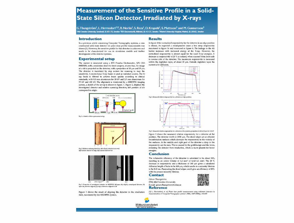
This presentation describes earlier, current and future dosimetric and image quality analysis methods, necessary to correspond to the rapidly developing CT-techniques.

1. Introduction During the first 40 years since the introduction of CT, a tremendous technical development has been made (Fig 1, grey milestones). Dose levels from CT examinations have also increased [1], but have now finally begun to decline due to major technical improvements.
Courtesy of John Boone
Fig 1 Evolution of CT scanners and dosimetry [6]

At the same time the dosimetry has been more or less the same based on measurements of CTDI alone until about 2010. (Fig1 dark blue)
A dramatically change of standards, regulations and proposed guide lines has occurred as a result of discussions in the industry and user communities during the last 15 months.
Courtesy of John Boone
Fig 1 Evolution of CT scanners and dosimetry [6]
Here is a list of reports known by the author (member of the IEC 62BC CT MT30 CT and IEC 62C WG3 (Dosimetry) that have been provided on the market recently:
• AAPM TG111, [11] Start the new dose metric using also helical scan to go further from old CTDI metrics, 2010
• AAPM TG 200, [11] New wider CT phantoms, 2011
• IAEA human health reports no. 5. [12] Status of computed tomography dosimetry for wide cone beam scanners, 2011
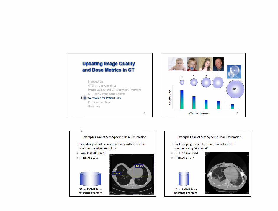
• AAPM TG204, [11] Correct for patient size using a new metric, 2011
• RP162: [10], Criteria for acceptability of medical radiological equipment, 2012

• IEC 60601-2-44-: Edition 3.0 amendment 1, [8] CT Safety Standard, accept alternative radiation detectors. Aug 2012.[8]
• IEC 62B/837/CDV [9] Extra oral (CBCT dental) FDIS Dec 2012.
• IEC 60601-2-68: [9] Particular requirements for basic safety and essential performance of X-ray based image guided radiotherapy equipment for use ….CDV, 2012
• ACR CT cookbook on its way 2013?
Aim 2013-2014 isotropic energy compensated point dose detector that fullfills IEC Diagnostic
dosemeter standard without software compensation

Lifecycle of an IEC publication • WD and CD documents (Preparatory and Committee stage)
• The development of a new publication has two possible starting points: either an existing publication is updated (maintenance), or a new one is created from scratch. Both scenarios are described in our page on the WD/CD documents.
• • CDV documents (Enquiry stage) • When a document reaches CDV stage, the IEC Editing and Document preparation team starts working on it.
For details on the interaction between the IEC and the TC at this stage, see our information on CDV documents.
• • FDIS documents (Approval stage) • When the IEC receives a draft FDIS, it goes through an IEC entry control to ensure it meets our
requirements, before being formatted and edited. For more details, see our information on FDIS documents.
• • Maintenance / updating a publication • For maintenance work on a publication, it is essential for the TC to start their work on the basis of
the publication files of the previous edition, not on intermediate versions in their possession. • • The TC can obtain the revisable files (Word version and, if available, editable images) through their IEC
Technical Officer - see Requesting files for the maintenance of an IEC publication. • • National adoptions • For national adoptions of a publication, National Committees can download an editable copy from the
IEC Revisable files database.
References 1. A Almén, S Richter, W Leitz, Swedish Radiation Protection Authority report 2008:03 Number of radiological
examinations in Sweden (in Swedish). SSI (Swedish Radiation Protection Authority) (2008) 2. S Mattsson. Radiation protection of the patient in diagnostic radiology and nuclear medicine - are we doing
enough? In: Proc Int Conf Medical Physics 2011 (Ed by D Adlienè) Technologija, Kaunas, 2011, pp 7-12.
3. Å Palm, E Nilsson, L Herrnsdorf. Absorbed dose and dose rate using the Varian OBI 1.3 and 1.4 CBCT system ,.J.ApplClin.Med.Phys. 11 (1), 2010
4. P-J.P Lin, L Herrnsdorf..Pseudohelical scan for the dose profile measurements of 160-mm-wide cone-beam MDCT, AJR; 194:897–902, American Roentgen Ray Society,2010
5. B Cederquist, S Sturesson, L Herrnsdorf. New trends in CT dosimetry using a narrow detector SEACOMP, 2010
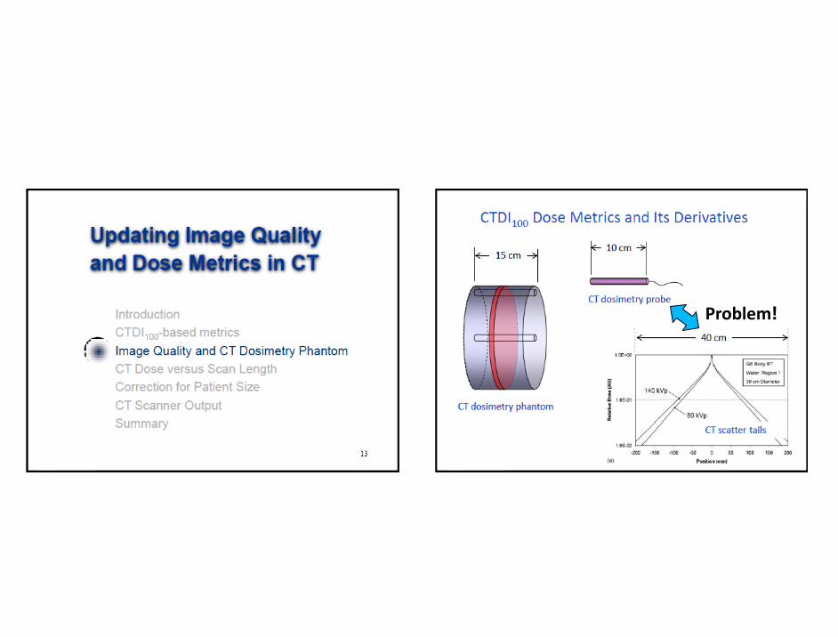
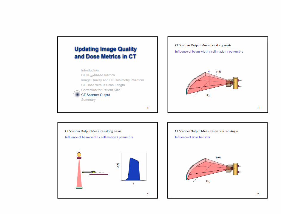
6. J Boone Updating image quality and dose metrics in CT, 2011 www.aapm.org/meetings/amos2/pdf/60-14863-35661-83.pdf
7. L Herrnsdorf, M Söderberg. A method to characterize the radiation output from a cone beam O-arm using a device for dose and dose profile scanning measurement. Accepted at SPIE, Orlando USA 2013
8. IEC 60601-2-44-: Edition 3.0 amendment 1, 2012-08 Particular requirements for the basic safety and essential performance of X-ray equipment for computed tomography
9. Geneva, Switzerland: International electrotechnical commission. http://www.iec.ch.
10. http://www.neyhqarc.nhs.uk/rp162/introductiontorp162project.aspx
11. http://www.aapm.org/pubs/reports/
12. http://www.iaea.org/books
2. A quick Review of the contents of some of the references listed earlier
in my talk:

Am1 to the CT safety standard IEC 60601-2-44 Ed 3.0 that now have
been accepted 20120831 and have few important change that is related
to dose measurement :
• Change from only accepting ion chambers to the more general term
RADIATION DETECTOR for measuring the CTDI and also accepting
alternative methods based on the measurement /sampling of the dose
profile.
• To be able to handle very large beam width like Toshiba A. 320 a
method to calculate the dose of broad fields of> 40 mm are made in a
new way .
It is based on a combination of measurement of the width of the true
dose profile free in air and measurement in standard dosimeter phantom.
• "Dose Alert" system is now introduced
• 32 cm phantom size recommended for dose measurement
• Last meeting was at FDA ,Washington 11-13 September 2012 and on
the agenda was to discuss the implementation of the new methods from
the new updated safety standard into the constancy and acceptance
standard for CT .
IEC working group 62BC MT30 CT present status :

One of the reasons for adaption of new metrics is the current diversity in methods to CT scan the patient. The table is not anymore moved during the examination for most new system for the wider beams.(Fig. 2) It is therefore an increasing need for a new type of dosimetry using more tailored dosemeters along with new way to control image quality [3,4,6,7]
Fig.2 Toshiba Aquilion 320
(320x0.5mm) =
160 mm wide beam

http://www.youtube.com/watch?v=t2o1ScMKzbY&feature=youtu.be
Point dose measurement using the moving table:

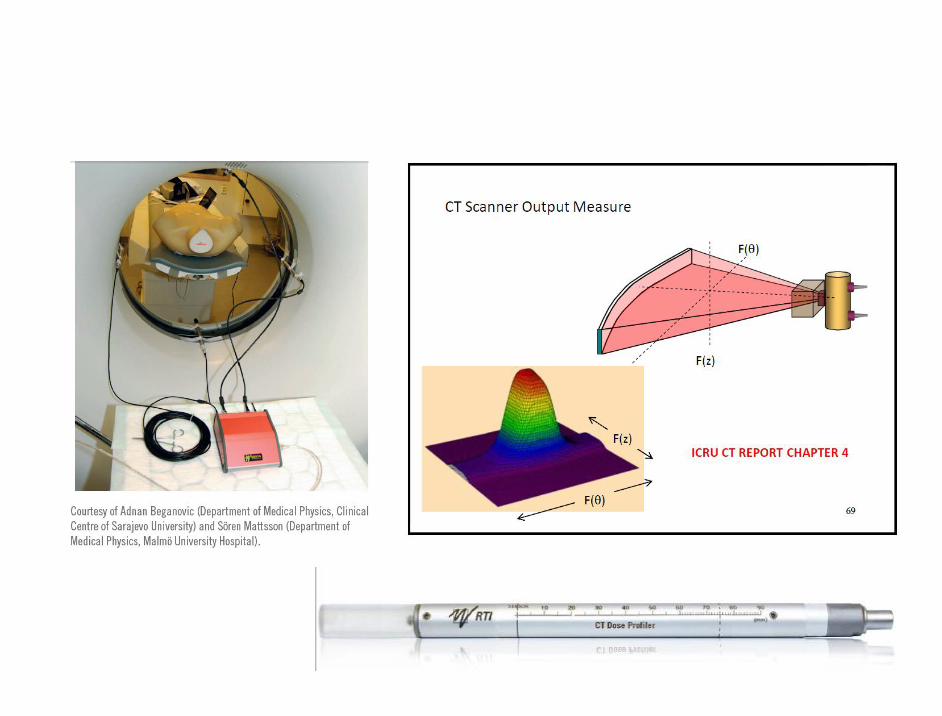
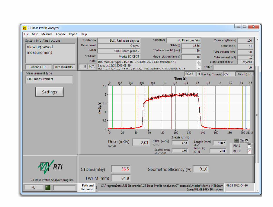
Example of hints and applications for best use of calibrated dose detectors; CT Dose profiler - design
Angle independent dose response <300um thick.

Example of hints and applications for best use of calibrated dose detectors; CT Dose profiler – angle independence
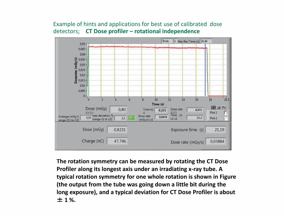
Example of hints and applications for best use of calibrated dose detectors; CT Dose profiler – rotational independence
The rotation symmetry can be measured by rotating the CT Dose Profiler along its longest axis under an irradiating x-ray tube. A typical rotation symmetry for one whole rotation is shown in Figure (the output from the tube was going down a little bit during the long exposure), and a typical deviation for CT Dose Profiler is about ± 1 %.
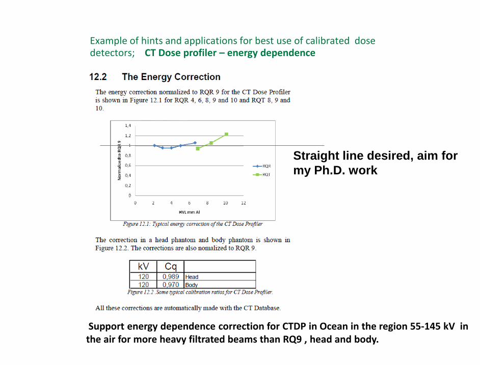
Example of hints and applications for best use of calibrated dose detectors; CT Dose profiler – energy dependence
Support energy dependence correction for CTDP in Ocean in the region 55-145 kV in the air for more heavy filtrated beams than RQ9 , head and body.
Straight line desired, aim for
my Ph.D. work

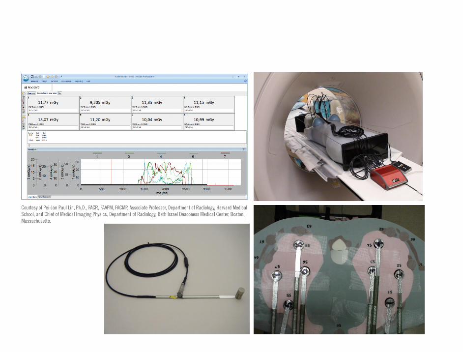
The pictures displays both detectors measured simultenously in a long TG200 phantom ~ 150 times higher sensitivity when comparing the calibration factor mGy/s/nA and ~100 times higher spatial resolution for CTDP compared to a standard Farmer chamber


Problem!
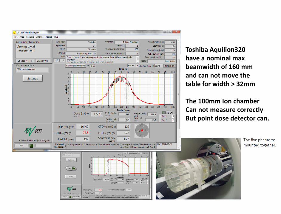
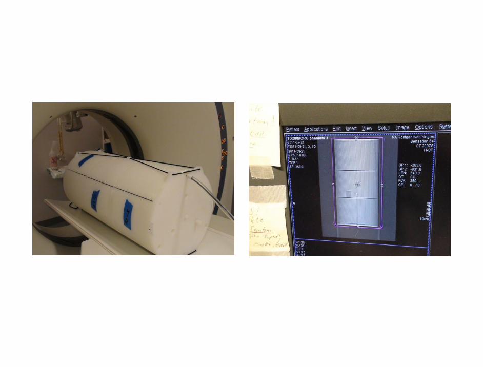
Toshiba Aquilion320 have a nominal max beamwidth of 160 mm and can not move the table for width > 32mm The 100mm Ion chamber Can not measure correctly But point dose detector can.

Solution:
Longer phantom and
Point dose Detector!


Based on a point dose detector! RTI CT Dose Profiler is a point dose detector.


Trends points for peak dose measurements.
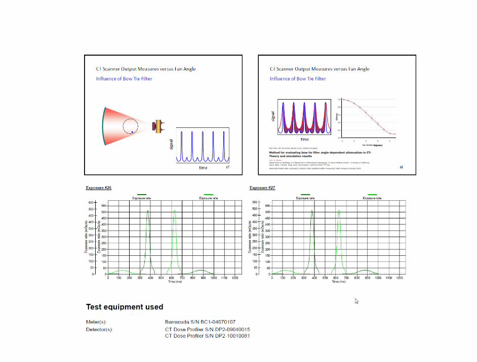
By putting the point dose detector peripheral free in air the attenuation of the bowtie filter can be studied
Dose is not following the square law when
using pencil chambers that is not fully
radiated!


Penelope Monte Carlo simulation with GPU Cuda support (20-40 faster )

3. CT and CBCT dose applikations
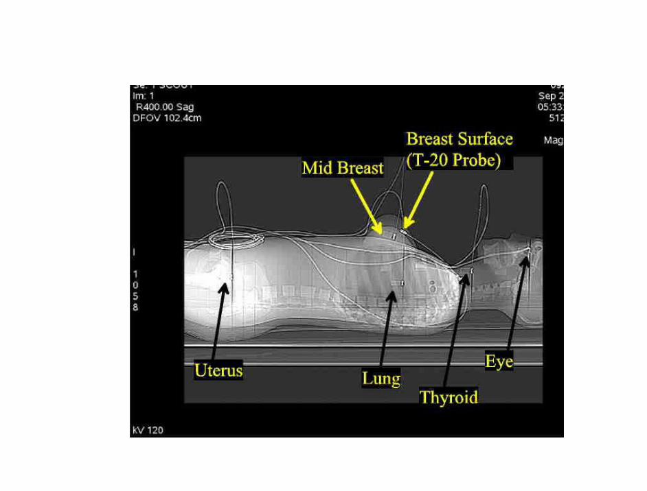
Barracuda/Ocean support up to 12 probes Evaluation of point dose phantom probes CT-Mover; no need to move the table Measuring in the TG200/ICRU phantom Measurement on a CBCT Dental system Measuring on a CBCT O-arm Measuring on a OBI Varian image CBCT

Evaluation of point dose phantom probes


CT Mover


47

48
CT-Mover

49
The pictures displays measurement of a doseprofile in two body phantoms (setup see earlier pages ) using the CT-Mover .

Example of hints and applications for best use of calibrated dose detectors; CT Dose profiler – field size dependent corrections

The pictures displays measurement of a 1,25 mm wide beam (NT=1,25 mm ) . Fo this small apertures the nominal NT is typically much smaller than the actual width. This small width can only be selected in axial mode so the CT –Mover was used.


AAPM/ICRU Dose/Image Phantom

Solution:
Longer phantom and
Point dose Detector!


The pictures displays both detectors measured simultenously in a long TG200 phantom ~ 150 times higher sensitivity when comparing the calibration factor mGy/s/nA and ~100 times higher spatial resolution for CTDP compared to a standard Farmer chamber

CBCT Dental application
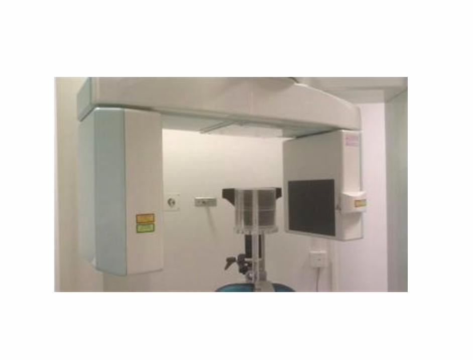


CBCT Dental video
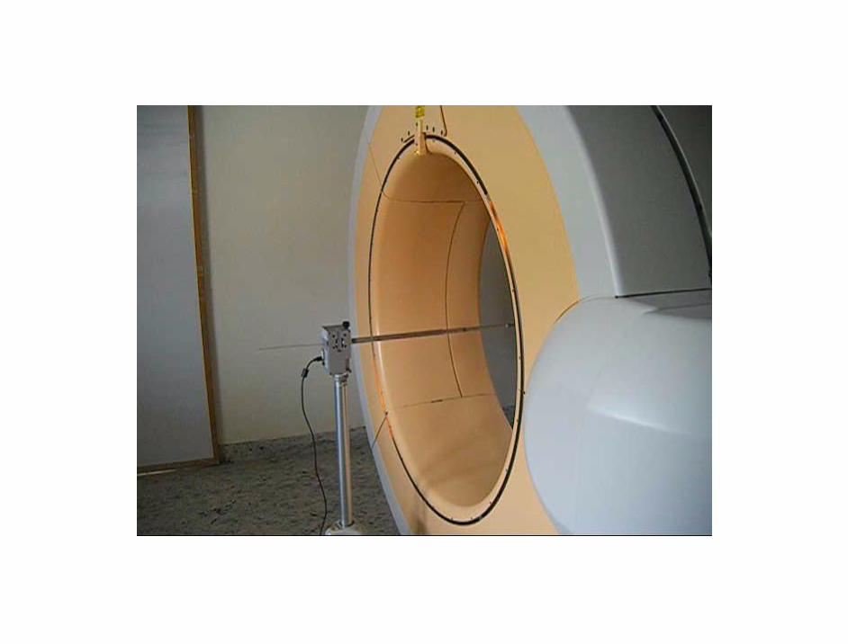
CBCT Mobile O-arm


Where is the beam ?

CBCT O-arm video
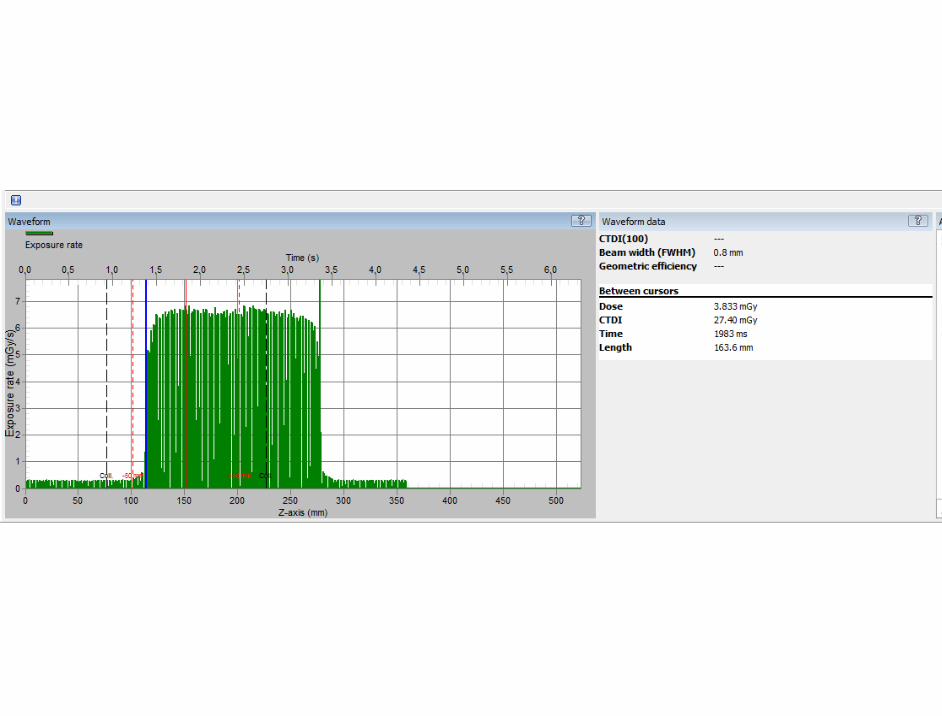
OBI Varian image CBCT mounted on the theraphy unit


4. Conclusions
It is important to actively adapt to the new dosimetry and
image quality protocols to be able to catch up with the
latest CT and CBCT technology and evaluate the type of
diagnostic radiation detector that could be used .

5. References 1. A Almén, S Richter, W Leitz, Swedish Radiation Protection Authority report 2008:03 Number of radiological
examinations in Sweden (in Swedish). SSI (Swedish Radiation Protection Authority) (2008) 2. S Mattsson. Radiation protection of the patient in diagnostic radiology and nuclear medicine - are we doing
enough? In: Proc Int Conf Medical Physics 2011 (Ed by D Adlienè) Technologija, Kaunas, 2011, pp 7-12.
3. Å Palm, E Nilsson, L Herrnsdorf. Absorbed dose and dose rate using the Varian OBI 1.3 and 1.4 CBCT system ,.J.ApplClin.Med.Phys. 11 (1), 2010
4. P-J.P Lin, L Herrnsdorf..Pseudohelical scan for the dose profile measurements of 160-mm-wide cone-beam MDCT, AJR; 194:897–902, American Roentgen Ray Society,2010
5. B Cederquist, S Sturesson, L Herrnsdorf. New trends in CT dosimetry using a narrow detector SEACOMP, 2010
6. J Boone Updating image quality and dose metrics in CT, 2011 www.aapm.org/meetings/amos2/pdf/60-14863-35661-83.pdf
7. L Herrnsdorf, M Söderberg. A method to characterize the radiation output from a cone beam O-arm using a device for dose and dose profile scanning measurement. Accepted at SPIE, Orlando USA 2013
8. IEC 60601-2-44-: Edition 3.0 amendment 1, 2012-08 Particular requirements for the basic safety and essential performance of X-ray equipment for computed tomography
9. Geneva, Switzerland: International electrotechnical commission. http://www.iec.ch.
10. http://www.neyhqarc.nhs.uk/rp162/introductiontorp162project.aspx
11. http://www.aapm.org/pubs/reports/
12. http://www.iaea.org/books

Thank You
Any Questions?