Journal of Optometry (2017) 10, 205---214 www.journalofoptometry.org REVIEW A Review of Mitochondrial Optic Neuropathies: From Inherited to Acquired Forms Yasmine L. Pilz * , Sherry J. Bass, Jerome Sherman State University New York, College of Optometry, New York, USA Received 4 July 2016; accepted 22 September 2016 Available online 28 December 2016 KEYWORDS optic neuropathy; mitochondria; retinal ganglion cells; Leber hereditary optic neuropathy; nutritional deficiencies Abstract In recent years, the term mitochondrial optic neuropathy (MON) has increasingly been used within the literature to describe a group of optic neuropathies that exhibit mito- chondrial dysfunction in retinal ganglion cells (RGCs). Interestingly, MONs include genetic aetiologies, such as Leber hereditary optic neuropathy (LHON) and dominant optic atrophy (DOA), as well as acquired aetiologies resulting from drugs, nutritional deficiencies, and mixed aetiologies. Regardless of an inherited or acquired cause, patients exhibit the same clinical manifestations with selective loss of the RGCs due to mitochondrial dysfunction. Various novel therapies are being explored to reverse or limit damage to the RGCs. Here we review the patho- physiology, clinical manifestations, differential diagnosis, current treatment, and promising therapeutic targets of MON. Published by Elsevier Espa˜ na, S.L.U. on behalf of Spanish General Council of Optometry. This is an open access article under the CC BY-NC-ND license (http://creativecommons.org/licenses/ by-nc-nd/4.0/). PALABRAS CLAVE neuropatía óptica; mitocondria; células ganglionares de la retina; Neuropatía Óptica Hereditaria de Leber; deficiencias nutricionales Revisión de las neuropatías ópticas mitocondriales: de las formas hereditarias a las adquiridas Resumen En los últimos a˜ nos, se ha incrementado el uso en la literatura del término neu- ropatía óptica mitocondrial (MON), para describir un grupo de neuropatías ópticas que presentan una disfunción mitocondrial en las células ganglionares de la retina (RGC). De manera intere- sante, las MON incluyen etiologías genéticas, tales como Neuropatía Óptica Hereditaria de Leber (LHON) y Atrofia Óptica Dominante (DOA), así como etiologías adquiridas derivadas del consumo de drogas, deficiencias nutricionales y etiologías mixtas. Independientemente de que la causa sea hereditaria o adquirida, los pacientes presentan las mismas manifestaciones clínicas, con pérdida selectiva de RGCs debido a la disfunción mitocondrial. Se están explorando diversas terapias novedosas para revertir o limitar el da˜ no a las RGC. En este documento revisamos la ∗ Corresponding author. E-mail address: [email protected](Y.L. Pilz). http://dx.doi.org/10.1016/j.optom.2016.09.003 1888-4296/Published by Elsevier Espa˜ na, S.L.U. on behalf of Spanish General Council of Optometry. This is an open access article under the CC BY-NC-ND license (http://creativecommons.org/licenses/by-nc-nd/4.0/). Document downloaded from http://www.elsevier.es, day 12/09/2017. This copy is for personal use. Any transmission of this document by any media or format is strictly prohibited. Document downloaded from http://www.elsevier.es, day 12/09/2017. This copy is for personal use. Any transmission of this document by any media or format is strictly prohibited.
Transcript
Journal of Optometry (2017) 10, 205---214
www.journalofoptometry.org
REVIEW
A Review of Mitochondrial Optic Neuropathies:
From Inherited to Acquired Forms
Yasmine L. Pilz ∗, Sherry J. Bass, Jerome Sherman
State University New York, College of Optometry, New York, USA
Received 4 July 2016; accepted 22 September 2016Available online 28 December 2016
Abstract In recent years, the term mitochondrial optic neuropathy (MON) has increasinglybeen used within the literature to describe a group of optic neuropathies that exhibit mito-chondrial dysfunction in retinal ganglion cells (RGCs). Interestingly, MONs include geneticaetiologies, such as Leber hereditary optic neuropathy (LHON) and dominant optic atrophy(DOA), as well as acquired aetiologies resulting from drugs, nutritional deficiencies, and mixedaetiologies. Regardless of an inherited or acquired cause, patients exhibit the same clinicalmanifestations with selective loss of the RGCs due to mitochondrial dysfunction. Various noveltherapies are being explored to reverse or limit damage to the RGCs. Here we review the patho-physiology, clinical manifestations, differential diagnosis, current treatment, and promisingtherapeutic targets of MON.Published by Elsevier Espana, S.L.U. on behalf of Spanish General Council of Optometry. This isan open access article under the CC BY-NC-ND license (http://creativecommons.org/licenses/by-nc-nd/4.0/).
PALABRAS CLAVEneuropatía óptica;mitocondria;células ganglionaresde la retina;Neuropatía ÓpticaHereditaria de Leber;deficienciasnutricionales
Revisión de las neuropatías ópticas mitocondriales: de las formas hereditarias a las
adquiridas
Resumen En los últimos anos, se ha incrementado el uso en la literatura del término neu-ropatía óptica mitocondrial (MON), para describir un grupo de neuropatías ópticas que presentanuna disfunción mitocondrial en las células ganglionares de la retina (RGC). De manera intere-sante, las MON incluyen etiologías genéticas, tales como Neuropatía Óptica Hereditaria de Leber(LHON) y Atrofia Óptica Dominante (DOA), así como etiologías adquiridas derivadas del consumode drogas, deficiencias nutricionales y etiologías mixtas. Independientemente de que la causasea hereditaria o adquirida, los pacientes presentan las mismas manifestaciones clínicas, conpérdida selectiva de RGCs debido a la disfunción mitocondrial. Se están explorando diversasterapias novedosas para revertir o limitar el dano a las RGC. En este documento revisamos la
http://dx.doi.org/10.1016/j.optom.2016.09.0031888-4296/Published by Elsevier Espana, S.L.U. on behalf of Spanish General Council of Optometry. This is an open access article under theCC BY-NC-ND license (http://creativecommons.org/licenses/by-nc-nd/4.0/).
Document downloaded from http://www.elsevier.es, day 12/09/2017. This copy is for personal use. Any transmission of this document by any media or format is strictly prohibited.Document downloaded from http://www.elsevier.es, day 12/09/2017. This copy is for personal use. Any transmission of this document by any media or format is strictly prohibited.
patofisiología, las manifestaciones clínicas, los diagnósticos diferenciales, el tratamiento actualy los prometedores objetivos terapéuticos de las MON.Publicado por Elsevier Espana, S.L.U. en nombre de Spanish General Council of Optometry.Este es un artıculo Open Access bajo la licencia CC BY-NC-ND (http://creativecommons.org/licenses/by-nc-nd/4.0/).
Introduction
With advances in the understanding of optic nerve diseases,a new term has emerged within the literature to describea group of diseases caused by mitochondrial dysfunction ofthe optic nerve: mitochondrial optic neuropathy (MON).
MON describes a group of optic neuropathies with similarclinical features and the same pathophysiology of mitochon-drial dysfunction. Regardless of the underlying inherited oracquired cause, MONs share the common feature of mito-chondrial dysfunction in the prelaminar area of the opticnerve. The subsequent selective damage and loss of reti-nal ganglion cells (RGCs) result in clinical signs of painless,progressive bilateral visual acuity loss, central or cecocen-tral scotomas, and color vision deficiencies. Although thesame clinical features exist in MONs, the prevalence of spe-cific aetiologies varies. In acquired MONs, the disease has nogender, racial, or age prevalence. In contrast, inherited aeti-ologies often affect children or young adults. Many have thepotential to be partially or completely reversed. Further-more, novel therapeutics appear promising in the treatmentof MONs.
Mechanism of Action
Whether from an inherited mutation or an acquired expo-sure, overwhelming evidence suggests a shared mechanismof mitochondrial dysfunction. The impairment in ATP pro-duction via alternations in oxidative phosphorylation andthe accumulation of reactive oxygen species (ROS) withinthe mitochondria trigger a decrease in the electricalpotential across the mitochondrial membrane. This causescytochrome C to leak out of the mitochondria into the cyto-plasm and bind to the apoptosis activating factor-1 (APAF-1)resulting in apoptosis of the RGC.1---4 In Leber hereditaryoptic neuropathy (LHON), mutations within the mitochon-drial DNA lead to dysfunction in the mitochondrial complexI and cause accumulation of ROS. Alcohol, tobacco, andother MON-causing toxins can increase exogenous ROS andthereby cause the same pathway to be activated.1 Whetherenergy depletion or over-production in ROS precipitates RCGloss remains to be understood and explains the variation intherapeutic targets used.
Susceptibility of prelaminar RGC in MON
MON selectively damages the RGCs within the prelami-nar area of the papillomacular bundle (PMB). The uniqueanatomical features of the pre-laminar area make the
PMB vulnerable to mitochondrial dysfunction in differentways.5 First, the pre-laminar area contains a high numberof mitochondria compared to the post-laminar area, demon-strating that the PMB has a high energy need.6 Secondly, PMBfibers contain a large number of unmyelinated RGC axonscompared to the post-laminar area which contains predom-inantly myelinated axons.2,5 This means that prelaminaraxons do not conduct electrical potential in an efficient waycompared post-laminar axons which uses saltatory conduc-tion. Thirdly, the PMB fibers are narrow in caliber, whichfurther contributes to decreased energy conduction.5 Thehigh-energy requirement of the PMB in conjunction with thelow energy production and slow axoplasmic transport leadsto susceptibility of the PMB when mitochondrial dysfunctionoccurs in MON.2,5,7,8
Examination
Clinical Presentation
Clinical manifestations of MON are summarized in Table 1.Patients with MON typically present with bilateral, painless‘‘fog’’ at the center of fixation. Visual acuities range approx-imately from 20/25 to 20/200.9 Only in rare cases such as inmethanol toxicity, can vision reach hand motion to no lightperception. While visual acuities range widely depending onthe severity of the disease, symmetry in visual acuity andophthalmological findings in both eyes is a hallmark featureof MON.
The loss of color vision, especially of red color, is a promi-nent feature of MON. Color vision loss may be more severethan visual acuity loss.2 Hardy Rand and Rittler (HRR) andFansworth D-15 are most useful because they establish blue-yellow or red-green deficits, whereas Ishihara only assessesred-green defects. Red cap test between two eyes is nor-mal due to the symmetrical nature of genetic or acquiredMON. Pupils react equally normal or slightly sluggish to light.Similarly, no relative afferent pupillary defect (RAPD) isobserved due to the symmetry of the disease. Reduced con-trast sensitivity at high spatial frequencies has been foundin sub-clinical cases of MON.10
Ophthalmic fundus evaluation varies depending on theextent of the disease. In early stages of MON, the opticnerves may appear normal, oedematous, or hyperemic.Peripapillary hemorrhages may be present. As the diseaseprogress, PMB loss and temporal optic disc pallor occurs. Inpatients with longstanding MON, diffuse bilateral optic discpallor and atrophy is apparent.
Document downloaded from http://www.elsevier.es, day 12/09/2017. This copy is for personal use. Any transmission of this document by any media or format is strictly prohibited.Document downloaded from http://www.elsevier.es, day 12/09/2017. This copy is for personal use. Any transmission of this document by any media or format is strictly prohibited.
Table 1 Clinical manifestations of mitochondrial opticneuropathy.
Relatively symmetric decrease in visual acuitiesCentral or cecocentral scotomasNormal to sluggish pupils without relative afferentpupillary defectColor vision changesNormal, oedematous, or hypermic optic nerve(early stage)Thickening of retinal nerve fiber layer temporally(early stage)Thinning of the ganglion cells on OCTPallor of optic nerve temporally then diffusely(late stage)
Ancillary Testing
In any patient suspected of MON, an Amsler Grid and visualfield 30-2 should be performed. In early stages of MON, sco-tomas may not be present. As the disease progresses, visualfield testing and Amsler Grid will reveal bilateral centralor cecocental scotomas. Often these defects are more pro-nounced with a red stimulus compared to a white-on-whitestimulus.9
Optical coherence tomography (OCT) is a very useful toolto monitor the subclinical cases and progression of MON. Inearly stages of MON, OCT may show temporal and inferior-temporal thickening of the retinal nerve fiber layer.11---13 Inlater stages, thinning especially temporally of the retinalnerve fiber layer and overall ganglion cell layer thinning canbe observed1.11,14---17 Visual-evoked potential (VEP) testingmay be useful in detecting subclinical MONs. A reduced P100amplitude with a normal or near-to-normal latency will beobserved in MON.18 This helps to differentiate a MON fromdemyelinating causes of optic neuropathy, such as multiplesclerosis (MS), in which the P100 amplitude is reduced andlatency is significantly delayed.18 It should be noted thatVEP is a non-specific test as there are multiple aetiologiesof reduced amplitudes including amblyopia, inflammatorydiseases, and compressive lesions.
Case History
An extensive case history is essential in a patient suspectedof having a genetic or acquired MON. Occupational and socialhistory may suggest possible exposure to toxins. A patientshould be questioned about occupational hazards includ-ing exposure to fumes (cyanide), organic solvents (ethyleneglycol, toluene, styrene, perchloroethylene), heavy metals(lead, mercury, thallium), and carbon dioxide. A social his-tory of alcohol, tobacco, and recreational drug consumptionshould be documented. Amount, frequency, and durationof intake of substances should be established. It is impor-tant to note the type of alcohol consumed since illicit liquormay contain methanol --- a life-threatening toxin. In cases ofdenial of consumption of substances, clinicians should verifyif there is a history of substance abuse.
Additional elements of the medical history should bereviewed carefully. As MON can be caused by nutritional
Table 2 Causes of acquired mitochondrial opticneuropathies.
deficiencies in vitamin B, establishing a patient’s relation-ship to food is important. Several systemic medicationscausing MON have been documented (Table 2). Symptomsof toxic peripheral neuropathy, such as numbness or tinglingof limbs or gait problems, should be noted. Metabolic dis-eases, such as diabetes mellitus, thyroid disease, or kidneyand liver failure may contribute to exacerbating severity ofMON. The presence of a metallic hip replacement shouldbe addressed. Patients with hip-replacements containingcobalt often exhibit symptoms of deafness, poor motor coor-dination, and numbness of limbs known as arthoprostheticcobaltism.19---21 A family history of bilateral reduced acuitiesor blindness may suggest the possibility of a genetic MON.
Genetic Aetiologies of MON
Leber Hereditary Optic Neuropathy (LHON)
About 90% of LHON cases are caused by one of threemitochondrial DNA (mDNA) point mutations: m.3460G>A(MTND1), m.11778G>A (MTND4), or m.14484T>C (MTND6).All LHON mutations affect the mitochondrial complex I sub-unit of the mitochondrial respiratory chain and lead to theaccumulation of cytochrome C as described in the commonmechanism above.
The minimum prevalence of the three most commonLHON mutations is 1 in 31,000 in North East of England.22
Similarly, the prevalence of LHON in the Netherlands hasbeen estimated to be 1 in 39,000.23 Age and sex are the mostcommon risk factors for developing LHON. Approximately95% of LHON carriers will manifest visual symptoms beforethe age of 50 years.24 Peak age of onset is usually within thesecond and third decade.
Not every person identified as having an LHON muta-tion will develop visual changes. A family history of visionloss in an LHON-carrier may be a predictor of vision loss.But in up to 40% of patients with LHON, no family his-tory is indicated.25 There is a strong sex bias for losingvision in LHON-carriers, known as incomplete penetrance.
Document downloaded from http://www.elsevier.es, day 12/09/2017. This copy is for personal use. Any transmission of this document by any media or format is strictly prohibited.Document downloaded from http://www.elsevier.es, day 12/09/2017. This copy is for personal use. Any transmission of this document by any media or format is strictly prohibited.
208 Y.L. Pilz et al.
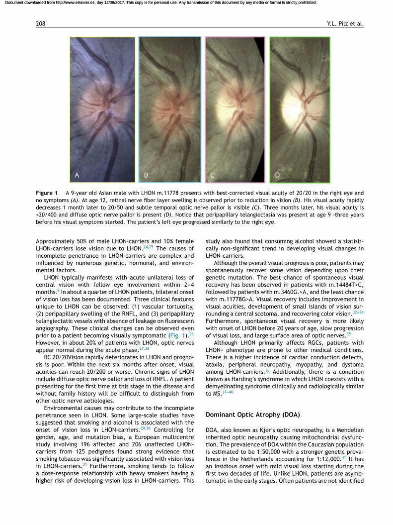
Figure 1 A 9-year old Asian male with LHON m.11778 presents with best-corrected visual acuity of 20/20 in the right eye andno symptoms (A). At age 12, retinal nerve fiber layer swelling is observed prior to reduction in vision (B). His visual acuity rapidlydecreases 1 month later to 20/50 and subtle temporal optic nerve pallor is visible (C). Three months later, his visual acuity is<20/400 and diffuse optic nerve pallor is present (D). Notice that peripapillary telangiectasia was present at age 9 -three yearsbefore his visual symptoms started. The patient’s left eye progressed similarly to the right eye.
Approximately 50% of male LHON-carriers and 10% femaleLHON-carriers lose vision due to LHON.24,25 The causes ofincomplete penetrance in LHON-carriers are complex andinfluenced by numerous genetic, hormonal, and environ-mental factors.
LHON typically manifests with acute unilateral loss ofcentral vision with fellow eye involvement within 2---4months.8 In about a quarter of LHON patients, bilateral onsetof vision loss has been documented. Three clinical featuresunique to LHON can be observed: (1) vascular tortuosity,(2) peripapillary swelling of the RNFL, and (3) peripapillarytelangiectatic vessels with absence of leakage on fluoresceinangiography. These clinical changes can be observed evenprior to a patient becoming visually symptomatic (Fig. 1).26
However, in about 20% of patients with LHON, optic nervesappear normal during the acute phase.27,28
BC 20/20Vision rapidly deteriorates in LHON and progno-sis is poor. Within the next six months after onset, visualacuities can reach 20/200 or worse. Chronic signs of LHONinclude diffuse optic nerve pallor and loss of RNFL. A patientpresenting for the first time at this stage in the disease andwithout family history will be difficult to distinguish fromother optic nerve aetiologies.
Environmental causes may contribute to the incompletepenetrance seen in LHON. Some large-scale studies havesuggested that smoking and alcohol is associated with theonset of vision loss in LHON-carriers.29,30 Controlling forgender, age, and mutation bias, a European multicentrestudy involving 196 affected and 206 unaffected LHON-carriers from 125 pedigrees found strong evidence thatsmoking tobacco was significantly associated with vision lossin LHON-carriers.31 Furthermore, smoking tends to followa dose-response relationship with heavy smokers having ahigher risk of developing vision loss in LHON-carriers. This
study also found that consuming alcohol showed a statisti-cally non-significant trend in developing visual changes inLHON-carriers.
Although the overall visual prognosis is poor, patients mayspontaneously recover some vision depending upon theirgenetic mutation. The best chance of spontaneous visualrecovery has been observed in patients with m.14484T>C,followed by patients with m.3460G.>A, and the least chancewith m.11778G>A. Visual recovery includes improvement invisual acuities, development of small islands of vision sur-rounding a central scotoma, and recovering color vision.32---34
Furthermore, spontaneous visual recovery is more likelywith onset of LHON before 20 years of age, slow progressionof visual loss, and large surface area of optic nerves.35
Although LHON primarily affects RGCs, patients withLHON+ phenotype are prone to other medical conditions.There is a higher incidence of cardiac conduction defects,ataxia, peripheral neuropathy, myopathy, and dystoniaamong LHON-carriers.36 Additionally, there is a conditionknown as Harding’s syndrome in which LHON coexists with ademyelinating syndrome clinically and radiologically similarto MS.37---40
Dominant Optic Atrophy (DOA)
DOA, also known as Kjer’s optic neuropathy, is a Mendelianinherited optic neuropathy causing mitochondrial dysfunc-tion. The prevalence of DOA within the Caucasian populationis estimated to be 1:50,000 with a stronger genetic preva-lence in the Netherlands accounting for 1:12,000.41 It hasan insidious onset with mild visual loss starting during thefirst two decades of life. Unlike LHON, patients are asymp-tomatic in the early stages. Often patients are not identified
Document downloaded from http://www.elsevier.es, day 12/09/2017. This copy is for personal use. Any transmission of this document by any media or format is strictly prohibited.Document downloaded from http://www.elsevier.es, day 12/09/2017. This copy is for personal use. Any transmission of this document by any media or format is strictly prohibited.
A Review of Mitochondrial Optic Neuropathies 209
until testing due to family history or diagnosis of bilateraloptic atrophy. Patients often present with visual acuities of20/30 or better. Although visual prognosis with DOA is highlyvariable, visual outcome is better compared to LHON withaverage visual acuities of 20/60 to 20/200.24
Approximately 50---60% of people with DOA have a muta-tion in the OPA-1(3q28-q29).42,43 OPA-1 is a critical pro-fusionprotein within the inner mitochondrial membrane. OPA-1
mutations cause mitochondrial defragmentation and leadto respiratory chain dysfunction and dysfunction within theATPase of the mitochondria.44,45
About 20% of patients with OPA-1 mutations will developadditional co-morbidities of deafness, chronic progressiveexternal ophthalmoplegia, ataxia, myopathy, peripheralneuropathy, and deafness. Deafness appears to be themost common systemic complication often presenting duringpuberty or young adulthood.46
Other Mitochondrial Syndromes
Several mitochondrial syndromes with prominent opticneuropathy exist including Charcot-Marie Tooth disease,hereditary spastic paraplegia, Friedrich ataxia, and Costeffsyndrome. Although each syndrome has its own characteris-tics, common signs of these mitochondrial syndromes aredistal limb weakness, decreased tendon reflexes, ataxia,myopathy, and deafness.27,33 In patients with suspectedmitochondrial syndromes, medical referrals should be ini-tiated immediately due to widespread systemic effects.
Acquired Aetiologies of MONs
Substances and Medication-Induced MONs
Heavy tobacco consumption, as well as heavy alcohol con-sumption, can lead to MON. Due to the co-morbidity ofdrinking and smoking, the condition was historically namedtobacco-alcohol amblyopia --- incorrectly named since thecondition has no amblyogenic factors.
Methanol, also known as wood alcohol, is considereda very potent toxin capable of rapid onset of MON.47,48
The main cause of poisoning is through adulterated liquorin which methanol is mixed with ethyl alcohol to pro-duce a potent alcoholic drink. Methanol is found inmany sources such as windshield wiper antifreeze, cannedheating sources, copy machine fluids, deicing fluid, fueladditives, paint remover, and varnish. Toxicity due tomethanol may result in symptoms of nausea, vomiting,dizziness, headaches, breathing difficulties, and changes inconsciousness.49 Metabolic acidosis may lead to coma anddeath. Thus, immediate hospitalization is recommended forpatients with methanol poisoning. Visual prognosis is alsodependent on prompt treatment.
Medication-induced MON may be caused byethambutol,50---54 linezolid,55---59 chloramphenicol,60,61
erythromycin.24 Less likely to cause MON are ciprofloxacin,streptomycin, isoniazid, antiretroviral drugs, amiodarone,infliximab, clioquinol, dapsone, quinine, pheniprazine, andsuramin. Severity of neuropathy depends on dosage andduration.
Nutrition-Deficient MONs
Nutrition-deficient MON may be caused by deficiencies inthiamine (vitamin B1), riboflavin (vitamin B2), pyridoxine(vitamin B6), folic acid (vitamin B9), or cobalamin (vita-min B12).62---64 In developing countries, the primary cause isundernourishment. In developed countries, vegan diets orweight-loss diets may result in vitamin B deficiencies caus-ing MON.63 In other cases, anorexia nervosa, alcoholism,gastrointestinal (GI) problems or GI surgeries may result innutritional malabsorption and must be confirmed via bloodtesting.65 Patients may present with pernicious cell anemiadue to vitamin B12 deficiency or Beriberi due to Vitamin B1
deficiency.
Mixed Aetiologies of MON
Mixed aetiologies of MON add to the complexity of identi-fying causal agents. Multiple toxins may be responsible forcausing an MON as seen in tobacco-alcohol amblyopia. Inother cases, toxic exposure may interact with nutritionaldeficits to cause MON. For instance, the Cuban EpidemicOptic Neuropathy (CEON) outbreak in 1991---1993 affecting500,000 people resulted from malnutrition due to the coun-try’s economic problems and a high prevalence of tobacco,particularly cigar smoking.66---68 Furthermore, genetic MONsmay be exacerbated or even triggered by toxic or nutritionalcauses of MON.
Differential Diagnosis
A comprehensive medical and social case history, as outlinedpreviously, may indicate potential causes of MON. However,MON is a diagnosis of exclusion and often multi-factorial.Thus, any case of suspected MON, an MRI of the brain andorbit is essential to rule out other aetiologies.
Compressive and demyelinating lesions may cause oede-matous or atrophic optic nerves. Symptoms of a compressivelesion may include headache and changes in mood, person-ality, or memory. An MRI of the brain and orbits will confirmdiagnosis.
Optic neuritis may be differentiated from MON due tothe pain upon eye movement. Visual recovery within weeksfurther confirms the diagnosis of optic neuritis, whereasvisual acuity loss and central scotomas will progressivelyworsen in MON. A brain MRI should be ordered due tothe strong association between optic neuritis and MS. Asmentioned previously, MON can co-exist with in a MS-likesyndrome known as Harding’s syndrome. Diffuse white mat-ter lesions on MRI suggest MS. VEPs also help differentiateMON from demyelinating causes of optic neuropathy in whichthe response is delayed and P100 amplitude is reduced.18
Ischemic optic neuropathy presents with oedema of theoptic nerves in the acute stage and diffuse optic atrophy inthe chronic stage. In cases of bilateral optic nerve oedemathe possibility of increased intracranial pressure (ICP) --- alife-threatening condition --- must be considered. Presenceof spontaneous venous pulsation within the optic nervesmay indicate normal ICP. However, the absence of venouspulsation occurs in about 30% of patients and if not previ-ously documented is not conclusive. Symptoms of increased
Document downloaded from http://www.elsevier.es, day 12/09/2017. This copy is for personal use. Any transmission of this document by any media or format is strictly prohibited.Document downloaded from http://www.elsevier.es, day 12/09/2017. This copy is for personal use. Any transmission of this document by any media or format is strictly prohibited.
210 Y.L. Pilz et al.
ICP may include headache, dizziness, nausea, and vomiting.In patients over 55 years, an artertic ischemic optic neu-ropathy (AION) should be ruled out by ordering C-reactiveprotein (CRP), erythrocyte sedimentation rate (ESR), andpotentially a temporal artery biopsy. Patients will normallyreport symptoms of jaw claudication, temporal headaches,and sensitivity to touch on temporal side of head.
Bilateral traumatic optic neuropathy may be suspectedwith a history of reported head or eye trauma. In thesecases, identifying trauma to other eye structures can beimportant in making diagnosis.
Normal Tension Glaucoma (NTG) and MONs share sim-ilar clinical characteristics of insidious visual acuity loss,paracentral defects, and thinning of neuroretinal rim. Par-ticularly, overlapping commonalities between NTG and DOAhave lead some researchers to postulate whether or notNTG is a hereditary optic neuropathy. Multiple studies havefound an association between OPA1 polymorphisms and nor-mal tension glaucoma (NTG).69---71 However, NTG manifestsitself in older population with an average of approximately64 years.72 Arcuate visual field defects, excavation of theinferior rim, and inferior optic nerve thinning also differen-tiate NTG from MONs.73
Differentiating an aetiology of MON requires differentblood tests. Laboratory blood tests for vitamin B1, B2, B6, B9,and B12 may establish nutritional causes for MON. Patientswith substance abuse should also have laboratory tests fornutritional deficiencies, as these two MON aetiologies oftenco-exist. If heavy metal toxicity is suspected, a 24-h urinetest to measure exposure to heavy metals can be conducted.A family history of bilateral vision loss may suggest a geneticaetiology of MON and may warrant further genetic testing. Itshould also be remembered that toxic or nutritional causesof MON may exacerbate a pre-existing genetic MON.
Optometric Management of MON
Baseline Testing and Monitoring
Baseline vision testing includes visual acuities, color vision,Amsler grid, visual fields, and ophthalmic evaluation of opticdiscs and retinal nerve fiber layer, and documentation withphotography or OCT. Genetic testing to rule out LHON andDOA should also be initiated.
Follow-up examinations are recommended every 4---6weeks in patients with reduced vision, a family history ofLHON or DOA, or patients with a strong suspicion of MON.Even if the patient is asymptomatic, close monitoring every4---6 weeks is essential. Patients should be strongly urged toreturn to clinic immediately once they notice a change invision. Furthermore, medical referrals may be necessary ifsubstance abuse, medication toxicity, or nutritional deficitsare suspected. For suspected LHON+ or DOA+ phenotypes,immediate medical referrals to address cardiovascular, neu-rological, motor or ear problems should be initiated.
Cessation of Agent inducing MON
Discontinuation of causal agent (e.g. medication, tobacco,alcohol, methanol) is recommended. In acquired MON, dis-continuation of toxicity-causing factor can lead to recovery
of visual acuity and central vision. Even with significant lossof the PMB, visual acuities can often remain on average20/50 after recovery and full visual recovery from substanceand medication-induced MONs has been documented in theliterature.
Genetic Counseling
Genetic testing is indicated for family members of a patientwith an inherited MON. Individuals identified with the muta-tion should undergo baseline testing as mentioned. Geneticcounseling may also be beneficial for individuals with aninherited MON who are planning on starting a family.
Clinical Trials
Introducing a patient to current clinical trials is an option.Particularly, a patient with an inherited MON may benefitfrom this treatment option. Generally speaking, early ther-apeutic intervention is recommended to lessen the chanceof irreversible damage.
Facilitating Access to Rehabilitation Resources andServices
Unfortunately patients with MONs, especially those withLHON, have a poor visual prognosis. Often LHON patientsare children or young adults who are otherwise systemicallyhealthy. Rapid vision loss is devastating and needs to beproperly addressed by the managing optometrist. Facilitat-ing access to rehabilitative services, such as low vision andoccupational therapy, can be valuable to a patient adaptingto vision loss. Family involvement and support during thisperiod is recommended.
Therapeutic Interventions
Coenzyme Q10 (CoQ10)
Various pharmacological targets are being explored to mit-igate the mitochondrial dysfunction that occurs in MON.Quinone analogs, such as CoQ10, help restore the elec-tron chain transport and promote ATP synthesis within themitochondria. They do this by bypassing mitochondrial com-plex I inhibition and shuttling electrons from the cytosoldirectly to complex III, thereby re-establishing ATP synthe-sis and ultimately reducing cytochrome C.8,74 Historically,CoQ10 has been used in mitochondrial disease due to itscellular mechanism of action.
Short-Chain Quinones: Idebenone
Recent studies have shifted attention toward idebenone andEPI-743, two shorter chain quinones, because they may bemore effective than CoQ10. Due to its lipophobic proper-ties, these two quinone analogs when orally administeredare able to be delivered more readily into the mitochondriaand have been reported to be more potent.
Over the last decade, studies have focused on idebenonetherapy for the treatment of LHON. In 1992, the first case
Document downloaded from http://www.elsevier.es, day 12/09/2017. This copy is for personal use. Any transmission of this document by any media or format is strictly prohibited.Document downloaded from http://www.elsevier.es, day 12/09/2017. This copy is for personal use. Any transmission of this document by any media or format is strictly prohibited.
A Review of Mitochondrial Optic Neuropathies 211
report was published reporting that 10-year-old boy withthe LHON showed visual recovery with a low daily dosage of90 mg idebenone.75 The Rescue of Hereditary Optic DiseaseOutpatient Study (RHODOS), a multi-center, double-blind,randomized, placebo-controlled study was conducted toinvestigate if daily dosages of 900 mg idebenone for 6months help slow progression in 85 patients with LHON muta-tions m.3460G4A and m.11778G4A.76 Although the RHODOSstudy failed to show a statistical difference in visual acu-ity between the two groups, idebenone treatment showeda trend in maintaining visual acuity over 6 months. Fur-thermore, patients with unequal visual acuities were morelikely to benefit from idebenone treatment, suggesting earlytreatment with idebenone may be important in preventingfellow eye involvement. In the follow-up study, visual acuityremained stable even 30 months after discontinuation of theof the 6-month long idebenone treatment.77
The same year, a retrospective study by Carelli et al.additionally demonstrated a trend in visual recovery inpatients using various dosages of idebenone within thefirst year of disease onset compared to untreated LHONpatients.78 Interestingly, LHON-patients with the mutationm.11778/ND4 showed statistically significant visual recov-ery when treated with idebenone whereas patients withm.3460/ND1 did not respond as well.
Differences in the two large studies of idebenone in 2011may be of clinical value. Carelli et al. only included LHON-patients who started idebenone treatment within 1 yearafter onset of the disease to control for the spontaneousrecovery, which occurs mostly in 2---5 years after LHON onset.The RHODOS study, on the other hand, included LHON-patients with onset of disease up to 5 years ago. Treatmentwith idebenone in RHODOS patients may have not been aseffective due to irreversible damage to RGCs with prolongedperiod of the disease. Thus, the importance of immediatetreatment with idebenone is stressed once diagnosis of LHONhas been made.
While treatment onset, duration, and dosing differedbetween these two studies, both support the therapeuticeffect of idebenone for LHON. As a result, the EuropeanMedicines Agency (EMA) approved idebenone under thetradename Raxone
®in September 2015 for the treatment
of LHON.The promising findings of idebenone in LHON treat-
ment have spurned interest in exploring the efficacy ofidebenone in DOA. In a small pilot study, seven DOA patientswith OPA-1 mutation received variable doses of idebenone(270---675 mg/day) for at least one year.79 Five out of sevenidebenone-treated patients showed an increase in visualacuities and four out of seven idebenone-treated patientsdemonstrated an improvement in color vision. A large-scale, randomized, placebo-controlled study is neededto fully evaluate visual recovery of DOA-patients usingidebenone.
Short-chain Quinones: EPI-743
Another short-chain quinone, EPI-743, has shown promise inthe treatment of LHON. In an open-labeled clinical trial, fourof five LHON-patients treated with EPI-743 within 90 daysafter disease onset for a period of at least 1 year, showed
variable improvements in visual acuity or visual field.80 Stud-ies to further explore the effects of EPI-743 in patients withDOA are needed.
Mitochondria-Targeted Quinones
To increase delivery into the mitochondria, short-chainquinones have been coupled to the lipophiliccation, triph-enylphosphonium (TPP). Mitoquinone mesylate (MitoQ) and10-(6′-plastoquinonyl) decyltriphenyl phosphonium (SkQ1)have been suggested as potential therapeutics. However,more evidence is needed to substantiate these claims.81
Gene Therapy
Gene therapy has shown potential in the treatmentof LHON. In vitro LHON studies have focused on theallotopic expression of mitochondrial-encoded genes deliv-ered via an adeno-associated virus (AVV) vector into themitochondria.82,83 In vivo studies have also demonstratedthat intravitreal injections of the AVV-mediated ND4 wild-type gene improves visual function in rodents.84,85 However,it remains controversial due to the questionable feasibilityof integration of ND4 wildtypes subunit into the mitochon-drial membrane.86 Recently, two other genetic engineeringstrategies have shown promising results. Direct delivery ofcircular mDNA into the mitochondria demonstrated restora-tion of cellular respiration and ATP production in vitro.87
Another genetic approach involving an AAV-vector contain-ing mDNA encoded with wildtype ND4 subunit tag showedsuccessful expression within the inner retinal layers and pre-vention of visual loss and optic atrophy in the LHON-mutantR340H ND4 mouse model.88
The groundwork has been laid for clinical gene ther-apy studies and promising results are emerging. In an openlabeled prospective study, five patients with the LHON car-rying the m.11778G>A mutation were evaluated after anintraocular injection of an AVV-mediated ND4 constructwas given.89 While the visual acuity of three of the par-ticipants remained unchanged after three months, two ofthe five patients experienced moderate improvements invisual acuity. No serious adverse effects were reported.In a related 9-month follow-up study, visual acuity of sixof the nine patients’ treated eye improved by at least0.3 log MAR.90
Other gene therapy clinical trials are currently inprogress. GenSight Biologics is currently sponsoring an openlabel trial (NCT02064569) to investigate the safety and tol-erability of intraocular injections of AAV2-mediated ND4 in21 LHON patients with the m.11778G>A mutation. Addition-ally, the company will evaluate the visual outcome of genetherapy in LHON patients with same mutation when visionloss is present (a) six months or less (NCT02652767) or (b)six to twelve months (NCT02652780). Another clinical trialconducted by the University of Miami will investigate thesafety in 27 patients with LHON using different doses of theAAV-ND4 gene therapy (NCT02161380) and is scheduled tobe completed in March 2019.
Gene therapy for treatment of DOA is in its infancy. It iswell known that DOA is linked to OPA-1 mutations and rodentmodels carrying vision-deficient OPA-1 mutations have been
Document downloaded from http://www.elsevier.es, day 12/09/2017. This copy is for personal use. Any transmission of this document by any media or format is strictly prohibited.Document downloaded from http://www.elsevier.es, day 12/09/2017. This copy is for personal use. Any transmission of this document by any media or format is strictly prohibited.
212 Y.L. Pilz et al.
generated.91---93 One approach is trying to develop intravit-real injections of an AAV vector to deliver wildtype OPA1 totarget RGCs in a DOA mouse model.
Antioxidants: MTP-131
MTP-131, also known as Bendavia, is a Szeto---Schiller (SS)peptide that selectively targets the inner mitochondrialmembrane. It acts as a potent antioxidant and enhanceselectron transport chain function. Since it has shown poten-tial in the treatment of MON clinical studies are currentlyunderway. A phase 2 clinical trial aims to evaluate if a1% ophthalmic solution of MTP-131 over 16 weeks is asafe and effective treatment for 12 subjects with LHON(NCT02693119).
Other Therapeutic Targets
Other therapeutic targets, out of the scope of this paper,have shown potential in the treatment of MON. They includeaestrogen, methylene blue, cyclosporine A, brimonidine,near-infrared therapy, and stem cell therapy.36,83,94
Conclusion
MON is an umbrella term used to describe inherited andacquired optic neuropathies with the same clinical man-ifestations. All MONs exhibit selective loss of the RGCsdue to mitochondrial dysfunction. Many acquired MONs arereversible and various novel therapeutics are on the horizonas treatment options.
Funding
No funding sources.
Conflict of interest
No conflict of interest is present.
References
1. Sadun AA, La Morgia C, Carelli V. Mitochondrial optic neu-ropathies: our travels from bench to bedside and back again.Clin Exp Ophthalmol. 2013;41:702---712.
tion as a cause of optic neuropathies. Prog Retin Eye Res.2004;23(January):53---89.
6. Bristow EA, Griffiths PG, Andrews RM, Johnson MA, Turnbull DM.The distribution of mitochondrial activity in relation to opticnerve structure. Arch Ophthalmol. 2002;120:791---796.
7. Pan BX, Ross-Cisneros FN, Carelli V, et al. Mathematically mod-eling the involvement of axons in Leber’s hereditary opticneuropathy. Invest Ophthalmol Vis Sci. 2012;53:7608---7617.
8. Sitarz KS, Chinnery PF, Yu-Wai-Man P. Disorders of the opticnerve in mitochondrial cytopathies: new ideas on patho-genesis and therapeutic targets. Curr Neurol Neurosci Rep.2012;12:308---317.
9. Grzybowski A, Zülsdorff M, Wilhelm H, Tonagel F. Toxicoptic neuropathies: an updated review. Acta Ophthalmol.2015;93:402---410.
10. Salmon JF, Carmichael TR, Welsh NH. Use of contrast sensitivitymeasurement in the detection of subclinical ethambutol toxicoptic neuropathy. Br J Ophthalmol. 1987;71:192---196.
11. Han J, Byun MK, Lee J, Han SY, Lee JB, Han SH. Longi-tudinal analysis of retinal nerve fibre layer and ganglioncell-inner plexiform layer thickness in ethambutol-inducedoptic neuropathy. Graefes Arch Clin Exp Ophthalmol.2015;253(December):2293---2299.
12. Barboni P, Carbonelli M, Savini G, et al. Natural history ofLeber’s hereditary optic neuropathy: longitudinal analysis ofthe retinal nerve fibre layer by optical coherence tomography.Ophthalmology. 2010;117:623---627.
13. Han J, Lee K, Rhiu S, Lee JB, Han SH. Linezolid-associatedoptic neuropathy in a patient with drug-resistant tuberculosis.J Neuroophthalmol. 2013;33:316---318.
14. Kim TW, Hwang JM. Stratus OCT in dominant optic atro-phy: features differentiating it from glaucoma. J Glaucoma.2007;16:655---658.
15. Milea D, Sander B, Wegener M, et al. Axonal loss occurs early indominant optic atrophy. Acta Ophthalmol. 2010;88:342---346.
16. Ito Y, Nakamura M, Yamakoshi T, Lin J, Yatsuya H, Terasaki H.Reduction of inner retinal thickness in patients with autosomaldominant optic atrophy associated with OPA1 mutations. Invest
Ophthalmol Vis Sci. 2007;48:4079---4086.17. Barboni P, Savini G, Valentino ML, et al. Retinal nerve fibre layer
evaluation by optical coherence tomography in Leber’s hered-itary optic neuropathy. Ophthalmology. 2005;112:120---126.
18. Kupersmith MJ, Weiss PA, Carr RE. The visual-evoked potentialin tobacco-alcohol and nutritional amblyopia. Am J Ophthal-
mol. 1983;95:307---314.19. Tower S. Arthroprosthetic cobaltism: identification of the at-
Zarattini G. Cobalt, chromium and molybdenum ions kineticsin the human body: data gained from a total hip replacementwith massive third body wear of the head and neuropathyby cobalt intoxication. Arch Orthop Trauma Surg. 2011;131:1299---1308.
21. Rizzetti MC, Liberini P, Zarattini G, et al. Loss of sight andsound. Could it be the hip? Lancet. 2009;373:1052.
22. Yu-Wai-Man P, Griffiths PG, Brown DT, Howell N, Turnbull DM,Chinnery PF. The epidemiology of Leber hereditary optic neu-ropathy in the North East of England. Am J Hum Genet.2003;72:333---339.
23. Spruijt L, Kolbach DN, de Coo RF, et al. Influence of mutationtype on clinical expression of Leber hereditary optic neurop-athy. Am J Ophthalmol. 2006;141:676---682.
26. Sherman J, Nath S. Retina Revealed: Case #38 Leber’s Hered-itary Optic Neuropathy. Review of Optometry. Available at:http://retinarevealed.com/case-38-lebers-hereditary-optic-neuropathy.aspx. Accessed 25.08.16.
27. Nikoskelainen EK, Huoponen K, Juvonen V, Lamminen T, Num-melin K, Savontaus ML. Ophthalmologic findings in Leberhereditary optic neuropathy, with special reference to mtDNAmutations. Ophthalmology. 1996;103:504---514.
Document downloaded from http://www.elsevier.es, day 12/09/2017. This copy is for personal use. Any transmission of this document by any media or format is strictly prohibited.Document downloaded from http://www.elsevier.es, day 12/09/2017. This copy is for personal use. Any transmission of this document by any media or format is strictly prohibited.
tigation of a large Brazilian pedigree of 11778/haplogroupJ Leber hereditary optic neuropathy. Am J Ophthalmol.2003;136:231---238.
31. Kirkman MA, Yu-Wai-Man P, Korsten A, et al. Gene-environmentinteractions in Leber hereditary optic neuropathy. Brain.2009;132(September (Pt 9)):2317---2326.
32. Mackey D, Howell N. A variant of Leber hereditary opticneuropathy characterized by recovery of vision and by anunusual mitochondrial genetic aetiology. Am J Hum Genet.1992;51:1218---1228.
33. Stone EM, Newman NJ, Miller NR, Johns DR, Lott MT, WallaceDC. Visual recovery in patients with Leber’s hereditary opticneuropathy and the 11778 mutation. J Clin Neuroophthalmol.1992;12:10---14.
34. Kline LB, Glaser JS, Stone EM, et al. Visual recovery in patientswith Leber’s hereditary optic neuropathy and the 11778 muta-tion. J Clin Neuroophthalmol. 1992;12:10---14.
35. Barboni P, Savini G, Valentino ML, et al. Leber’s hereditary opticneuropathy with childhood onset. Invest Ophthalmol Vis Sci.2006;47:5303---5309.
36. Yu-Wai-Man P, Votruba M, Moore AT, Chinnery PF. Treatmentstrategies for inherited optic neuropathies: past, present andfuture. Eye (Lond). 2014;28(May):521---537.
37. Harding AE, Sweeney MG, Miller DH, et al. Occurrence of amultiple sclerosis-like illness in women who have a Leber’shereditary optic neuropathy mitochondrial DNA mutation.Brain. 1992;115(Pt 4):979---989.
38. Palace J. Multiple sclerosis associated with Leber’s hereditaryoptic neuropathy. J Neurol Sci. 2009;286:24---27.
39. Vanopdenbosch L, Dubois B, D’Hooghe MB, Meire F, Car-ton H. Mitochondrial mutations of Leber’s hereditary opticneuropathy: a risk factor for multiple sclerosis. J Neurol.2000;247:535---543.
40. Kellar-Wood H, Robertson N, Govan GG, Compston DA, HardingAE. Leber’s hereditary optic neuropathy mitochondrial DNAmutations in multiple sclerosis. Ann Neurol. 1994;36:109---112.
41. Lyle WM. Genetic Risks: A Reference For Eye Care Practition-
ers. Waterloo, Canada: University of Waterloo Press; 1990.42. Alexander C, Votruba M, Pesch UEA, et al. Identification of the
gene responsible for dominant optic atrophy (OPA1) on chro-mosome 3q28. Am J Hum Genet. 2000;67:40.
43. Delettre C, Lenaers G, Griffoin JM, et al. Nuclear gene OPA1,encoding a mitochondrial dynamin-related protein, is mutatedin dominant optic atrophy. Nat Genet. 2000;26:207---210.
44. Lenaers G, Olichon A, Delettre C, Hamel C, Belenguer P. Func-tions and dysfunctions of the human dynamin OPA1. Biochim
Biophys Acta. 2004;1657:37.45. Chevrollier A, Guillet V, Loiseau D, et al. Hereditary optic neu-
ropathies share a common mitochondrial coupling defect. Ann
rological disease is common in patients with OPA1 mutations.Brain. 2010;133:771---786.
47. Sanaei-Zadeh H, Zamani N, Shadnia S. Outcomes of visualdisturbances after methanol poisoning. Clin Toxicol (Phila).2011;49:102---107.
48. Sharma R, Marasini S, Sharma AK, Shrestha JK, Nepal BP.Methanol poisoning: ocular and neurological manifestations.Optom Vis Sci. 2012;89:178---182.
49. Barceloux DG, Bond GR, Krenzelok EP, Cooper H, Vale JA.American Academy of Clinical Toxicology Ad Hoc Committee
on the Treatment Guidelines for Methanol Poisoning. Amer-ican Academy of Clinical Toxicology practice guidelines onthe treatment of methanol poisoning. J Toxicol Clin Toxicol.2002;40:415---446.
50. Kozak SF, Inderlied CB, Hsu HY, Heller KB, Sadun AA. The role ofcopper on ethambutol’s antimicrobial action and implicationsfor ethambutol-induced optic neuropathy. Diagn Microbiol
Infect Dis. 1998;30:83---87.51. Sharma P, Sharma R. Toxic optic neuropathy. Indian J Ophthal-
mol. 2011;59:137---141.52. Fraunfelder FW, Sadun AA, Wood T. Update on
ethambutol optic neuropathy. Expert Opin Drug Saf.2006;5(September):615---618.
53. De Palma P, Franco F, Bragliani G, et al. The incidence of opticneuropathy in 84 patients treated with ethambutol. Metab
Pediatr Syst Ophthalmol. 1989;2:80---82.54. Delacoux E, Moreau Y, Godefroy A, Evstigneef T. Prevention of
ocular toxicity of ethambutol: study of zincaemia and chro-matic analysis. J Fr Ophtalmol. 1978;1:191---196.
55. Azamfirei L, Copotoiu SM, Branzaniuc K, Szederjesi J, Copo-toiu R, Berteanu C. Complete blindness after optic neuropathyinduced by short-term linezolid treatment in a patient suf-fering from muscle dystrophy. Pharmacoepidemiol Drug Saf.2007;16:402---404.
56. Joshi L, Taylor SRJ, Large O, Yacoub S, Lightman S. Acase of optic neuropathy after short-term linezolid use ina patient with acute lymphocytic leukemia. Clin Infect Dis.2009;48:73---74.
58. Rucker JC, Hamilton SR, Bardenstein D, Isada CM, LeeMS. Linezolid-associated toxic optic neuropathy. Neurology.2006;66:595---598.
59. Javaheri M, Khurana RN, O’hearn TM, Lai MM, Sadun AA.Linezolid-induced optic neuropathy: a mitochondrial disorder?Br J Ophthalmol. 2007;91:111---115.
60. Harley RD, Huang NN, Macri CH, Green WR. Optic neuri-tis and optic atrophy following chloramphenicol in cysticfibrosis patients. Trans Am Acad Ophthalmol Otolaryngol.1970;74:1011---1031.
61. Lazar M. Chloramphenicol optic neuropathy. Arch Ophthalmol.1980;98:1417---1421.
65. Thompson RE, Felton JL. Nutritional amblyopia associatedwith jejunoileal bypass surgery. Ann Ophthalmol. 1982;14:848---850.
66. Arnaud J, Fleites-Mestre P, Chassagne M, et al. Vitamin B intakeand status in healthy Havanan men, 2 years after the Cubanneuropathy epidemic. Br J Nutr. 2001;85:741---748.
67. The Cuba Neuropathy Field Investigation Team. Epidemic opticneuropathy in Cuba ---- clinical characterization and risk factors.N Engl J Med. 1995;333:1176---1182.
68. Barnouin J, Perez Cristia R, Chassagne M, et al. Vitamin andnutritional status in Cuban smokers and nonsmokers in the con-text of an emerging epidemic neuropathy. Int J Vitam Nutr Res.2000;70:126---138.
69. Aung T, Ocaka L, Ebenezer ND, et al. A major marker for normaltension glaucoma: association with polymorphisms in the OPA1gene. Hum Genet. 2002;110:52---56.
70. Yu-Wai-Man P, Stewart JD, Hudson G, et al. OPA1 increases therisk of normal but not high tension glaucoma. J Med Genet.2010;47(February):120---125.
Document downloaded from http://www.elsevier.es, day 12/09/2017. This copy is for personal use. Any transmission of this document by any media or format is strictly prohibited.Document downloaded from http://www.elsevier.es, day 12/09/2017. This copy is for personal use. Any transmission of this document by any media or format is strictly prohibited.
71. Guo Y, Chen X, Zhang H, et al. Association of OPA1 poly-morphisms with NTG and HTG: a meta-analysis. PLoS ONE.2012;7:e42387.
72. Anderson DR, Drance SM, Schulzer M. Natural history of normal-tension glaucoma. Ophthalmology. 2001;108:247---253.
73. Buono LM, Foroozan R, Sergott RC, Savino PJ. Is normaltension glaucoma actually an unrecognized hereditary opticneuropathy? New evidence from genetic analysis. Curr Opin
Ophthalmol. 2002;13(December):362---370.74. Haefeli RH, Erb M, Gemperli AC, et al. NQO1-dependent redox
cycling of idebenone: effects on cellular redox potential andenergy levels. PLoS ONE. 2011;6:e17963.
75. Mashima Y, Hiida Y, Oguchi Y. Remission of Lebers hereditaryoptic neuropathy with Idebenone. Lancet. 1992;340:368---369.
76. Klopstock T, Yu-Wai-Man P, Dimitriadis K, et al. A randomizedplacebo-controlled trial of idebenone in Leber’s hereditaryoptic neuropathy. Brain. 2011;134:2677---2686.
77. Klopstock T, Metz G, Yu-Wai-Man P, et al. Persistence of thetreatment effect of idebenone in Leber’s hereditary optic neu-ropathy. Brain. 2013:e260.
78. Carelli V, La Morgia C, Valentino ML, et al. Idebenone treatmentin Leber’s hereditary optic neuropathy. Brain. 2011;134:e188.
79. Barboni P, Valentino ML, La Morgia C, et al. Idebenone treat-ment in patients with OPA1-mutant dominant optic atrophy.Brain. 2013;136:e231.
80. Sadun AA, Chicani CF, Ross-Cisneros FN, et al. Effect of EPI-743 on the clinical course of the mitochondrial disease Leberhereditary optic neuropathy. Arch Neurol. 2012;69:331---338.
81. Gueven N, Nadikudi M, Daniel A, Chhetri J. Targeting mitochon-drial function to treat optic neuropathy. Mitochondrion. 2016.pii:S1567-7249(16)30115-5.
82. Guy J, Qi X, Pallotti F, et al. Rescue of a mitochondrial defi-ciency causing Leber Hereditary Optic Neuropathy. Ann Neurol.2002;52(November):534---542.
83. Bonnet C, Augustin S, Ellouze S, et al. The optimized allotopicexpression of ND1 or ND4 genes restores respiratory chain com-plex I activity in fibroblasts harboring mutations in these genes.Biochim Biophys Acta. 2008;1783(October):1707---1717.
84. Ellouze S, Augustin S, Bouaita A, et al. Optimized allotopicexpression of the human mitochondrial ND4 prevents blindness
in a rat model of mitochondrial dysfunction. Am J Hum Genet.2008;83:373---387.
85. Guy J, Qi X, Koilkonda RD, et al. Efficiency and safety ofAAV-mediated gene delivery of the human ND4 complex I sub-unit in the mouse visual system. Invest Ophthalmol Vis Sci.2009;50:4205---4214.
86. Perales-Clemente E, Fernández-Silva P, Acín-Pérez R, et al.Allotopic expression of mitochondrial-encoded genes in mam-mals: achieved goal, undemonstrated mechanism or impossibletask? Nucleic Acids Res. 2011;39:225---234.
87. Iyer S, Bergquist K, Young K, et al. Mitochondrial genetherapy improves respiration, biogenesis, and transcrip-tion in G11778A Leber’s hereditary optic neuropathy andT8993G Leigh’s syndrome cells. Hum Gene Ther. 2012;23:647---657.
88. Yu H, Koilkonda RD, Chou TH, et al. Gene delivery to mitochon-dria by targeting modified adeno-associated virus suppressesLeber’s hereditary optic neuropathy in a mouse model. Proc
Natl Acad Sci U S A. 2012;109:E1238---E1247.89. Feuer WJ, Schiffman JC, Davis JL, et al. Gene therapy for Leber
90. Wan X, Pei H, Zhao MJ, et al. Efficacy and safety of rAAV2-ND4treatment for Leber’s hereditary optic neuropathy. Sci Rep.2016;19(February):21587.
91. Alavi MV, Bette S, Schimpf S, et al. A splice site mutation in themurine Opa1 gene features pathology of autosomal dominantoptic atrophy. Brain. 2007;130:1029---1042.
92. Davies VJ, Hollins AJ, Piechota MJ, et al. Opa1 deficiency ina mouse model of autosomal dominant optic atrophy impairsmitochondrial morphology, optic nerve structure and visualfunction. Hum Mol Genet. 2007;16:1307---1318.
93. Sarzi E, Angebault C, Seveno M, et al. The humanOPA1(delTTAG) mutation induces premature age-relatedsystemic neurodegeneration in mouse. Brain. 2012;135:3599---3613.
94. Lopez Sanchez MI, Crowston JG, Mackey DA, Trounce IA. Emerg-ing mitochondrial therapeutic targets in optic neuropathies.Pharmacol Ther. 2016. pii:S0163-7258(16)30094-8.
Document downloaded from http://www.elsevier.es, day 12/09/2017. This copy is for personal use. Any transmission of this document by any media or format is strictly prohibited.Document downloaded from http://www.elsevier.es, day 12/09/2017. This copy is for personal use. Any transmission of this document by any media or format is strictly prohibited.