1 Laura E Ryan MD Review of Parathyroid Disease Review of Parathyroid Disease Laura E. Ryan, MD Assistant Director for Special Programs Center for Women’s Health Clinical Assistant Professor of Medicine Division of Endocrinology, Diabetes and Metabolism The Ohio State University Wexner Medical Center Objectives Objectives • Review the normal physiology of pathways influencing parathyroid hormone response • Distinguish between the clinical scenario and causes of primary vs. secondary hyperparathyroidism • Discuss indications for both conservative management and parathyroid surgery • Develop an algorithm for maximizing the success of parathyroid surgery Calcium Balance Calcium Balance Diet 1000 mg 900 mg 300 mg 175 mg 500 mg Bone ECF Ca++ Gut Feces 825 mg Urine 175 mg 125 mg 175 mg 9,825 mg 500 mg 10,000 mg Parathyroid Hormone Parathyroid Hormone • Maintains normal extracellular serum calcium • Peptide hormone: – Stored and secreted as PTH 1-84 (intact PTH) – Plasma half life 10 min (bioactive) • Cleaved in liver and kidney to: – N-terminal PTH (short-lived, active) – COOH-terminal PTH (long-lived, inactive) • C-terminal PTH cleared by kidney – Increased with renal dysfunction
Transcript
1
Laura E Ryan MD
Review of Parathyroid DiseaseReview of Parathyroid Disease
Laura E. Ryan, MDAssistant Director for Special Programs
Center for Women’s HealthClinical Assistant Professor of Medicine
Division of Endocrinology, Diabetes and MetabolismThe Ohio State University Wexner Medical Center
ObjectivesObjectives• Review the normal physiology of pathways
influencing parathyroid hormone response
• Distinguish between the clinical scenario and causes of primary vs. secondary hyperparathyroidism
• Discuss indications for both conservative management and parathyroid surgery
• Develop an algorithm for maximizing the success of parathyroid surgery
Calcium BalanceCalcium BalanceDiet1000 mg
900 mg
300 mg
175 mg
500 mg
Bone
ECF Ca++
Gut
Feces825 mg
Urine175 mg
125 mg175 mg
9,825 mg
500 mg
10,000 mg
Parathyroid HormoneParathyroid Hormone• Maintains normal extracellular serum calcium
PTH ActionsPTH Actions↑ Bone resorptionLiberates Ca & PO4↓Ca
↑ PTH ↑ Ca reabsorption↓ PO4 reabsorption↑ 1,25(OH)2 D
↑ Ca absorption↑ PO4 absorption
* Indirect effectVia ↑ 1,25 (OH)2D
↑ Ca+
**
UVB
Vitamin D3
Cutaneous Formation ofVitamin D
77--DHC, DHC, provitaminprovitamin DD33
SKIN
KIDNEY
1,25(OH)2 Vitamin D
LIVERVitD-25-hydroxylase
25(OH)VitD
1-α hydroxylase
Monday morning, 8:15Monday morning, 8:15• 58yo WF presents with worsening bone loss• She has lost 2” of height from her youth, but denies
a history of fractures• She does not smoke, does not require chronic
steroid therapy, and she has no strong family history of hip fracture
• She has a long-standing history of intermittent diarrhea, nausea, constipation and has been tolddiarrhea, nausea, constipation and has been told that she has “irritable bowel syndrome”
• Renal disease• Poor 1-α hydroxylase• Phosphorous and
magnesium abnormalities
L di tiElderly
• Ethnicity• Calcium deficiency
• Poor intake, malabsorption
• Hypercalciuria• Idiopathic• Bartter syndrome• High sodium diet
• Loop diuretics• Also causes
hypercalciuria• Anti-epileptics
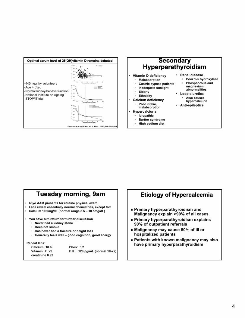
Tuesday morning, 9amTuesday morning, 9am• 65yo AAM presents for routine physical exam• Labs reveal essentially normal chemistries, except for:• Calcium 10.9mg/dL (normal range 8.5 – 10.5mg/dL)
• You have him return for further discussion• Never had a kidney stone• Does not smoke• Has never had a fracture or height loss• Generally feels well – good cognition, good energy
• Cause – unclear– Increased “setpoint” of serum calcium– Increased # of functional PTH cells
• Genetic predisposition is rare – can be seen in cases of MEN, especially MEN1
• Risk factors – possibly prior radiation to the neck
6
Natural History of Primary HyperparathyroidismNatural History of Primary Hyperparathyroidism
Silverberg SJ et al. N Silverberg SJ et al. N EnglEngl J Med 1999;341:1249J Med 1999;341:1249--1255.1255.
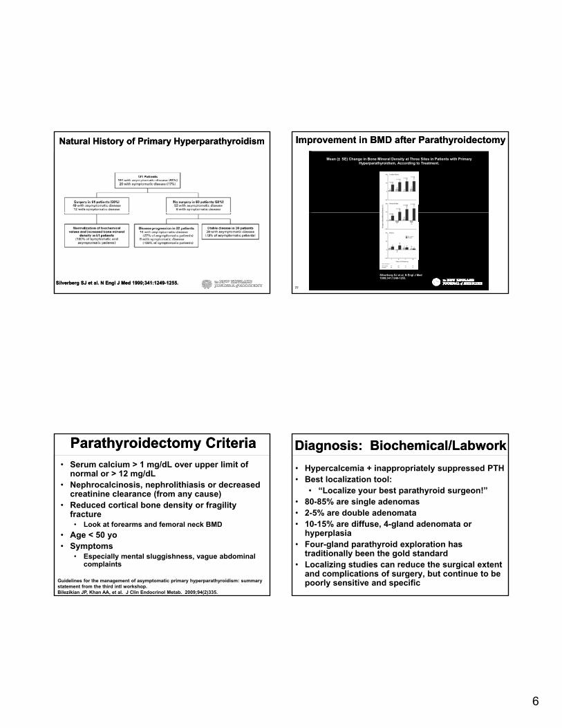
Improvement in BMD after ParathyroidectomyImprovement in BMD after Parathyroidectomy
Mean (± SE) Change in Bone Mineral Density at Three Sites in Patients with Primary Hyperparathyroidism, According to Treatment.
22
Silverberg SJ et al. N Engl J Med 1999;341:1249-1255.
Parathyroidectomy CriteriaParathyroidectomy Criteria• Serum calcium > 1 mg/dL over upper limit of
normal or > 12 mg/dL• Nephrocalcinosis, nephrolithiasis or decreased
creatinine clearance (from any cause)• Reduced cortical bone density or fragility
fracturefracture• Look at forearms and femoral neck BMD
• Age < 50 yo• Symptoms
• Especially mental sluggishness, vague abdominal complaints
Guidelines for the management of asymptomatic primary hyperparathyroidism: summary statement from the third intl workshop.Bilezikian JP, Khan AA, et al. J Clin Endocrinol Metab. 2009;94(2)335.
• Hypercalcemia + inappropriately suppressed PTH• Best localization tool:
• “Localize your best parathyroid surgeon!”• 80-85% are single adenomas• 2-5% are double adenomata2 5% are double adenomata• 10-15% are diffuse, 4-gland adenomata or
hyperplasia• Four-gland parathyroid exploration has
traditionally been the gold standard• Localizing studies can reduce the surgical extent
and complications of surgery, but continue to be poorly sensitive and specific
7
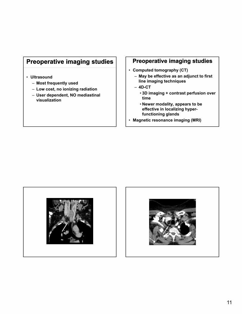
Imaging for Primary HyperparathyroidismImaging for Primary
Hyperparathyroidism• Sestamibi scanning
• Technitium-99m-methoxyisobutylisonitrile
• Combined with SPECT has highest PPV
• Negative sestamibi occurs in
Ultrasound ofNormal left thyroid bed
Negative sestamibi occurs in 12-25% of patients with disease
• Often unrevealing in 4-gland hyperplasia and in those with coexisting thyroid disease
• SPECT successfully detects 96% single adenomas, but only 45% of multiglandular disease
Ultrasound for localization
Ultrasound for localization
• Noninvasive, inexpensive, reproducible in the OR
• Particularly helpful in re-operative cases
Small adenoma in rightInferior thyroid bed
• Can help identify thyroid pathology to facilitate operative planning
• Moderate sensitivity for single-gland adenoma (70-82%)
• Operator dependent
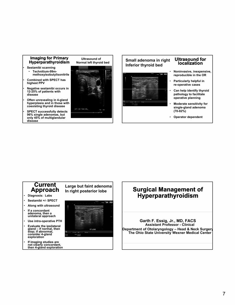
Current ApproachCurrent
Approach• Diagnosis: Labs
• Sestamibi +/- SPECT
• Along with ultrasound
• If a concordant adenoma, then a unilateral approach
Large but faint adenomaIn right posterior lobe
unilateral approach
• Use intra-operative PTH
• Evaluate the ipsilateral gland – if normal, then stop; if abnormal, consider 4-gland exploration
• If imaging studies are not clearly concordant, then 4-gland exploration
Surgical Management of Surgical Management of HyperparathyroidismHyperparathyroidism
Garth F. Essig, Jr., MD, FACSAssistant Professor - Clinical
Department of Otolaryngology – Head & Neck SurgeryThe Ohio State University Wexner Medical Center
8
OutlineOutline
• Anatomy
• Localization studies
• Surgical options• Surgical options
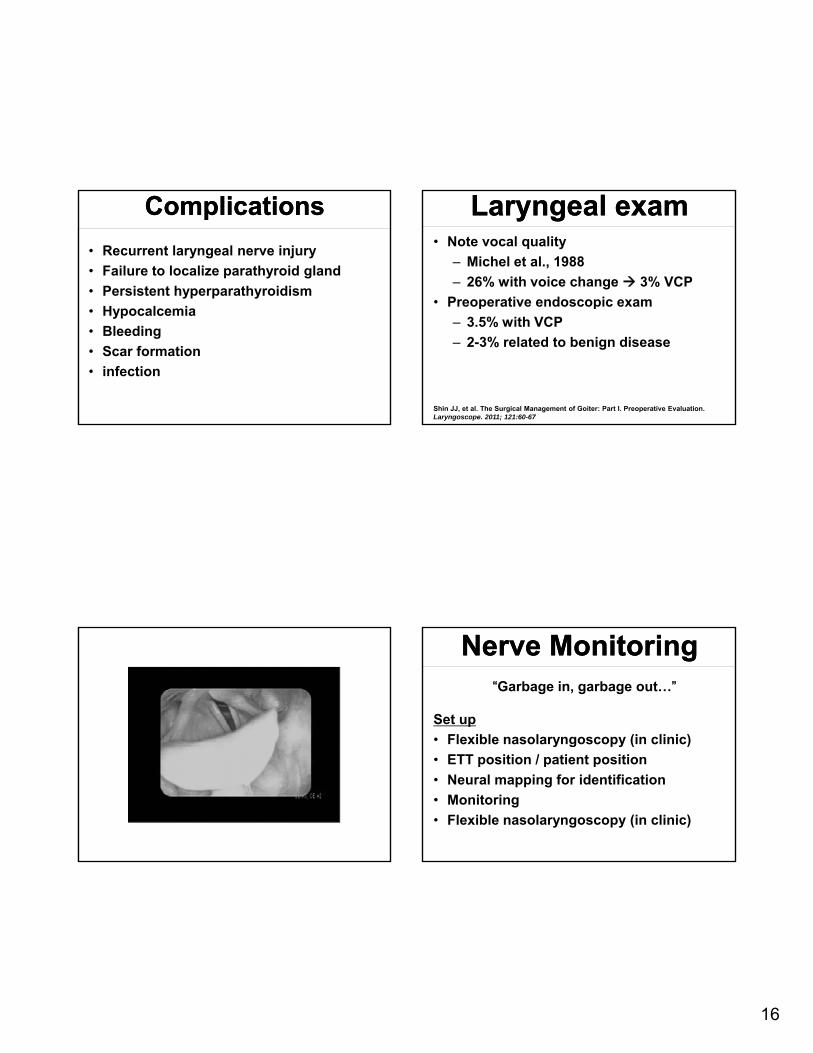
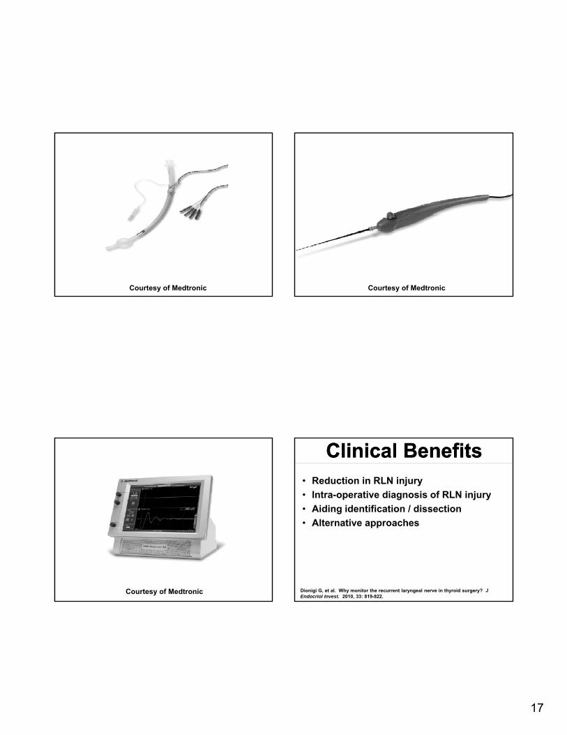
• Recurrent laryngeal nerve monitoring
• complications
GoalsGoals
1. Find and treat ALL abnormal parathyroids
2. Minimize dissection
3 Attain durable cure3. Attain durable cure
HistoryHistory
• 1925 – Mandl, first successful parathyroidectomy
• 1963 – Berson et al., measured human PTH
• 1988 – Nussbaum et al., reported use of rapid PTH
• 1990s – Irvin, IOPTH
Anatomy and EmbryologyAnatomy and Embryology
• Usually 4 glands
– Designated by location right/left, superior/inferior
• Inferior parathyroid gland
3rd h l h ( h l d t i– 3rd pharyngeal pouch (cephalad to superior glands)
Clinical PresentationClinical Presentation"Bones, stones, abdominal groans, and psychic moans.”
•Physical exam
– Usually non contributory
– Laryngeal
•Laboratory analysis
•Imaging studies
Surgical IndicationsSurgical Indications• Surgery is advised in patients with overt manifestation of
primary hyperparathyroidism or inability to comply with annual surveillance.
National Institutes of Health (NIH) consensus panel forsurgery in asymptomatic primary hyperparathyroidism
Serum calcium (above normal) 1.0mg/dL( ) g
Creatinine Clearance <60 cc/min
Bone mineral density T-score <−2.5 at any site and/or previous fracture fragility
Age <50
Bilezikian JP, et al. Guidelines for the Management of Asymptomatic Primary Hyperparathyroidism: Summary Statement from the Third International Workshop. J Clin Endocrinol Metab. Feb 2009; 94(2): 335–339
ObservationObservation
• For those who do not undergo surgery
– Calcium every 6 months
– Annual serum creatinine
– Annual bone density (lumbar spine, hip, distal third of radius)
Localization StudiesLocalization Studies
• Localization of diseased parathyroid is the major limitation of a minimally invasive approach (MIP)approach (MIP)
• Sestamibi scan– Relies on differential washout times (thyroid
vs abnormal parathyroid)
– Thyroid pathology can result in false positivespositives
– Frequently used in conjunction with other anatomic imaging modality
• Single photon emission computed tomography (SPECT)– Superposition of the two radiomarkers results
in more accurate localization
Intraoperative AdjunctsIntraoperative Adjuncts
• Radioguided
• Methylene blue
• Rapid IOPTH
13
Intraoperative PTHIntraoperative PTH• Extensively described in patients with
primary HPT
– Confirms complete excision of hyperfunctioning gland prior to leaving the OR
– Alerts surgeon to additional hypersecreting glands
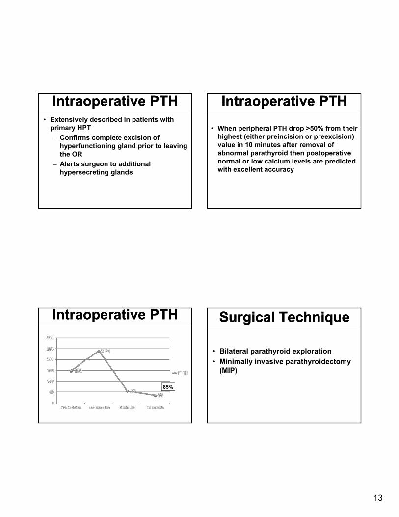
Intraoperative PTHIntraoperative PTH
• When peripheral PTH drop >50% from their highest (either preincision or preexcision) value in 10 minutes after removal of b l th id th t tiabnormal parathyroid then postoperative
normal or low calcium levels are predicted with excellent accuracy


![4. PARATHYROID HORMONE.ppt [Read-Only]ocw.usu.ac.id/.../mk_end_slide_parathyroid_hormone.pdf · Parathyroid Hormone (PTH) Peptide hormone secreted by parathyroid glands, which are](https://static.documents.pub/doc/80x56/5fd9a3fa6d8805309b4bc740/4-parathyroid-read-onlyocwusuacidmkendslideparathyroidhormonepdf.jpg)


