1 LOWER EXTREMITY PRACTICE QUESTIONS 1. ____ A skier went off a down hill course and caught one ski under a log. X ray after the accident showed that he had fractured the tibia. A cast was placed on the leg that went from the knee to the foot. When the cast was removed, the patient dragged his foot and was unable to lift it from the ground. This condition most likely resulted from pressure of the cast on which of the following nerves? A. Femoral B. Obturator C. Superficial Peroneal D. Common Peroneal E. Tibial 2. ____A football player was tackled from the lateral side while attempting an end around run in a tie game. The foot on that leg was planted on the ground and the tackle was made by another player who weighed 312 pounds and was running at the rate of 3.5 miles per hour. MRI of the patient's knee (arrow above) shows a tear in which of the following structures (note position of patella)? A. Tibial Collateral ligament B. Fibular Collateral ligament C. Anterior Cruciate ligament. D. Posterior Cruciate ligament. E. Semitendinosus tendon
Transcript
1
LOWER EXTREMITY PRACTICE QUESTIONS 1. ____ A skier went off a down hill course and caught one ski under a log. X ray after the accident showed that he had fractured the tibia. A cast was placed on the leg that went from the knee to the foot. When the cast was removed, the patient dragged his foot and was unable to lift it from the ground. This condition most likely resulted from pressure of the cast on which of the following nerves? A. Femoral B. Obturator C. Superficial Peroneal D. Common Peroneal E. Tibial
2. ____A football player was tackled from the lateral side while attempting an end around run in a tie game. The foot on that leg was planted on the ground and the tackle was made by another player who weighed 312 pounds and was running at the rate of 3.5 miles per hour. MRI of the patient's knee (arrow above) shows a tear in which of the following structures (note position of patella)? A. Tibial Collateral ligament B. Fibular Collateral ligament C. Anterior Cruciate ligament. D. Posterior Cruciate ligament. E. Semitendinosus tendon
2
3. ____A cross country runner was attempting to pass another runner in a race and stepped off the path. His foot landed on a small stump resulting in hyperinversion of the foot. Subsequent x-ray showed no fractures of the tarsal bones, distal tibia or fibula but the ankle was swollen and painful. Which of the following structures was (were) most likely to have been damaged? A. deltoid ligament. B. long plantar ligament. C. spring ligament. D. calcaneofibular and anterior talofibular ligaments. E. calcaneofibular and posterior talofibular ligaments.
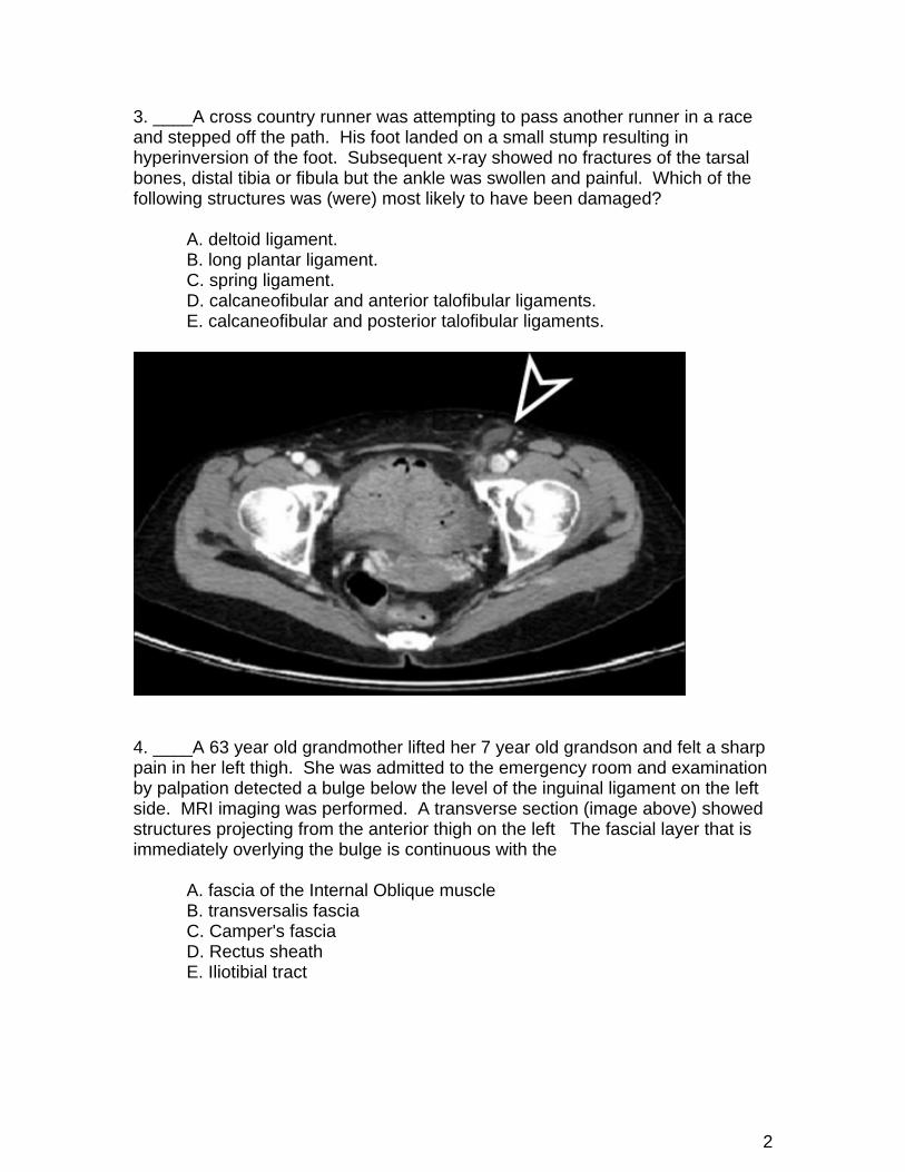
4. ____A 63 year old grandmother lifted her 7 year old grandson and felt a sharp pain in her left thigh. She was admitted to the emergency room and examination by palpation detected a bulge below the level of the inguinal ligament on the left side. MRI imaging was performed. A transverse section (image above) showed structures projecting from the anterior thigh on the left The fascial layer that is immediately overlying the bulge is continuous with the A. fascia of the Internal Oblique muscle B. transversalis fascia C. Camper's fascia D. Rectus sheath E. Iliotibial tract
3
5. ____A runner accelerated toward the finish line of a race and suddenly felt a pop on the back of his thigh. He then fell down in excruciating pain. Xray of the pelvis (image above) showed that a small piece of bone had been fractured and avulsed by muscle tendons. This piece of bone is part of which of the following structures? A. pubis B. ischial spine C. ischial tuberosity D. acetabulum E. ilium 6. ____ Following hip replacement surgery on the left side of the body, an adult patient complains that he has difficulty walking. He is also very unstable when standing if he lifts his right leg. When the patient is observed while walking in a physician's office, the pelvis sways considerably and tilts toward the right when the right leg is lifted. Which of the following nerves was likely to have been damaged in the hip surgery? A. Left Inferior Gluteal Nerve B. Right Inferior Gluteal Nerve C. Left Sciatic Nerve D. Left Superior Gluteal Nerve E. Right Superior Gluteal Nerve
4
7. ____ While on a hunting trip, a teenage patient falls and the hunting knife in his belt penetrates his upper thigh. After being rushed to an emergency room, Inspection of the wound shows a deep cut 1.5 inches below the inguinal ligament that is bleeding profusely. The physician suspects that the femoral artery has been severed and ligates the Femoral artery immediately below the inguinal ligament. The lower limb is still able to receive a sufficient supply of arterial blood because of which of the following anastomoses. A. Inferior Gluteal artery with the Medial and Lateral Femoral Circumflex arteries. B. Internal Pudendal artery with the Medial and Lateral Femoral Circumflex arteries. C. Superficial Circumflex Iliac artery with the Inferior Gluteal artery. D. Inferior Epigastric artery with the Medial and Lateral Femoral Circumflex arteries. E. Inferior Epigastric artery with the Inferior Gluteal artery.
8. ____ A 76-year-old woman is walking down the stairs of her house and falls. She is in pain and has difficulty walking but she does not see a physician. After one week, the pain has become unbearable and she goes to the emergency room of her local hospital. An xray of the thigh (image above) shows a fracture in the neck of the femur and degenerative changes in the femoral head. The blood supply from which of the following arteries is likely to be compromised by the fracture and result in insufficient blood supply to the head of the femur? A. Lateral Femoral Circumflex artery B. Medial Femoral Circumflex artery C. Inferior Epigastric artery D. Inferior Gluteal artery E. Superficial External Pudendal artery
5
9. ____ A carpenter is working on a building site and a large beam falls on the lateral side of his foot. An xray image of the foot (above) shows fractures to the lateral bones of the foot. Healing of the fracture indicated by the arrow at right could be complicated because the tendon of leg muscle inserts at this point. Which of the following muscles inserts at the point indicated by the right arrow (Note: not in review sheet but this was a question on the last board exam)? A. Tibialis posterior B. Peroneus longus C. Tibialis anterior D. Peroneus brevis E. Extensor digiti minimi
6
10. ____ A patient complains that the medial side of the sole of his foot is painful when he stands or walks. The xray of his foot (above) shows a substantial decrease in the height of the medial arch. Weakness in which the following structures could produce this condition? A. Plantar calcaneonavicular ligament B. Long plantar ligament C. Anterior talofibular ligment D. Deltoid ligament E. Posterior talofibular ligament
7
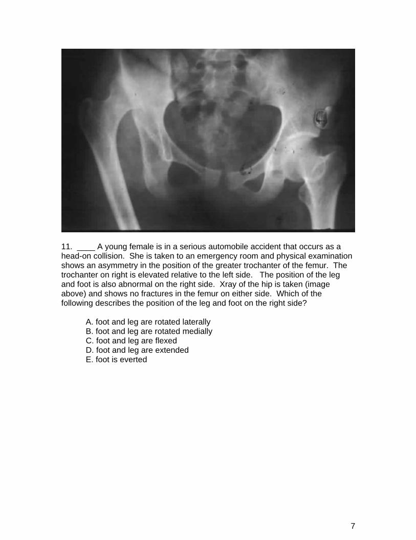
11. ____ A young female is in a serious automobile accident that occurs as a head-on collision. She is taken to an emergency room and physical examination shows an asymmetry in the position of the greater trochanter of the femur. The trochanter on right is elevated relative to the left side. The position of the leg and foot is also abnormal on the right side. Xray of the hip is taken (image above) and shows no fractures in the femur on either side. Which of the following describes the position of the leg and foot on the right side? A. foot and leg are rotated laterally B. foot and leg are rotated medially C. foot and leg are flexed D. foot and leg are extended E. foot is everted
8
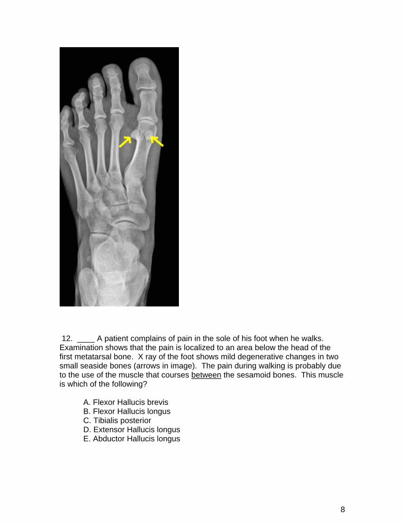
12. ____ A patient complains of pain in the sole of his foot when he walks. Examination shows that the pain is localized to an area below the head of the first metatarsal bone. X ray of the foot shows mild degenerative changes in two small seaside bones (arrows in image). The pain during walking is probably due to the use of the muscle that courses between the sesamoid bones. This muscle is which of the following? A. Flexor Hallucis brevis B. Flexor Hallucis longus C. Tibialis posterior D. Extensor Hallucis longus E. Abductor Hallucis longus
9
13. ____ A patient suffers a deep knife cut to his shin (anterior compartment). He does not seek medical attention but wraps it in bandages. In succeeding days, the wound does not heal and bleeding is persistent. Examination by an emergency room physician shows extensive infection. The wound is opened and cleaned (photo attached). Damage to an arterial branch is repaired and care is taken not to damage the nerve that accompanies the artery. Which of the following are the artery and nerve? A. Anterior Tibial Artery, Superficial Peroneal Nerve B. Anterior Tibial Artery, Deep Peroneal Nerve C. Peroneal Artery, Tibial Nerve D. Peroneal Artery, Superficial Peroneal Nerve D. Popliteal Artery, Common Peroneal Nerve
10
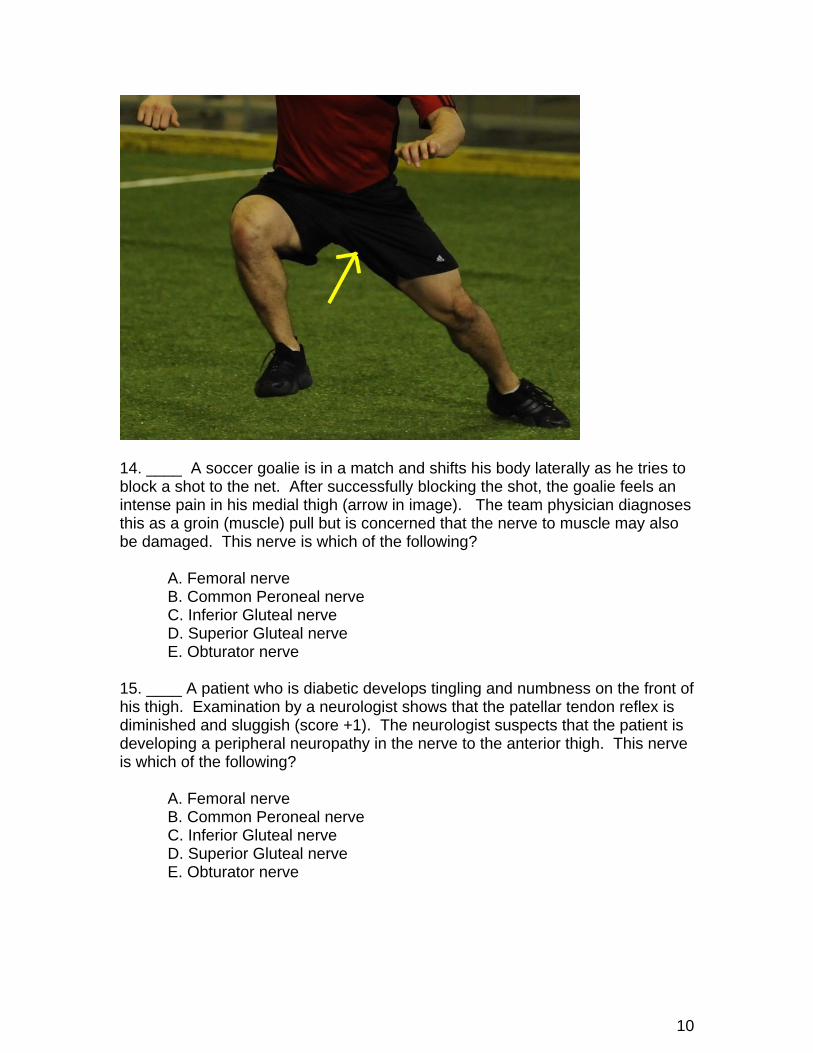
14. ____ A soccer goalie is in a match and shifts his body laterally as he tries to block a shot to the net. After successfully blocking the shot, the goalie feels an intense pain in his medial thigh (arrow in image). The team physician diagnoses this as a groin (muscle) pull but is concerned that the nerve to muscle may also be damaged. This nerve is which of the following? A. Femoral nerve B. Common Peroneal nerve C. Inferior Gluteal nerve D. Superior Gluteal nerve E. Obturator nerve 15. ____ A patient who is diabetic develops tingling and numbness on the front of his thigh. Examination by a neurologist shows that the patellar tendon reflex is diminished and sluggish (score +1). The neurologist suspects that the patient is developing a peripheral neuropathy in the nerve to the anterior thigh. This nerve is which of the following? A. Femoral nerve B. Common Peroneal nerve C. Inferior Gluteal nerve D. Superior Gluteal nerve E. Obturator nerve
11
16. ____ A tennis player is in an intense match and runs toward the net. He feels a 'snap' in his calf. After the match he feels intense pain deep in his posterior calf. Physical examination shows that the Tendo Calcaneus (Achilles tendon) has not been ruptured or sprained but the pain is intensified if the patient attempts to plantar flex the foot. The physician suspects that the symptoms are due to damage to a deep muscle that inserts on the Achilles tendon. Which of the following muscles is most likely to be damaged? A. Plantaris B. Tibialis Posterior C. Flexor Hallucis longus D. Peroneus Tertius E. Flexor Digitorum longus
12
LOWER EXTREMITY ANSWER KEY 1. D 2. C 3. D 4. B 5. C 6. D 7. A 8. B 9. D 10. A 11. B 12. B 13. B 14. E 15. A 16. A