Insertion of an intravascular catheter is one of the most common invasive procedures in hospitals worldwide. These intravascularlines are crucial in resuscitation, allow vital medication to be administered, and can be used to monitor the patients’ real-time vitalparameters. There is, however, growing recognition of potential risks to life and limb associated with their use. Medical literatureis now replete with isolated case reports of complications succinctly described by Garden and Laussen (2004) as “An unendingsupply of “unusual” complications from central venous catheters.”This paper reviews complications of venous and arterial cathetersand discusses treatment approaches and methods to prevent complications, based on current evidence and endeavours to provideinformation and guidance that will enable practitioners to prevent, recognise, and successfully treat extravasation injuries in adults.
1. Definition
Extravasation injury is defined as the damage caused bythe efflux of solutions from a vessel into surrounding tissuespaces during intravenous infusion. The damage can extendto involve nerves, tendons, and joints and can continue formonths after the initial insult. If treatment is delayed, surgicaldebridement, skin grafting, and even amputation may be theunfortunate consequences of such an injury [1].
2. Incidence
Extravasation is not as rare as many people think, and itmay occur even in the most closely monitored situations.A study which investigated extravasation over a five-weekperiod in a UK hospital established an incidence of 39% inadults, almost double that of previously published reports[2]. Two percent of the Medical Defence Union cases involv-ing anaesthetic-related events between 1970–1982 (excludingdeaths) were due to extravasation injuries [3], and for thosepatients who received a course of cytotoxic injections 5%experienced extravasations [4]. Despite this, extravasationinjuries remain uncommon, with an estimated incidencepublished in the literature of between 0.1% and 6% in patientsreceiving chemotherapy [1]. The published rate is likely
an underestimation, however, as many cases of extravasationgo unreported.
3. Aetiology
Sites most often implicated in extravasation injuries includethe dorsum of the hand and foot [5], ankle, antecubital fossa[6], and near joints or joint spaces [7] where there is littlesoft tissue protection for underlying structures [8]. Limbswith local vascular problems such as lymphoedemamay havereduced venous flow causing pooling and potential leakageof infusates around the site of cannulation [5, 9]. Peripheralrather than central venous administration of antineoplasticagents is more likely to be associated with frequent cannula-tion which is a risk factor for extravasation, and this shouldbe avoided [5, 7].
More extravasations occur at night and often go unno-ticed [10]; however, data from the National ExtravasationInformation Service green card reporting database shows that44% of extravasations occur between the hours of 2 pm and10 pm, 10% occur between 10 pm and 6 am, and 38% occurbetween 6 am and 2 pm [11]. Inexperienced personnel maypose a higher risk, particularly during cytotoxic administra-tion.
There are various patient factors that contribute to theaetiology of extravasation injuries. Veins of people receiving
2 ISRN Dermatology
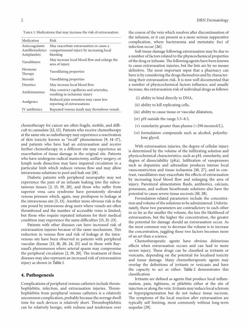
Table 1: Medications that may increase the risk of extravasation.
May exacerbate extravasation or cause acompartmental injury by increasing localbleeding
Vasodilators May increase local blood flow and enlarge thearea of injury
HormoneTherapy Vasodilating properties
Steroids Vasodilating propertiesDiuretics May increase local blood flow
Antihistamines May constrict capillaries and arterioles,resulting in ischaemic injury
Analgesics Reduced pain sensation may cause lessreporting of extravasations
IV antibiotics Repeated venous insult may thrombose vessels
chemotherapy for cancer are often fragile, mobile, and diffi-cult to cannulate [12, 13]. Patients who receive chemotherapyat the same site as radiotherapymay experience a reactivationof skin toxicity known as “recall” phenomenon [9, 14–17],and patients who have had an extravasation and receivefurther chemotherapy in a different site may experience anexacerbation of tissue damage in the original site. Patientswho have undergone radical mastectomy, axillary surgery, orlymph node dissection may have impaired circulation in aparticular limb which reduces venous flow and may allowintravenous solutions to pool and leak out [18].
Diabetic patients with peripheral neuropathy may notexperience the pain of an infusate leaking into the subcu-taneous tissues [2, 13, 19, 20], and those who suffer fromsuperior vena cava syndrome have persistently elevatedvenous pressure which, again, may predispose to leakage atthe intravenous site [5, 13]. Another more obvious risk is theone posed by intravenous drug users where vessels are oftenthrombosed and the number of accessible veins is limited,but those who require repeated infusions for their medicalcondition may experience the same difficulties [13, 21–23].
Patients with other diseases can have increased risk ofextravasation injuries because of the same mechanism. Thisreduction in venous flow and risk of leakage at the intra-venous site have been observed in patients with peripheralvascular disease [13, 18, 20, 24, 25] and in those with Ray-naud’s phenomenon where arterial spasm may compromisethe peripheral circulation [2, 19, 20]. The treatment of thesediseases may also represent an increased risk of extravasationinjury as shown in Table 1.
4. Pathogenesis
Complications of peripheral venous catheters include throm-bophlebitis, infection, and extravasation injuries. Throm-bophlebitis from peripheral venous catheters is a relativelyuncommon complication, probably because the average dwelltime for such devices is relatively short. Thrombophlebitiscan be relatively benign, with redness and tenderness over
the course of the vein which resolves after discontinuation ofthe infusion, or it can present as a more serious suppurativecomplication, where bacteraemia and metastatic foci ofinfection occur [26].
Soft tissue damage following extravasation may be due toa number of factors related to the physicochemical propertiesof the drug or infusate.The following agents have been knownto cause extravasation injuries, but the lists are by no meansdefinitive. The most important input that a pharmacy canhave is by considering the drugs themselves and by character-ising their extravasation risk. It is now well documented thata number of physicochemical factors influence, and usuallyincrease, the extravasation risk of individual drugs as follows:
(i) ability to bind directly to DNA,(ii) ability to kill replicating cells,(iii) ability to cause tissue or vascular dilatation,(iv) pH outside the range 5.5–8.5,(v) osmolarity greater than plasma (>290mosmol/L),(vi) formulation compounds such as alcohol, polyethy-
lene glycol.
With extravasation injuries, the degree of cellular injuryis determined by the volume of the infiltrating solution andphysicochemical characteristics, such as pH, osmolarity, anddegree of dissociability (pKa). Infiltration of vasopressorssuch as dopamine and adrenaline produces intense localvasoconstriction and tissue ischaemia [10, 27], and in con-trast, vasodilators may exacerbate the effects of extravasationby increasing local blood flow and enlarging the area ofinjury. Parenteral alimentation fluids, antibiotics, calcium,potassium, and sodium bicarbonate solutions also have thepotential to cause severe tissue necrosis [10, 28].
Formulation-related parameters include the concentra-tion and volume of the solutions to be administered. Unfortu-nately, these two parameters are contradictory to each otherin so far as the smaller the volume, the less the likelihood ofextravasation, but the higher the concentration, the greaterthe potential for damage should an extravasation occur. Asthe most common way to decrease the volume is to increasethe concentration, juggling these two factors becomes moreof an art than a science.
Chemotherapeutic agents have obvious deleteriouseffects when extravasation occurs and can lead to moresevere injury. These drugs can be classified as irritants orvesicants, depending on the potential for localised toxicityand tissue damage. Many chemotherapeutic agents mayoverlap the definitions of irritants or vesicants and havethe capacity to act as either. Table 2 demonstrates thisclassification.
Irritants are defined as agents that produce local inflam-mation, pain, tightness, or phlebitis either at the site ofinjection or along the vein. Irritantsmay induce local sclerosisor hyperpigmentation but do not induce tissue necrosis.The symptoms of the local reaction after extravasation aretypically self limiting, most commonly without long-termsequelae [29].
ISRN Dermatology 3
Table 2: Classification of chemotheraputic agents into irritants(bold), vesicants (italic) and both irritant and vesicant (normal).
are vesicant (i.e., produce blisters) [7, 9], and as well ascausing immediate injury may also bind to tissue DNA [30]so that the drug is continually released from dying to healthycells, resulting in a slow increase in ulcer size over time.Doxorubicin, for example, has been shown to remain in tissuefor 5 months after extravasation [31] which means that theinjury can present late with extensive tissue destruction [32].Ulcers caused by these highly vesicant agents usually donot heal and often require plastic surgery and skin grafting[15].
The full effect of the extravasation injury is not usuallyimmediately apparent but may evolve over days or weeks.Early local symptoms of a vesicant extravasation resemblethose of an irritant extravasation: local pain, erythema,
burning, pruritus, or swelling [33, 34]. Over the course ofthe reaction, however, as tissue necrosis evolves and becomesclinically apparent, progressive erythema, discolouration,blistering, or desquamation may develop. The severity of thelocal reactionmay vary both upon the agent extravasated andupon the total dose of extravasated material.
The pathogenesis of the severe tissue damage that vesi-cant chemotherapeutic agents cause is not fully understood.Agents that bind to DNA induce more damage than non-DNA-binding drugs [34, 35]. These agents are taken up bythe surrounding cells, causing progressive and prolongedlocal damage [35]. This has particularly been suggested to bethe case for the severe tissue damage seen with doxorubicinextravasation [28]. Additionally, the significant free-radicalformation of vesicant agents is suggested as a potentialmechanism of the severe necrotic effect [35].
In order for the extravasated compound to do damage, itmust move out from the initial site of extravasation [20]. Thefact that this movement occurs is evident when we considerthat the resultant area of damage is often considerablylarger than the initial physical appearance at the time ofextravasation.
An understanding of drug or infusate cellular transportprocess may allow us to better predict the spectrum ofdamage that may be expected. Some forms of transportmechanism may directly cause cell death because of the rateat which they affect the local cellular environment. Osmoticpressure is such a factor and this is directly related to theosmolality of the administered drug. Osmotic pressure cancause cell death and hence tissue necrosis by cell implosionfrom hypertonic solutions, or cell explosion from hypotonicsolutions; however, the former of these cellular fates is by farthe most common. Some substances have the potential tocause tissue damage by having an osmolality greater than thatof serum (281–289mosmol/L) [36].
Hyperosmolar substances such as hypertonic glucosesolutions or X-ray contrast media draw fluid from cellsresulting in cell death by dehydration, whereas calcium andpotassium salts cause cell death by fluid overload. Hyper-tonic solutions which contain ions and are also acidic areparticularly damaging to tissues because they are capableof killing cells by precipitating cell proteins [36]. Calciumchloride, for example, has caused full-thickness skin necro-sis, and hypertonic saline is the most common sclerosantassociated with necrosis. Parenteral nutrition extravasationis reported more often in children [18] and can cause skinsloughs [10] and limb contractures particularly in prematureinfants.
The pH of a substance outside of the physiological rangemay have an adverse effect on tissue [2, 18, 37, 38]. Thiopen-tone and phenytoin, for example, are highly alkaline andhave caused severe injuries including amputations [39].Otheragents are shown as follows:acidic agents include
Extravasation of intravenous fluids is marked initially by painand swelling, which then progresses to blanching, blistering,and discolouration of the skin. Pain is the most usefulsymptom to alert the administrator to the possibility of acomplication.
Induration, erythema, venous discolouration, or swellingmay be observed at the site, but it is worth noting thatdiscolouration alone may not indicate extravasation as dox-orubicin, epirubicin, andmitozantrone have all been reportedto produce this effect when administered intravenously.
Persistent induration often progresses to a dry blackeschar in 1 or 2 weeks, which then usually sloughs to revealan ulcer. Objective staging of extravasations is useful forquality improvement purposes and for deciding the degreeof intervention required [40].
6. Recognition
A summary of the signs and symptoms above is presented asfollows.
Recognition of an extravasation is through
(i) pain,(ii) erythema,(iii) swelling,(iv) tenderness,(v) local blistering (indicative of at least a partial-
thickness skin injury),(vi) mottling/darkening of skin,(vii) firm Induration,(viii) ulceration (usually not evident until 1-2 weeks after
injury),(ix) no capillary filling (a white appearance with non-
Note that not all of the above symptoms may be present.As well as these signs demonstrated from clinical examina-tion, an awareness of peripheral factors, such as attachedpumps and monitoring equipment, may highlight a problemearlier.
A reduced rate of flow may be observed when using aninfusion pump, and therefore, close observation is necessary.Increased resistance to the administration, once possiblechanges in the position of the body for example bending ofwrist or elbow, cannula support, or bandaging, have beenexcluded as possible causes, indicates a displaced cannulaand the possibility of extravasation. Once the alternative
diagnoses have been considered and excluded and one ormore of the symptoms are present, the practitioner shouldproceed on the basis of a diagnosis of extravasation.
A lack of blood return from the cannula is commonlyquoted as a sign that extravasation has occurred. It is however,the most misleading of all signs and has been implicated in anumber of serious incidents. If there has been extravasationinjury and the cannula has become displaced, the act oftrying to draw back test for blood return can move thecannula back into the vein while a hole remains in the veinwall in the proximity of the cannula tip. If administrationrecommences, a larger and more significant extravasationinjury then ensues. Alternatively, the bevel of the needle canpuncture the veinwall during venepuncture, allowing drug toescape into the tissue whilst the lumen of the needle may stillremain in the blood vessel and allow adequate blood return.
7. Management
Treatment is determined by the stage of extravasation, thenature of the infiltrating solution, and the availability ofspecific antidotes. In all cases of infiltration, the intravenousinfusion should be stopped promptly, and any constrictingbands or tapes should be removed. Treatment protocols forsevere extravasations vary from conservative to aggressivemanagement of the acute injury [28, 39, 41, 42], with addi-tional variations in wound management [10, 43, 44].
There is no standard treatment for the acute phase of thisextravasation injury. However, once it is detected, emergencymanagementmust be taken immediately.The infusion shouldbe stopped and the intravenous cannula should be aspirated.Any collection or palpable effusion in the subcutaneoustissues should be drained and the limb should be immobilisedand elevated above the heart level. Many authors prefer theconservative treatment until lesions evolve for at least 1 week[13, 34, 45, 46]. On the other hand, with full-thickness skinnecrosis, ulcer, or persistent pain, many surgeons suggestearly aggressive debridement because the chronicity and thenature of the wound can cause patients to suffer delayed treat-ment of primary disease (i.e., carcinoma) and morphofunc-tional damage [13, 45, 46]. In these situations, surgical inter-ventionwith radical debridement andwound coveragewouldbe required [39]. A proposed algorithm for approaching thetreatment of extravasation injuries is shown in Figure 1.
Treatment of a vesicant extravasation includes immediatecessation of infusion, aspiration of asmuch extravasated drugas possible through the still-intact catheter, and attempts forthe aspiration of the extravasated agent in the surroundingtissue. This aspiration may help to limit the extent of tissuedamage. Application of cold packs provides symptomaticpain relief. Hot packs increase local vasodilation, diluting theextravasated drug. Cold packs should not be administeredin the event of extravasation of vinca alkaloids as increasedtissue ulceration has been demonstrated in animal modelswith the use of cold packs [47].
The local application of antidotes to different chemother-apeutic agents is based on very limited data. Sodiumthiosulfate is recommended as an effective antidote for
ISRN Dermatology 5
Symptoms• Pain• Burning• Stinging
Signs•Induration•Erythema•Swelling
Peripheral line Central line
•• Disconnect drip• Do not remove cannula
• Stop infusionStop infusion• Aspirate drug from line
Mark extravasated area Leave central line in situ
• Aspirate the extravasated drug• Attempt to draw blood back from the cannula• Injection of 0.9% sodium chloride may aidthis
Inform doctor Inform doctor
Extravasation in nontunneled section
Refer to plastic surgery
Extravasation in tunneled section
Follow individual management instruction∗
Figure 1: Proposed treatment algorithm. ∗Individual management instructions. (I) Aspirate extravasation injuries and inject steroidhydrocortisone subcutaneously to the affected area and IV if large-scale inflammation, flare, or fracturing along the vein has occurred.(II) Treatment is then characterised as either (A) spread and dilute (1) using normal saline or hyaluronidase, (2) keep limb warm, (3)use continuous compression and elevation of the limb or (B) localise and neutralise (1) use antidote if available, (2) use intermittent coldcompression.
mechlorethamine cisplatin. Hyaluronidase has beenrecommended for extravasation of vinca alkaloids [9]. Themechanism of action in prevention of tissue damage isnot fully understood and has not been extensively studied.Hyaluronidase has been suggested to act via temporarybreakdown of hyaluronic acid, which holds together tissueplanes, and subsequent facilitation of drug dispersement anddilution [48].
Topical application of dimethylsulfoxide (DMSO) hasbeen proposed to help prevent significant tissue necrosisin animal and human models. The pathophysiology of theinteraction is not known, although free-radical scavengingand facilitation of elimination of drug from local tissuesare postulated pathways of efficacy [49]. Procedures such asliposuction or saline flushout have been proposed througha single-institution series but have not been met withwidespread usage [50].
Dexrazoxane, employed for protection of anthracycline-induced cardiotoxicity, has been evaluated in animal models
and demonstrated to be protective against local tissue damageand ulceration in anthracycline extravasation [51]. Potentfree-radical scavenging effects are suggested as the mecha-nism of protection from tissue damage.
The indications for surgery in an extravasation injurypatient include full-thickness skin necrosis, chronic ulcer,and persistent pain [13, 45]. When the patient has fulfilledthe indication for surgery, a surgical treatment is necessaryas early as possible to decrease the morbidity, suffering,and delayed treatment of primary disease of the patient.It is imperative that complete or radical excision of allnecrotic tissues must be performed until the bleeding isobserved and only healthy tissue is left for wound cover-age. Some authors use the intraoperative fluorescent dyeinjection to detect the doxorubicin HCl in the tissue toensure complete excision [52]. Immediate or delayed sur-gical reconstruction could then be successfully performed[13].
6 ISRN Dermatology
Although case reports of local interventions includingglycerine, chlorhexidine, and dimethylsulfoxide (DMSO)have been published for the treatment of docetaxel extrava-sation, it is not clear whether the application of an antidotefor irritant extravasation ismore effective than local palliativemeasures [15].
8. Prevention
Measures to prevent extravasation include careful insertionof peripheral venous cannulae, flushing with sterile saline toensure patency, and suitable dressing to prevent movement,without obscuring possible swelling or erythema. Regularinspection of the site and regulated delivery of intravenousfluids from continuous infusion pumps (usually limited toan hour at a time) may prevent the inadvertent infiltrationof a large amount of fluid before detection, but it is helpful toremember that although occlusion alarms on infusion pumpsmay be set to the lowest limit possible, increased pressure isnot always registered [53].
Hyperosmolar fluids, acidic or alkaline solutions, orinfusates with irritant or vesicant properties should be giventhrough central venous lines, if possible, or should be dilutedor neutralised appropriately. The addition of heparin eitherto flush solutions or to continuous infusions has not beenshown to prolong peripheral catheter patency or to reduce theincidence of infiltration or extravasations conclusively and isnot recommended [54].
The site for cannulation must be chosen appropriately toreduce risk of extravasation.Thismust be in an areawhere thedevice can be introduced easily and fastened securely, whereit is always in view for regular inspection. Taking these factorsinto account, the most appropriate site is considered to bethe forearm. However, it has to be accepted that this is notalways going to be an available area.The vessels in the dorsumof the hand are probably the next most appropriate locationto consider. As a general rule joints and creases should beavoided as these often represent a “small” anatomical space,with nerves and tendons present.
For slow infusion of high-risk drugs, a central line orperipherally inserted central catheter (PICC) line should beused, but if administration through a peripheral cannulais necessary, it is best to administer cytotoxics through arecently sited cannula after ensuring its patency with a salineflush. When administering vesicants by slow intravenousinjection, a push into the side-arm port of a fast-runningintravenous infusion of compatible solution is recommended.If administering more than one infusion sequentially, themost vesicant drug should be administered first. A frequentassessment of the peripheral site is required, watching forsigns of redness or swelling.
If there are any doubts concerning the patency of anintravascular catheter, the infusion must be stopped pendinginvestigation. It is recommended to re-site the cannula if thereis any uncertainty about its patency.
Some investigators suggest delaying the administration ofantiemetics until after vesicant administration as the sedativeand anti-inflammatory effects of antiemetics often mask
the early warning signs of extravasation and may impede thepatient’s ability to report any sensation at the infusion site. Itis important to never hurry and to administer drugs slowlyto allow the drug to be diluted by the carrier solution whilecareful assessment of the IV site is undertaken. Documenta-tion of the rate of administration, location and condition ofsite, verification of patency, and patient’s responses, is advisedwhen giving any drugs with the potential to extravasate.
The elimination of human error can be considered tobe impossible but systems can be put in place to decreasepotential risks and to avoid a “failure to rescue” scenario.Systems that can be used tominimise this risk include the useof good training and educational policy, not only as stand-alone courses, but importantly, on a continuing educationalbasis.
9. Prognosis
Local necrosis may heal with conservative management,leaving minimal long-term sequelae, or may progress to sig-nificant eschar formation and tissue ulceration that ultimatelyrequires surgical debridement and further intervention, withlong-term morbidity for the patient.
Ulceration after vesicant extravasation is typicallymarkedby delayed healing. Morbidity may consist of cosmeticdefects, chronic pain, or loss of function secondary tocontractures or neuropathy, even in the absence of ulcerationof skin [48]. Published patient series have estimated thatonly approximately one third of vesicant extravasations willprogress to tissue ulceration [50]. Repeated infusion ofthe offending agent, even in another limb, may induce arecall reaction at the site of extravasation [14]. One case ofsquamous cell carcinoma of the skin was documented at thesite of a doxorubicin extravasation 10 years previously [50].
When extravasation occurs, there is no certain way ofpredicting the pattern of damage that will ensue. Heckler [55]proposed clinical staging based on 1 to 4 clinical stages ofthe extravasation injury. In stages 1 and 2, no signs of skindamage and loss are observed, whereas in stages 3 and 4, thesoft tissue damage ismore extensive andmay include skin andunderlying tissue necrosis.
Although there is little direct literature on the effectof time from occurrence to either treatment or extent ofmaximum injury, all authors make the generalised statementthat the sooner an extravasation injury is treated, the betterthe outcome, and the smaller the affected area. However, ourability to define and characterise the mechanism and rate ofmovement of individual compounds in the subcutaneous tis-sues will allow us to better predict the extent of extravasationinjuries.
10. Discussion
The consequences of iatrogenic injuries such as those fromextravasation are potentially limb-threatening and havesevere ongoing consequences for the patient. Prevention, asalways, is better than cure, but despite our aim to eliminateerrors such as these from our hospitals, the data on incidence
ISRN Dermatology 7
suggests that an approach to management needs to be clari-fied. There are many factors influencing the pathophysiologyof the condition including those that the patient carries andthose of the offending infusate. In-depth knowledge of thesefactors allows a tailored approach to treatment.
The initial management of extravasation injuries asoutlined in Figure 1 reflects current practice in the fieldand provides a foundation on which to add more invasivetreatments. A knowledge of the causative agent is crucial, asis an awareness of the potential for the introduction of anantidote drug. As with any iatrogenic injury, communicationwith the patients and their relatives is the key for maintainingtrust. A firm grasp of the options for management and thecurrent evidence for such choices alongside an appreciationof the potential progression of the injury and the prognosisfor the patient aids this process.
In adults, early first aid and inclusion of the plasticsurgery team for specialist advice of benefit. The type ofoffending agent, volume extravasated, and various patientfactors influence the type of treatment that is required.This intricate mix of factors makes it difficult to accuratelypredict the progression of the injury and therefore the mostappropriate treatment. It is the senior authors’ approach toperform early surgery in the presence of skin ulcers, full-thickness skin necrosis and persistent pain. Regular review isadvised until healing is achieved. This approach then allowsfor reconstructive surgery to follow, allowing repair of thedefect and restoration of function.
11. Conclusion
Extravasation injury is very dangerous. It increasesmorbidity,causes delayed treatment of the primary disease, and haslong-term sequelae. Prevention is better than cure, butwhere extravasation injury does occur in adults, the authors’preferred method is one of theearly surgical interventionswith regular followup for the consideration of reconstructivesurgery.
References
[1] M. E. MacCara, “Extravasation. A hazard of intravenous ther-apy,” Drug Intelligence and Clinical Pharmacy, vol. 17, no. 10, pp.713–717, 1983.
[2] A. M. Jones and A. Stanley, “Probe High Extravasation Rates.An investigation of extravasation in City Hospital NHS Trust,Birmingham,”TheNational Extravasation Information Service,http://www.extravasation.org.uk/probe.htm.
[3] J. E. Utting, “Pitfalls in anaesthetic practice,” British Journal ofAnaesthesia, vol. 59, no. 7, pp. 877–890, 1987.
[4] J. J. Wang, E. Cortes, L. F. Sinks, and J. F. Holland, “Therapeuticeffect and toxicity of adriamycin in patients with neoplasticdisease,” Cancer, vol. 28, no. 4, pp. 837–843, 1971.
[5] D. Gault and J. Challands, “Extravasation of Drugs,” in Anaes-thesia Review, L. Kaufman and R. Ginsburg, Eds., vol. 13,Churchill Livingstone, Edinburgh, UK, 1997.
[6] A. S. Brown, D. J. Hoelzer, and S. A. Piercy, “Skin necrosis fromextravasation of intravenous fluids in children,” Plastic andReconstructive Surgery, vol. 64, no. 2, pp. 145–150, 1979.
[7] A. L. Garden and P. C. Laussen, “An unending supply of“unusual” complications from central venous catheters,” Paedi-atric Anaesthesia, vol. 14, no. 11, pp. 905–909, 2004.
[8] R. Smith, “Prevention and treatment of extravasation,” BritishJournal of Parenteral Therapy, vol. 6, no. 5, pp. 114–118, 1985.
[9] G. Bertelli, “Prevention and management of extravasation ofcytotoxic drugs,” Drug Safety, vol. 12, no. 4, pp. 245–255, 1995.
[10] A. S. Brown, D. J. Hoelzer, and S. A. Piercy, “Skin necrosis fromextravasation of intravenous fluids in children,” Plastic andReconstructive Surgery, vol. 64, no. 2, pp. 145–150, 1979.
[11] I. Hawley, “Statistics from the National Extravasation Infor-mation Service green card reporting database,” 1987–2006,http://www.extravasation.org.uk/stats.htm.
[12] L. S.Wood and S.M. Gullo, “IV vesicants: how to avoid extrava-sation,” American Journal of Nursing, vol. 93, no. 4, pp. 42–46,1993.
[13] S. M. Shenaq, E. H. A. Abbase, and J. D. Friedman, “Soft-tissuereconstruction following extravasation of chemotherapeuticagents,” Surgical Oncology Clinics of North America, vol. 5, no.4, pp. 825–845, 1996.
[14] J. Shapiro andG. E. Richardson, “Paclitaxel-induced “recall” softtissue injury occurring at the site of previous extravasation withsubsequent intravenous treatment in a different limb,” Journalof Clinical Oncology, vol. 12, no. 10, pp. 2237–2238, 1994.
[15] D. S. Alberts and R. T. Dorr, “Case report: topical DMSO formitomycin-C-induced skin ulceration,” Oncology NursingForum, vol. 18, no. 4, pp. 693–695, 1991.
[16] S. S. Donaldson, J. M. Glick, and J. R. Wilbur, “Adriamycinactivating a recall phenomenon after radiation therapy,” Annalsof Internal Medicine, vol. 81, no. 3, pp. 407–408, 1974.
[17] D. Baer and S.Wilkinson, “Daunomycin, adriamycin, and recalleffect,” Annals of Internal Medicine, vol. 85, pp. 259–260, 1975.
[18] M. E. Gil and J. Mateu, “Treatment of extravasation fromparenteral nutrition solution,” Annals of Pharmacotherapy, vol.32, no. 1, pp. 51–55, 1998.
[19] J. L. Chen andM. O’Shea, “Extravasation injury associated withlow-dose dopamine,” Annals of Pharmacotherapy, vol. 32, no. 5,pp. 545–548, 1998.
[20] M. P. Federle, P. J. Chang, S. Confer, and B. Ozgun, “Frequencyand effects of extravasation of ionic and nonionic CT contrastmedia during rapid bolus injection,” Radiology, vol. 206, no. 3,pp. 637–640, 1998.
[21] E. Kassner, “Evaluation and treatment of chemotherapy extrav-asation injuries,” Journal of Pediatric Oncology Nursing, vol. 17,no. 3, pp. 135–148, 2000.
[22] D. F. Brown, M. J. Muirhead, P. M. Travis et al., “Mode ofchemotherapy does not affect complications with an implant-able venous access device,” Cancer, vol. 80, no. 5, pp. 966–972,1997.
[23] T. Kerrison and J. Woodhull, “Reducing the risk of throm-bophebitis: comparison of Teflon and Vialon cannulae,” Profes-sional Nurse London, vol. 9, pp. 662–662, 1994.
[24] T. Modena, B. Conti, I. Genta et al., “Hyaluronidase-injectablemicroparticles intended for the treatment of extravasation,”Journal of Microencapsulation, vol. 15, no. 1, pp. 85–92, 1998.
[25] R. J. Ignoffo and M. A. Friedman, “Therapy of local toxicitiescaused by extravasation of cancer chemotherapeutic drugs,”Cancer Treatment Reviews, vol. 7, no. 1, pp. 17–27, 1980.
[26] E. A. Khan, A. G. Correa, and C. J. Baker, “Suppurative throm–bophlebitis in children: a ten-year experience,” Pediatric Infec-tious Disease Journal, vol. 16, no. 1, pp. 63–67, 1997.
8 ISRN Dermatology
[27] M. Subhani, S. Sridhar, and J. D. DeCristofaro, “Phentolamineuse in a neonate for the prevention of dermal necrosis causedby dopamine: a case report,” Journal of Perinatology, vol. 21, no.5, pp. 324–326, 2001.
[28] C. E.Wilkins and A. J. B. Emmerson, “Extravasation injuries onregional neonatal units,” Archives of Disease in Childhood, vol.89, no. 3, pp. F274–F275, 2004.
[29] W. S. Susser, D. L. Whitaker-Worth, and J. M. Grant-Kels,“Mucocutaneous reactions to chemotherapy,” Journal of theAmerican Academy of Dermatology, vol. 40, no. 3, pp. 367–398,1999.
[30] M. J. Soble, R. T. Dorr, P. Plezia, and S. Breckenridge, “Dose-dependent skin ulcers inmice treatedwithDNAbinding antitu-mor antibiotics,” Cancer Chemotherapy and Pharmacology, vol.20, no. 1, pp. 33–36, 1987.
[31] M. Garnick, M. Israel, V. Khetarpal, and J. Luce, “Persistence ofanthracycline levels following dermal and subcutaneous adri-amycin extravasation,” Proceedings of the American Associationfor Cancer Research, vol. 22, p. 685, 1981.
[32] J. Bhawan, J. Petry, and M. E. Rybak, “Histologic changesinduced in skin by extravasation of doxorubicin (adriamycin),”Journal of Cutaneous Pathology, vol. 16, no. 3, pp. 158–163, 1989.
[33] P. Berghammer, R. Pohnl, M. Baur, and C. Dittrich, “Docetaxelextravasation,” Supportive Care in Cancer, vol. 9, no. 2, pp. 131–134, 2001.
[34] R. Rudolph and D. L. Larson, “Etiology and treatment ofchemotherapeutic agent extravasation injuries: a review,” Jour-nal of Clinical Oncology, vol. 5, no. 7, pp. 1116–1126, 1987.
[35] C. Sauerland, C. Engelking, R. Wickham, and D. Corbi,“Vesicant extravasation Part I: mechanisms, pathogenesis, andnursing care to reduce risk,” Oncology Nursing Forum, vol. 33,no. 6, pp. 1134–1141, 2006.
[36] S. E. Zimmet, “The prevention of cutaneous necrosis followingextravasation of hypertonic saline and sodium tetradecyl sul-fate,” Journal of Dermatologic Surgery and Oncology, vol. 19, no.7, pp. 641–646, 1993.
[37] D. T. Gault, “Extravasation injuries,” British Journal of PlasticSurgery, vol. 46, no. 2, pp. 91–96, 1993.
[38] V. K. Rao, P. D. Feldman, and D. G. Dibbell, “Extravasationinjury to the hand by intravenous phenytoin: report of threecases,” Journal of Neurosurgery, vol. 68, no. 6, pp. 967–969, 1988.
[39] B. K. Siwy and A. M. Sadove, “Acute management of dopamineinfiltration injury with Regitine,” Plastic and ReconstructiveSurgery, vol. 80, no. 4, pp. 610–612, 1987.
[40] R. J. Kumar, S. P. Pegg, and R. M. Kimble, “Management ofextravasation injuries,” ANZ Journal of Surgery, vol. 71, no. 5,pp. 285–289, 2001.
[41] P. A. Harris, S. Bradley, and A. L. H. Moss, “Limiting thedamage of iatrogenic extravasation injury in neonates,” Plasticand Reconstructive Surgery, vol. 107, no. 3, pp. 893–894, 2001.
[42] J. Friedman, “Plastic surgical problems in the neonatal intensivecare unit,” Clinics in Plastic Surgery, vol. 25, no. 4, pp. 599–617,1998.
[43] P. A. Falcone, D. T. Barrall, D. R. Jeyarajah, and J. A. I. Gross-man, “Nonoperative management of full-thickness intravenousextravasation injuries in premature neonates using enzymaticdebridement,” Annals of Plastic Surgery, vol. 22, no. 2, pp. 146–149, 1989.
[44] K. E. Zenk, I. Dungy, and G. R. Greene, “Nafcillin extravasationinjury. Use of hyaluronidase as an antidote,” American Journalof Diseases of Children, vol. 135, no. 12, pp. 1113–1114, 1981.
[45] N. Scuderi and M. G. Onesti, “Antitumor agents: extravasation,management, and surgical treatment,”Annals of Plastic Surgery,vol. 32, no. 1, pp. 39–44, 1994.
[46] R. G. Dufresne, “Skin necrosis from intravenously infusedmaterials,” Cutis, vol. 39, no. 3, pp. 197–198, 1987.
[47] G. Bertelli, D. Dini, G. B. Forno et al., “Hyaluronidase as anantidote to extravasation of vinca alkaloids: clinical results,”Journal of Cancer Research and Clinical Oncology, vol. 120, no.8, pp. 505–506, 1994.
[48] K. A. Denkler and B. E. Cohen, “Reversal of dopamine extrava-sation injury with topical nitroglycerin ointment,” Plastic andReconstructive Surgery, vol. 84, no. 5, pp. 811–813, 1989.
[49] S. J. Phelps and R. A. Helms, “Risk factors affecting infiltrationof peripheral venous lines in infants,” Journal of Pediatrics, vol.111, no. 3, pp. 384–389, 1987.
[50] R. Lauvin, L. Miglianico, and R. Hellegouarc’h, “Skin canceroccurring 10 years after the extravasation of doxorubicin,” NewEngland Journal of Medicine, vol. 332, no. 11, p. 754, 1995.
[51] D. A. Millam, “Managing complication of i.v. therapy,” Nursing,vol. 18, pp. 34–43, 1988.
[52] J. Davies, D. Gault, and R. Buchdahl, “Preventing the scars ofneonatal intensive care,” Archives of Disease in Childhood, vol.70, no. 1, pp. F50–F51, 1994.
[53] P. S. Shah, E. Ng, and A. K. Sinha, “Heparin for prolongingperipheral intravenous catheter use in neonates,” CochraneDatabase of Systematic Reviews, no. 4, Article ID CD002774,2005.
[54] R. A. Ener, S. B. Meglathery, and M. Styler, “Extravasation ofsystemic hemato-oncological therapies,” Annals of Oncology,vol. 15, no. 6, pp. 858–862, 2004.
[55] F. R. Heckler, “Current thought on extravasation injuries,”Clinics in Plastic Surgery, vol. 16, pp. 557–563, 1989.