Page 1
1
Revision 1 1
2
Mineral Precipitation and Dissolution in the Kidney 3
Michael G Hill1, Erich Königsberger and Peter M May 4
Chemical and Metallurgical Engineering and Chemistry 5
Murdoch University 6
6150 Murdoch, 7
Western Australia 8
Abstract 9
The formation of kidney stones is a significant 10
human health problem. Calcium minerals are 11
involved in a majority of these stones. Despite 12
much research, the processes involved in stone 13
formation remain poorly understood and hence, 14
reliable procedures for preventing their formation 15
have yet to be developed. However, recent 16
advances point to some key steps in mineral 17
formation and transformation involving calcium 18
phosphates, which can help to illuminate these 19
issues. A computer model has been developed to 20
1 Author for correspondence
Page 2
2
express the current status of literature data 21
succinctly and to illustrate that computer 22
modelling is a powerful tool for calculating 23
mineral solubilities and for providing insight into 24
the processes involved. Determining the nature of 25
the initial solid phase of calcium phosphate formed 26
is evidently important. 27
Keywords: Kidney Disease, Urolithiasis, Apatite, Brushite, 28
Whewellite, Weddellite 29
Introduction 30
Urolithiasis denotes the pathological crystallisation of minerals that are 31
deposited in the form of calculi or 'stones' in the urinary tract, especially 32
in the kidney. In contrast to the biologically-controlled formation of 33
bone and teeth, urolithiasis is a spontaneous process resembling the 34
formation of minerals in low-temperature, aqueous geochemical 35
environments. This review explores the thermodynamic and kinetic 36
aspects of mineral-urine interactions, together with pathological 37
preconditions of urolithiasis. Various calcium phosphate minerals are 38
crucially involved in kidney stone pathology but many chemical and 39
Page 3
3
mineralogical issues relating to them remain unclear. We summarize 40
what is currently known and identify the most important areas for future 41
work. Progress is unlikely unless current understanding can be made 42
more quantitative. 43
Kidney stone formation is a worldwide problem (Linder and Little, 44
1986; Grases et al., 1999; Moe, 2006), and is very painful (Grases et al., 45
1998; Thomas and Hall, 2005). There is a high economic cost associated 46
with the condition as a result of hospitalization and days taken off work 47
(Linder and Little, 1986; Grases et al., 1998, 1999; Parks and Coe, 48
1996). Although surgical treatments have improved, there is a high, and 49
increasing, incidence of the pathology (Romero et al., 2010; Tiselius, 50
2011b). Despite much research, the underlying causes are still not well 51
understood; prevention has therefore proved difficult (Söhnel and 52
Grases, 1995; Grases et al., 1998; Grases and Costa-Bauza, 2006; Evan 53
et al, 2015; Tiselius, 2015). 54
Some risk factors are, however, well known. Incidence is age and gender 55
dependent, being twice as common in males than in females (Hesse 56
et al., 1986; Moe, 2006; Hughes, 2007; Romero et al., 2010; Tiselius, 57
2011b), with a peak age of presentation at 20 to 50 years (Robertson 58
et al., 1981; Hesse et al., 1986; Hughes, 2007). Dietary factors are 59
Page 4
4
significant, especially increasing risk are diets high in animal protein 60
(Abdel-Halim, 2005; Tiselius, 2011b) and fat (Tiselius, 2011b). 61
Insufficient fluid intake, resulting in a more concentrated urine 62
significantly exacerbates the problem (Tiselius, 2011b). Obesity is 63
another well known risk factor (Abdel-Halim, 2005; Hughes, 2007; 64
Romero et al., 2010; Tiselius, 2011b; Rendina et al., 2013). The 65
environment also has an effect: risk is increased for those living in hot 66
climates and in periods of hotter weather (Soucie et al., 1994; Moe, 67
2006; Hughes, 2007; Romero et al., 2010). Genetic influences are 68
known to be important and differences have been noted in rates of 69
urolithiasis between different racial groups. Incidence and prevalence is 70
highest in Caucasians, decreasing in Hispanics and Asians and lowest in 71
Africans (Soucie et al., 1994; Hughes, 2007; Romero et al., 2010; 72
Moran, 2014). In fact, kidney stones are very rare in most of 73
Sub-Saharan Africa (Kumar and Muchmore, 1990; Rodgers, 2006). 74
Physiology 75
The kidneys perform the vital function of removing unwanted 76
substances from the blood. To understand kidney stone formation it is 77
necessary to consider first the processes of filtration and reabsorption. 78
The balance between these two plays a key role in the potential 79
Page 5
5
nucleation of stone forming minerals. Filtration starts with an 80
unselective separation, where the fluid that is blood plasma passes 81
through an ultrafiltration membrane into the tubules of the kidney. This 82
is then followed by (a) a selective reabsorption process, in which 83
metabolically useful substances are returned from the filtrate back into 84
the blood, and (b) secretion, in which unwanted substances are 85
transferred into the fluid in the tubule, and thus ultimately become 86
excreted in the urine. 87
The basic functional unit of the kidney is called a nephron. A nephron is 88
a tube, through which flows the fluid being processed by the kidney. 89
Each nephron consists of a number of sections for adding or removing 90
substances from the fluid in the tube to perform the overall extraction of 91
urine from the blood. The most important sections for present purposes 92
are the Bowman’s capsule (BC), the proximal tubule (PT), the loop of 93
Henle (LH), the distal tubule (DT), the collecting tubule (CT) and the 94
collecting duct (CD). The loop of Henle is made up of a thin descending 95
limb, a thin ascending limb and a thick ascending limb, as shown in 96
Figure 1. Nephrons vary in length. The ‘long’ ones have a longer loop of 97
Henle and there will be some differences between the composition of the 98
fluid in long and short nephrons. The output from a number of collecting 99
ducts flows through the duct of Bellini, which is located in a papilla. The 100
Page 6
6
papillae protrude into a calyx (CX), which is a space where urine 101
collects before exiting the kidney via the ureter (Bell et al., 1968; 102
Guyton and Hall, 2000; Kerr, 1999; Atherton, 2006b). 103
Although most of the filtrate entering the Bowman’s capsule is 104
reabsorbed, the reabsorption occurs unevenly along the length of the 105
nephron (Atherton, 2006a). Some segments reabsorb more water than 106
solutes, and reabsorption of the solutes takes place to varying extents in 107
different sections (Guyton and Hall, 2000). This results in marked 108
changes in solution composition and concentration as the fluid flows 109
along the nephron (Asplin et al, 1996). As a result of the depletion of 110
water, the solutes become more concentrated and in certain cases can 111
become increasingly supersaturated with respect to various minerals. 112
The final result of the process is a solution containing all the substances 113
to be excreted emerging at the urine-forming end of the kidney tubules. 114
Both the composition and daily volume of urine are very variable, both 115
inter-individual and intra-individual (Saude et al., 2007). The pH of 116
urine also varies from around 4.8 to 7.2 (Kok, 1997). Concentrations are 117
dependent on daily urine volume which can vary significantly. Figures 118
for typical daily urine volume range from 0.99 to 2.3 litres (Diem and 119
Lentner, 1970; Taylor and Curhan, 2007; Eisner et al., 2010; Taylor 120
Page 7
7
et al., 2010). The values in Table 1 have been calculated by dividing the 121
average value in mmol per 24 hours by the volume to obtain 122
concentration values for normal subjects. 123
124
Mineralogy 125
Minerals often occur naturally within biological structures. Multicellular 126
entities are frequently made up of soft tissue supported by hard 127
structures. In the case of vertebrates, these hard structures are normally 128
composed of minerals, and biological mechanisms are generally 129
required in order to construct and maintain these structures. Pathological 130
calcifications, as in the formation of kidney stones and calculi formed in 131
other parts of the body, such as the gall bladder, pancreas and salivary 132
glands, may or may not involve active biological processes. 133
The minerals of particular relevance to this review are apatite, brushite, 134
octacalcium phosphate, whewellite and weddellite. Apatite comprises a 135
group of minerals with the general formula Ca5(PO4)3(F,Cl,OH) 136
(Tiselius, 2011b). The minerals hydroxyapatite (hereafter abbreviated as 137
HAP) and flouroapatite are found ubiquitously in the body as part of the 138
Page 8
8
building blocks of bones and teeth (Söhnel and Grases, 2011). As a 139
result of the need to form these structures, blood plasma, and many other 140
biofluids, are supersaturated with hydroxyapatite (Taunton et al, 2010; 141
Söhnel and Grases, 2011; Holt et al, 2014). Calcium compounds 142
predominate in the majority of kidney stones; 85% of all kidney stones 143
contain calcium salts. Most (about 80%) have calcium oxalate as the 144
major component (Grases et al., 1999; Tiselius, 2011b). Other stones 145
formed are typically either calcium phosphate or mixed calcium 146
oxalate/calcium phosphate (Coe et al., 2011). 147
As well as being absorbed from food, oxalate (like uric acid) is a 148
metabolic end product (Williams, 1978; Knight et al., 2006). An 149
important function of the kidneys is therefore to excrete oxalate from the 150
body. Given the well known insolubility of many oxalate salts, this 151
introduces a range of possible precipitates. The calcium oxalate 152
compounds predominantly found in kidney stones are whewellite 153
(calcium oxalate monohydrate), and weddellite (calcium oxalate 154
dihydrate). Calcium oxalate has three different crystal forms – the 155
monohydrate (COM), the dihydrate (COD), and the trihydrate (COT). 156
The literature frequently describes the monohydrate as the most stable 157
compound whereas the trihydrate is considered to be metastable and the 158
dihydrate unstable (Tomazic and Nancollas, 1980; Grases et al., 1998; 159
Page 9
9
Rodgers et al., 2011). This is probably due to the fact that COD cannot 160
be precipitated from solutions that contain only calcium and oxalate ions 161
(Tomazic and Nancollas, 1980). However, COD can be precipitated 162
from artificial and real urine and consequently often appears in kidney 163
stones (Werness et al., 1979; Tomazic and Nancollas, 1980). The 164
solubilities of these three hydrates follow the order COM < COD < COT 165
(Streit et al., 1998). As a result, solutions saturated with either COD or 166
COT are supersaturated with respect to COM. Both COT and COD 167
transform into COM (Tomazic and Nancollas, 1980). 168
Kidney Stone Formation 169
The passage of fluid through the kidney causes significant changes in 170
concentration and hence also ionic strength (Bell et al., 1968; Guyton 171
and Hall, 2000; Atherton, 2006a). These changes, which can potentially 172
result in supersaturation, are illustrated in Figures 2, 3, 4 and 5 (Asplin et 173
al, 1996; Hojgaard and Tiselius, 1999; Kok, 1997; Rodgers et al, 2011; 174
Tiselius et al, 2009), showing plots of published values of calcium, 175
oxalate, phosphate and pH in different nephron segments. Kok gives 176
probable ranges, shown as min and max in the plots. In fact, most urine 177
samples are always supersaturated with respect to calcium oxalate and 178
the calcium phosphates (Asplin et al., 1996; Grases et al., 1999). 179
Page 10
10
It is known that hydroxyapatite is supersaturated throughout the length 180
of the nephron and that there is a risk of calcium phosphate precipitation 181
both in the ascending limb of the loop of Henle and the distal tubule 182
(Tiselius, 2011b). Calculations have shown that precipitation of 183
hydroxyapatite can cause the other salts to become unsaturated (Rodgers 184
et al., 2011). However, it is not known which phase of calcium 185
phosphate is the first to precipitate (Tiselius, 1997a). Our suggestion is 186
based on Ostwald’s Rule of Stages which holds that the formation of the 187
least stable phases precedes the thermodynamically stable phase (Söhnel 188
and Grases, 2011; Sawada, 1997): this identifies the first substance to 189
precipitate in the formation of hydroxyapatite as one of (a) amorphous 190
calcium phosphate (ACP), having the formula CaxHy(PO4)z·nH2O, (b) 191
octacalcium phosphate (OCP), Ca8H2(PO4)6·5H2O, or (c) brushite (Bru), 192
CaHPO4·2H2O (Luptak et al., 1994; Asplin et al., 1996; Tiselius, 1997a; 193
Grases et al, 1997; Söhnel and Grases, 2011). Knowing this 194
initially-formed phase would obviously be important in establishing 195
how the process of kidney stone formation begins. 196
Urinary supersaturation with calcium oxalate monohydrate is apparently 197
never sufficient to result in homogeneous nucleation; thus, 198
heterogeneous nucleation must be taking place on some nucleating 199
substrate (Söhnel and Grases, 1995; Grases et al., 2012). 200
Page 11
11
Hydroxyapatite, brushite, and uric acid are all likely candidates as 201
substrates for calcium oxalate monohydrate nucleation (Robertson et al., 202
1981; Söhnel and Grases, 1995; Tiselius, 1997a; Højgaard and Tiselius, 203
1999; Tiselius et al., 2009; Grases et al., 2012). 204
Most calcium oxalate stones contain a small proportion of calcium 205
phosphate, often in the core of the stone, indicating that calcium 206
phosphate is a common initial crystal phase (Tiselius, 2011b; Højgaard 207
and Tiselius, 1999). Recent work has suggested that calcium oxalate 208
stone formation is based on calcium phosphate precipitation higher up in 209
the nephron, which highlights the importance of understanding the 210
particular mechanism involved (Tiselius, 2011a; Coe et al., 2011; 211
Tiselius, 2015). High levels of supersaturation of calcium phosphate and 212
higher pH can be found in the ascending limb of the loop of Henle and 213
the distal tubule, especially in the long nephrons, which may in 214
particular result in the precipitation of calcium phosphate (Tiselius, 215
2011a; Rodgers et al., 2011). Precipitated calcium phosphate may then 216
either continue to move along in the nephron tubule, or be internalized 217
by the nephron cells, in what appears to be a defense mechanism, hence 218
building up solid in the interstitial tissue (Tiselius, 2011a). This 219
precipitated calcium phosphate in the interstitial tissue acts as a 220
precursor of ‘Randall’s Plaque’ (Tiselius, 2011a) , which is a result of 221
Page 12
12
tissue damage that is most likely associated with oxidative stress (Khan 222
and Canales, 2015; Grases et al., 2015; Grases et al., 2016). Following 223
loss of the normal urothelial covering of the renal papilla, the 224
calcification of the interstitial tissue at the end of the nephron becomes 225
exposed to urine, resulting in the formation of Randall’s Plaque (Evan, 226
2010). There is thus strong evidence linking the presence of Randall’s 227
Plaque to the formation of attached calcium oxalate papillary kidney 228
stones (Coe et al., 2011; Evan et al, 2015) since almost all calcium 229
oxalate stones show some signs of attachment (Coe et al., 2011). In most 230
cases the point of attachment is the papilla where the protective 231
glycosaminoglycan layer becomes damaged or defective (Söhnel and 232
Grases, 1995). These glycosaminoglycan layers have strong 233
anti-adherent properties (Coe et al., 2011) so most calcium oxalate 234
stones seem to be formed on Randall’s Plaque instead. Indeed, the 235
conditions required for the formation of the most common type of stone 236
are the presence of Randall’s Plaque and damage to the protective 237
glycosaminoglycan layer (Tiselius, 2011b, 2011a). 238
When calcium phosphate crystals are transported further along in the 239
nephron tubule, the influence of pH change becomes important. If the 240
pH is sufficiently low in the collecting duct, the calcium phosphate 241
which has remained within the nephron tubule dissolves and brings 242
Page 13
13
about sufficiently high levels of calcium and oxalate concentration for 243
crystal nucleation to occur (Kok, 1997; Højgaard and Tiselius, 1999). In 244
the case where all of the calcium phosphate crystals dissolve, the 245
resultant stone will be pure calcium oxalate but, a mixed stone results 246
where some of the calcium phosphate remains undissolved. Whether, 247
and how, the initial calcium phosphate precipitation can be counteracted 248
is not yet known but has become an active focus of research (Tiselius, 249
2011b). 250
Besides the Randall’s Plaque mechinism, there are two hypotheses to 251
explain the formation of the initial entity that may lead to the formation 252
of a kidney stone (Kok and Khan, 1994). In one model, the stone starts to 253
grow as a free particle within the fluid in the kidney, and in the other the 254
particle is attached from the outset to the wall of a duct within the 255
kidney. Finlayson and Reid (1978) developed a quantitative model to 256
describe fluid flow through the kidney and concluded that it was not 257
possible for a kidney stone to form from a free particle. Kok and Khan 258
(1994) examined the issue by updating the Finlayson and Reid model 259
with more accurate data on nephron dimensions, differences between 260
long and short nephrons, taking into account varying levels of oxalate 261
concentration and considering the effect of crystal agglomeration, which 262
had been left out of the original model. This study concluded that it 263
Page 14
14
could be possible for a particle to grow large enough to become trapped 264
within the transit time of fluid through the nephron provided crystal size 265
is increased by agglomeration. Robertson (2004) further enhanced the 266
model of Kok and Khan by including the effects of drag on fluid and 267
particles travelling close to the wall and gravity acting on particles in 268
upward draining nephrons. The results in this case indicated that even 269
without agglomeration the particle may still become large enough to 270
become trapped within the lumen before reaching the end of the 271
nephron. In the alternative ‘fixed particle model’, crystals become 272
attached, usually due to renal cell injury, at the opening of the duct of 273
Bellini, where they may subsequently grow into the so-called ‘Randall’s 274
Plugs’ that obstruct the lumen of the nephron and result in stones often 275
projecting into a minor calyx (Evans, 2010). The formation of Randall’s 276
Plugs generally requires abnormally high supersaturation with respect to 277
HAP and COM (Khan and Canales, 2015). 278
279
People who suffer from calcium phosphate stones have been found to 280
have decreased calcium reabsorption, as well as decreased HCO3– 281
reabsorption in the thick ascending limb of the loop of Henle resulting in 282
a higher pH in the distal parts of the nephron (Coe et al., 2011). As 283
Page 15
15
calcium phosphate precipitates only at high pH, this leads to calcium 284
phosphate crystals being preferentially formed in the collecting ducts. It 285
has been shown that these stones can be almost completely made up of 286
calcium phosphate (Tiselius, 2011b). 287
Those who suffer from calcium oxalate stones have been found to have 288
decreased calcium reabsorption in the proximal tubule of the nephron 289
(Coe et al., 2011). This results in high calcium concentrations within the 290
loop of Henle and hence increased entry of calcium into the medullary 291
interstitium and likelihood of calcium crystal nucleation in the thin limb 292
basement membranes. The formation of Randall’s Plaque is thus 293
accellerated. 294
The Issue of Supersaturation 295
Supersaturation with respect to the stone constituents is a requirement 296
for stone formation (Robertson and Nordin, 1976; Finlayson, 1978; 297
Grases et al., 1999; Tiselius, 2011a). The composition of stones formed 298
have been found to correspond to the supersaturation levels in the urine 299
of the patient (Parks et al., 1997). Thus, knowledge of the state of 300
saturation of various minerals in the ultrafiltrate as it passes through the 301
Page 16
16
nephron is evidently essential for an understanding of the genesis of 302
kidney stones. 303
Urine is always supersaturated with respect to calcium oxalate 304
(Robertson and Nordin, 1976; Luptak et al., 1994). In the case of the 305
calcium phosphates urine supersaturation is not as frequent (Robertson 306
and Nordin, 1976) and is dependent on higher pH levels (Tiselius, 307
2011b). 308
Generally speaking, a number of carbonates (particularly calcium 309
carbonates) in biofluids appear to be supersaturated in vivo. However, 310
these calcium carbonates have not been found in kidney stones, even 311
though they are known to form sometimes in other organs – for example, 312
they can occur in pancreatic, salivary and gall bladder stones, where 313
vaterite, the least stable of these minerals has been found (Königsberger 314
and Königsberger, 2006). One possible explanation for this difference is 315
the more acidic pH of urine but a complete understanding of these 316
observations awaits elucidation. 317
The calcium oxalate hydrates are sparingly soluble substances 318
(Königsberger and Königsberger, 2006). The results of experiments to 319
determine the solubility of sparingly soluble salts can be influenced by 320
numerous factors including the techniques used to approach equilibrium 321
Page 17
17
between solid and solution and physical characteristics of the sample, 322
affecting particle size for example (Gamsjäger and Königsberger, 2003). 323
Accurate measurement of the solubility of these salts is therefore 324
difficult: published values of their solubilities are accordingly rather 325
variable (Hodgkinson, 1980; Königsberger and Tran-Ho, 1997, Hummel 326
et al., 2005). 327
The solubility products of sparingly soluble electrolytes are frequently 328
measured as conditional solubility constants, or concentration products 329
(Ksp), at constant ionic strength I (Gamsjäger and Königsberger, 2003). 330
These values are functions of I and show specific ion effects at higher I 331
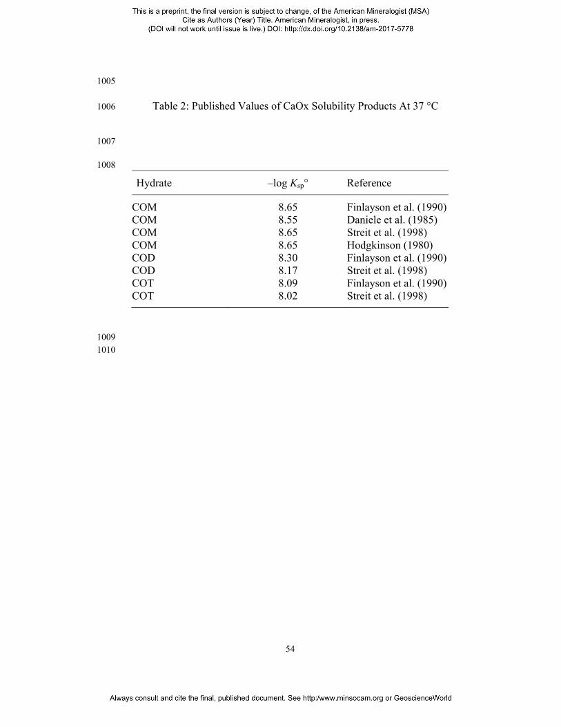
(Figure 6). In a recent review (Hummel et al., 2005), solubility products 332
for calcium oxalates have been critically evaluated and extrapolated to I 333
= 0 (infinite dilution) using the SIT approach for the calculation of 334
activity coefficients (see section below). In addition to the increase of 335
CaOx solubility products with ionic strength as an effect of changing 336
activity coefficients, Figure 6 compares selected experimental data for 337
NaCl and KCl background electrolytes with recent critical evaluations 338
(Hummel et al, 2005). A list of some values of solubility products at I = 0 339
(Ksp°) published for the calcium oxalate hydrates is also shown in 340
Table 2. 341
Page 18
18
The nature of urine increases the achievable supersaturation of the salts 342
of interest well above the measured experimental value for solubility of 343
sparingly soluble salts. Many ions present in urine, such as Mg2+, citrate 344
and HPO42–, increase the solubility of CaOx by forming complexes with 345
either the Ca2+ or the C2O42– ions (Hodgkinson, 1980; Gutzow et al., 346
1993; Streit et al., 1998). Such complex formation generally results in an 347
additional increase in solubility. 348
The solubility of CaOx in a 0.20 mol/L sodium chloride solution is 1.94 349
× 10-4 mol/L, while in an artificial urine solution this is increased to 2.98 350
× 10-4 mol/L (Streit et al., 1998). 351
Page 19
19
For increasing concentrations of Mg2+ of 2.5, 5.0 and 7.5 mmol/L, the 352
respective ion product values for calcium oxalate are 1.0 × 10⁻⁷, 1.35 × 353
10−7 and 2.02 × 10⁻⁷ (Elliot and Ribeiro, 1973). Thus, urine contains 354
much higher concentrations of calcium and oxalate in solution than are 355
present in a saturated solution of calcium oxalate in water. In addition to 356
the dissolved salts, urine contains macromolecules. A number of 357
proteins and similar substances are secreted into the tubule by the 358
tubular cells (Söhnel and Grases, 1995; Tiselius, 1997a; Højgaard and 359
Tiselius, 1999). Tamm-Horsfall Protein (THP) is the most abundant 360
protein in human urine (Devuyst et al, 2005), with a concentration of the 361
order of magnitude of 10−7 mol/L in urine (Glauser et al, 2000; Laube 362
et al, 2001); it therefore cannot bind a significant amount of calcium. 363
While the macromolecules have sometimes been shown to promote 364
crystal nucleation, they are also known to inhibit crystal growth 365
(Rodgers et al., 1993). This process is mainly via the action of binding to 366
calcium-rich centres on the crystal surface (Tiselius, 2011b). Phytic 367
acid, present at micromolar levels in urine, is another substance that 368
has been shown to inhibit the growth of calcium oxalate crystals (Söhnel 369
and Grases, 1995), presumably by mechanisms akin to those mentioned 370
above for macromolecules. 371
Page 20
20
As already mentioned, the risk of stone formation can be determined 372
from the supersaturated state of the stone forming salts. Methods involve 373
measurement of ratios of concentrations of certain substances (Tiselius, 374
1997b) and determining how much is required to initiate precipitation 375
following the addition of the ions of interest to a sample of urine. For 376
example, adding calcium chloride or ammonium oxalate induces CaOx 377
precipitation (Luptak et al., 1994; Laube et al, 2000). 378
Quantitative Chemical Speciation Modelling 379
It is now widely accepted that the application of geochemical techniques 380
to "predict, identify and quantify minerals in low temperature aqueous 381
environments can be adapted" to the study of biofluids (Taunton et al, 382
2010). Thermodynamic calculations have been used routinely to 383
investigate the state of saturation of substances in urine (Linder and 384
Little, 1986; Asplin et al., 1996; Parks et al., 1997; Laube et al., 2002; 385
Königsberger and Tran-Ho, 1997; Milosevic et al., 1998; Rodgers et al., 386
2006; Pak et al., 2009; Rodgers et al., 2011) providing a useful 387
alternative to the induction of precipitation by substance addition. This 388
technique uses measurements of substance concentrations to estimate 389
free ion concentrations and supersaturation states so that risk can be 390
evaluated. 391
Page 21
21
The most widely used program to perform such calculations has been 392
EQUIL. This program was developed by Finlayson in 1977 (Finlayson, 393
1977; Brown and Purich, 1992). EQUIL2 is an updated version of this 394
program, which included translation from FORTRAN to BASIC, 395
making it available on a larger number of computers (Werness et al, 396
1985). Enhancements led to a newer version, EQUIL93 (Brown et al, 397
1994), which increased the number of ions and complexes that could be 398
represented and updated the thermodynamic database with data from the 399
Martell and Smith critically evaluated compilation of equilibrium 400
constants (Martell and Smith, 1974-1982), and other sources. 401
A chemical speciation program (confusingly, also called EQUIL) was 402
developed by Ting-Po and Nancollas in 1972 (Ting-Po and Nancollas, 403
1972), but this program appears to be unrelated to that described above. 404
Another program frequently used in urolithiasis research is the Joint 405
Expert Speciation System (JESS) software package (May and Murray, 406
1991a, 1991b). 407
In addition to EQUIL and JESS, other software has also occasionally 408
been used. Prywer and Mielniczek-Brzoska (2016) used HySS 409
(Alderighi et al, 1999) to model chemical speciation in the formation of 410
struvite kidney stones resulting from bacterial infection. 411
Page 22
22
412
Grases et al. (1997) first used JESS to model the supersaturation of 413
calcium and magnesium phosphates in artificial urine in 1997. In this 414
work, citrate and oxalate were considered in addition to the inorganic 415
salts. All possible complexes whose formation constants were available 416
in the JESS thermodynamic database were thus considered. In addition, 417
one of the then built-in activity coefficient models of JESS was used 418
(Davies equation). Considering the number of species (213), reactions 419
(265) and thermodynamic quantities (more than 4000, including 420
enthalpy, free energy and heat capacity values), this urine model was 421
possibly the largest at that time. After incorporating solubility constants 422
(log Ks0) determined in their laboratory (Streit et al, 1998), Königsberger 423
and Tran-Ho (Königsberger and Tran-Ho, 1997) employed this model to 424
calculate solubilities of the three calcium oxalate hydrates in NaCl(aq) 425
and urine-like liquors. Subsequently, the JESS urine model was 426
extended to include uric acid and cystine (Königsberger and 427
Königsberger, 2001), resulting in a considerable increase in the number 428
of species (280), reactions (380), and thermodynamic quantities (some 429
7200, mainly equilibrium constants but also standard potentials, Gibbs 430
energies, enthalpies, and heat capacities). The effect of complexing 431
species such as citrate and magnesium ions on calcium oxalate 432
Page 23
23
solubilities helped to identify conditions for reducing its supersaturation 433
in urine (Königsberger and Tran-Ho, 1997; Königsberger and 434
Königsberger, 2001). Significant effects of urine composition on uric 435
acid (Königsberger and Wang, 1999) and cystine (Königsberger et al, 436
2000) solubilities were not predicted nor found experimentally. 437
Furthermore, the JESS modelling suggested regions of thermodynamic 438
and kinetic control of calcium oxalate crystallisation that correlated well 439
with a clinical test (Grases et al, 2000). 440
In order to calculate the degree of saturation of a dissolved substance, 441
values for the ion activity coefficients have to be determined. A number 442
of empirical models can be used for this. The Davies equation (1) is an 443
extension of Debye-Hückel theory without adjustable parameters, it has 444
no theoretical foundation, but often works fairly well for ionic strengths 445
up to 0.1 mol kg–1 (Grenthe et al, 1997). 446
At 25º C, 447
log = −0.51 − 0.3 (1) 448
where: 449
γi is the activity coefficient of ion i 450
Page 24
24
Zi is the charge of ion i 451
Im is the ionic strength on molal scale. 452
Specific Ion Interaction Theory (SIT), Equation (2), is a semi-empirical 453
model based on Brønsted-Guggenheim-Scatchard models. It contains a 454
number of parameters that have some theoretical basis (Grenthe et al, 455
1997). 456
log = − . + ∑ ( , ) (2) 457
where: 458
A is the Debye-Hückel parameter for activity coefficient 459
ε(i,k) are interaction coefficients for oppositely charged aqueous ions i 460
and k; 461
mk is the molality of ion k. 462
The current method used by JESS is the SIT-like equation, shown in 463
Equation (3). 464
log ′ = log + ∆ √. √ + (3) 465
where: 466
Page 25
25
K0 is the equilibrium constant at infinite dilution 467
K' is the conditional equilibrium constant at finite ionic strength 468
A and ΔZ2 are the Debye-Hückel parameter and a function of the ionic 469
charges respectively 470
B is a temperature dependent parameter 471
(May, 2000) 472
The JESS software package calculates log(SI) values: 473
Ksp
IAPSI log)log( = 474
where 475
IAP is the ion-activity product 476
Ksp is the solubility product 477
Using these methods, estimates of supersaturation of the calcium 478
phosphate and calcium oxalate compounds have been calculated for 479
final urine, as well as for the different nephron segments (Robertson and 480
Nordin, 1976; Luptak et al., 1994; Tiselius, 1997; Rodgers et al., 2011; 481
Page 26
26
Robertson, 2015). It has been determined that for the calcium 482
phosphates, supersaturation and therefore the risk of crystallization is 483
higher in the proximal and distal tubules (Luptak et al., 1994; Asplin 484
et al., 1996; Tiselius, 1997a; Rodgers et al., 2011; Robertson, 2015). For 485
calcium oxalate, supersaturation levels are higher in the collecting duct 486
(Luptak et al., 1994; Rodgers et al., 2011; Robertson, 2015). The 487
variation in the values on which these calculations are based, as 488
discussed above, indicates that the quantitative results from such 489
calculations cannot be regarded as exact. In general, computational 490
models should be used to gain insight into the working of a process, 491
rather than in attempts to obtain individual numerical results that can be 492
taken as the definitive answer to the problem (May, 2015). 493
Using published data about concentrations of the solutes in the different 494
nephron segments (Rodgers et al., 2011), shown in Table 3, some 495
calculations performed using the JESS software package give the values 496
shown in Table 4. 497
While some earlier work concentrated on the behaviour of minerals 498
under simulated lung fluid conditions, with a focus of assessing mineral 499
durability and secondary mineral formation (Taunton et al, 2010) we 500
prefer to concentrate instead on the implications of Ostwald's Rule of 501
Page 27
27
Stages (Chung et al., 2009), which is known to work well for systems 502
which reach equilibrium too rapidly to apply conventional reaction path 503
analysis, which is in constrast to the long-term time-frame for minerals 504
resident in the lungs. 505
The results in Table 4 indicate that brushite is the supersaturated 506
substance with the lowest SI value under the conditions in the distal 507
portion of the collecting duct and thus, brushite seems from Ostwald’s 508
Rule of Stages to be the substance most likely to precipitate. Brushite 509
has indeed been found in some kidney stones (Grases and Costa-Bauza, 510
2006), particularly in overgrowths of a calculus that had ‘plugged’ the 511
duct of Bellini (Evan et al., 2015). The core of that specimen contained 512
hydroxyapatite, the most stable calcium phosphate phase, which may 513
well have been formed by recrystallization of brushite. Another instance 514
of stone plugging in the duct of Bellini contained COD (Grases et al., 515
2016), which is less stable than COM. Both of these stones were 516
associated with renal tissue damage probably acting as heterogeneous 517
nucleant. We conclude that the crystallyzation of metastable phases 518
according to Ostwald’s Rule of Stages can be applied to the growth of 519
stones on ‘Randall’s Plugs’, which are usually associated with excessive 520
supersaturation with respect to the stable phases (Khan and Canales, 521
2015). The metastable phases brushite and COD were also found in 522
Page 28
28
cavities of low urodynamic efficacy, in which heterogeneous nucleants 523
(organic matter and calcium phosphate crystals respectively) become 524
trapped and high supersaturation is maintaned (Grases and Costa-Bauza, 525
2006). 526
In contrast, the growth of papillary stones induced by Randall’s Plaque 527
inevitably proceeds even at the low supersaturation prevailing in urine of 528
normal composition. Such stones contain the stable phases HAP and 529
COM (Grases et al., 2015; Grases et al., 2016). However, (metastable) 530
amorphous calcium phosphates were found as possible precursors of 531
Randall’s Plaque (Evan, 2010), which indicates high supersaturation and 532
the applicability of Ostwald’s Rule of Stages in interstitial tissue. 533
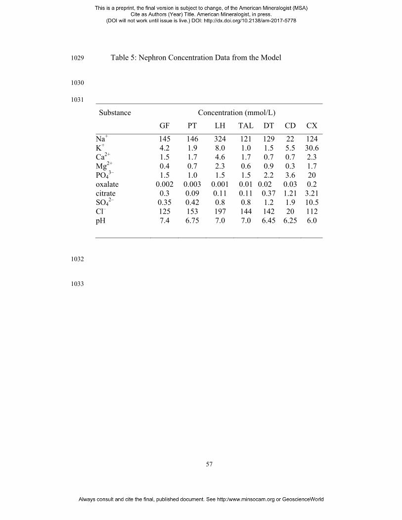
As a result of these findings, we have developed a model to calculate the 534
concentration changes along the path of the nephron. The calculations 535
are based on published values of concentration, reabsorption and 536
excretion of different substances within the sections of the nephron 537
(Luptak et al, 1994; Asplin et al, 1996; Kok, 1997; Tiselius, 1997; 538
Hojgaard and Tiselius, 1999; Rodgers et al, 2011; Rodgers et al, 2013). 539
Output from the model for normal kidney filtration is shown in Table 5. 540
The model allows different conditions to be investigated by changing 541
input values which respresent blood plasma concentrations of the 542
Page 29
29
substances under consideration and changes in how much of a particular 543
substance is reabsorbed in a given nephron section. For example, it has 544
been discovered that calcium oxalate stone formers often have reduced 545
calcium reabsorption in the proximal tubule (Coe et al, 2011; Worcester 546
et al, 2013), and the model allows simulation of such scenarios. 547
Using calculated concentrations for the different nephron sections 548
log(SI) values for substances of interest can be determined using JESS. 549
It should be stressed again that JESS calculates the chemical speciation, 550
and hence log(SI), by considering all complex species whose formation 551
constants are contained in its database. 552
Figure 7 shows log(SI) values for brushite for three different senarios, 553
normal kidney filtration with a plasma calcium concentration of 1.5 554
mmol/L and oxalate concentration of 1.75 µmol/L, a high plasma 555
calcium concentration of 3.0 mmol/L, and reduced calcium reabsorption 556
in the proximal tubule together with the increased plasma calcium 557
concentration. The second two situations result in an increased SI for the 558
brushite all along the nephron. Log(SI) for brushite is above zero in the 559
loop of Henle and the collecting duct, indicating an increased risk of 560
precipitation in those locations. 561
562
Page 30
30
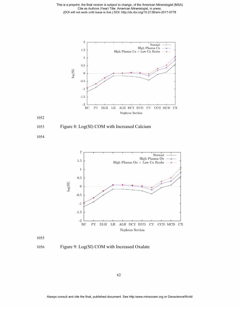
Figure 8 shows the log(SI) values for the calcium oxalate monohydrate 563
for the same three conditions described above. This shows an increased 564
risk of precipitation towards the end of the nephron. 565
Figure 9 shows the log(SI) values for the calcium oxalate monohydrate 566
for normal, a high plasma concentration of oxalate of 3.0 µmol/L, and 567
the same high value of plasma oxalate together with the reduction in 568
reabsorption of calcium in the proximal tubule. 569
Both these simulations show log(SI) COM increasing in the proximal 570
tubule to reach a peak in the ascending loop of Henle before decreasing 571
toward the distal tubule, and then increasing steadily in the collecting 572
duct. The risk of crystal formation, where log(SI) > 0, is only seen with 573
higher than normal calcium or oxalate plasma levels, and increased with 574
a pathological reabsorption profile. This is in good agreement with the 575
results of Robertson (2015). 576
JESS Version 8.3 used in this work leads to the same general conclusion 577
as the previous work by Rodgers et al. (Rodgers et al., 2011), although 578
there are small quantitive differences due to changes in the way weak ion 579
associations are handled (May, 2015). The absolute values of the 580
saturations calculated by different JESS versions change to a small 581
extent over time, but in almost all cases their pattern through the 582
Page 31
31
different compartments is the same and no large discrepancies have been 583
found. It can thus be concluded that the changes are due to updates that 584
have been made to the database. Further information about how JESS 585
approaches the selection of equilibrium constants is given in The JESS 586
Primer, available via the website http://jess.murdoch.edu.au (May, 587
2015). 588
However, this issue is complicated by a number of theoretical and 589
practical factors. These include the fact that protein binding of calcium 590
ions is still not well characterised (Taunton et al, 2010; Holt et al., 2014), 591
calcium buffering and the observation that coating of seeds by proteins 592
can cause inhibition of crystal growth. Another factor is that the 593
saturation state of relevant minerals may alter under physiological 594
conditions (Miller et al, 1958; Streit et al, 1998). Note also that in this 595
regard chemical speciation calculations using ion association 596
frameworks have well known limitiations (May, 2015). The absolute SI 597
values calculated by JESS are therefore interesting but need to be 598
interpreted with caution. However, it is clear that their changes across 599
the nephron are significant and must be taken into account. 600
601
Page 32
32
602
Implications 603
604
Kidney stone formation is a serious medical problem for which the 605
underlying mechanisms are poorly understood. In the human body, 606
hydroxyapatite must be kept supersaturated to allow the processes that 607
form bones and teeth to operate. As a result of this, a number of calcium 608
phosphate minerals tend to be supersaturated in various biofluids. A 609
delicate biological balance is therefore required between preventing the 610
formation of solid structures where and when they are harmful and 611
producing them as required. 612
613
Understanding kidney stone formation requires the investigation of 614
mineral formation in a complex environment, where the changes that are 615
taking place are often very difficult to observe directly. Interdisciplinary 616
research in particular can be of great value in medical science. 617
Combining techniques from biology, geochemistry, thermodynamics, 618
mathematics and computer science, comprehensive models can now be 619
developed to investigate and explain processes taking place in the 620
human body. Computer modelling techniques are a powerful tool that 621
Page 33
33
can be utilized to overcome the problems with experimental in vivo and 622
in vitro investigations. Thermodynamic calculations have been shown to 623
be useful, especially in improving understanding of the processes 624
involved in kidney stone formation. Much insight can be gained into the 625
processes taking place and the interactions between them. As more and 626
better data are included in the databases that these computer models use, 627
the results obtained from the models can be expected to improve. Basic 628
mineralogical theory and experiments provide the pre-requisite building 629
blocks for these databases. The modelling is then able to combine theory 630
and experiment to simulate the complex interactions between the 631
components of the system being investigated. Similar issues arise in 632
geochemical complex aqueous environments, where metastable 633
equilibria and kinetic restrictions often prevail. Insights obtained by 634
geochemical modellers may therefore also be helpful in improving the 635
computational area of kidney stone research. 636
637
Determining the details of the initial solid phase formation in the loop of 638
Henle, or distal tubule, would probably be of greatest value as this 639
information would help to show how the risk of this initial particle 640
formation can be reduced. Thus, investigation of crystal seed formation 641
is likely to be a key area for future research. Better understanding of the 642
Page 34
34
thermodynamics, kinetics and morphology of the minerals involved is 643
therefore needed to improve prospects in this medical arena. 644
645
Acknowledgements 646
The authors would like to thank the reviewers Allen Rodgers and Greg 647
Druschel for their suggestions for the improvement of this manuscript. 648
References 649
Abdel-Halim, R. (2005). Urolithiasis in adults: Clinical and 650
biochemical aspects. Saudi Medical Journal, 26:705 – 713. 651
Alderighi, L., Gans, P., Ienco, A., Peters, D., Sabatini, A., and 652
Vacca, A. (1999). Hyperquad simulation and speciation (HySS): a 653
utility program for the investigation of equilibria involving soluble 654
and partially soluble species. Cordination Chemistry Reviews, 655
184:311 – 318. 656
Asplin, J.R., Mandel, N.S., and Coe, F.L. (1996). Evidence for 657
calcium phosphate supersaturation in the loop of Henle. American 658
Journal of Physiology, 270:F604 – F613. 659
Page 35
35
Atherton, J.C. (2006a). Function of the nephron and the formation 660
of urine. Anaesthesia and Intensive Care Medicine, 7:221–226. 661
Atherton, J.C. (2006b). Renal blood flow, glomerular filtration and 662
plasma clearance. Anaesthesia and Intensive Care Medicine, 663
7:216–220. 664
Bell, G.H., Davidson, J.N., and Scarborough, H. (1968). Textbook 665
of Physiology and Biochemistry. E and S Livingstone Ltd, 666
Edinburgh and London, 7th edition. 667
Brown, C.M., Ackerman, D.K., and Purich, D.L (1994). EQUIL93: 668
A tool for experimental and clinical urolithiasis. Urological 669
Research, 22:119 – 126. 670
Brown, C.M., and Purich, D.L. (1992). Physical-chemical 671
processes in kidney stone formation. In F. L. Coe and M. J. Favus, 672
editors, Disorders of Bone and Mineral Metabolism, chapter 29, 673
pages 613 – 624. Raven Press Ltd, New York. 674
Chung, S.-Y., Kim, Y.-M., Kim, J.-G., and Kim, Y.-J. (2009) 675
Multiphase transformation and Ostwald’s rule of stages during 676
crystallization of a metal phosphate. Nature Physics, 5:68 – 73. 677
Page 36
36
Coe, F. L., Evan, A., and Worcester, E. (2011). 678
Pathophysiology-based treatment of idiopathic calcium kidney 679
stones. Clinical Journal of the American Society of Nephrology, 680
6:2083 – 2092. 681
Daniele, P. G., Sonego, S., Rozani, M., and Marangella, M. (1985). 682
Ionic strength dependence of formation constants. Part 8. Solubility 683
of calcium oxalate monohydrate and calcium hydrogenphosphate 684
dihydrate in aqueous solution, at 37 ∘ C and different ionic 685
strengths. Annali di Chimica, 75:245 – 251. 686
Devuyst. O., Dahan, K., and Pirson, Y. (2005) Tamm-Horsfall 687
protein or uromodulin: new ideas about an old molecule. 688
Nephrology Dialysis Transplantation, 20:1290 – 1294. 689
Diem, K., and Lentner, C., editors (1970). Documenta Geigy 690
Scientific Tables. JR Geigy SA, Basle, 7th edition. 691
Eisner, B.H., Eisenberg, M.L., and Stoller, M.L. (2010). The 692
relationship between body mass index and quantitative 24-hour 693
urine chemistries in patients with nephrolithiasis. Urology, 75:1289 694
– 1293. 695
Elliot, J.S., and Ribeiro, M.E. (1973). The effect of varying 696
Page 37
37
concentrations of calcium and magnesium upon calcium oxalate 697
solubility. Investigative Urology. 10:295 – 297. 698
Evan, A.P. (2010). Physiopathology and etiology of stone 699
formation in the kidney and the urinary tract. Pediatric Nephrology, 700
25:831–841. 701
Evan, A.P., Worcester, E.M., Coe, F.L., Williams, J.J., and 702
Lingeman, J. E. (2015). Mechanisms of human kidney stone 703
formation. Urolithiasis, 43 (Supplement 1):S19 – S32. 704
Finlayson, B. (1977). Calcium stones: Some physical and clinical 705
aspects. In D. S. David, editor, Calcium Metabolism in Renal 706
Failure and Nephrolithiasis Perspectives in nephrology and 707
hypertension), chapter 10, pages 337 – 382. Wiley and Sons, New 708
York. 709
Finlayson, B. (1978). Physicochemical aspects of urolithiasis. 710
Kidney International, 13:344–360. 711
Finlayson, B., and Reid, F. (1978). The expectation of free and 712
fixed particles in urinary stone disease. Investigative Urology, 713
15:442 - 448. 714
Page 38
38
Finlayson, B., Khan, S.R., and Hackett, R.L. (1990). Theoretical 715
chemical models of urinary stones. In J.E.A. Wickham and A.C. 716
Buck., editors, Renal Tract Stone, chapter 10, pages 133 – 147. 717
Churchill Livingstone, Edinburgh. 718
Gamsjäger, H., and Königsberger, E. (2003). Solubility of sparingly 719
soluable ionic solids in liquids. In G. Hefter and R. Tomkins, 720
editors, The Experimental Determination of Solubilities, chapter 721
4.2, pages 315 – 358. John Wiley and Sons Ltd, Chichester. 722
Glauser, A., Hochreiter, W., Jaeger, P., and Hess, B. (2000). 723
Determinants of urinary excretion of Tamm-Horsfall protein in 724
non-selected kidney stone formers and healthy subjects. 725
Nephrology Dialysis Transplantation, 15:1580– 1587. 726
Grases, F., and Costa-Bauza, A. (2006). Mechanisms of renal and 727
salivary calculi formation and development. In E. Königsberger and 728
L-C. Königsberger, editors, Biomineralization - Medical Aspects 729
of Solubility, chapter 2, pages 39 – 69. Wiley, Chichester, UK. 730
Grases, F., Costa-Bauza, A., and Garcia-Ferragut, L. (1998). 731
Biopathological crystallization: a general view about the 732
Page 39
39
mechanisms of renal stone formation. Advances in Colloid and 733
Interface Science, 74:169 – 194. 734
Grases, F., Costa-Bauza, A., Bonarriba, C.R., Pieras, E.C., 735
Fernández, R. A., and Rodríguez, A. (2015). On the origin of 736
calcium oxalate monohydrate papillary renal stones. Urolithiasis, 737
43 (Supplement 1):S33 – S39. 738
Grases, F., Costa-Bauza, A., Gomila, I., Ramis, M., Garcia-Raja, 739
A., and Prieto, R. M. (2012). Urinary pH and renal lithiasis. 740
Urological Research, 40:41 – 46. 741
Grases, F., Costa-Bauza, A., Königsberger, E. and Königsberger, 742
L-C. (2000). Kinetic versus thermodynamic actors in calcium renal 743
lithiasis. International Urology and Nephrology , 32:19-27. 744
Grases, F., Söhnel, O., and Costa-Bauza, A. (1999). Renal stone 745
formation and development. International Urology and 746
Nephrology, 31:591 – 600. 747
Grases, F., Söhnel, O., and Costa-Bauza, A., Servera, A., and 748
Benejam, J. (2016). A case of Randall’s Plugs associated to calcium 749
oxalate dihydrate calculi. Urology Case Reports, 7:37–38. 750
Page 40
40
Grases, F., Villacampa, A.I., Söhnel, O., Königsberger, E., and 751
May, P.M. (1997). Phosphate composition of precipitates from 752
urine-like liquors. Crystal Research and Technology, 32:707 – 715. 753
Grenthe, I., Plyasunow, A. V., and Spahiu, K. (1997). Modelling in 754
aquatic chemistry. In I. Grenthe and I. Puigdomenech, editors, 755
Modelling in Aquatic Chemistry, chapter IX, pages 325 – 426. 756
Nuclear Energy Agency, Organisation for Economic Co-operation 757
and Development. 758
Gutzow, I., Atanassova, S., Budevsky, G., and Neykov, K. (1993). 759
Solubility, inhibited growth and dissolution kinetics of calcium 760
oxalate crystals in solutions, containing hippuric acid. Urological 761
Research, 21:181 – 185. 762
Guyton, A. C., and Hall, J. E. (2000). Textbook of Medical 763
Physiology. WB Saunders Company, Philadelphia, 10th edition. 764
Hammarsten, G. (1929). On calcium oxalate and its solubility in the 765
presence of inorganic salts with special reference to the occurrence 766
of oxaluria. Comptes Rendus des Travaux du Laboratoire 767
Carlsberg, 17:1 – 85. 768
Page 41
41
Hesse, A., Classen, A., Knoll, M., Timmermann, F., and 769
Vahlensieck, W. (1986). Dependence of urine composition on the 770
age and sex of healthy subjects. Clinica Chimica Acta, 160:79–86. 771
Hodgkinson, A. (1980). Solubility of calcium oxalate in human 772
urine, simulated urine, and water. Investigative Urology, 18:123 – 773
126. 774
Højgaard, I., and Tiselius, H.-G. (1999). Crystallization in the 775
nephron. Urological Research, 27:397 – 403. 776
Holt, C., Lenton, S., Nylander, T., Sørensen, E. S., and Teixeira, 777
S. C. (2014). Mineralisation of soft and hard tissues and the stability 778
of biofluids. Journal of Structural Biology, 185:383 – 396. 779
Hughes, P. (2007). Kidney stones epidemiology. Nephrology, 780
12:S26 – S30. 781
Hummel, W., Anderegg, G., Rao, L., Puigdomenech, I., and 782
Tochiyama, O. (2005). Chemical Thermodynamics of Compounds 783
and Complexes of U, Np, Pu, Am, Tc, Se, Ni and Zr with Selected 784
Organic Ligands. OECD-NEA, Elsevier, Amsterdam. 785
Kerr, J. (1999). Atlas of Functional Histology. Mosby, London. 786
Page 42
42
Khan, S.R., and Canales, B.K. (2015) Unified theory on the 787
pathogenesis of Randall’s plaques and plugs. Urolithiasis, 43 788
(Supplement 1):S109–S123. 789
Knight, J., Jaing, J., Assimos, D., and Holmes, R. (2006). 790
Hydroxyproline ingestion and urinary oxalate and glycolate 791
excretion. Kidney International, 70:1929 – 1934. 792
Kok, D. J. (1997). Intratubular crystallization events. World Journal 793
of Urology, 15:219 – 228. 794
Kok, D. J., and Khan, S. R. (1994). Calcium oxalate nephrolithiasis, 795
a free or fixed particle disease. Kidney International, 46:847 – 854. 796
Königsberger, E., and Königsberger, L.-C. (2001). Thermodynamic 797
modeling of crystal deposition in humans. Pure and Applied 798
Chemistry, 73:785-797. 799
Königsberger, E., and Königsberger, L.-C. (2006). Solubility 800
phenomena related to normal and pathological biomineralization 801
processes. In E. Königsberger and L-C. Königsberger, editors, 802
Biomineralization - Medical Aspects of Solubility, chapter 1, pages 803
1 – 37. Wiley, Chichester, UK. 804
Page 43
43
Königsberger, E., and Tran-Ho, L.-C. (1997). Solubility of 805
substances related to urolithiasis - experiments and computer 806
modelling. Current Topics in Solution Chemistry, 2:183 – 202. 807
Königsberger, E., and Wang, Z. (1999). Solubility of uric acid in 808
salt solutions and artificial urine. Monatshefte fur Chemie, 809
130:1067 - 1073. 810
Königsberger, E. and Wang, Z. and Königsberger, L.-C. (2000). 811
Solubility of L-Cystine in NaCl and Artificial Urine Solutions. 812
Monatshefte fur Chemie, 131:39 - 45. 813
Kumar, S., and Muchmore, A. (1990). Tamm-Horsfall 814
protein-uromodulin (1950-1990). Kidney International, 37:1395 – 815
1401. 816
Laube, N., Glatz, S., and Hesse, A. (2001). The relation of urinary 817
Tamm-Horsfall-Protein on CaOx-crystallization under the scope of 818
the Bonn-Risk-Index. Urological Research, 29:45–49. 819
Laube, N., Labedzke, V., Hergarten, S., and Hesse, A. (2002). 820
Determination of urinary calcium-oxalate formation risk with 821
BONN-Risk-Index and EQUIL applied to a family. Journal of 822
Chemical Information and Computer Sciences, 42:633 – 639. 823
Page 44
44
Laube, N., Schneider, A., and Hesse, A. (2000). A new approach to 824
calculate the risk of calcium oxalate crystallization from unprepared 825
native urine. Urological Research, 28:274 – 280. 826
Linder, P. W., and Little, J. C. (1986). Prediction by computer 827
modelling of the precipitation of stone-forming solids from urine. 828
Inorganica Chimica Acta, 123:137 – 145. 829
Luptak, J., Bek-Jensen, H., Fornander, A.-M., Hojgaard, I., Nilsson, 830
M.-A., and Tiselius, H. (1994). Crystallization of calcium oxalate 831
and calcium phosphate at superstauration levels corresponding to 832
those in different parts of the nephron. Scanning Microscopy, 8:47 833
– 62. 834
Martell, A.E., and Smith, R.M. (1974-1982) Critical stability 835
constants, vols 1-6. Plenum Press, New York. 836
May, P., and Murray, K. (1991a). JESS, A Joint Expert Speciation 837
System - I raison d’être. Talanta, 38:1409 – 1417. 838
May, P., and Murray, K. (1991b). JESS, A Joint Expert Speciation 839
System - II the thermodynamic database. Talanta, 38:1419 – 1426. 840
Page 45
45
May, P.M. (2000). A simple, general and robust function for 841
equilibria in aqueous electrolyte solutions to high ionic strength and 842
temperature. Chemical Communications, pages 1265 – 1266. 843
May, P.M. (2015). JESS at thirty: Strengths, weaknesses and future 844
needs in the modelling of chemical speciation. Applied 845
Geochemistry 55:3-16. 846
Miller, G.H., Vermeulen, C.W., and Moore, J.D. (1958). Calcium 847
oxalate solubility in urine: Experimental urolithiasis XIV. The 848
Journal of Urology, 79:607 – 612. 849
Milosevic, D., Batinic, D., Konjevoda, N.B.P., Stambuk, N., 850
Votava-Raic, A., Fumic, V. B. K., Rumenjak, V., 851
Stavljenic-Rukavina, A., Nizic, L., and Vrljicak, K. (1998). 852
Determination of urine supersaturation with computer program 853
Equil 2 as a method for estimation of the risk of urolithiasis. Journal 854
of Chemical Information and Computer Sciences, 38:646 – 650. 855
Moe, O.W. (2006). Kidney stones: pathophysiology and medical 856
management. The Lancet, 367:333 – 344. 857
Moran, M.E. (2014). Urolithiasis: A Comprehensive History. 858
Springer, New York. 859
Page 46
46
Pak, C. Y. C., Maalouf, N. M., Rodgers, K., and Poindexter, J. R. 860
(2009). Comparison of semi-emperical and computer derived 861
methods for estimating urinary saturation of calcium oxalate. The 862
Journal of Urology, 182:2951 – 2956. 863
Parks, J.H., and Coe, F. L.,. (1996). The financial effects of kidney 864
stone prevention. Kidney International, 50:1706 – 1712. 865
Parks, J. H., Coward, M., and Coe, F. L. (1997). Correspondence 866
between stone composition and urine supersaturation in 867
nephrolithiasis. Kidney International, 51:894 – 900. 868
Prywer, J., and Mielniczek-Brzoska, E. (2016). Chemical equilibria 869
of complexes in urine. a contribution to the physicochemistry of 870
infectious urinary stone formation. Fluid Phase Equilibria, 425:282 871
– 288. 872
Rendina, D., De Filippo, G., De Pascale, F., Zampa, G., 873
Muscariello, R., De Palma, D., Ippolito, R., and Strazzullo, P. 874
(2013). The changing profile of patients with calcium 875
nephrolithiasis and the ascendancy of overweight and obesity: a 876
comparison of two patient series observed 25 years apart. 877
Nephrology Dialysis Transplantation, 28:iv146 – iv151. 878
Page 47
47
Robertson, W. G. (2004). Kidney models for calcium oxalate stone 879
formation. Nephron Physiology, 98:21 - 30. 880
Robertson, W. G. (2015). Potential role of fluctuations in the 881
composition of renal tubular fluid through the nephron in the 882
initiation of Randall’s plugs and calcium oxalate crystalluria in a 883
computer model of renal function. Urolithiasis, 43(Supplement 884
1):S93 – S107. 885
Robertson, W., and Nordin, B. (1976). Physio-chemical factors 886
governing stone formation. In D. I. Williams and G. D. Chisholm, 887
editors, Scientific Foundations of Urology, chapter 37, pages 254 – 888
267. London Heinemann Medical. 889
Robertson, W. G., Scurr, D. S., and Bridge, C. M. (1981). Factors 890
influencing the crystallisation of calcium oxalate in urine - critique. 891
Journal of Crystal Growth, 53:182 – 194. 892
Rodgers, A. (2006). The riddle of kidney stone disease: lessons 893
from Africa. Urological Research, 34:92 – 95. 894
Rodgers, A., Allie-Hamdulay, S., and Jackson, G. (2006). 895
Therapeutic action of citrate in urolithiasis explained by chemical 896
Page 48
48
speciation: increase in pH is the determinant factor. Nephrology 897
Dialysis Transplantation, 21:361–369. 898
Rodgers, A. L., Allie-Hamdulay, S., Jackson, G., and Tiselius, 899
H.-G. (2011). Simulating calcium salt precipitation in the nephron 900
using chemical speciation. Urological Research, 39:245–251. 901
Rodgers, A. L., Allie-Hamdulay, S., Jackson, G. E., and Durbach, I. 902
(2013). Theoretical modeling of the urinary supersaturation of 903
calcium salts in healthy individuals and kidney stone patients: 904
Precursors, speciation and therapeutic protocols for decreasing its 905
value. Journal of Crystal Growth, 382:67 – 74. 906
Rodgers, A. L., Ball, D., and Harper, W. (1993). Urinary 907
macromolecules are promoters of calcium oxalate nucleation in 908
human urine: turbidimetric studies. Clinica Chimica Acta, 220:125 909
– 134. 910
Romero, V., Akpinar, H., and Assismos, D. G. (2010). Kidney 911
stones: A global picture of prevalence, incidence, and associated 912
risk factors. Reviews in Urology, 12:e86 – e96. 913
Page 49
49
Saude, E. J., Adamko, D., Rove, B. H., Marrie, T., and Sykes, B. D. 914
(2007). Variation of metabolites in normal human urine. 915
Metabolomics, 3:439 – 451. 916
Sawada, K. (1997). The mechanisms of crystallization and 917
transformation of calcium carbonates. Pure and Applied Chemistry, 918
69:921 – 928. 919
Siener, R., Jahnen, A., and Hesse, A. (2004). Influence of a mineral 920
water rich in calcium, magnesium and bicarbonate on urine 921
composition and the risk of calcium oxalate crystallization. 922
European Journal of Clinical Nutrition, 58:270 – 276. 923
Söhnel, O., and Grases, F. (1995). Calcium oxalate monohydrate 924
renal calculi. formation and development mechanism. Advances in 925
Colloid and Interface Science, 59:1 – 17. 926
Söhnel, O., and Grases, F. (2011). Supersaturation of body fluids, 927
plasma and urine, with respect to biological hydroxyapatite. 928
Urological Research, 39:429 – 436. 929
Soucie, J. M., Thun, M. J., Coates, R. J., William, M., and Austin, 930
H. (1994). Demographic and geographic variability of kidney 931
stones in the United States. Kidney International, 46:893 – 899. 932
Page 50
50
Streit, J., Tran-Ho, L.-C., and Königsberger, E. (1998). Solubility of 933
the three calcium oxalate hydrates in sodium chloride solutions and 934
urine-like liquors. Monatshefte für Chemie, 129:1225–1236. 935
Taunton, A. E., Gunter, M. E., Druschel, G. K., and Wood, S. A. 936
(2010). Geochemistry in the lung: Reaction-path modeling and 937
experimental examination of rock-forming minerals under 938
physiologic conditions. American Mineralogist, 95:1624 – 1635. 939
Taylor, E. N., and Curhan, G. C. (2007). Differences in 24-hour 940
urine composition between black and white women. Journal of the 941
American Society of Nephrology, 18:654–659. 942
Taylor, E. N., Stampfer, M. J., Mount, D. B., and Curhan, G. C. 943
(2010). DASH-Style diet and 24-hour urine composition. Clinical 944
Journal of the American Society of Nephrology, 5:2315 – 2322. 945
Thomas, B., and Hall, J (2005). Urolithiasis. Surgery, 23(4):129 – 946
133 947
Ting.-Po., I., and Nancollas, G.H. (1972). EQUIL - a general 948
computational method for the calculation of solution equilibria. 949
Analytical Chemistry, 44:1940 – 1950. 950
Page 51
51
. 951
Tiselius, H., Lindbäck, B., Fornander, A.-M., and Nilsson, M.-A. 952
(2009). Studies on the role of calcium phosphate in the process of 953
calcium oxalate crystal formation. Urological Research, 37:181 – 954
192. 955
Tiselius, H. G. (1997a). Estimated levels of supersaturation with 956
calcium phosphate and calcium oxalate in the distal tubule. 957
Urological Research, 25:153 – 159. 958
Tiselius, H. G. (1997b). Risk formulas in calcium oxalate 959
urolithiasis. World Journal of Urology, 15:175 – 186. 960
Tiselius, H.-G. (2011a). A hypothesis of calcium stone formation: 961
an interpretation of stone research during the past decades. 962
Urological Research, 39:231 – 243. 963
Tiselius, H.-G. (2011b). Who forms stones and why? European 964
Urology Supplements, 10:408 – 414. 965
Tiselius, H.-G. (2015). Should we modify the principles of risk 966
evaluation and recurrence preventive treatment of patients with 967
calcium oxalate stone disease in view of the etiologic importance of 968
Page 52
52
calcium phosphate? Urolithiasis, 43:S47 – S57. 969
Tomazic, B. B., and Nancollas, G. H. (1980). The kinetics of 970
dissolution of calcium oxalate hydrates. Investigative Urology, 971
18:97 – 101. 972
Werness, P. G., Duckworth, S. C., and Smith, L. H. (1979). 973
Calcium oxalate hydrate crystal growth. Investigative Urology, 974
17:230 – 233. 975
Williams, H. E. (1978). Oxalic acid and the hyperoxaluric 976
syndromes. Kidney International, 13:410 – 417. 977
Worcester, E. M., Bergsland, K. J., Gillen, D. L., and Coe, F. L. 978
(2013). Evidence for increased renal tubule and parathyroid gland 979
sensitivity to serum calcium in idiopathic hypercalciuria. American 980
Journal of Physiology. Renal Physiology, 305(6):F853 – F860. 981
982
Figure 1: Schematic Depicting the Nephron Structure and Function 983
Figure 2: Calcium Concentrations in the Nephron 984
Figure 3: Oxalate Concentrations in the Nephron 985
Figure 4: Phosphate Concentrations in the Nephron 986
Figure 5: pH Variation in the Nephron 987
Figure 6: Ionic strength dependence of Ksp for COM at 37 °C 988
Page 53
53
Figure 7: Log(SI) Brushite with Increased Calcium 989
Figure 8: Log(SI) COM with Increased Calcium 990
Figure 9: Log(SI) COM with Increased Oxalate 991
992
993
994 995 996 997
998
Table 1: Substance Concentrations in Urine. A:Rodgers et al. (2006), 999 B:Kok (1997), C:Diem and Lentner (1970), D:Siener et al. (2004) 1000
1001
1002
Substance Selected Selected Range References Concentration
(mmol/L) Reference (mmol/L)
Na+ 151 A 45 to 582 B K+ 32.0 A 20 to 260 B Ca2+ 2.25 A 0.5 to 7.5 B Mg2+ 3.35 A 0.5 to 12.5 B PO4
3– 19.9 A 5 to 75 B Cl– 104.9 A 118.2 to
236.5 C
oxalate 0.108 A 0.1 to 1 B sulfate 12.2 D 14.8 to 34.5 C citrate 2.0 A 0.1 to 7.5 B urea 338.3 C 206.7 to
469.2 C
1003 1004
Page 54
54
1005
Table 2: Published Values of CaOx Solubility Products At 37 °C 1006
1007
1008
Hydrate –log Ksp° Reference
COM 8.65 Finlayson et al. (1990) COM 8.55 Daniele et al. (1985) COM 8.65 Streit et al. (1998) COM 8.65 Hodgkinson (1980) COD 8.30 Finlayson et al. (1990) COD 8.17 Streit et al. (1998) COT 8.09 Finlayson et al. (1990) COT 8.02 Streit et al. (1998)
1009 1010
Page 55
55
1011
Table 3: Nephron Concentration Data from Rodgers et al. (2011) 1012
1013
1014
Substance Concentration (mmol/L) GF PT LH DTp DTd CDm CDd
Na+ 135 135 278 79 93 94 109 K+ 3.8 3.0 13.8 0.90 58.0 53.0 63.7 Ca2+ 1.50 2.78 3.47 1.32 0.94 1.60 4.50 Mg2+ 0.54 0.19 0.24 0.12 0.40 1.45 3.85 PO4
3– 0.80 0.80 1.00 1.00 3.34 12.1 32.3 oxalate 0.0015 0.01 0.013 0.013 0.04 0.12 0.32 citrate 0.07 0.09 0.11 0.11 0.37 1.21 3.21 SO4
2– 1.4 3.1 3.9 3.9 13.0 7.8 20.8 Cl– 139 139 293 101 145 146.6 170.0 pH 7.40 6.75 6.50-7.306.38-7.006.45-7.005.00-6.25 5.50-6.70 av pH 7.40 6.75 6.90 6.69 6.725 5.625 6.1
1015 1016
Page 56
56
1017 1018
1019
Table 4: log SI Values for the Stone Forming Salts 1020
1021
1022
Salt pH CaOx Bru HAP OCP
GF 7.40 -1.267 -0.592 9.043 1.754 PT 6.75 -0.236 -0.458 7.365 1.118 LH 6.50 -0.291 -0.643 5.972 0.230 LH 7.30 -0.294 -0.516 9.546 2.208 DTp 6.38 -0.247 -0.726 4.413 -0.788 DTp 7.30 -0.256 -0.425 8.973 1.944 DTd 6.45 -0.145 -0.547 4.716 -0.328 DTd 7.00 -0.176 -0.400 7.294 1.183 CDm 5.00 0.506 -0.921 -1.694 -4.096 CDm 6.25 0.438 0.035 5.970 1.172 CDd 5.50 1.084 0.184 4.212 0.550 CDd 6.70 0.853 0.681 9.961 4.182
1023
1024
1025
1026
1027
1028
Page 57
57
Table 5: Nephron Concentration Data from the Model 1029
1030
1031
Substance Concentration (mmol/L) GF PT LH TAL DT CD CX
Na+ 145 146 324 121 129 22 124 K+ 4.2 1.9 8.0 1.0 1.5 5.5 30.6 Ca2+ 1.5 1.7 4.6 1.7 0.7 0.7 2.3 Mg2+ 0.4 0.7 2.3 0.6 0.9 0.3 1.7 PO4
3– 1.5 1.0 1.5 1.5 2.2 3.6 20 oxalate 0.002 0.003 0.001 0.01 0.02 0.03 0.2 citrate 0.3 0.09 0.11 0.11 0.37 1.21 3.21 SO4
2– 0.35 0.42 0.8 0.8 1.2 1.9 10.5 Cl– 125 153 197 144 142 20 112 pH 7.4 6.75 7.0 7.0 6.45 6.25 6.0
1032
1033
Page 58
58
1034
Figure 1: Schematic Depicting the Nephron Structure and Function 1035
1036
Page 59
59
1037
Figure 2: Calcium Concentrations in the Nephron 1038
1039
1040
Figure 3: Oxalate Concentrations in the Nephron 1041
1042
Page 60
60
1043
Figure 4: Phosphate Concentrations in the Nephron 1044
1045
1046
Figure 5: pH Variation in the Nephron 1047
Page 61
61
1048
Figure 6: Ionic strength dependence of Ksp for COM at 37 °C 1049
1050
Figure 7: Log(SI) Brushite with Increased Calcium 1051
Page 62
62
1052
Figure 8: Log(SI) COM with Increased Calcium 1053
1054
1055
Figure 9: Log(SI) COM with Increased Oxalate 1056