REVISTA ESPAÑOLA DE REVISTA ESPAÑOLA DE Q uimioterapia Q uimioterapia ISSN: 0214-3429 Volumen 28 Número 2 Abril 2015 Páginas: 61-115 SPANISH JOURNAL OF CHEMOTHERAPY Publicación Oficial de la Sociedad Española de Quimioterapia
Transcript
R E V I S T A E S P A Ñ O L A D ER E V I S T A E S P A Ñ O L A D EQuimioterapiaQuimioterapiaISSN: 0214-3429Volumen 28Número 2Abril 2015Páginas: 61-115
SPANISH JOURNALOF CHEMOTHERAPY
Publicación Oficialde la Sociedad Españolade Quimioterapia
QuimioterapiaR E V I S T A E S P A Ñ O L A D E
Revista Española de Quimioterapia tiene un carácter multidisciplinar y está dirigida a todos aquellos profesionales involucrados en la epidemiología, diagnóstico, clínica y tratamiento de las enfermedades infecciosas
Fundada en 1988 por la Sociedad Española de Quimioterapia
Indexada enScience Citation IndexExpanded (SCI),Index Medicus (MEDLINE), Excerpta Medica/EMBASE,Índice Médico Español (IME), Índice Bibliográfico en Ciencias de la Salud (IBECS)
Secretaría técnica Dpto. de MicrobiologíaFacultad de MedicinaAvda. Complutense, s/n28040 [email protected] en Internet:www.seq.es
Publicidad y SuscripcionesSociedad Española de QuimioterapiaDpto. de MicrobiologíaFacultad de MedicinaAvda. Complutense, s/n28040 Madrid
Atención al clienteTeléfono 91 394 15 12Correo electró[email protected]
Consulte nuestra página webwww.seq.es
Publicación que cumple los requisitos desoporte válido
ISSN0214-3429
e-ISSN 1988-9518
Depósito LegalM-32320-2012
ComposiciónImpresos y Revistas, S.A.Herreros, 4228906 Getafe (Madrid)Arte y DiseñoVicente Aparisi (Edycom)
ImpresiónEspaña
Esta publicación se imprime en papel no ácido. This publication is printed in acid free paper.
LOPDInformamos a los lectores que, según la Ley 15/1999 de 13 de diciembre, sus datos personales forman parte de la base de datos de la Sociedad Española de Quimioterapia (si es usted socio)
Si desea realizar cualquier rectificación o cancelación de los mismos, deberá enviar una solicitud por escrito bien a la Sociedad Española de Quimioterapia
Reservados todos los derechos.Queda rigurosamente prohibida,sin la autorización escrita deleditor, la reproducción parcialo total de esta publicaciónpor cualquier medio oprocedimiento, comprendidos lareprografía y el tratamientoinformático, y la distribución deejemplares mediante alquiler opréstamo públicos, bajo lassanciones establecidas por la ley
Sociedad Española de Quimioterapia
QuimioterapiaR E V I S T A E S P A Ñ O L A D E
DirectorJ. Barberán López
Comité Editorial
F. Álvarez Lerma (Barcelona)F. Baquero Mochales (Madrid)
E. Bouza Santiago (Madrid)J. A. García Rodríguez (Salamanca)M. Gobernado Serrano (Valencia)
Consejo Editorial
G. Acuña (Chile)J. M. Aguado (Madrid)L. Aguilar (Madrid)J. I. Alós (Madrid)J. R. Azanza (Pamplona)J. Aragón (Las Palmas de Gran Canaria) A. Artero (Valencia)J. Campos (Madrid)F.J. Candel (Madrid)E. Cantón (Valencia)R. Cantón (Madrid)J. A. Capdevila Morell(Barcelona)E. Carreras (Barcelona)M. Casal (Córdoba)J. Castillo (Zaragoza) J. J. Castón (Ciudad Real)R. Cisterna (Bilbao)J. Cobo Reinoso (Madrid) J. Cordero (Madrid)P. Courvalin (Francia)J. L. del Pozo (Navarra)R. De la Cámara (Madrid)M. De la Rosa (Granada)J. De la Torre (Córdoba)A. Delgado (Bilbao)A. Domínguez-Gil Hurlé(Salamanca)
A. Ramos (Madrid)C. Ramírez Ronda (Estados Unidos)J. Reina (Palma de Mallorca)M. A. Ripoll (Ávila)E. Rodríguez Noriega (México)J. L. Rodríguez Tudela (Madrid)J. Sabbaj (Guatemala)M. Sabriá (Barcelona)M. Salavert (Valencia)B. Sánchez Artola (Madrid)J. I. Santos (México)M. A. Sanz (Valencia)M. Segovia (Murcia)R. Serrano (Madrid)P. M. Shah (Alemania)D. Sevillano (Madrid)A. Soriano (Barcelona)A. Tomasz (Estados Unidos)J. R. Toral Revuelta (Madrid)J. Tuells (Alicante)C. Vallejo (Oviedo)K. Ueno (Japón)J. Vila (Barcelona)J. Yuste (Madrid)
J. E. Losa Garcia (Madrid)J. R. Maestre Vera (Madrid)A. M. Martín Sánchez (Las Palmas)I. Martínez Gil (Madrid)L. Martínez Martínez (Santander)E. Maseda (Madrid)T. Mazzei (Italia)M. A. Menéndez (Madrid)R. Menéndez (Valencia)R. Meyer (Estados Unidos)P. Muñoz (Madrid)J. L. Muñoz Bellido (Salamanca)A. Navarro (Madrid)V. Navarro (Alicante)R. Negroni (Argentina)C. E. Nord (Suecia)A. Novelli (Italia)V. Olmo (Las Palmas)A. Orero (Madrid)R. Ortiz de Lejarazu (Valladolid)J. A. Oteo (Logroño)E. Palencia Herrejón (Madrid) J. Parra (Granada)A. Pascual Hernández (Sevilla)J. Pasquau (Sevilla)J. Pemán (Valencia)C. Pérez Giraldo (Badajoz)J. E. Perea (Sevilla)B. Pérez-Gorricho (Madrid)
J. Eiros (Valladolid)M. C. Fariñas Álvarez (Santander)C. Fariñas (Santander)S. M. Finegold (Estados Unidos)J. Fortún (Madrid)X. Garau (Barcelona)E. García Sánchez (Salamanca)I. García García (Salamanca)J. García Rodríguez (Madrid)J. E. García Sánchez (Salamanca)E. García Vázquez (Murcia)H. Giamarellou (Grecia)A. C. Gómez García (Badajoz)J. Gómez Gómez (Murcia)M. L. Gómez-Lus (Madrid)J. González del Castillo (Madrid)F. González Romo (Madrid)E. Gotuzzo (Perú)J. J. Granizo (Madrid)S. Grau (Barcelona)J. Guinea (Madrid)X. Guirao (Barcelona)N. Gutierrez Zufiaurre (Salamanca)J. Hernández Quero (Granada)J. P. Horcajada Gallego (Barcelona)R. Isturiz (Venezuela)J. Kosmidis (Grecia)H. Lecour (Portugal)J. Liñares (Barcelona)
J. Mensa Pueyo (Barcelona)J. J. Picazo de la Garza (Madrid)
J. Prieto Prieto (Madrid)B. Regueiro García (Santiago de Compostela)
A. Torres Martí (Barcelona)
Secretario de RedacciónLuis Alou Cervera
Sumario
Tigeciclina: 10 años de historia y en plena vigencia 61José Barberán, Santiago Salso, Almudena Alhambra
Análisis de la asociación entre genotipos y resistencia a 79múltiples antimicrobianos en aislados de Staphylococcus aureus resistente a meticilinaVicente Aguadero, Carmen González-Velasco, Ana Vindel, Miguel González-Velasco, Juan José Moreno
Detección de uropatógenos inusuales durante un periodo 86 de 3 años en un hospital regionalCristina Gómez-Camarasa, Carmen Liébana-Martos, José María Navarro-Marí, José Gutiérrez-Fernández
Mortalidad entre los portadores de Staphylococcus aureus resistente 92 a la meticilina en centros sociosanitariosEster Vendrell, Josep A Capdevila, Pilar Barrufet, Lluís Force, Goretti Sauca, Encarna Martínez, Elisabet Palomera, Mateu Serra-Prat, Jordi Cornudella, Annabel Llopis, MªAsunción Robledo, Crisóstomo Vázquez
Evaluación de métodos fenotípicos para la detección 98 de Staphylococcus aureus resistente a meticilinaGertrudis Horna, Lizeth Astocondor, Jan Jacobs, Coralith García
Diversidad genotípica y fenotípica en Enterococcus faecalis: 101 ¿Es la invasión en agar un marcador de patogenicidad?Fabio Cafini, Fernando Gómez-Aguado, María Teresa Corcuera, Carmen Ramos, Pedro Bas, Luis Collado, María Luisa Gómez-Lus, José Prieto
Aparición de Enterococcus faecium resistente a daptomicina 109 en un paciente crítico con sepsis intraabdominal postoperatoriaFrederic Ballester, Isabel Pujol, Pitter Cueto, Conxita Rovira, Simona Iftimie, Jordi Camps
Bacteriemia por Helicobacter cinaedi en paciente infectado 111 por virus hepatitis C tratado con triple terapiaBeatriz Lacruz, María Pilar Huarte, Gregorio Tiberio, Luis Torroba, Iván Quispe
Diagnóstico de paludismo en un hospital regional con asistencia 113 a inmigrantes y viajerosGemma Jiménez-Guerra, Consuelo Miranda-Casas, José María Navarro-Marí, José Gutiérrez-Fernández
Revisión
Cartas al Director
Originales
QuimioterapiaR E V I S T A E S P A Ñ O L A D E
Volumen 28Número 2Abril 2015
Contents
QuimioterapiaR E V I S T A E S P A Ñ O L A D E
Volume 28Number 2April 2015
10 years of tigecycline and still in full force 61José Barberán, Santiago Salso, Almudena Alhambra
An analysis of the association between genotype and antimicrobial 79 resistance in methicillin-resistant Staphylococcus aureus clinical isolatesVicente Aguadero, Carmen González-Velasco, Ana Vindel, Miguel González-Velasco, Juan José Moreno
Detection of unusual uropathogens during a period of three years 86 in a regional hospitalCristina Gómez-Camarasa, Carmen Liébana-Martos, José María Navarro-Marí, José Gutiérrez-Fernández
Mortality among methicillin-resistant Staphylococcus aureus carriers 92 in long-term care facilitiesEster Vendrell, Josep A Capdevila, Pilar Barrufet, Lluís Force, Goretti Sauca, Encarna Martínez, Elisabet Palomera, Mateu Serra-Prat, Jordi Cornudella, Annabel Llopis, MªAsunción Robledo, Crisóstomo Vázquez
Phenotypic methods for detection of methicillin-resistant Staphylococcus aureus 98Gertrudis Horna, Lizeth Astocondor, Jan Jacobs, Coralith García
Genotypic and phenotypic diversity in Enterococcus faecalis: 101 is agar invasion a pathogenicity score?Fabio Cafini, Fernando Gómez-Aguado, María Teresa Corcuera, Carmen Ramos, Pedro Bas, Luis Collado, María Luisa Gómez-Lus, José Prieto
Emergence of daptomycin-resistant Enterococcus faecium in a critically 109 ill patient with postoperative intra-abdominal sepsis Frederic Ballester, Isabel Pujol, Pitter Cueto, Conxita Rovira, Simona Iftimie, Jordi Camps
Helicobacter cinaedi–associated bacteremia in hepatitis C patient 111 under triple therapyBeatriz Lacruz, María Pilar Huarte, Gregorio Tiberio, Luis Torroba, Iván Quispe
Diagnosis of malaria in a regional hospital with assistance 113 to immigrants and travelersGemma Jiménez-Guerra, Consuelo Miranda-Casas, José María Navarro-Marí, José Gutiérrez-Fernández
Review
Letters to the editor
Originals
bacterias se inhiben con concentraciones entre 0,25 y 1 mg/L (inferiores a los puntos de corte establecidos) con independen-cia de que produzcan o no BLEE (betalactamasas de espectro extendido), ampC o carbapenemasas, y sean o no resistentes a quinolonas, Otro tanto ocurre para Shigella spp. y Salmonella spp. Sin embargo, su actividad es menor frente a Proteus spp., Providencia spp. y Morganella morganii ya que la CMI90 se en-cuentra por encima del punto de corte más permisivo que es el de la FDA, por eso el EUCAST no considera a tigeciclina un antibiótico adecuado para el tratamiento de estos microorga-nismos. Serratia marcescens se sitúa en un punto intermedio entre Escherichia coli y Morganella morganii con una CMI90 de 2-4 mg/L. Por lo que se refiere a bacilos gramnegativos no fermentadores el principal problema de actividad es Pseudo-monas aeruginosa. Prácticamente más del 90% de las cepas no se inhiben con concentraciones de tigeciclina por debajo de los valores habituales de los puntos de corte de sensibilidad. Ac-tualmente se considera un microorganismo con una resistencia intrínseca a tigeciclina que está mediada por la bomba de flujo MexXY9,10. La situación de Stenotrophomonas maltophilia es bastante buena con más del 95% de las cepas sensibles (CMI ≤ 2 mg/L)11,12. Asimismo, se considera que tigeciclina es razo-nablemente activa frente a Acinetobacter baumannii y algunas otras especies de Acinetobacter, con CMI90 entre 0,5 y 2 mg/L9 . Tigeciclina también es activa frente a algunos de los patógenos respiratorios como Haemophilus influenzae o Moraxella cata-rrhalis, y sobre Neisseria spp., sin influir el que sea sensibile o resistente a tetraciclinas. Asimismo, se ha observado una acti-
ASPECTOS MICROBIOLÓGICOS
Tigeciclina es un derivado de minociclina con un radical butilglicilamido en el anillo D de la molécula (figura 1) que le confiere una afinidad por la subunidad 30S del ribosoma bacteriano cinco veces mayor y por tanto una capacidad in-trínseca superior para inhibir la síntesis proteica mediante el bloqueo de la entrada del ADN de transferencia en el sitio A1,2. Este radical también impide la captación del fármaco por la mayoría de las proteínas de las bombas de flujo que afectan a los compuestos de su misma grupo, generalmente asociadas a la hiperexpresión de un gen regulador3,4. Este mecanismo de acción hace que tigeciclina no tenga resistencia cruzada con los fármacos más utilizados en las infecciones nosocomiales para grampositivos (vancomicina, daptomicina y linezolid) y gramnegativos (betalactámicos y aminoglucósidos)5.
La interpretación de las pruebas de sensibilidad se hace en función de los puntos de corte establecidos por la FDA (Food and Drug Administration) y el EUCAST (European Committee on Antimicrobial Susceptibility Testing) que son similares con la excepción de los de las enterobacterias (CMI ≤2 mg/L para la FDA y CMI ≤1 mg/L para el EUCAST)6-8 (tablas 1 y 2). Además, el EUCAST ha establecido un punto de corte de sensibilidad no relacionado con la especie CMI ≤0,25 mg/L (tabla 2)8. Estas di-ferencias hacen que en ocasiones una bacteria pueda conside-rarse sensible o resistente según el criterio que se elija.
El espectro de tigeciclina es uno de los más amplios9. En-tre los cocos grampositivos se encuentran Staphylococcus spp. (sensibles y resistentes a meticilina y Staphylococcus aureus con sensibilidad intermedia a vancomicina [VISA]), cuya CMI es la más baja de todos los antiestafilocócicos, Enterococcus spp., tanto E. faecalis como E. faecium (sensibles y resistentes a vancomicina), y Streptococcus spp. (sensibles y resistentes a penicilina y tetraciclinas) (tabla 3). La mayoría de las entero-
Tigeciclina: 10 años de historia y en plena vigencia
Hospital Madrid Universitario Montepríncipe. Universidad CEU San Pablo. Madrid
José BarberánSantiago SalsoAlmudena Alhambra
Correspondencia:José BarberánHospital Madrid Universitario MontepríncipeUniversidad CEU San PabloMadridE-mail: [email protected]
Revisión
Figura 1 Estructura molecular de tigeciclina
Radical butilglicilamido
Rev Esp Quimioter 2015;28(2): 61-78 61
vidad relativamente buena en Eikenella corrodens (tabla 4). De entre las bacterias anaerobias con relevancia clínica son en ge-neral sensibles Clostridium difficile y Clostridium perfringens, otras bacterias grampositivas y las del grupo fragilis, si bien no lo es tanto Bacteroides fragilis (tabla 5). Además, tigecicli-na tiene buena actividad sobre micobacterias de crecimiento rápido (Mycobacterium abscessus, Mycobacterium chelonae y Mycobacterium fortuitum) sean sensibles o resistentes a te-traciclinas, Chlamydophyla pneumoniae, Mycoplasma hominis y Mycoplasma pneumoniae. Pero es reducida frente a otras micobacterias como Mycobacterium avium complex, Myco-bacterium lentiflavum, Mycobacterium marinum y Mycobac-terium kansasii, y Ureaplasma urealyticum9 (tabla 6). Tigeci-clina es más activa que minocilina sobre Nocardia spp., como ha demostrado un estudio con más de 50 aislados13. Lo mismo ocurre sobre corinebacterias y bacterias coreniformes, incluido Rodococcus equi, y Listeria monocytogenes14. La actividad de tigeciclina también es excelente en grupos de bacterias multi-rresistentes como enterobacterias productoras de BLEE, de be-talactamasas ampC plasmídicas, carbapenemasas y carentes de porinas, no sensibles a muchos compuestos como quinolonas y betalactámicos15-17.
A. baumannii, por su multirresistencia, es una de las bac-terias que mayor interés ha suscitado para ser tratadas con tigeciclina. En un estudio europeo en el que más del 60% de las cepas de A. baumanni estudiadas en nuestro país eran re-
sistentes a imipenem, la CMI de tigeciclina fue de 1 mg/L18. En otro trabajo en el que se han estudiado 5127 aisados de Acinetobacter spp., pertenecientes a 140 hospitales de 30 países de Norteamérica, Sudamérica, Europa y la re-gión del pacífico de Asia, la sensibilidad global a tigeciclina (CMI ≤2 mg/L) fue de al menos el 90%. De las 60 muestras españolas más del 80% tenían una CMI ≤1 mg/L a tigeciclina y el 90% estaban incluidas cuando el punto de cor-te establecido era ≤ 219. En un centro español (n= 142) el 88% de las cepas fueron sensibles a tigecicilina (CMI ≤2 mg/L)11. En un estudio multicéntrico realizado en nuestro país pos-teriormente en 2010, con participación de 43 hospitales y en el que se analizaron 456 ais-lados de Acinetobacter spp., sólo el 24% eran resistentes a tigeciclina (sensible: CMI90 ≤1 mg /L), frente a más del 80% a carbapenémicos y tetraciclinas20. Sin embargo, la interpretación de estos datos publicados es controvertida de-rivada por un lado de la falta de definición de un punto de corte de sensibilidad, si bien en la mayoria de los estudios se aplican los acepta-dos por la FDA y EUCAST para las enterobac-terias19, y por otro del problema metodológico que subyace en la determinación de la activi-dad in vitro de tigeciclina en Acinetobacter spp. ya que se han detectado variaciones considera-
bles en función del medio y condición21-24. Bolmström et al. en un trabajo observaron que la determinación de la actividad de tigeciclina medida por Etest o microdilución eran práctiamente equivalentes25. Pero estos resultados han sido cuestionados en estudios posteriores26,27. En uno de ellos la concordancia entre estas técnicas se observó sólo en un pequeño número de casos y en la mayoría la CMI obtenida por Etest superaba en 2-8 di-luciones a la alcanzada por microdilución26. Por tanto, es posi-ble que no sean ciertas las CMI de A. baumannii determinadas por Etest en muchos trabajos. Algo parecido ocurre también en este microorganismo al comparar la difusión en disco con la microdilución e incluso se han observado diferencias en la CMI entre varios medios sólidos21,23. En general, parece que el método de microdilución es bastante fiable para establecer la CMI de Acinetobacter spp. a tigeciclina, y que los medios sóli-dos lo son menos o bastante menos, tanto en Acinetobacter spp. como en Enterobacter spp y Serratia marcescens28,29. Las causas de estas variaciones no se conocen bien todavía, pero parece influir la oxidación de tigeciclina con el paso del tiem-po en los medios de cultivo que la hacen menos activa con el consiguiente aumento de la CMI30. Asimismo, se ha observado que el contenido de ciertos cationes en los medios de cultivo, en particular de manganeso, también hace aumentar la CMI de tigeciclina. El antibiótico y el manganeso forman un complejo que reduce la actividad in vitro del primero31,32. Desde el punto de vista clínico, el cambio de la CMI de tigeciclina en 2-4 veces, dependiendo del método o de sus condiciones, tiene un enor-
Tigeciclina: 10 años de historia y en plena vigencia J. Barberán, et al.
Tabla 1 Puntos de corte de tigeciclina de la FDA6,7
CMI (mg/L) Halo (mm)
S I R S I R
S. aureus ≤ 0,5 - - ≥ 16 - -
Streptococcus spp. ≤ 0,25 - - ≥ 16 - -
E. faecalis ≤ 0,25 - - ≥ 16 - -
Enterobacterias ≤ 2 4 ≥ 8 ≥ 16 15-18 ≤ 14
Anaerobios ≤ 4 8 ≥ 16 - - -
Tabla 2 Puntos de corte de tigeciclina del EUCAST8
CMI (mg/L) Halo (mm)
S R S R
S. aureus ≤ 0,5 > 0,5 ≥ 18 < 18
Streptococcus spp., distinto a S. pneumoniae ≤ 0,25 > 0,5 ≥ 19 < 16
Enterococcus spp. ≤ 0,25 > 0,5 ≥ 18 < 15
Enterobacteriasa ≤ 1 > 2 ≥ 18 < 15
No relacionado con la especie ≤ 0,25 > 0,5 - -
a Salvo a Proteus, Providencia y Morganella spp.
Rev Esp Quimioter 2015;28(2): 61-78 62
me impacto final a la hora de considerar que la bacteria sea sensible o resistente.
En cuanto a la evolución temporal de la actividad in vi-tro de tigeciclina sobre los microorganismos (grampositivos y gramnegativos aerobios, y anaerobios), los estudios de vigilan-cia muestran estabilidad, de tal manera que las tasas de resis-tencia no suelen superar el 5%, excepto en A. baumannii 33-40. No obstante, en enterobacterias, particularmente en Klebsiella pneumoniae, se han comunicado cifras mayores en algunos trabajos clínicos41-48.
La resistencia bacteriana a tigeciclina está mediada gene-ralmente por bombas de flujo de amplio espectro o multifár-macos, algunas de ellas pertenecientes a la familia resistencia-nodulación-división y con frecuencia se asocian a la hiperex-presión de un gen regulador4,48. Se encuentran en distintas especies de bacterias, con resistencia intrínseca a tigeciclina como P. aeruginosa (MexXY–OprM)10 y P. mirabilis (AcrAB)49, y excepcionalmente en microorganismos inicialmente sensibles
como E. coli, K. pneumoniae, Enterobacter cloacae y Salmo-nella enterica (sistema AcrAB)4,50,51, y S. marcesecens (SdeXY)52. En el caso de Acinetobacter spp. la resistencia se asocia a una sobreexpesión de los sistemas AdeABC, AdeIJK y TetA53-56. El sistema TetX se ha implicado en la resistencia observada en Bacteroides48,57. Los mecanismos de resistencia de los gram-positivos a tigeciclina son menos conocidos, aunque se ha re-lacionado a S. aureus con el sistema MATE58. Las resistencias por medio de estos mecanismos pueden ser seleccionados por otros antibióticos, por lo que la reserva del uso de tigeciclina no preserva su futura actividad59.
En estudios in vitro, la combinación de tigeciclina con
Tigeciclina: 10 años de historia y en plena vigencia J. Barberán, et al.
Rev Esp Quimioter 2015;28(2): 61-78
Tabla 3 Actividad de tigeciclina frente a grampositivos9
Microorganismo CMI90 (mg/L)
Staphylococcus aureus
SASM
SARM
VISA
0,125-1
0,25-0,5
0,25-1
0,5
Staphylococcus coagulasa-negativa
Sensible a meticilina
Resistente a meticilina
0,25-1
0,25-1
0,25-1
Enterococcus faecalis
Enterococcus faecium
0,13-0,5
0,13-0,25
Streptococcus pneumoniae
Sensible a penicilina
Intermedio a penicilina
Resistente a penicilina
Sensible a tetraciclina
Resistente a tetraciclina
≤0,02-0,5
0,13-0,25
0,06-0,5
0,13-0,25
0,03
0,03
Streptococcus grupo A 0,06-0,25
Streptococcus grupo B 0,06-0,25
Streptococcus viridans
Sensible a penicilina
Resistente a penicilina
Sensible a tetraciclina
Resistente a tetraciclina
0,03-0,5
0,25
0,06
0,06
0,13
SASM: S. aureus sensible a meticilinaSARM: S. aureus resistente a meticilinaVISA: S. aureus con sensibilidad intermedia a vancomicina
Tabla 4 Actividad de tigeciclina frente a gramnegativos9
BLEE: betalactamasa de espectro extendido
Microorganismo CMI90 (mg/L)
E. coli
BLEE (-)
BLEE (+)
Sensible a ciprofloxacino
Resistente a ciprofloxacino
0,25-1
0,25-1
0,5-1
1
1
K. pneumoniae
BLEE (-)
BLEE (+)
1-2
1-2
1-2
M. morganii 4
P. mirabilis
P. vulgaris
8
4
Providencia spp. 8
Shigella spp. 0,5
Salmonella spp. 1
Citrobacter spp. 0,5-2
Enterobacter spp. 1-2
S. marcescens 2-4
S. maltophilia 2-4
P. aeruginosa 16-32
Acinetobacter spp. 0,5-8
B. cepacia 4-32
H. influenzae 0,5-2
M. catarrhalis 0,13-0,5
N. gonorrhoeae
Sensible a tetraciclina
Resistente a tetraciclina
0,13-1
0,13
0,5
E. corrodens 2
63
rifampicina, amikacina, colistina, cotrimoxazol, daptomicina y algunos betalactámicos ha puesto de manifiesto sinergia en alrededor del 50% en grampositivos y gramnegativos, in-cluido Acinetobacter spp. Raramente se ha observado anta-gonismo60-65. En biopelículas estafilocócicas, tigeciclina se ha mostrado muy activa con CMI de las más bajas, eficiente en la inhibición de estas estructuras y sinérgica con rifampicina y N-acetilcisteína66-69.
La actividad de tigeciclina sobre la mayoría de los mi-croorganismos nosocomiales, incluidos los multirresistentes, donde a veces es la única opción terapéutica, hace necesaria para los clínicos, hoy más que nunca, la posibilidad de cono-cer los perfiles de sensibilidad bacteriana a este antibiótico que emiten los laboratorios de microbiología de los hospitales.
FARMACOCINÉTICA Y FARMACODINAMIA
Tigeciclina es un fármaco de gran liposolubilidad que va a condicionar sus propiedades farmacocinéticas: acceso rápido a los tejidos y al interior de la células desde el plasma, de los que se va eliminando posteriormente de forma lenta70,71.
Por su perfil farmacocinético, se pueden diferenciar dos volúmenes de distribución, uno central (plasmático) que es de 1,15 l/kg y otro periférico (tisular) mayor de 7,2 l/kg, a pesar de su elevada unión proteica (71-89%)70,71. En relación con otros antimicrobianos, el volumen de distribución de tigeciclina só-lo es superado por los macrólidos y azoles. Tigeciclina alcanza concentraciones muy altas, en comparación con la plasmática, en la vía biliar, en la bilis y la mucosa del colon72. En otras lo-calizaciones como pulmón y hueso son más bajas, pero aún así mayores que las plasmáticas. Sin embargo, en algunas lo-calizaciones como el líquido sinovial y cefalorraquíeo las con-centraciones son inferiores a las séricas (tabla 7)72. La causa de estas diferencias en las concentraciones viene determinada por la celularidad que hay en cada uno de estos sitios ana-tómicos. La concentración intracelular de tigeciclina es 20-30 veces superior a la extracelular73. A pesar de esta tendencia del
fármaco a distribuirse en los tejidos, su concentración plas-mática, sobre todo tras dosis múltiples, está por encima de los puntos de corte de sensibilidad establecidos para la mayoría de los cocos grampositivos, pero no en el caso de las entero-bacterias, para las que habría que administrar dosis ≥100 mg/día6,8,70. Sin embargo, hay que tener en cuenta que los puntos de corte de sensibilidad establecidos son sólo una ayuda para el tratamiento pero no una regla matemática. En este sentido, es bien conocida la efectividad de los betalactámicos en el tra-tamiento de las infecciones respiratorias bajas, aún cuando su concentración en el esputo está muy por debajo 8 mg/l, punto que separa en estos casos la sensibilidad de la resistencia74.
La eliminación plasmática de tigeciclina se realiza sólo en un 10% por vía renal. El 50% lo hace de forma activa por el circuito bilis-heces con poca circulación enterohepática70,71. Como consecuencia de la escasa eliminación renal de tigeci-clina, sus parámetros farmacocinéticos no varían en la insu-ficiencia renal grave ni con la hemodiálisis. La hemofiltración tampoco modifica al fármaco por el poco tiempo que perma-nece en plasma70,71,75,76. Estos hechos hacen que en el paciente con insuficiencia renal y en el crítico se recomiende la dosis normal y puede constituir una ventaja para el tratamiento. Por el contrario, el ajuste de dosis si es necesario en la insuficiencia hepática estadio Child C por la acumulación del fármaco y en concreto se recomienda reducir la dosis de mantenimiento a la mitad (25 mg/12 h), pero no la de carga8,74.
La metabolización hepática de tigeciclina es del 30% y de
Tigeciclina: 10 años de historia y en plena vigencia J. Barberán, et al.
Rev Esp Quimioter 2015;28(2): 61-78
Tabla 5 Actividad de tigeciclina frente a anaerobios9
Microorganismo CMI90 (mg/L)
B. fragilis
Bacteroides del grupo fragilis
2
0,13-2
C. perfringens 0,25-1
C. difficile 0,03-0,13
P. acnes 0,06
Peptostreptococcus spp. 0,03-0,25
Fusobacterium spp. 0,06
Prevotella spp. 0,06-1
Porphyromonas spp. 0,06
Tabla 6 Actividad de tigeciclina frente a micobacterias y bacterias atípicas9
Microorganismo CMI90 (mg/L)
M. abscessus
Sensible a tetraciclina
Resistente a tetraciclina
0,25
0,25
M. chelonae
Sensible a tetraciclina
Resistente a tetraciclina
≤0,13
≤0,13
M. grupo fortuitum
Sensible a tetraciclina
Resistente a tetraciclina
≤0,13
≤0,13
M. avium complex > 32
M. lentiflatum > 32
M. marinum 3-16
M. kansasii 32
C. pneumoniae 0,13
M. hominis 0,5
M. pneumoniae 0,25
U. urealyticum 8
64
tipo no microsomal (glucuronización, N-acetilación y citocro-mos distintos al CYP450), lo que evita la aparición de interac-ciones medicamentosas. Hay descrita una con anticoagulantes orales sin relevancia clínica ni modificación del INR70,71. La se-mivida de eliminación de tigeciclina es alta, en torno a las 37 horas, lo que permitiría administrarla una vez al día, sin em-bargo se recomienda cada 12 horas para mejorar la tolerancia digestiva del fármaco70,71. Las condiciones farmacocinéticas de tigeciclina no se ven alteradas por razones de edad y sexo70,71.
El parámetro farmacocinético/farmacodinámico (FC/FD) que mejor predice la eficacia clínica y la erradicación microbio-lógica de tigeciclina es el ABC24h (área bajo la curva)/CMI71,75. A partir de varios estudios con este antibiótico en infecciones complicadas de piel y partes, intraabdominales y neumonía co-munitaria se han identificado unos puntos de corte para esta-filococos y estreptococos (11,4-17,9), enterobacterias (6,96) y neumonía comunitaria (≥ 12,8)77-79. El potencial de estos pun-tos de corte ha sido analizado posteriormente en una simula-ción Monte Carlo. Con el punto de corte ≥ 17,9 para S. aureus, la probabilidad media de erradicación microbiológica fue 94% y 66% para una CMI ≤ 0,25 y ≥ 0,5 mg/L, respectivamente80. En el caso de las enterobacterias para un punto de corte ≥ 6,96, la probabilidad media de curación clínica fue del 94% con una CMI ≤ 0,25 mg/L, del 84% con una CMI de 0,5 mg/L y del 67% con una CMI de 8 mg/L81. Al relacionar estos valores con el ABC24h de tigeciclina en plasma que oscila entre 4,18 y 5,35 mg×h/l, se observa que si la CMI es de 0,5 mg/L el parámetro FC/FD se cumple, pero si la CMI es de 1 mg/L estaría en el límite inferior72. Por el contrario, la situación es más favorable en el foco infeccioso tisular, donde la relación ABC/CMI es mucho mayor, ya que el ABC0-24 h de tigeciclina en bilis, vesícula biliar, colon y pulmón son 537, 23, 2,6 y 2 veces superiores a la sérica, respectivamente72. Del conocimiento de estos hechos y la falta de puntos de corte para el resto de microorganismos, se puede deducir que en infecciones por bacterias multirresistentes con CMI altas, sobre todo localizadas en el torrente circulatorio, se-ría conveniente el uso de tigeciclina a dosis más altas de las convencionales, en torno a los 150-200 mg/día o recurrir a la terapia combinada con otros antibióticos82,83.
EXPERIENCIA CLÍNICA
Tigeciclina ha sido aprobada por la FDA para el tratamiento de las infecciones compli-cadas de piel y partes blandas (IPPBc), intraab-dominales (IIA) y la neumonía adquirida en la comunidad (NAC). En Europa tiene las mismas indicaciones con la excepción de la NAC. Sendos ensayos clínicos de no inferioridad, multicén-tricos, aleatorizados y doble ciego en fase 3 en los que tigeciclina se comparó con vancomicina más aztreonam, imipenem y levofloxacino, res-pectivamente han permitido estas indicaciones clínicas84-86.
Infección de piel y partes blandas
El ensayo clínico pivotal en IPPBc con 1.100 pacientes se compone de dos estudios multicéntricos, aleatorizados, doble ciego, en fase 3. Las tasas de curación clínica para tigeciclina y vancomicina más aztreonam fueron del 79,7% y 81,9% en la valoración por intención de tratar y del 86,5% y 88,6% en los pacientes clínicamente evaluables84. En un estudio en fase 3, mulicéntrico, aleatorizdo 3:1 y doble ciego, se ha compa-rado la eficacia de tigeciclina frente a vancomicina en IPPBc causadas por S. aureus resistente a meticilina (SARM). La tasas de curación clínica fueron equiparables tanto en la valoración por intención de tratar [78,6% (55/70) vs. 87% (20/23)] como en la población microbiológicamente evaluable [86,4% (51/59) vs, 86,9%(20/23)]87. Más adelante, en un ensayo clínico en fase 3b/4, aleatorizado y abierto, tigeciclina se ha comparado a am-picilina-sulbactam o amoxicilina-clavulánico en IPPBc. En los pacientes clínicamente evaluables tratados con tigeciclina cu-raron 162 de 209 (77,5%) frente a los 152 de 196 (77,6%) con el comparador. La erradicación en los pacientes microbiológi-camente evaluables fue del 79,2% para tigeciclina y del 76,8% para el betalactámico88. Tigeciclina también se ha comparado con delafloxacino, a dos dosis diferentes, en IPPBc en un ensa-yo clínico en fase 2, multicéntrico, aleatorizado y doble ciego. El principal microoganismo causal fue S. aureus con predo-minio de los SARM. En los pacientes clínicamente evaluables las tasas de curación fueron las siguientes: 91,2% tigeciclina, 94,3% delafloxacino 300 mg/12 y 92,5% delafloxacino 450 mg/12 h89. El último ensayo clínico de tigeciclina en IPPBc es uno en fase 3, doble ciego, de no inferioridad frente a ertape-nem ± vancomicina en el pie diabético. La dosis de tigeciclina y ertapenem fueron de 150 mg y 1 g diarios, respectivamen-te. En el mismo tigeciclina no reunió los criterios establecidos de no inferioridad en relación con el comprador y además sus efectos adversos (náuseas y vómitos) y tasa de discontinuación fueron significativamente más elevados90 (tabla 8).
Asimismo, se han publicado varios estudios observaciona-les de tipo prospectivo con tigeciclina en pacientes críticos y no críticos con tasas de curación que van del 63% al 82%91-95. En los primeros las infecciones suelen afectar a los tejidos pro-fundos, con frecuencia son necrosantes y precisan cirugía93-95.
Tigeciclina: 10 años de historia y en plena vigencia J. Barberán, et al.
Rev Esp Quimioter 2015;28(2): 61-78
LCR: líquido cefalorraquídeo
Tabla 7 Concentraciones de tigeciclina a las 24 horas de la infusión en suero, tejidos y líquidos corporales72
Sitio de muestra Suero (mg/L) Sitio (mg/L o mg/kg) Relación sitio/suero
Una revisión sobre estos trabajos realizados en Europa entre los años 2006 y 2011 con inclusión de 254 pacientes, docu-menta el uso de tigeciclina en la práctica clínica diaria. Los pacientes que recibieron este fármaco superaban los 60 años, la mayoría presentaban alguna comorbilidad, más de la mitad tenían una infección nosocomial y el 34% estaban en unida-des de cuidados intensivos. El APACHE II y SOFA medio fueron de 15,0±7,9 y 5,8±3,9, respectivamente. El agente causal más frecuente fue S. aureus (52,7%), el 32% de las infecciones eran polimicrobianas y el 30% de los microorganismos tenían algún tipo de resistencia. Más del 70% de los pacientes recibieron ti-geciclina en monoterapia y la duración media del tratamiento fue de 12 días. La respuesta clínica global fue del 80% y del
86% cuando se administró tigeciclina en solitario. Estas tasas fueron inferiores en las infecciones más graves: APACHE II ≥15 (75%) y SOFA ≥7 (58%)96.
Infección intraabdominal
El ensayo clínico pivotal en IIA es el resultado de otros dos estudios aleatorizados, doble ciego, en fase 3. El número de casos aleatorizados superó el millar y la curación clínica en los pacientes microbiológicamente evaluables fue del 86% para tigeciclina e imipenem85. Tigeciclina también se ha compara-do a imipenem en otro ensayo clínico en fase 3, multicéntri-co, aleatorizado y abierto, realizado en China (n= 199). La tasa de curación en pacientes microbiológicamente evaluables fue
Tigeciclina: 10 años de historia y en plena vigencia J. Barberán, et al.
Rev Esp Quimioter 2015;28(2): 61-78
Tabla 8 Eficacia y seguridad de tigeciclina en IPPBc en ensayos clínicos
a Náusea, vómitos y diarreab Sólo frente a vancomicina
Tabla 9 Eficacia y seguridad de tigeciclina en IIA en ensayos clínicos
AutorTipo
estudioN
Tigeciclina(mg)
ComparadorRespuesta clínica
pEfectos adversosa
p
Babinchak85 Doble ciego 1642100 inicial50/12 h
Imipenem/cilasttina NS < 0,01
Chen97 Abierto 199100 inicial50/12 h
Imipenem/cilasttina NS < 0,005
Towfigh98 Abierto 473100 inicial50/12 h
Ceftriaxona+
metronidazolNS 0,014b
Qvist99 Abierto 473100 inicial50/12 h
Ceftriaxona+
metronidazolNS NS
a Náusea, vómitos y diarreab Sólo para náuseas
66
de 86,5% para tigeciclina y 97,6% para imipenem, y de 81,7% y 90,9%, respectivamente, en la valoración por intención de tratar97. Dos ensayos clínicos de características similares al an-terior han comparado tigeciclina con ceftriaxona más metro-nidazol en IIA sin hallar diferencias en la eficacia98,99. En el pri-mero, con 473 pacientes, la curación clínica fue del 70,4% vs. 74,3% y la erradicación microbiológica del 68,1% vs. 71,5%98. En el segundo, los porcentajes de curación clínica fueron de 81,8% (162/198) vs. 79,4% (150/189) para tigeciclina y ceftria-xona más metronidazol, respectivamente, y los de erradicación del 82,4% (98/119) para tigeciclina y del 79,6% (86/108) para el comparador99 (tabla 9).
Con respecto a los estudios observacionales en IIA, la res-puesta a tigeciclina en pacientes críticos y no criticos va del 54% al 88%91-95,100,101. En el de Maseda et al, hecho en nuestro país en pacientes críticos, tigeciclina en combinación (pipera-cilina-tazobactam 43,5% y fluconazol 52,2%) fue considerado un tratamiento empírico adecuado en el 95% de los casos con una tasa de respuesta favorable del 78%101. Una revisión sobre estos trabajos realizados en Europa mostró que los 785 pacien-tes con IIA que recibieron tigeciclina se caracterizaban por te-ner una edad media de 63 años, comorbilidad (88%), infección de origen nosocomial (65%), estancia en unidades de cuidados intensivos (56%) y peritonitis secundaria (65%). La media del APACHE II fue del 16,9±7,6 y del SOFA de 7,0±4,2. Los microor-ganismos más aislados fueron E. coli (41%) y E. faecium (40%). El 49% de las infecciones fueron polimicrobianas con 17% de patógenos resistentes. El 54% de los pacientes recibieron tige-ciclina en monoteraia y la duración media del tratamiento fue de 10,6 días. La respuesta clínica global alcanzó el 77%, más
alta en los tratamientos en monoterapia (80%) y sólo del 54% cuando el SOFA era ≥ 7102.
La revisión de los estudios observacionales europeos, tan-to de IIA como de IPPBc, ha puesto de manifiesto que tigeci-clina sólo fue la primera opción de tratamiento en el 36% de los casos y como monoterapia en el 50%. Las principales razo-nes para su uso fueron el fracaso de otros tratamientos (46%), hacer una cobertura de amplio espectro (41%) y sospecha de patógenos resistentes (39%)103.
Neumonía
El ensayo pivotal de la NAC que compara tigeciclina con levofloxacino es suma de otros dos estudios y está formado por más de 800 pacientes, 47% con grados III a V de la es-cala de Fine. La respuesta clínica de tigeciclina y levofloxaci-no fue similar en los pacientes clínicamente evaluables [89% (253/282) vs. 86% (252/292)] y en la valoración por intención de tratar [81% (319/394) vs. 79% (321/403)]86. La mayor dife-rencia entre ambos antibióticos (22,9), favorable a tigecilina, se produjo en el grupo de pacientes diabéticos104. Tampoco hubo diferencias significativas en la estancia hospitalaria, duración del tratamiento y en la tasa de rehospitalización86. Cuando el ABC24h/CMI de tigeciclina era ≥ 12,8 la desaparición de la fiebre se produce significativamente antes79. Otro ensayo clínico do-ble ciego en fase 3 con 418 pacientes, también compara tigeci-clina frente a levofloxcino en la NAC. Las tasa de curación fue-ron comparables entre los dos grupos de tratamiento tanto en pacientes clínicamente evaluables (90,6% vs. 87,2%) como en la modificación por intención de tratar (78% vs. 77,8%)105. En
Tigeciclina: 10 años de historia y en plena vigencia J. Barberán, et al.
Rev Esp Quimioter 2015;28(2): 61-78
Tabla 10 Eficacia y seguridad de tigeciclina en neumonía en ensayos clínicos
AutorTipo
estudioN
Tigeciclina(mg)
ComparadorRespuesta clínica
pEfectos adversosa
p
Tanaseanu86 Doble ciego 859100 inicial50/12 h
Levofloxacino NS < 0,01
Bergallo105 Doble ciego 418100 inicial50/12 h
Levofloxacino NS < 0,05
Dartois104 Doble ciego 574100 inicial50/12 h
Levofloxacino NS -
Ramírez106 Doble ciego 425100 inicial50/12 h
Levofloxacino NS -
Freire107 Doble ciego 954100 inicial50/12 h
Imipenem/cilastatina < 0,05b < 0,05
Ramírez108 Doble ciego 108150 inicial
75/12 ho
200 inicial100/12 h
Imipenem/cilastatina NS -
a Náusea, vómitos y diarreab Sólo en NVM
67
un ensayo clínico en fase 3, prospectivo, aleatorizado y doble ciego en NAC tratadas con tigeciclina o levofloxacino, se ha es-tudiado el tiempo necesario para poder hacer terapia secuen-cial con levofloxacino oral. En los pacientes clínicamente eva-luables la proporción de este cambio fue de 89,9% (124/138) para tigeciclina y 87,8% (137/156) para levofloxacino, con un tiempo medio necesario de 5 días. Los porcentajes de curación fueron comparables, 96,8% para tigecilina y 95,6% para levo-floxacino106 (tabla 10).
La eficacia y seguridad de tigeciclina también ha sido ex-plorada en la neumonía hospitalaria (NH) en un ensayo clínico en fase 3, multicéntrico, aleatorizado y doble ciego de no in-ferioridad. Se incluyeron 945 pacientes, de los que 235 tenían una neumonía asociada a ventilación mecánica (NVM). En el mismo, la asociación de tigeciclina y ceftazidina fue comparada con imipenem más vancomicina. Las tasas de curación globales fueron equiparables, del 67,9% para el grupo de tigeciclina y del 78,2% para el de imipenem en los pacientes clínicamente evaluables y del 62,7% y 67,6% en la valoración por intención de tratar, respectivamente. Sin embargo, en los pacientes con
NVM, la eficacia de tigecilina (47,9%) fue inferior a la de imi-penem (70,1%) con una diferencia que excedía el 15%, criterio estadístico preestablecido de no inferioridad entre los grupos de tratamiento. Además, los pacientes con NVM tratados con tigeciclina tenían una mortalidad superior a la de imipenem. Por otro lado, la media del ABC24h/CMI de tigeciclina en los pacientes ventilados (1,7) fue significativamente inferior al al-canzado en los casos que no necesitaron este procedimiento (4,3)107. Posteriormente, basado en la conveniencia de aumen-tar el radio ABC24h/CMI, se ha llevado a cabo un ensayo clínico en fase 2 multicéntrico, aleatorizado y doble ciego en la NH, incluida la NVM, que compara dosis altas de tigeciclina (dosis inicial de 200 mg seguido de 100 mg/12 h y dosis inicial de 150 mg seguido de 75 mg/12 h) frente a 1 g/8 h de imipenem/cilastatina. Las tasas de curación en la población clínicamente evaluable fueron del 85% (17/20), 69,6% (16/23) y 75% (18/24), respectivamente. La media del ABC/CMI en los pacientes cura-dos fue de 24,3±20,4 y en los que hubo fracaso de 22,8±9,5108
(tabla 10). En los estudios observacionales la respuesta de la NH a tigeciclina ha oscilado entre el 45% y 88%92,95,109.
Tigeciclina: 10 años de historia y en plena vigencia J. Barberán, et al.
Rev Esp Quimioter 2015;28(2): 61-78
Tabla 11 Eficacia de tigeciclina en terapia combinada
Autor Infección N Monoterapia Terapia combinadaRespuesta
clínicaMortalidad
p
Tumbarello120 Bacteriemia por KPC 125 TigeciclinaColistina
Gentamicina
Biterapiaa
Triterapiab
- 0,02
Zarkotou121 Bacteriemia por KPC 53 TigeciclinaColistina
GentamicinaCarbapenem
Tigeciclina +c - 0,001
Qureshi122 Bacteriemia por KPC 41 TigeciclinaColistina
GentamicinaCarbapenemPiper/tazoAmox/clav
Tigeciclina +colistina
carbapenemaminoglucósido
- 0,01
Di Carlo124 IIA 30 Tigeciclina100 mg incial50 mg / 12 h
KPC: K. pnemoniae productora de carbapenemasasPiper/tazo: piperacilina/tazobactamAmox-clav: amoxicilina/clavulánicoa Tigeciclina + colistina, gentamicina o meropenemb Tigeciclina + colistina + meropenem o tigeciclina + gentamicina + meropenemc Colistina, gentamicina, carbapenem o amikacina; colistina + gentamicina o colistina + carbapenem
68
Infecciones por patógenos multirresistentes
El incremento de microorganismos multirresistentes (MMR), incluidos los del grupo ESKAPE (E. faecium, S. aureus, K. pneumoniae, A. baumannii, P. aeruginosa y Enterobacter spp.), que se ha producido en nuestro entorno, ha limitado de forma significativa las posibilidades terapéuticas en la actuali-dad110-113. En este contexto, los clínicos han fijado la atención en tigeciclina que ha empezado a usarse fuera de indicación en monoterapia o terapia combinada en infecciones graves cau-sadas por estas bacterias, especialmente en los casos de Kleb-siella spp. productoras de carbapenemasas (KPC) y Acinetobac-ter spp. con resultados satisfactorios.
Cocos grampositivos. Además del ensayo clínico de Florescu et al., en SARM, antes comentado87, la eficacia de tigecilina también ha sido analizada en este microorganismo (incluidas cepas comunitarias productoras de la leucidina de Panton-Valentine) en un trabajo de conjunto sobre 6 estudios clínicos (4 randomizados doble ciego y 2 abiertos). Se han in-cluido 378 pacientes con IPPBc, 79 correspondían a infeccio-nes del pie diabético (IPD) sin osteomielitis. La dosificación de tigeciclina fue la habitual (100 mg de carga, seguida de 50 mg/12 h) excepto en un estudio con 150 mg/24 h. La duración del tratamiento osciló entre 14 y 28 días. Las tasas de cura-ción clínica y microbiológica de tigeciclina fueron del 75,6% y 69,7%, respectivamente, y en la comparación con vancomicina no hubo diferencias significativas114.
En un estudio observacional prospectivo en pacientes gra-ves con infecciones causadas por SARM (n= 132), la eficacia global de tigeciclina alcanzó el 61% y de forma particular: IPPBc 91% (50), IPD 100% (30), NH 70% (21), IIA 27% (19), bacteriemia 68% (15) y NAC 84% (11). En este mismo trabajo la eficacia global de tigeciclina, generalmente usada combina-ción, en infecciones por enterobacterias productoras de BLEE (67) fue del 31% y del 50% en el caso particular de IIA (35)115.
Bacilos gramnegativos. En un estudio en fase 3 multi-céntrico, no comparativo y abierto se ha analizado la respues-ta a tigeciclina de varios bacilos gramnegativos (A. baumannii 47%, E. coli 25%, K. pneumoniae 16,7% y Enterobacter spp. 11%) causantes de múltiples infecciones (IPPB 67%, IIA 14% y NH 14%). La tasa de curación clínica fue del 72,2% (26/36) y la erradicación en los pacientes microbiológicamente evalua-bles del 66,7% (24/36). En el caso de A. baumannii estas tasas fueron del 82,4% (14/17) y 64,7% (11/17), respectivamente116.
En un estudio retrospectivo en pacientes críticos con di-versos tipos de infecciones (21 NVM/asociada a sistema sani-tario, 10 bacteriemias y 14 infecciones quirúrgicas) causadas por bacilos gramnegativos multirresistentes (A. baumannii 28 y K. pneumoniae 23), la eficacia de tigeciclina en monoterapia (100 mg de dosis de carga y 50 mg/12 h de mantenimiento) y en combinación (generalmente con colistina) fueron similares: 81% y 78%, respectivamente117.
En el tratamiento de las KPC hay varios trabajos con tige-ciclina en monoterapia con dosis convencionales y elevadas, y terapia combinada. En una cohorte retrospectiva de casos de bacteriuria por KPC (n= 87) se comparó el aclaramiento mi-
crobiológico de varios antibióticos con el siguiente resultado: aminoglucósido 88% que fue significativamente mayor que el de polimixina B (64%, P= 0,02), tigeciclina (43%, p < 0,001) y el del grupo que no recibió tratamiento (36%, p ≤ 0,001)118. En una serie retrospectiva de 16 infecciones por KPC (neumo-nía 31%, infección urinaria 31%, peritonitis 20%, bacteriemia asociada a catéter 12% y meningitis 6%) en pacientes críticos, los que recibían una dosis de mantenimiento de tigeciclina de 200 mg tenían una mortalidad a los 30 días del 20%, mien-tras que ascendía al 33% cuando sólo se dministraban 100 mg (p= 0,55)119. La terapia combinada parece ser más efectiva que la monoterapia según muestran los resultados de algunos trabajos publicados. El estudio retrospectivo de una cohorte de 125 pacientes con bacteriemias por KPC de tres hospita-les italianos, ha mostrado que la mortalidad a los 30 días es significativamente menor con la terapia combinada (tigeciclina + colistina o gentamicina ± meropenem) que con la mono-terapia (tigeciclina o colistina) (34,1% vs. 54,3%, p= 0,02)120. Otros dos estudios de las mismas características, uno griego y otro norteamerícano, con menor número de pacientes, han llegado a las mismas conclusiones. En el primero el tratamien-to combinado más frecuente fue tigeciclina con colisitina, y en el segundo colistina o tigeciclina con un carbapenem121,122. Además de en la bacteriemia, la terapia combinada también se ha investigado en infecciones de otra localización por KPC. En una serie retrospectiva de 26 infecciones por KPC (NVM 16, bacteriemia 7, infección urinaria 2 y peritonitis 1) en pacientes críticos politraumáticos sin comorbilidad, la combinación de tigeciclina (100 mg/12 h) con gentamicina o colistina, y la adi-ción a veces de fosfomicina como tercer antibiótico, fue efec-tiva en el 96% (24/26) de los casos, con una mortalidad cruda a los 30 días del 14%123. En un trabajo prospectivo de 30 casos de infecciones postquirúrgicas (15 abscesos intraabdominales, 8 de fuga anastomótica, 4 del sitio quirúrgico y 3 peritonitis) en pacientes críticos se valoró la combinación de colistina con tigeciclina a dosis estándar frente a altas dosis (dosis inicial de 200 mg seguido de 100 mg/12 h). Los 12 casos más graves recibieron la segunda pauta y en comparación con el resto tu-vieron una significativa mejor respuesta clínica (p= 0,0035) y menor mortalididad (p=0,005)124 (tabla 11).
La experiencia de tigeciclina en A. baumannii ha ido au-mentado en los últimos años por las múltiples resistencias que este microorganismo expresa125,126. En dos series de NVM por A. baumannii las tasas de curación oscilaron entre 69% y 80%127,128. Más recientemente en otra serie retrospectiva de 55 NVM por A. baumannii resistentes a carbapenems, la respuesta clínica a otras alternativas fue la siguiente: ampicilina-sul-bactam 60%, colistina 66%, aminoglucósido 80% y tigeciclina 90%, aunque en este último caso apareció resistencia interme-dia en 4 de 6 casos129. Procedente de Taiwan, se ha publicado una serie retrospectiva de tigeciclina a dosis convencionales en pacientes críticos con neumonía por A. baumannii, don-de la resolución clínica ha sido significativamente menor en las infecciones monomicrobianas que polimicrobianas (14/31, 45,2% vs. 56/85, 65,9%; p= 0,044)130.
En una serie turca de NVM, con bacteriemias en ocasio-
Tigeciclina: 10 años de historia y en plena vigencia J. Barberán, et al.
Rev Esp Quimioter 2015;28(2): 61-78 69
nes, por A. baumannii, la respuesta clínica de tigeciclina fue del 76% (23/30) y la microbiológica del 50%. En 13 casos hubo superinfecciones, sobre todo por P. aeruginosa131. En otra se-rie retrospectiva en el Reino Unido formada por 34 infeccio-nes (bacteriemia 9, infección respiratoria 9, osteoarticular y de piel y partes blandas 10, intraabdominales 5 e intracraneal 1), la respuesta clíncia de tigeciclina fue del 68%, aunque tres
tuvieron nuevos episodios de bacteriemia por bacilos gramne-gativos mientras estaban en tra-tamiento132. Peores resultados clínicos (28%) y microbiologicos (44%) fueron vistos en otra se-rie retrospectiva con 29 pacien-tes críticos, donde la neumonía fue la infección más frecuente (83%)133. En otro estudio retros-pectivo con 27 pacientes con in-fecciones por A. baumannii, 17 fueron tratados con tigeciclina en monoterapia, llegando a la curación microbiológica y clíni-ca en el 72% y 59%, respectiva-mente134.
Los datos de estudios comparativos de tigeciclina en Acinetobacter spp. son más bien escasos. En un trabajo re-trospectivo se ha comparado tigeciclina frente colistina en infecciones causadas mayori-tariamente por A. baumannii y en menor medida por entero-bacterias resistentes a carbape-nems (neumonía 143, IPPBc 76, urinaria 24 y bacteriemia 23). El fracaso microbiológico fue equi-valente entre los grupos de tra-tamiento, pero la mortalidad en el hospital y a los tres meses ha sido significativamente más baja con tigeciclina que con colistina (30%) o la asociación de colis-tina más tigeciclina (P= 0,014 y P= 0,005, respectivamente), sin embargo, los pacientes que recibieron colistina tenían sig-nificativamente mayor gravedad (p= 0,02) y retraso en el inicio del tratamiento antibiótico (p≤ 0,001)135.
Otro estudio retrospectivo en infecciones relacionadas con el sistema sanitario (n= 386) producidas por A. baumannii ha comparado la eficacia de tigeci-
clina en monoterapia (n= 108) o en combinación (ceftazidima, ceftriaxona, piperacilina-tazobactam o un carbapenem) (n= 158) con imipenem más sulbactam (n= 120), sin observar diferencias en la mortalidad a los 30 días. Pero la respuesta clínica fue signi-ficativamente mejor (p ≤ 0,05) con tigeciclina que con el grupo comparador. Además, el tratamiento con tigeciclina fue uno de factores predictores más significativos de respuesta clínica136.
Tigeciclina: 10 años de historia y en plena vigencia J. Barberán, et al.
Rev Esp Quimioter 2015;28(2): 61-78
Tabla 12 Eficacia y mortalidad de tigeciclina frente a comparadores en meta-análisis.
Autor Nº estudios Nº pacientes Infecciones Eficacia Mortalidad
Iarikov141 8 5599Aprobadas yno aprobadas
Menor Mayor
Cai144 8 4561 Aprobadas Similar Similar
Yahav145 15 7654Aprobadas yno aprobadas
Menor Mayor
Tasina146 14 7409Aprobadas yno aprobadas
Similara Similar
Prasad147 13 7434Aprobadas yno aprobadas
Menor Mayor
McGovern148 17 3788Aprobadas yno aprobadas
- Mayor
Vardakas149 11 5268 Aprobadas Similarb Similar
aMenor en modificación por intención de tratarbMenor en las indicaciones no aprobadas
Tabla 13 Mortalidad de tigeciclina por tipo de infección150
IPPBc: infección de piel y partes blandas complicadasIIA: infección intraabdominalNAC: neumonía adquirida en la comunidadNAH: neumonía adquirida en el hospitalNo NVM: No neumonía asociada a ventilación mecánicaNVM: neumonía asociada a ventilación mecánicaMMR: microorganismos multirresistentesIPD: infección del pie diabético
Tipo de infecciónTigeciclina Comparador
% Riesgo de diferencia (IC 95%)n/N % n/N %
IPPBc 12/834 1,4 6/813 0,7 0,7 (−0,3 a 1,7)
IIA 42/1382 3,0 31/1393 2,2 0,8 (−0.4 a 2,0)
NAC 12/424 2,8 11/422 2,6 0,2 (−2,0 a 2,4)
NAH
No NVM
NVM
66/467
41/336
25/131
14,1
12,2
19,1
57/467
42/345
15/122
12,2
12,2
12,3
1,9 (−2,0 a 6,3)
0,0 (−4,9 a 4,9)
6,8 (−2,1 a 15,7)
MMR 11/128 8,6 2/43 4,7 3,9 (−4,0 a 11,9)
IPD 7/553 1,3 3/508 0,6 0,7 (−0.5 a 1,8)
Global 150/3788 4,0 110/3646 3,0 0,6 (0,1 a 1,2)
70
La neumonía del paciente crítico por A. baumannii también ha sido últimamente escenario para comparar tigeciclina frente a colistina en dosis estándar. En un estudio retrospectivo con 294 casos la mortalidad de tigeciclina (60,7%,) fue mayor que la de colistina (44%), pero esta diferencia sólo alcanzó la significación cuando la CMI90 para tigeclicina era ≥ 2 mg/L (10/12 vs. 37/84, P = 0,01)137. El estudio prospectivo de una cohorte de sepsis (50% neumonias y 68% asociadas a ventilación mecánica) por A. bau-mannii procedente de 28 hospitales españoles que recibió trata-miento activo durante más de 48 horas, de acuerdo a los patro-nes de sensibilidad, ha servido para comparar en nuestro medio la monoterapia (n= 68, 67,3%)) frente a la terapia combinada (N= 33, 32,7%). Colistina (67%) y carbapenems (14%) fueron los antibioticos mas usados en monoterapia. Las combinaciones ad-ministradas con más frecuencia fueron colistina más tigecilina (27%) y carbapenem más tigeciclina (12%). La mortalidad cruda a los 30 días fue similar: moterapia 23,5% y terapia combinada 24%. En el estudio multivariante tampoco hubo diferencias en ambas modalidades de tratamiento138.
En último lugar, en cuanto a combinaciones, cabe destacar un ensayo clínico en pacientes hematológicos con neutropenia febril que compara la asociación de piperacilina-tazobactam y tigeciclina (100 mg de carga, seguido de 50 mg/12 h) fren-te piperacilina-tazobactam en monoterapia. En la valoración por intención de tratar la respuesta fue favorable en el 67,9% (127/187) para la combinación y 44,3% (90/203) para la mono-terapia (p≤ 0,001). No hubo diferencias en la mortalidad ni en los efectos adversos139 (tabla 11).
Meta-análisis
La experiencia clínica (eficacia y mortalidad) existente con tigeciclina a lo largo de estos años ha sido analizada en varios meta-análisis (tabla 12)140-149. La FDA en los años 2010 y 2013 y la EMEA en el 2011 emitieron una advertencia sobre el ma-yor riesgo de mortalidad de tigeciclina observado en un análisis de conjunto de varios ensayos clínicos en IPPBc, infecciones del pie diabético, IIA, NH y NVM, desaconsejando su uso en infec-ciones graves140-143. En otros tres meta-análisis también se ha visto que la mortalidad global de tigeciclina ha sido superior a la de sus comparadores, aunque no siempre con significación estadística145-147. Las diferencias encontradas entre ellos pueden atribuirse a la metodología usada, el número de ensayos inclui-dos y la disponibilidad de datos. Más recientemente, un cuar-to meta-análisis en el que además de los estudios se tuvieron en cuenta los datos de los pacientes, ha puesto de manifiesto que la mortalidad parece relacionarse con el empeoramiento o complicaciones de la infección y las enfermedades de base de los pacientes, y no con tigeciclina. No obstante, los pacientes con bacteriemia o con NVM bacteriémicos tenían mayor riesgo de muerte en el grupo de tigeciclina148. Por último, un trabajo ha analizado por separado las infecciones donde tigeciclina está o no aprobada. En las primeras no se han observado diferencias significativas frente a los comparadores en la eficacia clínica ni en la mortalidad, pero fuera de indicación tigeciclina fue signifi-cativamente menos eficaz149. Esta mayor mortalidad de tigecicli-
na sobre sus comparadores ya había sido detectada a lo largo de los ensayos clínicos. En las indicaciones aprobadas oscila entre el 0,2% en NAC y el 0,8% en la IIA, pero se eleva a casi el 7% en la NVM (tabla 13)150.
Tigeciclina en dosis altas
La administración de dosis más altas de tigeciclina parece ser una práctica cada vez más común cuando se administra fuera de indicación para el tratamiento de infecciones noso-coliales por MMR108,119,123,124,151-155. En un estudio retrospectivo en pacientes críticos con infecciones graves por MMR, 54 ca-sos recibieron tigeciclina a dosis estándar (50 mg/12 h) y 46 a dosis elevadas (100 mg/12 h). La tolerencia fue similar y en ningún paciente hubo que retirar el fármaco o reducir la do-sis. En los pacientes con NVM la dosis elevada fue un factor predictor independiente de curación clínica (p= 0,009)154. En un estudio de revisión de 8 trabajos y 263 pacientes (58% críticos) se han comparado dos regímenes de de tigeciclina a dosis altas: 1) 200 mg de carga seguido de 100 mg/12 h y 2) 150 mg de carga seguido de 75 mg/12 h. La respuesta clínica fue del 80% (17/20) y 69,6% (16/23), respectivamente (P= 0,4). La mortalidad también fue más baja con las dosis más eleva-das (3/20; 8,6% vs 7/23; 19,6%), y al contrario sucede con los efectos adversos (diarrea, náuseas y vómitos), pero sin alcanzar significación156.
SEGURIDAD
En los diferentes ensayos clínicos (tablas 8, 9 y 10) y es-tudios observacionales publicados los efectos adversos (EA) observados con tigeciclina son leves y reversibles. Los más fre-cuentes son de tipo digestivo (náuseas, vómitos y diarrea) y generalmente han sido superiores a los observados en los an-tibióticos con los que se ha comparado, llegando en algunos casos a alcanzar significación estadística84,85,87-90,97-99,104,107,157. Lo mismo ha ocurrido en los meta-análisis145,146,157, llegando en al-gún caso a alcanzar la significación145, y cuando tigeciclina se ha usado dosis más altas157. La incidencia de las náuseas está en torno al 20% y algo menor para los vómitos (13%), pero la interrupción del tratamiento por estos motivos es inferior al 5%. Con mucha menor frecuencia (≤ 5%) se han comunicado otros EA como exantema, incluido el síndrome de Stevens Jo-hnson, y lesiones en órganos (hígado, páncreas, riñón, etc)114. Una publicación reciente ha informado de un descenso del fi-brinógeno en suero y un aumento del INR (international nor-malised ratio) y del tiempo de tromboplastina parcial activado en pacientes críticos que recibían dosis altas de tigeciclina. Este efecto reversible se ha relacionado con la eliminación de la flo-ra intestinal y el consiguiente déficit de vitamina K, aunque no se pueden descartar otros mecanismos como un efecto directo del fármaco en la cascada de la coagulación158.
CONCLUSIONES
El tiempo y la experiencia han confirmado la eficacia y se-
Tigeciclina: 10 años de historia y en plena vigencia J. Barberán, et al.
Rev Esp Quimioter 2015;28(2): 61-78 71
guridad de tigecilina en las indicaciones aprobadas (IPPBc, IIA y NAC). Pero el panorama actual de resistencias bacterianas que condiciona el tratamiento antibiótico, agravado por la falta de nuevos agentes, viene demandando, cada vez más, el empleo de tigeciclina en indicaciones fuera de aprobación. En estos últimos supuestos, varios meta-análisis y la propia compañía han señalado la mayor mortalidad del tratamiento con tigeci-clina administrada a la dosis aconsejada en los ensayos clínicos frente a los tratamientos considerados de elección (fundamen-talmente carbapenems). Aún aceptando estos hechos, discu-tibles en varios aspectos (ensayos clínicos no diseñados para medir la mortalidad, mezcolanza en los subgrupos de estudio, ausencia de valoración de datos de pacientes, etc,.), la realidad es que en ocasiones tigeciclina es la única alternativa que le queda al clínico para tratar con garantía de éxito infecciones nosocomiales por MMR, sobre todo por bacilos gramnegativos. Por tanto, a día de hoy la discusión no se centra en cuándo utilizar tigeciclina, sino en como optimizar su rendimiento, buscando la combinación más adecuada con otros fármacos y/o incrementando la dosis. Desde el punto de vista microbio-lógico la mayoría de las enterobacterias son sensibles a tigeci-clina, con independencia de que produzcan BLEE, ampC o car-bapenemasas, e igual sucede con Acinetobacter spp., aunque su punto de corte no está bien establecido. Con respecto a la farmacocinética, tigeciclina penetra bien en los tejidos, pero su concentración sérica a dosis estándar puede no superar la CMI de los bacilos gramnegativos. Estas dudas pueden explicar los resultados contradictorios obtenidos en la NVM por Acineto-bacter spp. y la bacteriemia por bacilos gramnegativos. El co-nocimiento del parámetro que predice la eficacia de tigeciclina (ABC24h/CMI) puede ayudar a mejorar su efectividad, como ha quedado patente con el aumento de la dosis en la NVM sin estar condicionado por el deterioro de función renal y hepática leve-moderada. La combinación es aconsejable por dos mo-tivos: 1) dar cobertura a microorganismos fuera del espectro de tigeciclina, como P. aeruginosa en la NVM y 2) mejorar la actividad en MMR con CMI al borde del punto de corte. Un buen ejemplo de la asociación es la signifcativa mejora de efi-cacia en el paciente neutropénico febril cuando se ha combi-nado con piperacilina-tazobactam. Por último, otros aspectos interesantes del empleo de tigeciclina son la diversificación del tratamiento y la reducción de la presión selectiva de los car-bapenems de lo que caben esperarse beneficios a medio-largo plazo y su utilidad en pacientes alérgicos a betalactámicos.
BIBLIOGRAFÍA
1. Bauer G, Berens C, Projan SJ, Hillen W. Comparison of tetracycline and tigecycline binding to ribosomes mapped by dimethylsulphate and drug-directed Fe2+ cleavage of 16S rRNA. J Antimicrob Che-mother 2004; 53:592–9.
2. Olson MW, Ruzin A, Feyfant E, Rush TS, O’Connell J, Bradford PA. Functional, Biophysical, and Structural Bases for Antibacterial Activity of Tigecycline. Antimicrobial Agents Chemother 2006; 50:2156-2166.
3. Someya Y, Yamaguchi A, Sawai T. A novel glycylcycline,9-
(N,Ndimethylglycylamido)-6-demethyl-6-deoxytetracycline, is neither transported nor recognized by the transposon Tn10-en-coded metal–tetracycline/H+ antiporter. Antimicrob Agents Che-mother 1995; 39:247–9.
4. Keeney D, Ruzin A, McAleese F, Murphy E, and Bradford PA. Ma-rA-mediated overexpression of the AcrAB efflux pump results in decreased susceptibility to tigecycline in Escherichia coli. J Antimi-crob Chemother 2008; 61:46-53.
5. Sorlózano A, Gutiérrez J, Salmerón A, Luna JD, Martínez-Checa F, Román J et al. Activity of tigecycline against clinical isolates of Staphylococcus aureus and extended-spectrum beta-lactamase-producing Escherichia coli in Granada, Spain. Int J Antimicrob Agents. 2006; 28:532-6.
6. Pillar CM, Draghi DC, Dowzicky MJ, and Sahm DF. In Vitro Activity of Tigecycline against Gram-Positive and Gram-Negative Patho-gens as Evaluated by Broth Microdilution and Etest. J Clin Micro-biol 2008; 46:2862-7.
7. Papaparaskevas J, Tzouvelekis LS, Tsakris A, Pittaras TE, Legakis NJ; Hellenic Tigecycline Study Group. In vitro activity of tigecycline against 2423 clinical isolates and comparison of the available in-terpretation breakpoints. Diagn Microbiol Infect Dis. 2010; 66:187-94.
9. Noskin GA. Tigecycline: a new glycylcycline for treatment of se-rious infections. Clin Infect Dis. 2005; 41 Suppl 5:S303-14.
10. Dean CR, Visalli MA, Projan SJ, Sum PE, Bradford PA. Efflux-media-ted resistance to tigecycline (GAR-936) in Pseudomonas aerugino-sa PAO1. Antimicrob Agents Chemother 2003; 47:972–8.
11. Insa R, Cercenado E, Goyanes MJ, Morente A, Bouza E. In vitro activity of tigecycline against clinical isolates of Acinetobacter baumannii and Stenotrophomonas maltophilia. J Antimicrob Che-mother 2007; 59:583-5.
12. Farrell DJ, Sader HS, Jones RN. Antimicrobial Susceptibilities of a WorldwideCollection of Stenotrophomonas maltophilia Isolates Tested against Tigecycline and Agents Commonly Used for S mal-tophilia Infections. Antimicrob Agents Chemother 2010; 54:2735–7.
13. Cercenado E, Marín M, Sánchez-Martínez M, Cuevas, Martínez-Alarcón J and Bouza E. In Vitro Activities of Tigecycline and Eight Other Antimicrobials against Different Nocardia Species Identi-fied by Molecular Methods. Antimicrob. Agents Chemother 2007; 51:1102-1104.
14. Salas C, Calvo J and Martínez-Martínez L. Activity of Tigecycline against Coryneform Bacteria of Clinical Interest and Listeria mo-nocytogenes. Antimicrob Agents Chemother 2008; 52:1503-1505.
15. Morosini M, García-Castillo M, Coque TM, Valverde A, Novais A, Loza E et al. Antibiotic Coresistance in Extended-Spectrum-ß-Lactamase-Producing Enterobacteriaceae and In Vitro Activity of Tigecycline Antimicrob Agents Chemother 2006; 50:2695-2699.
16. Conejo MC, Mata C, Navarro F, Pascual A; GEMARA collaborative group. Detection and reporting beta-lactam resistance phenotypes in Escherichia coli and Klebsiella pneumoniae: a multicenter profi-
Tigeciclina: 10 años de historia y en plena vigencia J. Barberán, et al.
72Rev Esp Quimioter 2015;28(2): 61-78
ciency study in Spain. Diagn Microbiol Infect Dis. 2008; 62:317-25
17. Castanheira M, Sader HS, Deshpande LM, Fritsche TR and Jones RN. Antimicrobial Activities of Tigecycline and Other Broad-Spec-trum Antimicrobials Tested against Serine Carbapenemase- and Metallo-β-Lactamase-Producing Enterobacteriaceae: Report from the SENTRY Antimicrobial Surveillance Program. Antimicrob Agents Chemother 2008; 52:570-573.
18. Rodloff AC, Leclercq R, Debbia EA, Cantón R, Oppenheim BA, Dowzicky MJ. Comparative analysis of antimicrobial susceptibility among organisms from France, Germany, Italy, Spain and the UK as part of the tigecycline evaluation and surveillance trial. Clin Mi-crobiol Infect. 2008; 14:307-14.
19. Mendes RE, Farrella DJ, Sadera HS, Ronald N. Jones RN. Compre-hensive assessment of tigecycline activity tested against a world-wide collection of Acinetobacter spp. (2005–2009). Diagn Micro-biol Infect Dis 2010; 68:307-11.
20. Fernández-Cuenca F, Tomás Carmona M, Caballero Moyano F, Bou G, Martínez Martinez L, Vila J et al. Actividad de 18 agentes anti-microbianos frente a aislados clínicos de Acinetobacter baumannii: segundo estudio nacional multicéntrico (proyecto GEIH-REIPI-Ab 2010). Enferm Infecc Microbiol Clin. 2013; 31:4–9.
21. Thamlikitkul V, Tiengrim S, Tribuddharat C. Comment on: High tigecycline resistance in multidrug-resistant Acinetobacter bau-mannii. J Antimicrob Chemother 2007; 60: 177–8.
22. Curcio D, Fernandez F. Acinetobacter spp. susceptibility to tige-cycline: a worldwide perspective. J Antimicrob Chemother 2007; 60:449–50.
23. Tenorio-Abreu A, Eiros Bouza JM, Rodríguez- Molins E, Andaluz Ojeda D, Bobillo de Lamo F, Domínguez-Gil González M et al. Varia-bilidad en la sensibilidad de tigeciclina frente a Acinetobacter bau-mannii en diferentes medios de cultivo. Rev Esp Quimioter 2010; 23:76-80.
24. Tejero R, Causse M, Moreno MA, Solis F, Rodríguez-López, Casal M. Evaluación de la variabilidad en la sensibilidad de Acinetobac-ter baumannii a tigeciclina en un mismo medio de cultivo con dos métodos de difusión cuantitativos comerciales diferentes. Rev Esp Quimioter 2012; 25:189-93
25. Bolmström A, Karlsson A, Engelhardt A, Ho P, Petersen PJ, Bradford PA et al. Validation and reproductibility assessment of tigecycline MIC determinations by Etest. J Clin Microbiol. 2007; 45:2474-9.
26. Casal M, Rodríguez F, Johnson B, Garduno E, Tubau F, de Lejarazu RO et al. influence of testing methodology on the tigecycline acti-vity profile against presumably tigecycline-non-susceptible Acine-tobacter spp. J Antimicrob Chemother. 2009; 64:69-72.
27. Marchaim D, Pogue JM, Tzuman O, Hayakawa K, Lephart PR, Salim-nia H et al. Major variation in MICs of tigecycline in Gram-nega-tive bacilli as a function of testing method. J Clin Microbiol 2014; 52:1617-21.
28. Cohen Stuart J, Mouton JW, Diederen BM, Al Naiemi N, Thijsen S, Vlaminckx BJ et al. Evaluation of Etest to determine tigecycli-ne MICs for Enterobacter species. Antimicrob Agents Chemother 2010; 54:2746-7.
29. Torrico M González N, Giménez MJ, Alou L, Sevillano D, Navarro D
et al. Influence of media and testing methodology on susceptibility to tigecycline of Enterobacteriaceae with reported high tigecycline MIC. J Clin Microbiol. 2010; 48:2243-6.
30. Bradford PA, Petersen PJ, Young M, Jones CH, Tischler M and O’Connell J. Tigecycline MIC Testing by Broth Dilution Requires Use of Fresh Medium or Addition of the Biocatalytic Oxygen-Reducing Reagent Oxyrase To Standardize the Test Method. Antimicrob Agents Chemother 2005; 49: 3903-3909.
31. Fernández-Mazarrasa C Mazarrasa O, Calvo J, del Arco A, Martínez-Martínez L. High concentrations of manganese in Mueller-Hinton agar increase MICs of tigecycline determined by Etest. J Clin Mi-crobiol. 2009; 47:827-9.
32. Veenemans J, Mouton JW, Kluytmans JA, Donnely R, Verhulst C, van Keulen PH. Effect of manganese in test media on in vitro sus-ceptibility of Enterobacteriaceae and Acinetobacter baumannii to tigecycline. J Clin Microbiol 2012; 50:3077-9.
33. Tubau F, Liñares J, Rodríguez MD, Cercenado E, Aldea MJ, Gonzá-lez-Romo F et al. Susceptibility to tigecycline of isolates from sam-ples collected in hospitalized patients with secondary peritonitis undergoing surgery. Diagn Microbiol Infect Dis. 2010; 66:308-13.
34. Dowzicky MJ, Chmelarova E. Global in vitro activity of tigecycline and linezolid against Gram-positive organisms collected between 2004 and 2009. Int J Antimicrob Agents 2011; 37:562–6.
35. Namdari H, Tan TY, Dowzicky MJ. Activity of tigecycline and com-parators against skin and skin structure pathogens:global results of the Tigecycline Evaluation and Surveillance Trial, 2004–2009. Intern J Infect Dis 2012; 16:e60-e66
36. Sader HS, Farrell DJ, Jones RN. Tigecycline activity tested against multidrug-resistan Enterobacteriaceae and Acinetobacter spp. iso-lated in US medical centers (2005–2009). Diagn Microbiol Infect Dis 2011; 69:223–7.
37. Andrasevica AT, DowzickybIn MJ. In vitro activity of tigecycline and comparators against Gram-negative pathogens isolated from blood in Europe (2004–2009). Intern J Antimicrob Agents 2012; 39:115-23.
38. Mayne D, Dowzicky MJ. In vitro activity of tigecycline and com-parators against organisms associated with intra-abdominal infec-tions collected as part of TEST (2004–2009). Diagn Microbiol Infect Dis 2012; 74:151-7.
39. Hawser SP, Lob S, Hoban D, Bouchillon S, Badal R. Susceptibility of global intraabdominal Enterobacteriaceae isolates to tigecycline (TEST 2007–2010). J Infect 2012; 64:620-22.
40. Denys GA, Callister SM, Dowzicky MJ. Antimicrobial susceptibility among gram-negative isolates collected in the USA between 2005 and 2011 as part of the Tigecycline Evaluation and Surveillance Trial (T.E.S.T.). Ann Clin Microbiol Antimicrob 2013; 12:24.
41. Falagas ME, Maraki S, Karageorgopoulos DE, Kastoris AC, Mavro-manolakis E, Samonis G. Antimicrobial susceptibility of multidrug-resistant (MDR) and extensively drug-resistant (XDR) Enterobac-teriaceae isolates to fosfomycin. Int J Antimicrob Agents 2010; 35:240–3.
42. Livermore DM, Warner M, Mushtaq S, Doumith M, Zhang J, Woo-dford N. What remains against carbapenem-resistant Enterobac-
Tigeciclina: 10 años de historia y en plena vigencia J. Barberán, et al.
Rev Esp Quimioter 2015;28(2): 61-78 73
teriaceae? Evaluation of chloramphenicol, ciprofloxacin, colistin, fosfomycin, minocycline, nitrofurantoin, temocillin and tigecycli-ne. Int J Antimicrob Agents 2011; 37:415–19.
43. Woodford N, Hill RL, Livermore DM. In vitro activity of tigecycli-ne against carbapenem-susceptible and -resistant isolates of Kle-bsiella spp. and Enterobacter spp. J Antimicrob Chemother 2007; 59:582–3.
44. Naesens R, Ursi JP, Van Schaeren J, Jeurissen A. In vitro activity of tigecycline against multidrug-resistant Enterobacteriaceae iso-lates from a Belgian hospital. Eur J Clin Microbiol Infect Dis 2009; 28:381–4.
45. Sekowska A, Gospodarek E. Susceptibility of Klebsiella spp. to ti-gecycline and other selected antibiotics. Med Sci Monit 2010; 16:BR193–6.
46. Bercot B, Poirel L, Dortet L, Nordmann P. In vitro evaluation of an-tibiotic synergy for NDM-1-producing Enterobacteriaceae. J Anti-microb Chemother 2011; 66:2295–7.
47. Vázquez MF, Romero ED, García MI, Rodríguez JA, Bellido JL. Com-parative in vitro activity of tigecycline against enterobacteria pro-ducing two or more extended-spectrum b-lactamases. Int J Anti-microb Agents 2008; 32:541–3.
48. Sun Y, Cai Y, Liu X, Bai N, Liang B, Wang R. The emergence of clini-cal resistance to tigecycline. Int J Antimicrob Agents 2013; 41:110-6.
49. Visalli MA, Murphy E, Projan SJ, Bradford PA. AcrAB multidrug eff-lux pumpis associated with reduced levels of susceptibility to ti-gecycline (GAR-936) in Proteus mirabilis. Antimicrob Agents Che-mother 2003; 47:665–9.
50. Ruzin A, Visalli MA, Keeney D, Bradford PA. Influence of transcrip-tional activator RamA on expression of multidrug efflux pump AcrAB and tigecycline susceptibility in Klebsiella pneumoniae. An-timicrob Agents Chemother 2005; 49:1017–22.
51. Keeney D, Ruzin A, Bradford PA. RamA, a transcriptional regulator, and AcrAB, an RNDtype efflux pump, are associated with decrea-sed susceptibility to tigecycline in Enterobacter cloacae. Microb Drug Resist 2007; 13:1–6.
52. Hornsey M, Ellington MJ, Doumith M, Hudson S, Livermore DM, Woodford N. Tigecycline resistance in Serratia marcescens associa-ted with up-regulation of the SdeXY–HasF efflux system also acti-ve against ciprofloxacin and cefpirome. J Antimicrob Chemother 2010; 65:479–82.
53. Peleg AY, Adams J, Paterson DL. Tigecycline efflux as a mecha-nism for nonsusceptibility in Acinetobacter baumannii. Antimicrob Agents Chemother 2007; 51:2065–9.
54. Sun JR, Chan MC, Chang TY, Wang WY, Chiueh TS. Overexpression of the adeB gene in clinical isolates of tigecycline-nonsusceptible Acinetobacter baumannii without insertion mutations in adeRS. Antimicrob Agents Chemother 2010; 54:4934–8.
55. Ruzin A, Immermann FW, Bradford PA. RT-PCR and statistical analyses of ade-ABC expression in clinical isolates of Acinetobac-ter calcoaceticus–Acinetobacter baumannii complex. Microb Drug Resist 2010; 16:87–9.
56. Rumbo C, Gato E, López M, Ruiz de Alegría C, Fernández-Cuenca
F, Martínez-Martínez L et al. Contribution of Efflux Pumps, Porins, and b-Lactamases to Multidrug Resistance in Clinical Isolates of Acinetobacter baumannii. Antimicrob Agents Chemother 2013; 57:5247-57.
57. Volkers G, Palm GJ, Weiss MS, Wright GD, Hinrichs W. Structural basis for a new tetracycline resistance mechanism relying on the TetX monooxygenase. FEBS Lett 2011; 585:1061–6.
58. McAleese F, Petersen P, Ruzin A, Dunman PM, Murphy E, Projan SJ, et al. A novel MATE family efflux pump contributes to the re-duced susceptibility of laboratory-derived Staphylococcus au-reus mutants to tigecycline. Antimicrob Agents Chemother 2005; 49:1865–71.
59. Stein GE, Babinchak T. Tigecycline: an update. Diagn Microbiol In-fect Dis 2013; 75:331–336.
60. Petersen PJ, Labthavikul P, Hal Jones C, Bradford PA. In vitro an-tibacterial activities of tigecycline in combination with other anti-microbial agents determined by chequerboard and time-kill kinetic analysis. J Antimicrob Agents 2006; 57:573-6.
61. Vouillamoz J, Moreillon P, Giddey M, Entenza JM. In vitro activities of tigecycline combined with other antimicrobials against multire-sistant Gram-positive and Gram-negative pathogens. J Antimicrob Chemother 2008; 61:371–374
62. Entenza JM, Moreillon P. Tigecycline in combination with other an-timicrobials: a review of in vitro, animal and case report studies. Intern J Antimicrob Agents 2009; 34: 8.e1–8.e9
63. Pournaras S, Vrioni G, Neou E, Dendrinos J, Dimitroulia E, Poulou A et al. Activity of tigecycline alone and in combination with colis-tin and meropenem against Klebsiella pneumoniae carbapenemase (KPC)-producing Enterobacteriaceae strains by time–kill assay. In-tern J Antimicrob Agents 2011; 37:244-7.
64. Silvestri C, Cirioni O, Arzeni D, Ghiselli R, Simonetti O, Orlando F et al. In vitro activity and in vivo efficacy of tigecycline alone and in combination with daptomycin and rifampin against Gram-positive cocci isolated from surgical wound infection. Eur J Clin Microbiol Infect Dis. 2012; 31:1759-64.
65. Betts JW, Phee LM, Hornsey M, Woodford N, Wareham DW. In vitro and in vivo activities of tigecycline-colistin combination therapies against carbapenem-resistant Enterobacteriaceae. Antimicrob Agents Chemother. 2014; 58:3541-6.
66. Labthavikul P, Petersen PJ, Bradford PA. In vitro activity of tige-cycline against Staphylococcus epidermidis growing in an adhe-rent-cell biofilm model. Antimicrob Agents Chemother. 2003; 47:3967-9.
67. Molina Manso D, del Prado G, Ortiz Pérez A, Manrubia Cobo M, Gómez Barrena E, Cordero Ampuero J et al. In vitro susceptibility to antibiotics of staphylococci in biofilms isolated from orthopaedic infections. Intern J Antimicrob Agents 2013; 41:521-3.
68. Tang HJ, Chen CC, Cheng KC, Wu KY, Lin YC, Zhang CC et al. In vitro efficacies and resistance profiles of rifampin-based combination regimens for biofilm-embedded methicillin-resistant Staphylococ-cus aureus. Antimicrob Agents Chemother 2013; 57:5717-20.
69. Aslam S, Trautner BW, Ramanathan V, Darouiche RO. Combination of tigecycline and N-acetylcysteine reduces biofilm-embedded
Tigeciclina: 10 años de historia y en plena vigencia J. Barberán, et al.
Rev Esp Quimioter 2015;28(2): 61-78 74
Tigeciclina: 10 años de historia y en plena vigencia J. Barberán, et al.
Rev Esp Quimioter 2015;28(2): 61-78
bacteria on vascular catheters. Antimicrob Agents Chemother 2007; 51:1556-8.
70. Muralidharan G, Micalizzi M, Speth J, Raible D, Troy S. Pharmaco-kinetics of tigecycline after single and multiple doses in healthy subjects. Antimicrob Agents Chemother. 2005; 49:220-9.
71. Meagher AK, Ambrose PG, Grasela TH, Ellis-Grosse EJ. The Pharma-cokinetic and Pharmacodynamic Profile of Tigecycline. Clin Infect Dis 2005; 41 (Suppl. 5):S333–40
72. Rodvold KA, Gotfried MH, Cwik M, Korth-Bradley JM, Dukart G, Ellis-Grosse EJ. Serum, tissue and body fluid concentrations of tigecycline after a single 100 mg dose. J Antimicrob Chemother. 2006; 58:1221-9.
73. Ong CT, Babalola CP, Nightingale CH, Nicolau DP. Penetration, efflux and intracellular activity of tigecycline in human polymor-phonuclear neutrophils (PMNs). J Antimicrob Chemother. 2005; 56:498-501.
74. Klekner A, Bagyi K, Bognar L, Gaspar A, Andrasi M and Szabo J. Effectiveness of Cephalosporins in the Sputum of Patients with Nosocomial Bronchopneumonia. J Clin Microbiol 2006; 44:3418-21
76. Korth-Bradley JM, Troy SM, Matschke K, Muralidharan G, Fruncillo RJ, Speth JL et al. Tigecycline pharmacokinetics in subjects with various degrees of renal function. Effects of age and sex on single-dose pharmacokinetics of tigecycline in healthy subjects. J Clin Pharmacol. 2012; 52:1379-87.
77. Meagher AK, Passarell JA, Cirincione BB, Van Wart SA, Liolios K, Babinchak T et al. Exposure-response analyses of tigecycline effica-cy in patients with complicated skin and skin-structure infections. Antimicrob Agents Chemother. 2007; 51:1939-45.
78. Passarell JA, Meagher AK, Liolios K, Cirincione BB, Van Wart SA, Ba-binchak T et al. Exposure-response analyses of tigecycline efficacy in patients with complicated intra-abdominal infections. Antimi-crob Agents Chemother. 2008; 52:204-10.
79. Rubino CM, Bhavnani SM, Forrest A, Dukart G, Dartois N, Cooper A, et al. Pharmacokinetics-pharmacodynamics of tigecycline in patients with community acquired pneumonia. Antimicrob Agents Chemother 2012; 56:130–6.
80. Ambrose PG, Meagher AK, Passarell JA, Van Wart SA, Cirincione BB, Bhavnani SM et al. Application of patient population-derived pharmacokinetic-pharmacodynamic relationships to tigecycli-ne breakpoint determination for staphylococci and streptococci. Diagn Microbiol Infect Dis. 2009; 63:155-9.
81. Ambrose PG, Meagher AK, Passarell JA, Van Wart SA, Cirincione BB, Rubino CM et al. Use of a clinically derived exposure--response relationship to evaluate potential tigecycline-Enterobacteriaceae susceptibility breakpoints. Diagn Microbiol Infect Dis 2009; 63:38-42
82. Giamarellou H, Poulakou G. Pharmacokinetic and pharmacody-namic evaluation of tigecycline. Expert Opin Drug Metab Toxicol.
2011; 7:1459-70.
83. Sevillano D, Aguilar L, Alou L, Giménez MJ, González N, Torrico M et al. Exposure�response analysis of tigecycline in pharmacodyna-mic simulations using different size inocula of target bacteria. In-tern J Antimicrob Agents 2010; 36:137-44.
84. Ellis-Grosse EJ, Babinchak T, Dartois N, Rose G, Loh E, Tigecycline 300 and 305 cSSSI Study Groups. The efficacy and safety of ti-gecycline in the treatment of skin and skin-structure infections: results of 2 double-blind phase 3 comparison studieswith van-comycin-aztreonam. Clin Infect Dis 2005; 41 suppl 5:S341–53.
85. Babinchak T, Ellis-Grosse E, Dartois N, Rose GM, Loh E, Tigecycline 301 and 306 Study Groups. The efficacy and safety of tigecycli-ne for the treatment of complicated intra-abdominal infections: analysis of pooled clinical trial data. Clin Infect Dis 2005; 41:S354–67.
86. Tanaseanu C, Bergallo C, Teglia O, Jasovich A, Oliva ME, Dukart G, et al, 308 and 313 Study Groups. Integrated results of 2 phase 3 studies comparing tigecycline and levofloxacin in community-acquired pneumonia. Diagn Microbiol Infect Dis 2008; 61:329–38.
87. Florescu I, Beuran M, Dimov R, Razbadauskas A, Bochan M, Fi-chev G et al. Efficacy and safety of tigecycline compared with vancomycin or linezolid for treatment of serious infections with methicillin-resistant Staphylococcus aureus or vancomycin-resis-tant enterococci: a Phase 3, multicentre, double-blind, randomized study. J Antimicrob Chemother 2008; 62 Suppl 1:i17-28.
88. Matthews P, Alpert M, Rahav G, Rill D, Zito E, Gardiner D, et al. A randomized trial of tigecycline versus ampicillin-sulbactam or amoxicillin-clavulanate for the treatment of complicated skin and skin structure infections. BMC Infect Dis 2012; 12:297.
89. O’Riordan W, Mehra P, Manos P, Kingsley J, Lawrence L, Cammara-ta S. A randomized phase 2 study comparing two doses of dela-floxacin with tigecycline in adults with complicated skin and skin-structure infections. Intern J Infect Dis 2015; 30: e67-e73.
90. Lauf L, Ozsvár Z, Mitha I, Regöly-Mérei J, Embil JM, Cooper A et al. Phase 3 study comparing tigecycline and ertapenem in pa-tients with diabetic foot infections with and without osteomyelitis. Diagn Microbiol Infect Dis 2014; 78:469-80.
91. Avkan-Oguz V, Yapar N, Alp-Cavus S, Onder KD, Aktas E, Gulay Z, Cakır N. Clinical and microbiological efficacy of tigecycline for complicated skin-soft-tissue and intra-abdominal infections in a Turkish university hospital. Int J Clin Pract 2013; 67: 492–3.
92. Bassetti M, Nicolini L, Repetto E, Righi E, del Bobo V, Viscoli C. Tige-cycline use in serious nosocomial infections: a drug use evaluation. BMC Infect Dis 2010; 10:287.
93. Eckmann C, Heizmann WR, Leitner E, von Eiff C, Bodmann KF. Pros-pective, non-interventional, multi-centre trial of tigecycline in the treatment of severely ill patients with complicated infections: new insights into clinical results and treatment practice. Chemotherapy 2011; 57:275-84.
94. Bodmann KF, Heizmann WR, von Eiff C, Petrik C, Löschmann PA, Eckmann C. Therapy of 1,025 severely ill patients with complicated infections in a German multicenter study: safety profile and effica-cy of tigecycline in different treatment modalities. Chemotherapy
75
Tigeciclina: 10 años de historia y en plena vigencia J. Barberán, et al.
Rev Esp Quimioter 2015;28(2): 61-78
2012; 58:282-94.
95. Montravers P, Dupont H, Bedos JP, Bret P; Tigecycline Group. Tige-cycline use in critically ill patients: a multicentre prospective ob-servational study in the intensive care setting. Intensive Care Med 2014; 40:988-97.
96. Montravers P, Bassetti M, Dupont H, Eckmann C, Heizmann WR, Guirao X et al. Efficacy of tigecycline for the treatment of com-plicated skin and soft-tissue infections in real-life clinical practice from five European observational studies. J Antimicrob Chemother 2013; 68 Suppl 2: ii15–ii24
97. Chen Z, Wu J, Zhang Y, Wei J, Leng X, Bi J et al. Efficacy and safety of tigecycline monotherapy vs. imipenem/cilastatin in Chinese pa-tients with complicated intra-abdominal infections: a randomized controlled trial. BMC Infect Dis 2010; 10: 217.
98. Towfigh S, Pasternak J, Poirier A, Leister H, Babinchak T. A mul-ticentre, open-label, randomized comparative study of tigecycline versus ceftriaxone sodium plus metronidazole for the treatment of hospitalized subjects with complicated intra-abdominal infections. Clin Microbiol Infect 2010; 30:1274-81.
99. Qvist N, Warren B, Leister-Tebbe H, Zito ET, Pedersen R, McGovern PC et al. Efficacy of tigecycline versus ceftriaxone plus metronida-zole for the treatment of complicated intra-abdominal infections: results from a randomized, controlled trial. Surg Infect (Larchmt) 2012; 13:102-9.
100. Swoboda S, Ober M, Hainer C, Lichtenstern C, Seiler C, Wendt C et al. Tigecycline for the treatment of patients with severe sepsis or septic shock: a drug use evaluation in a surgical intensive care unit. J Antimicrob Chemother 2008; 61: 729–33.
101. Maseda E, Denis SE, Riquelme A, Gilsanz F. Use of tigecycline in critically ill patients with serious nosocomial intra-abdominal in-fections. Rev Esp Quimioter 2013; 26:56-63.
102. Eckmann C, Montravers P, Bassetti M, Bodmann KF, Heizmann WR, Sánchez García M et al. Efficacy of tigecycline for the treatment of complicated intraabdominal infections in real-life clinical practice from five European observational studies. J Antimicrob Chemother 2013; 68 Suppl 2: ii25–ii35.
103. Bassetti M, Eckmann C, Bodmann KF, Dupont H, Heizmann WR, Montravers P et al. Prescription behaviours for tigecycline in real-life clinical practice from five European observational studies. J Antimicrob Chemother 2013; 68 Suppl 2: ii5–ii14.
104. Bergallo C, Jasovich A, Teglia O, Oliva ME, Lentnek A, de Wouters L et al. Safety and efficacy of intravenous tigecycline in treatment of community-acquired pneumonia: results from a double-blind randomized phase 3 comparison study with levofl oxacin. Diagn Microbiol Infect Dis 2009; 63: 52–61.
105. Dartois N, Cooper CA, Castaing N, Gandjini H, Sarkozy D on behalf of the 308 and 313 Study Groups. Tigecycline Versus Levofloxacin in Hospitalized Patients With Community-Acquired Pneumonia: An Analysis of Risk Factors. Open Resp Med J 2013; 7:13-20.
106. Ramirez JA, Cooper AC, Wiemken T, Gardiner D, Babinchak T For the 308 Study Group. Switch therapy in hospitalized patients with community-acquired pneumonia: Tigecycline vs. Levofloxacin. BMC Infect Dis 2012, 12:159.
107. Freire AT, Melnyk V, Kim MJ, Datsenko O, Dzyublike O, Glumcher F et al. Comparison of tigecycline with imipenem/cilastatin for the treatment of hospital-acquired pneumonia. Diagn Microbiol Infect Dis 2010; 68:140–51.
108. Ramirez J, Dartois N, Gandjini H, Yan JL, Korth-Bradley J, McGovern PC. Randomized phase 2 trial to evaluate the clinical efficacy of two high-dosage tigecycline regimens versus imipenem-cilastatin for treatment ofhospital-acquired pneumonia. Antimicrob Agents Chemother 2013; 57:1756-62.
109. Curcio D, Whittle Vargas S, Ugarte Ubiergo S, Varón F, Rojas Suarez J, Paz Chávez C et al. Tigecycline Treatment of Critically Ill Patients: The Latinuser Experience. Curr Clinical Pharmacol 2011; 6:18-25.
110. Rice LB. Federal funding for the study of antimicrobial resistance in nosocomial pathogens: no ESKAPE. J Infect Dis 2008; 197:1079–81.
111. Boucher HW, Talbot GH, Bradley JS, Edwards JE Jr, Gilbert D, Rice LB et al. Bad Bugs, No Drugs: No ESKAPE! An Update from the In-fectious Diseases Society of America. Clin Infect Dis 2009; 48:1–12.
112. Oteo J, Saez D, Bautista V, Fernández-Romero S, Hernández-Mo-lina JM, Pérz Vázquez M et al. Carbapenemase-Producing Ente-robacteriaceae in Spain in 2012. Antimicrob Agents Chemother 2013, 57:6344.
113. Giménez MJ, García-Rey C, Barberán J, Aguilar L. Experiencia clíni-ca con tigeciclina en el tratamiento de infecciones nosocomiales producidas por aislados con mecanismos de resistencia prevalen-tes. Rev Esp Quimioter 2009; 22:48-56.
114. Puzniak LA, Quintana A, Wible M, Babinchak T, McGovern PC. Methicillin-resistant Staphylococcus aureus infection epidemiolo-gy and clinical response from tigecycline soft tissue infection trials. Diagn Microbiol Infect Dis 2014; 79:261-5.
115. Heizmann WR, Löschmann PA, Eckmann C, von Eiff C, Bodmann KF, Petrik C. Clinical efficacy of tigecycline used as monotherapy or in combination regimens for complicated infections with do-cumented involvement of multiresistant bacteria. Infection. 2015; 43:37-43.
116. Vasilev K, Reshedko G, Orasan R, Sanchez M, Teras J, Babinchak T, et al. A Phase 3, openlabel, non-comparative study of tigecycline in the treatment of patients with selected serious infections due to resistant Gram-negative organisms including Enterobacter species, Acinetobacter baumannii and Klebsiella pneumoniae. J Antimicrob Chemother 2008; 62 (Suppl. 1):i29–40.
117. Poulakou G, Kontopidou FV, Paramythiotou E, Kompoti M, Kat-siari M, Mainas E et al. Tigecycline in the treatment of infections from multi-drug resistant gram-negative pathogens. J Infect 2009; 58:273e284
118. Satlin MJ, Kubin CJ, Blumenthal JS, Cohen AB, Furuya EY, Wilson SJ et al. Comparative Effectiveness of Aminoglycosides, Polymyxin B, and Tigecycline for Clearance of Carbapenem-Resistant Klebsie-lla pneumoniae from Urine. Antimicrob Agents Chemother 2011; 55:5893-9.
119. Balandin Moreno B, Fernández Simón I, Pintado García V, Sánchez Romero I, Isidoro Fernández B, Romera Ortega MA et al. Tigecycli-ne therapy for infections due to carbapenemase-producing Kleb-
76
Tigeciclina: 10 años de historia y en plena vigencia J. Barberán, et al.
120. Tumbarello M, Viale P, Viscoli C, Trecarichi EM, Tumietto F, Mar-chese A et al. Predictors of Mortality in Bloodstream Infections Caused by Klebsiella pneumoniae Carbapenemase–Producing K. pneumoniae: Importance of Combination Therapy. Clin Infect Dis 2012; 55:943–50.
121. Zarkotou O, Pournaras S, Tselioti P, Dragoumanos V, Pitiriga V, Ra-nellou K et al. Predictors of mortality in patients with bloodstream infections caused by KPC-producing Klebsiella pneumoniae and impact of appropriate antimicrobial treatment. Clin Microbiol In-fect. 2011; 17:1798-803
122. Qureshi ZA, Paterson DL, Potoski BA, Kilayko MC, Sandovsky G, Sor-dillo E et al. Treatment outcome of bacteremia due to KPC-produ-cing Klebsiella pneumoniae: superiority of combination antimicro-bial regimens. Antimicrob Agents Chemother. 2012; 56:2108-13
123. Sbrana F, Malacarne P, Viaggi B, Costanzo S, Leonetti P, Leonildi A et al. Carbapenem-Sparing Antibiotic Regimens for Infections Cau-sed by Klebsiella pneumoniae Carbapenemase–Producing K. pneu-moniae in Intensive Care Unit. Clin Infect Dis 2013; 56:697–700
124. Di Carlo P, Gulotta G, Casuccio A, Pantuso G, Raineri M, Farulla CA et al. KPC - 3 Klebsiella pneumoniae ST258 clone infection in postoperative abdominal surgery patients in an intensive care set-ting: analysis of a case series of 30 patients. BMC Anesthesiology 2013, 13:13.
125. Garnacho-Montero J, Amaya-Villar R. Multiresistant Acinetobacter baumannii infections: epidemiology and management. Cur Opin Infect Dis 2010; 23:332–339
126. Pogue JM, Mann T, Barber KE, Kaye KS. Carbapenem-resistant Aci-netobacter baumannii: epidemiology, surveillance and manage-ment. Expert Rev Anti Infect Ther 2013; 11:383–393.
127. Schafer JJ, Goff DA, Stevenson KB, Mangino JE. Early experience with tigecycline for ventilator-associated pneumonia and bacte-remia caused by multidrug-resistant Acinetobacter baumannii. Pharmacotherapy 2007; 27:980–987.
128. Curcio D, Fernández F, Vergara J, Vázquez W, Luna CM. Late onset ventilator-associated pneumonia due to multidrug-resistant Aci-netobacter spp.: experience with tigecycline. J Chemother 2009; 21:58–62.
129. Chan JD, Graves JA, Dellit TH. Antimicrobial treatment and clini-cal outcomes of carbapenem-resistant Acinetobacter bauman-nii ventilator-associated pneumonia. J Intensive Care Med 2010; 25:343–348.
130. Ye JR, Lin HS, Kuo AJ, Leu HS, Chiang PC, Huang CT et al. The clini-cal implication and prognostic predictors of tigecycline treatment for pneumonia involving multidrug-resistant Acinetobacter bau-mannii. J Infect 2011; 63:351-61.
131. Guner R, Hasanoglu I, Keske S, Kalem AK, Tasyaran MA. Outcomes in patients infected with carbapenem-resistant Acinetobacter baumannii and treated with tigecycline alone or in combination therapy. Infection 2011; 39:515–518.
132. Gordon NC, Wareham DW. A review of clinical and microbiological outcomes following treatment of infections involving multidrug-
resistant Acinetobacter baumannii with tigecycline. J Antimicrob Chemother 2009; 63:775–780.
133. Gallagher JC, Rouse HM. Tigecycline for the treatment of Acineto-bacter infections: a case series. Ann Pharmacother 2008; 42:1188–94.
134. Shin JA, Chang YS, Kim HJ, Kim SK, Chang J, Ahn CM, Byun MK. Clinical outcomes of tigecycline in the treatment of multidrug-resistant Acinetobacter baumannii infection. Yonsei Med J. 2012; 53:974-84
135. Ku K, Pogue JM, Moshos J, Bheemreddy S, Wang Y, Bhargava A et al. Retrospective evaluation of colistin versus tigecycline for the treatment of Acinetobacter baumannii and/or carbapenem-resistant Enterobacteriaceae infections. Am J Infect Control 2012; 40:983–7.
136. Lee YT, Tsao SM, Hsueh PR. Clinical outcomes of tigecycline alo-ne or in combination with other antimicrobial agents for the treatment of patients with healthcare-associated multidrug-re-sistant Acinetobacter baumannii infections. Eur J Clin Microbiol Infect Dis 2013; 32:1211–20.
137. Chuang YC, Cheng CY, Sheng WH, Sun HY, Wang JT, Chenet YC et al. Effectiveness of tigecycline-based versus colistin- based the-rapy for treatment of pneumonia caused by multidrug-resistant Acinetobacter baumannii in a critical setting: a matched cohort analysis. BMC Infect Dis 2014, 14:102
138. López-Cortés LE, Cisneros JM, Fernández-Cuenca F, Bou G, Tomás M, Garnacho-Montero J et al. Monotherapy versus combination therapy for sepsis due to multidrug-resistant Acinetobacter bau-mannii: analysis of a multicentre prospective cohort. J Antimicorb Chemother 2014; 69:3119-26.
139. Bucaneve G, Micozzi A, Picardi M, Ballanti S, Cascavilla N, Salutari P et al. Results of a Multicenter, Controlled, Randomized Clinical Trial Evaluating the Combination of Piperacillin/Tazobactam and Tigecycline in High-Risk Hematologic Patients With Cancer With Febrile Neutropenia. J Clin Oncol 2014; 32:1463-71
140. FDA (2010) FDA drug safety communication: increased risk of death with Tygacil (tigecycline) compared to other antibiotics used to treat similar infections. http://www.fda.gov/Drugs/DrugSafety/ucm369580.htm. Accessed 12 march 2015.
141. Iarikov D, Alexander J, Charles J, Tracy L, Nambiar S. Analysis of an increase in all-cause mortality in tigecycline treated patients. 51st Interscience Conference on Antimicrobial Agents and Chemothe-rapy (ICAAC), 17-20 september 2011, Chicago IL, 2011, ASM Press, Washington, DC, Abstract K-1428.
142. FDA (2013) FDA drug safety communication: FDA warns of increa-sed risk of death with IV antibacterial Tygacil (tigecycline) and ap-proves new boxed warning. http://www.fda.gov/Drugs/DrugSafety/ucm369580.htm. Accessed 12 march 2015.
143. European Medicines Agency (2015). EPAR Summary for the Pu-blic: Tygacil CHMP variation assessment report Type II variation EMEA/H/C/000644/II/0057 P46 052.http://www.ema.europa.eu/ema/index.jsp?curl=pages/medicines/human/edicines/000644/human_med_001118.jsp&murl=menus/medicines/medicines.jsp&mid=WC0b01ac058001d125. Accessed 12 march 2015.
77
Tigeciclina: 10 años de historia y en plena vigencia J. Barberán, et al.
Rev Esp Quimioter 2015;28(2): 61-78
144. Cai Y, Wang R, Liang B, Bai N, Liu Y. Systematic review and me-ta-analysis of the effectiveness and safety of tigecycline for treatment of infectious disease. Antimicrob Agents Chemother. 2011; 55:1162-72.
145. Yahav D, Lador A, Paul M, Leibovici L. Efficacy and safety of tige-cycline: a systematic review and meta-analysis. J Antimicrob Che-mother 2011; 66:1963–71.
146. Tasina E, Haidich AB, Kokkali S, Arvanitidou M. Efficacy and safe-ty of tigecycline for the treatment of infectious diseases: a meta-analysis. Lancet Infect Dis 2011; 11:834–44.
147. Prasad P, Sun J, Danner RL, Natanson C. Excess Deaths Associated With TigecyclineAfter Approval Based on Noninferiority Trials. Clin Infect Dis 2012; 54:1699–709.
148. McGovern PC, Wible M, El-Tahtawy A, Biswas P, Meyer RD. All-cause mortality imbalance in the tigecycline phase 3 and 4 clinical trials. Intern J Antimicrob Agents 2013; 41:463-7.
149. Vardakas KZ, Rafailidis PI, Falagas ME. Effectiveness and Safety of Tigecycline: Focus on Use for Approved Indications. Clin Infect Dis 2012; 54:1672-3.
150. Tygacil-Pfizer. http://labeling.pfizer.com/showlabeling.aspx?id=491. Accessed 12 march 2015.
151. Cunha BA, McDermott B, Nausheen S. Single daily high-dose tige-cycline ther-apy of a multidrug-resistant (MDR) Klebsiella pneu-moniae and Enterobacter aerogenes nosocomial urinary tract in-fection. J Chemother 2007; 19:753–4.
152. Dandache P, Nicolau DP, Sakoulas G. Tigecycline for the treatment of multidrug-resistant Klebsiella pneumoniae meningitis. Infect Dis Clin Pract 2009; 17:66–8.
153. Humphries RM, Kelesidis T, Dien Bard J, Ward KW, Bhattacharya D, Lewinski MA. Successful treatment of pan-resistant Klebsie-lla pneumoniae pneumoniaand bacteraemia with a combina-tion of high-dose tigecycline and colistin. J Med Microbiol 2010; 59:1383–6.
154. De Pascale G, Montini L, Pennisi MA, Bernini V, Maviglia R, Bello G et al. High dose tigecycline in critically ill patients with severe infec-tions due to multidrug-resistant bacteria. Critical Care 2014, 18:R90
155. Maseda E, Suárez de la Rica A, Anillo V, Salgado P, Tamayo E, Gar-cía Bernedo CA et al. A practice-based observational study identi-fying factors associated with the use of high-dose tigecycline in the treatment of secondary peritonitis in severely ill patients. Rev Esp Quimioter 2015; 28:47-53.
156. Falagas ME, Vardakas KZ, Tsiveriotis KP, Triarides NA, Tansarli GS. Effectiveness and safety of high-dose tigecycline-containing regi-mens for the treatment of severe bacterial infections. Int J Antimi-crob Agents 2014; 44:1-7.
157. Guirao X, Sánchez García M, Bassetti M, Bodmann KF, Dupont H, Montravers P et al. Safety and tolerability of tigecycline for the treatment of complicated skin and soft-tissue and intra-abdomi-nal infections: an analysis based on five European observational studies. J Antimicrob Agents 2013; 68 Suppl 2: ii37–ii44.
158. Routsi C, Kokkoris S, Douka E, Ekonomidou F, Karaiskos I, Giamare-llou H. High-dose tigecycline-associated alterations incoagulation parameters in critically ill patientswith severe infections. Int J Anti-
microb Agentes 2015; 45:90-3.
78
Análisis de la asociación entre genotipos y resistencia a múltiples antimicrobianos en aislados de Staphylococcus aureus resistente a meticilina
RESUMEN
Los métodos de genotipaje son un recurso útil para la vig-ilancia, detención, prevención y control de agentes nosocomi-ales con multirresistencia antibiótica, como es Staphylococcus aureus resistente a meticilina (SARM). Nuestro grupo lleva a cabo el genotipaje mediante Electroforesis en Campo Pulsado (PFGE) de cepas SARM productoras de infección en la región de Extremadura (España), y realiza un estudio de asociación de los clones obtenidos, con respecto a la sensibilidad antibiótica mostrada por las cepas genotipadas, con el objetivo de conocer si existen resistencias que puedan asociarse específicamente a genotipos concretos. PFGE revela la existencia de 6 genoti-pos mayoritarios: E8a (25%), E7b (17%), E7a (12%), E8b (8%), E10 (6%), E20 (4%). A través del test Exacto de Fisher deter-minamos que los genotipos E8a y E10 se inclinan hacia ratios de resistencia mayores para levofloxacino en comparación a los mostrados por otros pulsotipos mayoritarios. De manera análoga ocurre para el pulsotipo E20 con mupirocina. Aunque no se encuentran cepas resistentes para vancomicina, tigecicli-na, linezolid y daptomicina, encontramos en los tres primeros, diferencias significativas en el valor medio de CMI obtenido en los diferentes genotipos mayoritarios. Concretamente E7b, E8b y E20 presentan CMI significativamente más altas con respec-to a tigeciclina, vancomicina y linezolid, respectivamente, en relación a los pulsotipos más sensibles. Además el perfil E8b muestra un mayor número de cepas con sensibilidad disminui-da a vancomicina (SDV) (CMI entre 1 y 2 mg/L) que los clones E10 y E8a, de manera significativa. Creemos que esta infor-mación puede resultar útil en la vigilancia de la sensibilidad antibiótica de SARM en nuestro medio, para evitar tratamien-tos inadecuados y/o futuras resistencias.
ABSTRACT
Genotyping methods are useful resources for the surveil-lance, detection, prevention and control of multidrug-resistant nosocomial agents, such as methicillin-resistant Staphylococ-cus aureus (MRSA). An understanding of the association be-tween genotype and antibiotic susceptibility in MRSA clones may be useful in the surveillance of MRSA and to avoid in-appropriate treatment future resistance. We genotyped MRSA clinical isolates from the Extremadura region of Spain using pulsed field electrophoresis (PFGE) and analyzed the spec-trum of antibiotic susceptibility for each isolate to determine whether resistance is associated with specific genotypes. PFGE revealed six major genotypes: E8a (25%), E7b (17%), E7a (12%), E8B (8%), E10 (6%), and E20 (4%). Isolates with the genotypes E8a and E10 exhibit higher resistance ratios for levofloxacin than isolates with the other major pulsotypes. Similar results were obtained for isolates with the E20 pulsotype with respect to mupirocin. Although we identified no vancomycin-, tigecy-cline-, linezolid- or daptomycin-resistant strains, we observed significant differences in the mean MIC values obtained for some of these drugs among the major genotypes. Specifical-ly, isolates with the E7b, E8b, and E20 genotypes have signif-icantly higher MICs of tigecycline, vancomycin and linezolid, respectively, than the most sensitive pulsotypes. Isolates with the E8b profile also exhibit a significantly higher rate of re-duced vancomycin susceptibility (RVS) (i.e., MIC between 1 and 2 mg/L) than clones with the E10 and E8a profiles. In conclu-sion, we report associations between genotype and antibiotic sensitivity that should be considered in programs for monitor-ing and controlling MRSA in health care settings.
An analysis of the association between genotype and antimicrobial resistance in methicillin-resistant Staphylococcus aureus clinical isolates
1Division of Clinical Microbiology, Mérida Hospital. Mérida, Badajoz, Spain.2 Division of Clinical Microbiology, Don Benito-Villanueva Hospital. Badajoz, Spain.3Nosocomial Infections Laboratory, National Centre of Microbiology. Majadahonda, Madrid, Spain. 4Mathematics Department, University of Extremadura. Badajoz, Spain.
Vicente Aguadero1 Carmen González-Velasco2 Ana Vindel3 Miguel González-Velasco4
Juan José Moreno1
Correspondence:Vicente Aguadero AceraDivision of Clinical Microbiology, Mérida Hospital. Miguel Servet Street S/N. 06800. Mérida (Badajoz, Spain). Phone: +34924381000.E-mail: [email protected]
Original
Rev Esp Quimioter 2015;28(2): 79-85 79
INTRODUCTION
Increasing resistance of microorganisms to antimicrobial drugs is a growing global public health problem, particularly among microorganisms that cause nosocomial infections. An-timicrobial resistance leads to increased morbidity, mortality, and hospital costs1-4. Methicillin-resistant Staphylococcus au-reus (MRSA) is one of the primary causes of nosocomial infec-tions. Because MRSA infection typically occurs in patients with serious health conditions, it is considered a health care-asso-ciated infection5,6. The progressive increase of glycopeptide-re-sistant MRSA isolates7,8 and isolates resistant to MRSA-specific antibiotics, such as daptomycin and linezolid9-11, highlights the importance of infection control measures in healthcare set-tings. Thus, genotyping methods have become essential ele-ments in epidemiological analyses, as powerful tools that com-plement the strategies used to fight the spread of infectious diseases and facilitate the implementation of new programs for the detection, control and prevention of outbreaks12,13.
The aim of our study is to assess the antimicrobial sen-sitivity of MRSA strains causing infection in the Extremadura region of Spain and to study the clonality of these strains by genotyping them using pulsed field electrophoresis (PFGE), the gold standard method for molecular typing of MRSA strains. We also asked whether antibiotic resistance is associated with a particular genotype, a question that has not been addressed by previous studies. The phenomenon of resistance to vanco-mycin is particularly important. Although in our setting, MRSA strains exhibiting vancomycin resistance (VRSA) or intermedi-ate resistance (VISA) have not been reported14,15, strains with a MIC for vancomycin between 1 and 2 mg/L are frequently iso-lated. Treatment of these strains with vancomycin is frequent-ly associated with an unsatisfactory clinical outcome10,16,17.
MATERIAL AND METHODS
Collection of bacterial isolates. Extremadura is located in the southwest region of Spain, with a population of 1 mi-llion habitants. Extremadura is divided into eight Health Areas, and each Health Area has a hospital. Between January and De-cember 2010, all MRSA isolates from clinical specimens pro-cessed during the routine work of each hospital were sent to the Division Microbiology at Merida Hospital. The strains origi-nated from general swabs, blood, urine, respiratory specimens, catheter, and nasal swabs. A total of 309 isolates were collec-ted, and we conducted stratified random sampling to select a sample of 100 strains, which proportionally reflected the eight Health Areas, for genotyping.
Antibiotic susceptibility study. Each isolate was subjec-ted to a susceptibility analysis using the following antibiotics: gentamicin, tobramycin, levofloxacin, erythromycin, clinda-mycin, quinupristin/dalfopristin (QD), teicoplanin, fosfomycin, nitrofurantoin, fusidic acid, mupirocin, rifampin, and trimetho-prim/sulfamethoxazole (SXT). Susceptibility was tested by mi-crodilution in broth using the AST-588 card in the automated Vitek 2® system (Biomerieux, Geneva, Switzerland). For each
isolate, E-test strips were used to determine the MICs for ti-gecycline, vancomycin, daptomycin and linezolid, which are specifically indicated for MRSA infections. For each strain, we prepared a microbial suspension in the range of 0.5-0.6 McFar-land standard. The suspensions were plated on 15 cm plates containing MH medium and the cross-shaped E-test strips for the four antibiotics. The plates were incubated for 24h at 37°C, and the MIC was determined for each antibiotic.
Genotyping using PFGE. The MRSA isolates were geno-typed using PFGE following SmaI digestion of chromosomal DNA, which was prepared using the protocol described by Cue-vas et al.18. Analysis of the gels was performed according to the criteria described by Tenover et al.19, and a dendrogram was constructed with Molecular Analyst Software® (Bio-Rad, Her-cules, California, USA) using the Dice correlation coefficient20 and the unweighted pair-group method with averages with a tolerance position of 0.8%. A PFGE type was assigned to each isolate according to the criteria described by Vindel et al.21.
Statistical analysis. Statistical tests, including the Chi-square test, Fisher’s exact test, ANOVA, and Tukey’s test, were performed using the SPSS 15.0. Software® (IBM, Armonk, New York, USA). The false discovery rate (FDR) test was performed using Statistical Software and Programming Language R 3.0.1 (R Foundation for Statistical Computing, Vienna, Austria).
RESULTS
The molecular analysis of the 100 MRSA strains revealed 27 pulsotypes. The following three genotypes accounted for 54% of the isolates: E8a (25%), E7b (17%) and E7a (12%). The remaining 46% of the isolates were distributed among 24 distinct genoty-pes. Of these genotypes, 8 genotypes corresponded to patterns of bands that were previously described and assigned names: E8b (8%), E10 (6%), E20 (4%), E13 (3%), A1 (1%), E16 (1%), E19 (1%), and E17 (1%). We obtained up to 16 patterns with sporadic pro-files that were represented by only a single isolate. Three sporadic patterns were observed in more than one isolate. These patterns were named “sporadic,” followed by a number designating their order of appearance. Thus, we observed Sporadic 1 in 3% of isola-tes, Sporadic 2 in 3% of isolates, and sporadic 3 in 2% of isolates. The 16 sporadic patterns that were represented by only a single isolate were not assigned a specific identification suffix.
Tables 1 and 2 show the susceptibility ratios for the an-tibiotics tested using the Vitek® system for each of the des-cribed pulsotypes. The Chi-square test revealed significant differences in susceptibility and resistance ratios between di-fferent genotypes for gentamicin (p = 0.001), levofloxacin (p = 0.001) and mupirocin (p = 0.001). However, when a post-hoc pairwise comparison of genotypes was performed using the FDR method, no significant differences were observed bet-ween pulsotypes for these three antibiotics. The combination of this pairwise comparison with Fisher’s exact statistic revea-led the following important trends: a) Isolates with the most frequently observed genetic profile (i.e., E8a) and with profiles ESP1 and E10 have a significantly higher rate of levofloxacin
An analysis of the association between genotype and antimicrobial resistance in methicillin-resistant Staphylococcus aureus clinical isolates
V. Aguadero, et al.
Rev Esp Quimioter 2015;28(2): 79-85 80
Table 2 Patterns of susceptibility antibiotic obtained in pulsotype represented by a single isolate, for antibiotics tested by microdilution in broth.
An analysis of the association between genotype and antimicrobial resistance in methicillin-resistant Staphylococcus aureus clinical isolates
V. Aguadero, et al.
81
resistance (40, 67, and 50% of isolates with intermediate sen-sitivity, respectively) than isolates with the genetic profiles E7b and E7a (E8a-E7b, p = 0.016; E8a-E7a, p = 0.015; ESP1-E7b, p = 0.029; ESP1-E7a, p = 0.029; E10-E7b, p = 0.040; E10-E7a, p = 0.025). b) A comparison of isolates with the E20 genotype to isolates with different genotypes revealed that isolates with the E20 genotype have a significantly higher rate of mupiro-cin resistance than isolates with the genotypes E8a (p = 0.015) and E7a (p = 0.050).
Differences in susceptibility and resistance ratios between pulsotypes were not statistically significant for the remaining antibiotics tested using the Vitek 2® system: tobramycin (p = 0.622), erythromycin (p = 0.150), clindamycin (p = 0.208), fos-fomycin (p = 0.257), fusidic acid (p = 0.993), and rifampicin (p = 0.993). It is worth noting that no resistant strains were observed for nitrofurantoin, QD, and SXT.
We observed highly variable sensitivity profiles for the set of pulsotypes represented by a single isolate (table 2). These profiles ranged from genotypes that were sensitive to most of the tested antibiotics, such as A1 (i.e., only mupirocin-re-sistant) and E19 (i.e., only tobramycin- and levofloxacin-resis-tant), to genotypes that were resistant to most of the test an-tibiotics, such as E17 (i.e., resistant to gentamicin, tobramycin, levofloxacin, erythromycin, clindamycin, mupirocin, and fos-fomycin).
The E-test method for the determination of MICs for anti-biotics that are particularly suitable for MRSA infection in each of the genotyped MRSA isolates failed to identify strains resis-tant to linezolid, tigecycline, daptomycin or vancomycin (ta-bles 3 and 4). However, 61% of the isolates were characterized as RVS strains (table 5).
To evaluate differences in the MICs for vancomycin and tigecycline between different genotypic profiles, ANOVA was used. The results of this test confirm that these differences are
An analysis of the association between genotype and antimicrobial resistance in methicillin-resistant Staphylococcus aureus clinical isolates
V. Aguadero, et al.
Tigecycline Daptomycin Vancomycin Linezolid
ESP 3
N 2 2 2 2
Maximum 0.125 0.094 1 0.5
Mean 0.125 0.079 1 0.44
E10
N 6 6 6 6
Maximum 0.19 0.19 1.5 2
Mean 0.142 0.136 1.2 1.042
ESP 2
N 3 3 3 3
Maximum 0.25 0.19 1.5 1.5
Mean 0.167 0.147 1.2 1.083
E13
N 3 3 3 3
Maximum 0.19 0.094 1 1
Mean 0.19 0.094 0.9 0.917
E20
N 4 4 4 4
Maximum 0.19 0.25 2 0.75
Mean 0.19 0.15 1.6 0.625
E7a
N 12 12 12 12
Maximum 0.25 0.38 2 2
Mean 0.196 0.157 1.4 1.386
E7b
N 17 17 17 17
Maximum 0.38 0.25 2 2
Mean 0.263 0.153 1.4 1.426
E8a
N 25 25 25 25
Maximum 0.38 0.19 1.5 2
Mean 0.205 0.123 1.3 1.12
E8b
N 8 8 8 8
Maximum 0.38 0.25 2 2
Mean 0.236 0.177 1.6 1.469
ESP 1
N 3 3 3 3
Maximum 0.25 0.19 1.5 2
Mean 0.23 0.147 1.5 1.5
Table 3 Statistical parameter values for each genotype represented by more than one isolated, relative to the MIC (mg/L) of particularly suitable antibiotics for MRSA obtained by E-test.
In bold, mean MIC significant by pairwise comparison with test Fisher´s exact.
Table 4 MIC values for particularly suitable antibiotics for MRSA infection, obtained for the pulsotypes represented by a single isolate.
MIC (mg/L)
Tigecycline Daptomycin Vancomycin Linezolid
A1 0.125 0.125 1 0.75
E16 0.25 0.19 1.5 2
E17 0.25 0.19 1.5 2
ESP 0.25 0.25 1.5 1.5
ESP 0.25 0.094 1 0.75
ESP 0.25 0.125 1.5 1.5
ESP 0.25 0.25 1.5 2
ESP 0.19 0.125 1.5 1
ESP 0.19 0.094 1 0.75
ESP 0.19 0.125 1.5 0.75
ESP 0.125 0.125 1 0.75
ESP 0.19 0.125 1 0.5
ESP 0.125 0.125 0.38 0.5
E19 0.125 0.125 1 0.38
ESP 0.25 0.125 1 2
ESP 0.25 0.094 1 0.75
ESP 0.19 0.125 1 0.5
Rev Esp Quimioter 2015;28(2): 79-85 82
statistically significant (vancomycin, p = 0.004; tigecycline, p = 0.010). We subsequently performed the post-hoc Tukey’s test, with the following significant results (table 3): a) No ma-jor genotype has an MIC of vancomycin or tigecycline that is significantly higher than that of any of the other major geno-types. b) The mean MIC value of tigecycline in isolates with the E7b pulsotype is significantly higher than that of isolates with the E10 pulsotype (p = 0.008). c) The mean MIC of vancomycin in isolates with the E8b pulsotype is significantly higher than that of isolates with the E8a (p = 0.018) and E10 (p = 0.030) pulsotypes.
We calculated the proportion of RVS isolates within each major genotype (table 5), and used Fisher’s exact test to iden-tify differences in these ratios between the major pulsotypes. The results indicated that these differences are significant (p = 0.002). The proportion of isolates with the E8b genetic profile that are characterized as RVS is significantly different from that of isolates with the E10 (p = 0.015) and E8a (p = 0.030) profiles, as indicated by the post-hoc pairwise compari-son using Fisher’s exact test (table 5). However, using the FDR method, no significant differences in the proportion of RVS isolates were obtained between pulsotypes.
To evaluate differences in the MICs values for linezolid and daptomycin between the major genotypic profiles, we used the Kruskal-Wallis test. The results indicated that these differences are statistically significant for linezolid (p = 0.034)
but not for daptomycin (p = 0.96). To further analyze diffe-rences in linezolid resistance, pairwise comparisons were per-formed using the FDR method. The results indicated that the mean MIC value for linezolid in isolates with the E20 pulsotype is significantly lower than that observed in isolates with the E7a (p = 0.05), E7b (p = 0.038), and E8b (p = 0.038) profiles (table 3).
DISCUSSION
An analysis of the statistical results obtained using the FDR method revealed no significant differences in the resist-ance ratios exhibited by different genotypes for any of the an-tibiotics tested using the Vitek® system. However, the results obtained using Fisher’s exact method indicated that the E8a and E10 genotypes exhibit higher resistance rates for levoflox-acin than the other major pulsotypes. Similar results were ob-tained for the E20 pulsotype with respect to mupirocin resist-ance. Genotype-associated susceptibility of MRSA isolates to gentamicin, tobramycin, erythromycin, clindamycin, fosfomy-cin, fusidic acid, and rifampin was not detected. We failed to detect resistance to the four antibiotics that are (i.e., vancomy-cin, tigecycline, daptomycin, and linezolid). However, because of the importance of antimicrobial sensitivity surveillance (i.e., monitoring increases in MIC) to therapeutic efficacy16,17¸22,23 it may be helpful to understand the associations between a particular genotype and reduced susceptibility to these anti-biotics, which are commonly used in the treatment of MRSA in outbreaks of hospital infections and in severe community infections24-26. For vancomycin, tigecycline and linezolid, we found significant differences between the mean MIC values of the major genotypes. Isolates with the E7b genotype exhibit a significantly elevated MIC of tigecycline, isolates with the E8b genotype exhibit a significantly elevated MIC of vancomycin, and isolates with the E20 genotype exhibit a significantly re-duced MIC of linezolid.
It is worth noting that none of the major genotypes had an MIC for any of the that was significantly higher than that of all the other major genotypes. Thus, the available data is not sufficient to establish priority antibiotics for MRSA treat-ment in a particular hospital or Health Area beyond the ther-apeutic indications attributed to each antibiotic according to patient clinical presentation (i.e., vancomycin in patients with bacteremia, linezolid in patients with pneumonia, tigecycline in patients with skin and soft tissue infections, and daptomy-cin in patients with bacteremia and endocarditis27). However, because the data obtained in this study indicate that isolates with certain pulsotypes exhibit higher MICs to some of these antibiotics, surveillance of antibiotic susceptibility in MRSA strains is important to prevent future resistance28, 29. Consist-ent with these conclusions, isolates with the E8b genotype ex-hibit a significantly elevated rate of RVS strains. In this case, 100% of the isolates have a vancomycin MIC of > 1mg/L. Be-cause this reduced susceptibility is associated with treatment failure16,17,30, we do not recommend treating a MRSA outbreak caused by this pulsotype with vancomycin. The same conclu-
Table 5 Proportions of RVS isolates on each majority genotype.
Vancomycin MIC (mg/L)
<1 >1
E10nº (%) 4 (66.7) 2 (33.3)
% of total 4 2
E7anº (%) 2 (16.7) 10 (83.3)
% of total 2 10
E7bnº (%) 4 (23.5) 13 (76.5)
% of total 4 13
E8anº (%) 12 (48) 13 (52)
% of total 12 13
E8bnº (%) 0 (0) 8 (100)
% of total 0 8
E20nº (%) 0 (0) 4 (100)
% of total 0 4
Minority profilesnº (%) 17(60.7) 11 (39.3)
% of total 17 11
TOTALnº (%) 39 (39) 61 (61)
% total 39 61
In bold, ratios significant by pairwise comparison with test E. Fisher.
An analysis of the association between genotype and antimicrobial resistance in methicillin-resistant Staphylococcus aureus clinical isolates
V. Aguadero, et al.
Rev Esp Quimioter 2015;28(2): 79-85 83
An analysis of the association between genotype and antimicrobial resistance in methicillin-resistant Staphylococcus aureus clinical isolates
V. Aguadero, et al.
Rev Esp Quimioter 2015;28(2): 79-85
sion can be made for isolates with the E20 pulsotype, of which 100% were also characterized as RVS. However, the low num-ber of isolates of this genotype (i.e., only four isolates) makes this phenomenon a clear trend, which should be confirmed with a larger size sample.
We observed the presence of two MRSA genotypes in Ex-tremadura, E17 and ESP, which are very infrequent (i.e., each observed in a single isolate) but highlight the existence of strains that exhibit antibiotic multiresistance. These isolates are resistant to gentamicin, tobramycin, levofloxacin, erythro-mycin, clindamycin, fosfomycin, and mupirocin (table 2) and exhibit reduced susceptibility to vancomycin and the highest recorded MIC of linezolid in the MRSA population studied (i.e., 2 mg/L) (table 4). These isolates have similar band profiles in PFGE, with only slight differences to distinguish them as dif-ferent pulsotypes, and they have identical antibiotic resistance patterns. It is possible that the ESP isolate is a phylogenetic derivative of the E17 genotype. Although no previous studies have directly associated the E17 genotype with multidrug re-sistance and the number of isolates described in our study is small, the results reported here should be considered for future MRSA outbreaks.
In conclusion, although the correlations between MRSA genotypes and antibiotic susceptibility were not significant in post-hoc studies, the observed tendencies should be analysed in futures studies with larger sample sizes.
REFERENCES
1. Stone PW, Braccia D, Larson E. Systematic review of economic analyses of health care-associated infections. Am J Infect Control 2005; 33:501-9.
2. Roberts RR, Scott RD, Hota B, Kampe LM, Abbasi F, Schabowski S, et al. Costs attributable to healthcare-acquiredin hospitalized adults and a comparison of economic methods. Med Care 2010; 48:1026-35.
3. Hassan M, Tuckman HP, Patrick RH, Kountz DS, Kohn JL. Cost of hospital-acquired. Hosp Top 2010; 88:82-9.
4. Gould IM, Reilly J, Bunyan D, Walker A. Costs of healthcare-asso-ciated methicillin-resistant Staphylococcus aureus and its control. Clin Microbiol Infect 2010; 16:1721-28.
5. Naimi TS, Le Dell K, Como-Sabetti K, Borchardt S, Boxrud D. Com-parison of community-and health care-associated methicillin re-sistant Staphylococcus aureus infection. JAMA 2003; 290: 2976-84.
6. Cercenado E, Ruiz de Gopegui E. Community-acquired methicil-lin-resistant Staphylococcus aureus. Enferm Infecc Microbiol Clin 2008; 26(S13):19-24.
7. Tenover FC, Biddle JW, Lancaster MV. Increasing resistance to van-comycin and other glycopeptide in Staphylococcus aureus. Emerg Infect Dis 2001; 7:327-32.
8. Appelbaum PC. Reduced glycopeptide susceptibility in methi-cillin-resistant Staphylococcus aureus (MRSA). Int J Antimicrob Agents 2007; 30:398-408.
9. Tsiodras S, Gold HS, Sakoulas G, Eliopoulos GM, Wennersten C, Venkataraman L, et al. Linezolid resistance in a clinical isolate of Staphylococcus aureus. Lancet 2001; 358:207-8.
10. Cercenado E. Update of antimicrobial resistance in Gram-positive microorganisms. Med Clin (Barc) 2010; 135(S3):10-5.
11. Picazo JJ, Betriu C, Culebras E, Rodríguez-Avial I, Gómez M, López-Fabal F; Grupo VIRA. Methicillin-resistant Staphylococcus aureus: changes in the susceptibility pattern to daptomycin during a 10-year period (2001-2010). Rev Esp Quimioter 2011; 4:107-11.
12. Versalovic J, Lupski JR. Molecular detection and genotyping of pathogens: more accurate and rapid answers. Trends Microbiol 2002; 10 (Suppl.): S15–S21
13. Singh A, Goering RV, Simjee S, Foley SL, Zervos MJ. Application of molecular techniques to the study of hospital infection. Clin Mi-crobiol Rev 2006; 19:512-30.
14. Picazo JJ, Betriu C, Rodríguez-Avial I, Culebras E, López F, Gómez M; Grupo VIRA. Comparative activity of daptomycin against clinical isolates of methicillin-resistant Staphylococcus aureus and coag-ulase-negative staphylococci. Enferm Infecc Microbiol Clin 2010; 28:13-6.
15. Picazo JJ, Betriu C, Rodríguez-Avial I, Culebras E, López-Fabal F, Gómez M; VIRA Study Group. Comparative activities of daptomy-cin and several agents against staphylococcal blood isolates. Gly-copeptide tolerance. Diagn Microbiol Infect Dis 2011; 70:373-9.
16. Howden BP, Ward PB, Charles PG, Korman TM, Fuller A, du Cros P, et al. Treatment outcomes for serious infections caused by methi-cillin-resistant Staphylococcus aureus with reduced vancomycin susceptibility. Clin Infect Dis 2004; 38:521–8.
17. Soriano A, Marco F, Martínez JA, Pisos E, Almela M, Dimova VP, et al. Influence of vancomycin minimum inhibitory concentration on the treatment of methicillin-resistant Staphylococcus aureus bacteremia. Clin Infect Dis 2008; 46:193-200.
18. Cuevas O, Cercenado E, Bouza E, Castellares C, Trincado P, Cabrera R, et al. Molecular epidemiology of methicillin-resistant Staphylo-coccus aureus in Spain: a multicentre prevalence. Clin Microbiol Infect 2007; 13:250-6.
19. Tenover FC, Arbeit R, Goering R, Mickelsen P, Murray B, Persing DH, et al. Interpreting chromosomal DNA restriction patterns produced by pulsed-field gel electrophoresis: criteria for bacterial strain typ-ing. J Clin Microbiol 1995; 33:2233-9.
20. Hunter PR, Gaston MA. Numerical index of the discriminatory abil-ity of typing systems: an application of Simpson’s index of diversi-ty. J Clin Microbiol 1988; 26:2465–6.
21. Vindel A, Trincado P, Gómez E, Cabrera R, Boquete T, Solá C, et al. Prevalence and evolution of methicillin-resistant Staphylococcus aureus in Spanish hospitals between 1996 and 2002. J Clin Micro-biol 2006; 44:266-270.
22. Livermore DM. Linezolid in vitro: mechanism and antibacterial spectrum. J Antimicrob Chemother 2003; 51 Suppl 2:ii9-16.
23. Sader HS, Fey PD, Limaye AP, Madinger N, Pankey G, Rahal J, et al. Evaluation of vancomycin andpotency trends (creep) against isolates collected in nine U.S. medical centers from 2002 to 2006. Antimicrob Agents Chemother 2009; 53:4127-32
84
An analysis of the association between genotype and antimicrobial resistance in methicillin-resistant Staphylococcus aureus clinical isolates
V. Aguadero, et al.
Rev Esp Quimioter 2015;28(2): 79-85
24. Pantosti A, Venditti M. What is MRSA?. Eur Respir J. 2009; 34:1190-6.
25. Gould IM, David MZ, Esposito S, Garau J, Lina G, Mazzei T, et al. New insights intomethicillin-resistant Staphylococcus aureus (MRSA) pathogenesis, treatment and resistance. Int J Antimicrob Agents 2012; 39:96-104.
26. Watkins RR, Lemonovich TL. Diagnosis and management of com-munity-acquired pneumonia in adults. Am Fam Physician 2011; 83:1299-306.
27. Micek ST. Alternatives to vancomycin for the treatment of methi-cillin-resistant Staphylococcus aureus infections. Clin Infect Dis 2007; 45:S184-S189.
28. Moise PA, Smyth DS, El-Fawal N, Robinson DA, Holden PN, Forrest A, et al. Microbiological effects of prior vancomycin use in patients with methicillin-resistant Staphylococcus aureus bacteraemia. J Antimicrob Chemother 2008; 61:85-90.
29. Gasch O, Camoez M, Dominguez MA, Padilla B, Pintado V, Almi-rante B, et al. Predictive factors for mortality in patients with me-thicillin-resistant Staphylococcus aureus bloodstream infection: impact on outcome of host, microorganism and therapy. Clin Mi-crobiol Infect 2013; 19:1049-57.
30. Abdelhady W1, Bayer AS, Seidl K, Nast CC, Kiedrowski MR, Hor-swill AR, et al. Reduced vancomycin susceptibility in an in vitro catheter-related biofilm model correlates with poor therapeutic outcomes in experimental endocarditis due to methicillin-resist-ant Staphylococcus aureus. Antimicrob Agents Chemother 2013; 57:1447-54.
85
period of 3 years (2011-2013) in the Microbiology Laboratory of the Hospital Virgen de las Nieves of Granada (Spain), fo-llowing the standard operating procedure, which we describe four cases caused by Trichosporon asahii, Aerococcus urinae, Pasteurella bettyae and Neisseria sicca. Hence the importance of having in the Clinical Microbiology Laboratory of the tools necessary to detection UTIs and reach a correct identification in all cases.
INTRODUCCIÓN
Las infecciones del tracto urinario (ITUs) constituyen una de las patologías infecciosas más frecuentes tanto en la comu-nidad como en el ámbito hospitalario1.
Su etiología se ha considerado bien establecida duran-te décadas. Escherichia coli continúa siendo el uropatógeno más frecuentemente aislado seguido de especies de Proteus, Enterococcus faecalis, Klebsiella pneumoniae, Pseudomonas aeruginosa, Enterobacter cloacae, Streptococcus agalactiae, Staphylococcus saprophyticus, Serratia marcescens y Morga-nella morganii2. El patrón de resistencia a los antibióticoss de estos microorganismos está cambiando3. Además diversos fac-tores como la edad, la existencia de enfermedades de base co-mo diabetes, trasplante de órganos, maniobras instrumentales como la cateterización urinaria o la exposición a antibióticos u hospitalizaciones previas hacen que la etiología de las ITUs varíe2-5. Para el diagnóstico de las ITUs el urocultivo continúa siendo el “gold standard”, permitiéndonos la cuantificación, identificación y realización del antibiograma, pero es un méto-do costoso y que requiere tiempo, ya que un alto porcentaje de los cultivos son negativos. Es por ello que se han introducido analizadores automáticos como el Sysmex UF-1000i (TOA Me-dical Electronics, Kobe, Japan) que realizan un cribado rápido, de forma que a las muestras que superan los puntos de corte establecidos6-8 se les realiza cultivo. Además gracias a la intro-ducción de nuevas tecnologías en la rutina de los Laboratorios de Microbiología, tal como el sistema Biotyper® de espectro-metría de masas (Bruker Daltonics, USA) es posible la identifi-cación rápida de uropatógenos menos comunes y difíciles de identificar por los métodos clásicos9. En este trabajo se revi-
RESUMEN
Las infecciones del tracto urinario (ITUs) constituyen una de las patologías infecciosas más frecuentes tanto en la comu-nidad como en el ámbito hospitalario. La etiología de las ITUs está bien establecida pero puede variar dependiendo de diver-sos factores como la edad, la existencia de enfermedades de base como diabetes, maniobras instrumentales como la cate-terización urinaria o la exposición a antibióticos y hospitaliza-ciones previas. Se revisaron retrospectivamente los casos diag-nosticados de ITUs por microorganismos poco usuales durante un periodo de 3 años (2011-2013) en el Laboratorio de Micro-biología del Hospital Virgen de las Nieves de Granada (España), siguiendo un procedimiento normalizado de trabajo, y descri-bimos 4 casos causados por Trichosporon asahii, Aerococcus urinae, Pasteurella bettyae y Neisseria sicca. Concluimos la importancia de disponer en los Laboratorios de Microbiología Clínica de las herramientas necesarias para llegar a una correc-ta detección de las ITUs e identificación de patógenos usuales e infrecuentes.
Detection of unusual uropathogens during a period of three years in a regional hospital
ABSTRACT
Urinary tract infections (UTIs) are one of the most fre-quent both in the community and in hospitals infectious disea-ses. The etiology of urinary tract infections is well established but may vary depending on various factors such as age, the presence of underlying diseases such as diabetes, instrumental procedures such as urinary catheterization or exposure to an-tibiotics or previous hospitalizations. UTIs diagnosed cases we-re retrospectively reviewed for unusual microorganisms over a
Detección de uropatógenos inusuales durante un periodo de 3 años en un hospital regional
1Laboratorio de Microbiología, Hospital Universitario Virgen de las Nieves, Granada.2Departamento de Microbiología, Facultad de Medicina. Universidad de Granada.
Cristina Gómez-Camarasa1
Carmen Liébana-Martos1
José María Navarro-Marí1
José Gutiérrez-Fernández1,2
Correspondencia:José Gutiérrez FernándezLaboratorio de Microbiología, Hospital Universitario Virgen de las NievesAvenida de las Fuerzas Armadas, 218021 GranadaE-mail: [email protected]
Original
Rev Esp Quimioter 2015;28(2): 86-91 86
saron retrospectivamente los casos diagnosticados en nuestro laboratorio de ITUs por microorganismos poco usuales.
MATERIAL Y MÉTODOS
Presentamos 4 casos de ITUs por microorganismos poco usuales: Trichosporon asahii, Aerococcus urinae, Pasteurella bettyae y Neisseria sicca detectados en nuestro Laboratorio de Microbiología mediante el procedimiento normalizado de trabajo (cribado automatizado Sysmex UF-1000i, cultivo en medio cromogénico CHROMagar Orientation® y agar Sangre Columbia® (Becton Dickinson) en los pacientes con enferme-dad renal de base, y espectrometría de masas durante los años 2011-2013.
RESULTADOS Y DISCUSIÓN
Las características más destacadas de estos cuatro casos se resumen en la tabla 1. Se trata de 4 varones con edades comprendidas entre los 28 y 69 años, dos de ellos trasplanta-dos renales, un paciente con cáncer de próstata y otro pacien-te parapléjico con hidronefrosis renal.
T. asahii y otros miembros del género Trichosporon son levaduras que se caracterizan por la producción de verdade-ras hifas o pseudohifas, artroconidias y blastoconidias10, y su distribución es muy ubicua: suelo, agua, restos vegetales, animales y aves. En el ser humano puede formar parte de la flora comensal de la boca, piel y uñas11. En un estudio realiza-do sobre 1004 varones sanos se ha visto que el 11% de ellos están colonizados por Trichosporon en la piel de la zona pe-rigenital (zona perianal, inguinal y escrotal)12. T. asahii es un agente oportunista emergente actualmente. Se han descrito casos de infecciones sistémicas y otras como infecciones del tracto urinario (sobre todo en pacientes con obstrucciones del tracto urinario, pacientes portadores de catéter o con terapia antibiótica prolongada13, agravando cuadros de disfunción re-
nal14), neumonitis, abscesos cerebrales, meningitis, otomicosis y peritonitis en individuos inmunodeprimidos, principalmente oncohematológicos, así como lesiones cutáneas y onicomicosis en individuos inmunocompetentes15-17.
En nuestro caso se trata de un paciente parapléjico de 43 años en tratamiento con baclofeno en perfusión continua por espasmos musculares, con hidronefrosis bilateral, que ingresa en la Unidad de Cuidados Intensivos por sospecha de sepsis de origen urinario. Al ingreso se solicita urocultivo y analítica mostrando leucocitosis con desviación a la izquierda y sedi-mento patológico, por lo que se inicia tratamiento con fosfo-micina 1 g/8h intravenoso. Se realiza radiografía de abdomen no observándose ninguna anormalidad. Se procede al envío de muestras de orina y hemocultivos al Laboratorio de Micro-biología para su estudio siendo el urocultivo positivo con > 100.000 UFC/ml de T. asahii y con > 40 leucocitos/mm3 y los hemocultivos negativos. Por lo que se inicia tratamiento con fluconazol intravenoso. El urocultivo se negativizó 10 días más tarde con buena evolución del paciente.
T. asahii crece en medio Sabouraud-dextrosa con incuba-ción a 30ºC durante 24 horas como colonias cremosas rugo-sas (figura 1). Existen pocos estudios sobre la sensibilidad a T. asahii, pero los azoles suelen mostrar buena actividad frente a él18-20. Los datos de sensibilidad antifúngica realizadas en nuestro caso mediante panel Sensititre YeastOne (Trek Diag-nostic Systems, Inc., Westlake, OH) se muestran en la tabla 2. El paciente fue tratado con fluconazol iv con resultados satis-factorios ya que en el urocultivo enviado 10 días más tarde la muestra resultó negativo y el paciente se dio de alta.
Aerococcus es un género que comprende 7 especies, de las cuales A. urinae y Aerococcus sanguinicola son patógenos humanos emergentes causantes de infección del tracto uri-nario y de endocarditis infecciosa21, linfadenitis22 y espondilo-discitis23.Hasta la introducción de la espectrometría de masas en los laboratorios de microbiología, el género Aerococcus se identificaba con dificultad y podía confundirse con el géne-
Detección de uropatógenos inusuales durante un periodo de 3 años en un hospital regionalC. Gómez-Camarasa, et al.
Tabla 1 Características de los pacientes con infección del tracto urinario.
Caso Sexo Edad Ingreso/Servicio Antecedentes personales Diagnóstico Agente causal Tratamiento Evolución
1 V 69 Urgencias Cáncer próstata ITU P. bettyae Fosfomicina Favorable
2 V 62 Consulta trasplante HTA, obesidad, artritis gotosa, insuficiencia renal crónica con trasplante renal.
Linfocele (causante compresión via urinaria)
A. urinae Ampicilina Favorable
3 V 28 Nefrología Insuficiencia renal crónica con trasplante renal, HTA secundaria, hiperparatiroidismo secundario, anemia.
Incontinencia urinaria permanente
N. sicca Desconocido Favorable
4 V 46 UCI Traumatología Paraplejía, hidronefrosis lateral
Síndrome de disreflexia autonómica e ITU
T. asahii Tratamiento inicial: Fosfomicina.
Evolución: fluconazol.
Favorable
*V, Varón; HTA: hipertensión arterial; ITU: infección del tracto urinario; UCI: unidad de cuidados intensivos.
Rev Esp Quimioter 2015;28(2): 86-91 87
intrínsicamente resistente a sulfonamidas25, característi-ca común al género Entero-coccus, pero según el medio utilizado, esta característica puede variar. Por lo que de-ben realizarse estudios de sensibilidad antibiótica a éste antes de ser adminis-trado en el tratamiento de la infección urinaria26. Además si estas infecciones no se tratan pueden causar serias complicaciones27.La mayoría de los pacientes con ITU pro-ducida por esta especie, son pacientes de avanzada edad y con factores predisponen-tes tales como hiperplasia de próstata, constricción uretral o diabetes mellitus21, 28-30.En nuestro caso se trata de un varón de 62 años trasplantado re-nal, con HTA, obesidad y artritis gotosa que presenta linfocele que le causa compresión de la vía urinaria presentando sinto-matología de ITU con leucocituria. Se envía muestra de orina al laboratorio de Microbiología para su estudio. Tras 24 horas de incubación se observó crecimiento de más de 100.000 UFC/ml de colonias puntiformes, alfa hemolíticas en agar sangre y de color gris-verdoso (figura 2). La identificación definitiva como A. urinae se realizó mediante espectrometría de masas. En nuestro caso el antibiograma se realizó mediante técnica de difusión en agar siendo sensible a amoxicilina, amoxicilina-cla-vulánico, vancomicina y cefotaxima y resistente a fosfomicina y levofloxacino. El paciente fue tratado con ampicilina oral con un resultado satisfactorio.
P. bettyae es un cocobacilo gram-negativo anaerobio fa-cultativo. Hasta la fecha no se han descrito casos de P. bettyae
ro Streptococcus. Esta diferencia es especialmente importan-te en los urocultivos donde el género Streptococcus suele ser contaminante habitual, mientras que el género Aerococcus es un pátogeno productor de ITU24.Se ha descrito A. urinae como
Detección de uropatógenos inusuales durante un periodo de 3 años en un hospital regionalC. Gómez-Camarasa, et al.
Rev Esp Quimioter 2015;28(2): 86-91
Figura 1 Colonias de T. asahii en medio Sabouraud-dextrosa.
CMI: concentración mínima inhibitoria.
CMI (mg/L)
Anfotericina B 0,5
Anidulafungina >8
Caspofungina 8
Flucitosina 2
Fluconazol 2
Itraconazol 0,25
Micafungina >8
Posaconazol 0,25
Voriconazol 0,03
Tabla 2 Estudio de sensibilidad in vitro de T. asahii.
Figura 2 Crecimiento y detalle de morfología de las colonias en agar sangre de A. urinae.
88
tolino42. No existen estudios donde impliquen a N. sicca como agente causal de in-fección del tracto urinario, por lo que estaríamos ante el primer caso descrito. En nuestro caso se trata de un varón de 28 años con insu-ficiencia renal crónica co-rregida con trasplante renal que presenta incontinencia urinaria permanente. Ade-más el paciente presenta hipertensión secundaria, hi-perparatiroidismo y anemia de los procesos crónicos.
La etiología de las in-fecciones del tracto urinario es muy diversa, y por tanto los laboratorios de Micro-biología deben disponer de las herramientas adecua-das para llegar a la identi-ficación correcta de todos aquellos agentes causantes
de infección. El empleo de un buen procedimiento normali-zado de trabajo ha sido capaz de detectar el crecimiento de microorganismos inusuales en orina en pacientes con impor-tantes patologías de base. Debemos tener presente nuevos o inusuales patógenos emergentes causantes de infecciones del tracto urinario, ya que su identificación y estudio de sensibi-lidad a los antimicrobianos es fundamental para instaurar un tratamiento rápido y adecuado al paciente, sobre todo en pa-cientes con enfermedades de base o con factores de riesgo.
2. Ochoa Sangrador C, Eiros Bouza JM, Mendez CP, Inglada Galiana L; Grupo de Estudio de los Tratamientos Antibioticos. The etiology of urinary tract infections and the antimicrobial susceptibility of urinary pathogens. Rev Esp Quimioter 2005; 18: 124-35.
3. Jimenez-Pacheco A, Sampedro A, Martinez-Brocal A, Miranda-Casas C, Navarro-Marí JM, Gutiérrez-Fernández J. Evolution of the resistance to antibiotics of bacteria involved in urinary tract infections: a 7-year surveillance study. Am J Infect Control 2014; 42:1033-8.
4. Golsdstein FW. Antibiotic susceptibility of bacterial strains isolated from patients with community-acquired urinary tract infections in France. Multicentre Stugy Group. Eur J Clin Microbiol Infect Dis 2000; 19: 112-7.
5. Ozkaya-Parlakay A, Karadag-Oncel E, CengizAB, Kara A, Yigit A, Gucer S, Gur D. Trichosporon asahii sepsis in a patient with pe-
como agente causal de infecciones, pero sí otras especies de Pasteurella como P. multocida. Ésta forma parte de la flora oral normal de perros y gatos31, y por tanto, pueden ser cau-santes de infecciones de heridas o de tejidos blandos tras sufrir mordeduras o arañazos, pero también se han descrito casos de abscesos, artritis, meningitis, endocarditis, peritonitis, neumo-nía y sepsis32-34. Igualmente se han descrito casos de infección sin antecentes de contacto previo con animales35. Los casos descritos de ITUs por P. multocida son muy raros36-40. Normal-mente ocurren en pacientes con enfermedades crónicas que tienen alguna alteración funcional o estructural del tracto uri-nario.
En nuestro caso se trata de un varón de 69 años con cáncer de próstata que sufre sintomatología de ITU. Se envía muestra de orina para su estudio al laboratorio de Microbio-logía la cual se procesa siguiendo el protocolo normalizado de trabajo. Tras 24 horas de incubación en medio cromogénico MPO crecieron colonias puntiformes de color azul-verdosas. La tinción de Gram mostró cocobacilos gramnegativos (figu-ra 3). Para la identificación definitiva se utilizó el sistema API NF (Biomerieux, Madrid, España) (figura 4) y espectrometría de masas. El antibiograma se realizó mediante técnica difusión en agar con discos de amoxicilina-clavúlanico, fosfomicina, cefu-roxima y levofloxacino. El microorganismo fue sensible a todos los antibióticos testados.
N. sicca forma parte de la flora normal del tracto respira-torio superior y ocasionalmente se han aislado como causantes de procesos infecciosos como endocarditis, bacteriemia, me-ningitis, neumonía41, artritis, peritonitis, conjuntivitis, así como causante de absceso hepático o absceso en la glándula de Bar-
Detección de uropatógenos inusuales durante un periodo de 3 años en un hospital regionalC. Gómez-Camarasa, et al.
Rev Esp Quimioter 2015;28(2): 86-91
Figura 3 Crecimiento en medio MPO, agar sangre y tinción de Gram de P. bettyae.
Figura 4 Identificación de P. bettyae mediante el sistema API NF (Biomerieux).
89
Detección de uropatógenos inusuales durante un periodo de 3 años en un hospital regionalC. Gómez-Camarasa, et al.
6. Gutiérrez-Fernández J, Lara A, Bautista MF, de Dios Luna J, Polo P, Miranda C, Navarro JM. Performance of the Sysmex UF100i system in screening for significant bacteriuria before quantitative culture of aerobic/facultative fast-growth bacteria in a reference hospital. J Appl Microbiol 2012; 113: 609-14.
7. Gutiérrez-Fernández J, Riazzo C, Sanbonmatsu S, de Dios Luna J, Sorlózano A, Miranda C, Navarro JM. Sysmex UF-1000i performan-ce for screening yeasts in urine. APMIS 2014; 122:324-8.
8. Muñoz-Algarra M, Martínez-Ruiz R, Orden-Martínez B. Evaluation of the Sysmex UF-1000i automated system for the diagnosis of uri-nary tract infection. Enferm Infecc Microbiol Clin 2013; 31: 29-31.
9. Sánchez-Juanes F, Siller Ruiz M, Moreno Obregón F, Criado Gon-zález M, Hernández Egido S, de Frutos Serna M, González-Buitrago JM, Muñoz-Bellido JL. Pretreatment of Urine Samples with SDS Improves Direct Identification of Urinary Tract Pathogens with Matrix-Assisted Laser Desorption Ionization-Time of Flight Mass Spectrometry. J Clin Microbiol 2014; 52: 335-8.
10. Heslop OD, Nyi Nyi MP, Abbott SP, Rainford LE, Castle DM, Coard KC. Disseminated trichosporonosis in a burn patient: meningitis and cerebral abscess due to Trichosporon asahii. J Clin Microbiol. 2011; 49: 4405-8.
11. Odero V, Galán-Sánchez F, García-Agudo L, García-Tapia AM, Guerrero-Lozano I, Rodríguez-Iglesias MA. Fungemia due to Tri-chosporon asahii in a patient with hematological malignancy. Rev Iberoam Micol 2013; S1130-406.
12. Silvestre AM, Miranda MAR, Camargo ZP. Trichosporon species iso-lated from the perigenital región, urineand catheters of a Brazilian population. Braz J Microbiol 2010; 41: 628-34.
13. Treviño M, García-Riestra C, Areses P, García X, Navarro D, Suárez FJ, López-Dequidt IA, Zaragoza O, Cuenca-Estrella M. Emerging Tri-chosporon asahii in elderly patients: epidemiological and molecu-lar analysis by the DiversiLab system. Eur J Clin Microbiol Infect Dis 2014; 33: 1497-503.
14. Febré N, Silva V, Medeiros EA, Wey SB, Colombo AL, Fischman O. Mi-crobiological characteristics of yeasts isolated from urinary tracts of intensive care unit patients undergoing urinary catheterization. J Clin Microbiol 1999; 37:1584-6.
15. Antachopoulos C, Papakonstantinou E, Dotis J, Bibashi E, Tamiolaki M, Koliouskas D, Roilides E. Fungemia due to Trichosporon asahii in a neutropenic child refractory to amphotericin B: clearance with voriconazole. J Pediatr Hematol Oncol 2005; 27: 283-5.
17. Rodrigues da S, de Faria RR, Guazzelli LS, Oliveira Fde M, Severo LC. Nosocomial infection due to Trichosporon asahii: clinical revisión of 22 cases. Rev Iberoam Micol 2006; 23: 85-9.
18. Walsh TJ, Melcher GP, Rinaldi MG, Lecciones J, McGough DA, Kelly P, Lee J, Callender D, Rubin M, Pizzo PA. Trichosporon beigelii: an emerging pathogen resistant to amphotericin B. J Clin Microbiol 1990; 28:1616-22.
19. Chowdary A, Ahmad S, Khan ZU, Doval DC, Randhawa HS. Trichos-poron asahii as an emerging etiologic agent of disseminated tri-
chosporonosis: a case report and an update. Ind J Med Microbiol 2004; 22: 16-22.
20. Rastogi VL, Nirvan PS. Invasive trichosporonosis due to Trichospo-ron asahii in a non-immunocompromised host: a rare case report. Ind J Med Microbiol 2007; 25: 59-61.
21. Shelton-Dodge K, Vetter EA, Kohner PC, Nyre LM, Patel R. Clinical significance and antimicrobial susceptibilities of Aerococcus san-guinicola and Aerococcus urinae. Diagn Microbiol Infect Dis 2011; 70: 448-51.
22. Santos R, Santos E, Goncalves S, Marques A, Sequeira J, Abecasis P, Cadete M. Lymphadenitis caused by Aerococcus urinae infection. Scand J Infect Dis 2003; 35: 353-4.
23. Astudillo L, Sailler L, Porte L, Lefevre JC, Massip P, Arlet-Suau E. spondylodiscitis due to Aerococcus urinae: first report. Scand J In-fect Dis 2003; 35: 890-1.
24. Rasmussen M. Aerococci and aerococcal infections. J Infect 2013; 66: 467-74.
25. Senneby E, Petersson AC, Rasmussen M. Clinical and microbiologi-cal features of bacteraemia with Aerococcus urinae. Clin Microbiol Infect 2012; 18: 546-50.
26. Humphries RM, Lee C, Hindler JA. Aerococcus urinae and trimetho-prim-sulfamethoxazole. J Clin Microbiol 2011; 49: 3934-5.
27. Grude N, Jenkins A, Tveten Y, Kristiansen BE. Identification of Ae-rococcus urinae in urine samples. Clin Microbiol Infect 2003; 9: 976-9.
28. Schuur PM, Sabbe L, van der Wouw AJ, Montagne GJ, Buiting AG. Three cases of serious infection caused by Aerococcus urinae. Eur J Clin Microbiol Infect Dis 1999; 18: 368-71.
29. Sierra-Hoffman M, Watkins K, Jinadatha C, Fader R, Carpenter JL. Clinical significance of Aerococcus urinae: a retrospective review. Diagn Microbiol Infect Dis 2005; 53: 289-92.
30. Cattoir V, Kobal A, Legrand P. Aerococcus urinae and Aerococcus sanguinicola, two frequently misidentified uropathogens. Scand J Infect Dis 2010; 42: 775-80.
31. Griego RD, Rosen T, Orengo if, Wolf JE. Dog, cat, and human bites: a review. J Am Acad Dermatol 1995; 33: 1019-29.
32. Weber DJ, Wolfson JS, Swartz MN, Hooper DC. Pasteurella mul-tocida infections: report of 34 cases and review of the literature. Medicine 1984; 63: 133-54.
33. Klein NC, Cunha BA. Pasteurella multocida pneumonia. Semin Res-pir Infect 1997; 12: 54-6.
34. Boerlin P, Siegrist HH, Burnens AP, et al. Molecular identification and epidemiological tracing of Pasteurella multocida meningitis in a baby. J Clin Microbiol 2000; 38: 1235-7.
35. Hubbert WT, Rosen MN. Pasteurella multocida infection in man unrelated to animal bite. Am J Public Health Nations Health 1970; 60: 1109-17.
42. Berger SA, Gorea A, Peyser MR, Edberg SC. Bartholin’s gland abscess caused by Neisseria sicca. J Clin Microbiol 1988; 26: 1589.
91
Mortalidad entre los portadores de Staphylococcus aureus resistente a la meticilina en centros sociosanitarios
RESUMEN
Introducción. La evolución natural de los pacientes con enfermedades crónicas y estables que son colonizados con Staphylococcus aureus resistente a la meticilina (SARM) es poco conocida. El objetivo es determinar el impacto de la co-lonización por SARM en la mortalidad entre los residentes de centros sociosanitarios (CSS).
Métodos. Se diseñó un estudio multicéntrico, prospectivo y observacional. Los residentes de 4 CSS tras ser clasificados según su estado de portador de SARM, fueron sometidos a se-guimiento durante 12 meses. Los portadores fueron tratados 5 días con mupirocina nasal.
Resultados. Entre 413 residentes se identificaron 93 por-tadores. Durante el período de estudio murieron 31 coloniza-dos, 11 de los cuales por infección. Predictores independientes de mortalidad incluyeron, a los 3 meses: insuficiencia cardíaca, neoplasia activa, colonización por SARM y enfermedad pulmo-nar obstructiva crónica; a los 12 meses incluyeron estos mis-mos factores y además: ictus, índice de Barthel <40, úlceras de presión, y edad más avanzada. La persistencia de SARM fue 35% y 62,5% a los 3 y 12 meses, respectivamente.
Conclusiones. La colonización por SARM entre los pa-cientes frágiles ingresados en CSS tiene una elevada prevalen-cia, y se ha asociado a mayor mortalidad. A pesar del trata-miento de los portadores, muchos permanecieron colonizados. Es necesario investigar mejor los factores que favorecen la per-sistencia de colonización por SARM, y el impacto de su modifi-cación sobre las tasas de mortalidad en estos pacientes.
Palabras clave: Mortalidad. Centros sociosanitarios. Staphylococus aureus resis-tente a la meticilina.
ABSTRACT
Introduction. Little is known about the natural course of patients with chronic stable illnesses colonized with methici-llin-resistant Staphylococcus aureus (MRSA). The aim is to de-termine the impact of MRSA colonization in mortality among long-term health care facility (LTHCF) residents.
Method. A multicenter, prospective, observational study was designed. Residents in 4 LTHCFs were classified accor-ding to MRSA carriage status and followed for 12 months. Treatment consisted of 5 days of nasal mupirocin in MRSA ca-rriers.
Results. Ninety-three MRSA-carriers among 413 resi-dents were identified. Thirty-one MRSA-colonized patients died during the study period, 11 of whom from an infectious disease. Independent predictors of their higher mortality rates included heart failure, current neoplasm, MRSA carriage and COPD at 3 months and these same factors plus stroke, Bar-thel index <40, pressure ulcers, and older age at 12 months. MRSA-persistence was 35% and 62.5% at 3 and 12 months, respectively.
Conclusions. MRSA colonization among frail LTHCFs resi-dents is highly prevalent, and is associated with higher morta-lity. Despite treatment of MRSA carriers, many remained colo-nized. Factors that promote persistence of MRSA colonization, and the impact of their modification on mortality rates in the-se patients, need further investigation.
Key words: Mortality. Nursing homes. Methicillin-Resistant Staphylococus au-reus.
Mortality among methicillin-resistant Staphylococcus aureus carriers in long-term care facilities
1Department of Internal Medicine, Hospital de Mataró, Consorci Sanitari del Maresme. Mataró. Barcelona, Spain. 2Department of Medicine. Universitat Autònoma de Barcelona. Bellaterra (Cerdanyola del Vallès). Barcelona, Spain. 3Department of Microbiology, Hospital de Mataró, Consorci Sanitari del Maresme. Mataró. Barcelona, Spain. 4Department of Epidemiology, Hospital de Mataró, Consorci Sanitari del Maresme. Mataró. Barcelona, Spain. 5Department of Geriatrics, El Casal de Curació. Vilassar de Mar. Barcelona, Spain.6Department of Geriatrics, Antic Hospital de Sant Jaume, Consorci Sanitari del Maresme. Mataró. Barcelona, Spain. 7Department of Geriatrics, Centre Geriàtric Maresme. Mataró. Barcelona, Spain. 8Department of Geriatrics, Residència Geriàtrica l’Estada. Sant Andreu de Llavaneres. Barcelona, Spain.
Ester Vendrell1,2
Josep A Capdevila1,2
Pilar Barrufet1
Lluís Force1
Goretti Sauca3
Encarna Martínez1
Elisabet Palomera4
Mateu Serra-Prat4
Jordi Cornudella5
Annabel Llopis6
MªAsunción Robledo7
Crisóstomo Vázquez8
Correspondence:Josep Anton Capdevila MorellInternal Medicine Unit. Hospital de Mataró. Ctra. Cirera, s/n. Mataró, 08304 Barcelona, Spain. Phone: +34 937417700 / +34 625166318. Fax: +34 937417702. E-mail: jcapdevila|@csdm.cat.
Original
Rev Esp Quimioter 2015;28(2): 92-97 92
INTRODUCTION
Methicillin-resistant Staphylococcus aureus (MRSA) in-fection has been shown to increase morbimortality and health-care costs as compared to other infections1. Data to support the previous statement come primarily from studies evaluating acute patients admitted to high-technology hospitals2. MRSA infection itself, however, has not been undoubtedly linked to the excess mortality across all studies3, as empirical treatments are frequently ineffective for this condition2,4,5 and MRSA ca-rriers exhibit frequent and severe comorbidities.
Current knowledge about the MRSA carrier state, irres-pective of the patients’ infection status, is scarce. Studying the evolution of MRSA-colonized patients in long-term care facili-ties may provide an accurate view of the significance of MRSA colonization, and help disentangle it from other short-term life expectancy predictors in acute patients. With this aim, we analyzed the 3-month and 1-year mortality in a cohort of MRSA colonized patients admitted to various long-term care facilities in a health region with a single acute-care reference hospital.
METHODS
During the month of December 2003 an epidemiological survey was carried out and samples for microbiological analy-sis were collected from all the patients admitted to any of 4 long-term health care facilities (LTHCF) in a single health re-gion. MRSA carriers were evaluated clinically and microbiolo-gically every 3 months for a year or to the time of their decea-se, whichever occurred first. These 4 LTCHF provide health care to a population of about 220,000 and share a common acute-care reference hospital. Each one of the centres (henceforth termed A, B, C, and D) provides a combination of long-term acute, rehabilitation and palliative care.
This work was approved by the reference Ethical Com-mittee and every patient (or a qualified proxy) gave his or her consent. MRSA detection was performed on swabs taken from the nasal fossae and any cutaneous ulcer participants were found to have. Samples were collected by designated members of the nursing staff in each centre using a swab that was sub-sequently introduced in a pipe filled with transport medium and sent to the microbiology laboratory of the reference hos-pital. All samples were cultured on standard media, including the Columbia blood-agar and the antibiotic supplemented Co-lumbia blood-agar (Columbia CNA agar) bases. Staphylococcus aureus was identified by the results of a gram stain and the catalase and coagulase tests; a disc diffusion antibiogram was performed and interpreted according to NCCLS standards6.
The date of decease of each participant was recorded. The cause of death was classified as “infectious” if it had been sus-pected as such (regardless of whether it had been microbio-logically proven or not), or as “not infectious” (any other or unknown causes).
The epidemiological survey to evaluate risk and persis-
tence factors for MARSA colonization was performed through personal interview and/or review of the medical records of each inpatient, and included: personal information, past me-dical history, clinical assessment of cognitive function, perfor-mance status and degree of dependence (expressed according to the Barthel index (BI)), nutritional assessment with the short form of the Mini Nutritional Assessment (SF-MNA), antibiotic or steroid treatment in the preceding 6 or chemotherapy in the previous 3 months, neutrophil count <500 cells/mL, skin lesions, any invasive procedure (including indwelling urinary, enteral or vascular catheters, parenteral nutrition, non-invasi-ve mechanical ventilation, with mention of their date if inser-tion/initiation and duration) and surgical drainage procedures. Basic laboratory results for each patient were also recorded.
A 2% mupirocin in a paraffin base was applied to the in-ner surface of each nostril three times daily for 5 days in every patient recognized as being MRSA-colonized. Eradication of colonization in other locations was not attempted, although all skin ulcers were kept thoroughly cleansed.
A post-hoc analysis of mortality associated to the MRSA-carrier state in patients who resided in a LTHCF at the be-ginning of the study, compared survivors to those who died during the follow-up period. Statistical analysis consisted in the comparison of survival curves of MRSA-carriers and non-carriers, and the assessment of risk factors for mortality at 3 and 12 months using contingency tables and, the t-Student or U-Mann-Whitney tests. Finally the analysis of independent predictors of mortality was performed entering those factors associated with mortality in a bivariate analysis, in a multiva-riate logistic regression model.
RESULTS
A total of 413 patients admitted in one of the LTHCFs at the beginning of the study period were included in the analy-sis. Ninety-three (22.5%) were identified as MRSA-carriers. The microorganism was isolated from the nares in 57 pa-tients (61.3% of carriers), from the nares and skin ulcers in 14 (15.1%) and from only ulcers in 22 (23.7%). The distribution of MRSA-carriers between the various centres was 35.5% (centre A), 25.8% (B), 21.5% (C) and 17.2% (D), which corresponded to a prevalence of MRSA carriage of 15.8%, 20.5%, 24.4% and 29.2%, respectively (p=0.116).
The epidemiological and clinical features of MRSA-carriers and non-colonized patients are shown in table 1. Our study disclosed statistically significant differences regarding varia-bles that have been classically associated to the MRSA carrier state, such as skin lesions, decubitus ulcers, urinary catheters and a greater degree of functional dependence (BI lower than 40).
The overall mortality rate at 12 months was 22% (90 pa-tients). MRSA-carriers had a greater mortality rate, both at 3 months (OR 3.18; 95% CI 1.54-6.60) and at 12 months (OR 2.21; 95% CI 1.32-3.70), as shown in the figure 1 (long rank test: p<.001) tables 1 and 2.
Mortality among methicillin-resistant Staphylococcus aureus carriers in long-term care facilitiesE. Vendrell Torra, et al.
Rev Esp Quimioter 2015;28(2): 92-97 93
Thirty-five percent of all patients who had been found to be MRSA-carriers at the beginning of the study period were found to be colonized at 3 months; at 6 and 9 months the pro-portion of patients who were colonized rose to 47% and 46%, respectively. Only 56 patients could be evaluated with nares and ulcer samples at the end of the 12 month follow-up, and at that time point, 35 patients (62.5%) were MRSA-carriers.
Table 2 displays the factors that were found to be associa-ted to mortality among patients admitted to LTHCFs at 3 and 12 months. Four individuals were known to have died during the study period, but they had to be excluded from analysis at 3 months, because their date of decease was not known. The independent predictors of mortality that were identified by the multivariate analysis included heart failure (OR 4.6; 95% CI 1.7-12.4), current neoplasm (OR 4.56; 95% CI 1.6-13.2), MRSA carriage (OR 3.41; 95% CI 1.3-8.9) and COPD (OR 2.79; 95% CI 1.1-7.3) at 3 months, and current neoplasm (OR 19.48; 95% CI 7.1-53.1), heart failure (OR 3.01; 95% CI 1.4-6.3), MRSA ca-rriage (OR 2.91; 95% CI 1.2-6.8), COPD (OR 2.13; 95% CI 1.0-4.6), stroke (OR 2.80; 95% CI 1.3-6.1), BI<40 (OR 3.38; 95% CI 1.6-7.2), pressure ulcers (OR 3.40; 95% CI 1.3-9.3), and older age (OR 1.39; 95% CI 1.0-1.1) at 12 months. Urinary catheters were associated with a lower mortality rate (OR 0.14; 95% CI 0.0-0.7). The model did not include steroid treatment, as its association to COPD could mask the results.
The cause of death of the 31 deceased MRSA-colonized patients was considered to be infection in 11 cases (4 with in-
fection of the skin ulcers, 6 with respiratory infection, and 1 with urinary tract infection) and not infectious in the re-maining 20 (6 progression of the current neoplasm, 4 heart failure, 2 rectorrhagia, 2 ischemic heart disease, 1 hyper-capnic respiratory failure, 1 deep venous thrombosis, 1 pe-ripheral arterial ischemia, and 3 unknown).
DISCUSSION
This observational study confirms there is a high pre-valence of MRSA colonization among patients with pluri-pathology who are admitted to LTHCFs for long time perio-ds7,8 and this colonization has prognostic relevance. Several patient-related factors, such as the existence of vascular or pressure ulcers, and institution-related factors, such as in-frastructure and organization charts, determine the obser-ved rates of MRSA colonization in medical centers9-11. The-se aspects have been previously evaluated in other studies covering risk and persistence factors for MRSA in different groups12,13, Bloemendaal et al. studied the transmission and colonization persistence of S. aureus comparing methici-llin-resistant to methicillin-sensible strains, and detected a more stable nasal colonization by MRSA14. Several authors emphasize the importance of mathematical models to in-vestigate transmission dynamics in nursing homes where MRSA is highly endemic13. Further studies are needed invol-ving nursing home residents and health care workers. The collaboration between different health institutions is also important in MRSA spread control13,15.
The most significant finding in this study is the high mor-tality rate observed both in the short and intermediate terms among MRSA carriers as compared to non-carriers. The co-rrelation observed with MRSA carriers and mortality is inde-pendently of other known factors associated with mortality in these colonized and institutionalized patients. Moreover, it should be stressed that this applies mainly to chronically ill pa-tients who are merely colonized, and not to acutely ill patients with active infections that adversely impact their short term prognosis. One out of 6 patients died within 3 months, and 1 in 3 did within 12 months of detecting the MRSA colonization, rates which are three times higher than those of non-coloni-zed patients in our sample. Other analyzed variables, such as various comorbidities and functional status, are also predic-tors of death among institutionalized patients. Nevertheless, among MRSA carriers, the relative risk of dying within 3 or 12 months was unchanged after adjusting for these co-variables using the Mantel-Haenszel stratified analysis.
A higher mortality rate among MRSA colonized patients in LTHCFs has been reported7,9,10,16,17, but a comprehensive explana-tion for this association has not been provided. In contrast, some authors found no relationship between MRSA colonization and mortality18-20. This lack of association has been accounted for by different factors. On one hand, the elderly living in a nursing home have prominent comorbidities and may require frequent admissions to acute care hospitals. Both the need for readmis-sion to acute care hospitals and the progression of functional
Mortality among methicillin-resistant Staphylococcus aureus carriers in long-term care facilitiesE. Vendrell Torra, et al.
Rev Esp Quimioter 2015;28(2): 92-97
Figure 1 Survival curves. MRSA(+) vs. MRSA(-) in residents of Long-term Health Care Facilities. Long rank test: p< 0.001, both at 3 and 12 months.
Patients MRSA+ in grey versus patients MRSA- in black
94
status index scores have been studied, but no relationship to the mortality rates has been disclosed16. On the other hand, they have more antibiotic pressure and frequent contacts with other residents or caregivers among which MRSA prevalence is high. Robust conclusions regarding the role of MRSA colonization among nursing home residents, however, are not available as studies in this field are scarce and have conflicting results12,19,21.
The evaluation of the causes of death that we retrospec-tively performed suggests that most patients did not die from
MRSA infection. Out of the 31 deceased patients, there was a suspicion of active infection in only 11 (35.5%). The microbiological etiology in these cases, however, could not be determined, as they died in a LTHCF where cultures are not readily performed22. However we cannot exclude MRSA as a direct cause of death in them, mainly from skin infections.
Several authors and consensus statements11 advocate for a proactive detection of MRSA in LTHCFs, and the implementation of isolation pro-cedures in an attempt to reduce the colonization and infection rates. Other documents15,23 preco-nize more general measures in LTHCFs, because, despite high prevalence of MRSA colonization, the risk of developing an active infection is con-sidered to be low18,24. Our observational study does not provide additional data to solve this dilemma, as only the higher mortality among MRSA carriers can be clearly deduced from our results.
A limitation in our study is the fact that MRSA was not systematically investigated in the perineum and the pharynx, and this may have underestimated the proportion of colonized re-sidents. Moreover, the possibility of de novo co-lonization of patients found to be not colonized at the beginning of the study period was also not evaluated.
Another potential limitation of our study design is the fact that no information on the time span from colonization to decease was available, because the initial evaluation was a prevalence survey (all participants were evalua-ted simultaneously) and not performed at the time of their admission to the LTHCF. This design allows for the detection of increased mortality rates, but not for variations in this measure with regard to the duration of the colonization.
It would be interesting to identify which factors promote persistence of MRSA coloniza-tion, and attempt to modify them, if possible, to determine whether this approach could modify mortality in this group of patients. Most MRSA positive patients in our study remained coloni-zed during the follow-up period, despite the fact
that all of them had been treated with topical mupirocin. On-ce colonized, nursing home residents usually remain colonized with the same MRSA strain, at least for 1 year25,26. In this study, colonized individuals were evaluated every 3 months for a year or until their death, and of the 56 MRSA-positive patients who were alive and could be contacted for control at 12 months, 35 (62.5%) remained positive.
It is well known that mupirocin has a low success rate in eradicating MRSA colonization, Strausbaugh et al. found that
Mortality among methicillin-resistant Staphylococcus aureus carriers in long-term care facilitiesE. Vendrell Torra, et al.
Rev Esp Quimioter 2015;28(2): 92-97
Table 1 Differential clinical and epidemiological characteristics of patients.
MRSA
(N=93)
Non MRSA
(N=320)p
Female, n (%) 57 (61.3) 205 (64.1) 0.625
Age (years) 76.7±11.5 76.6±13.3 0.738
Diabetes mellitus, n (%) 27 (29.0) 75 (23.4) 0.326
Heart failure, n (%) 28 (30.1) 80 (25.0) 0.366
Stroke, n (%) 21 (22.6) 82 (25.6) 0.474
COPD, n (%) 22 (23.7) 54 (16.9) 0.167
Arterial insufficiency, n (%) 16 (17.2) 40 (12.5) 0.285
Venous insufficiency, n (%) 17 (18.3) 74 (23.1) 0.279
Current neoplasm, n (%) 11 (11.8) 35 (10.9) 0.834
Liver cirrhosis, n (%) 4 (4.3) 10 (3.1) 0.533
Chronic renal failure, n (%) 1 (1.1) 11 (3.4) 0,310
Other comorbidities, n (%) 42 (45.2) 103 (32.2) 0.042
Cognitive impairment, n (%) 51 (54.8) 162 (50.6) 0.632
BI<40, n (%) 54 (58.1) 146 (45.6) 0.031
Possible malnutrition (SF-MNA<11) n (%) 56 (60.2) 231 (72.2) 0.537
Mortality at 3 months, n (%) 15 (16.1) 18 (5.6) 0.001
Mortality at 12 months, n (%) 31 (33.3) 59 (18.4) 0.002
COPD Chronic Obstructive Pulmonary Disease. BI Barthel indexa. SF-MNA short form of the mini nutritional assessment scaleb. g/L grams per liter. cells/mL cell count per milliliter.aBI score interpretation: <20 total dependence, 20-60 severe dependence, 65-90 moderate dependence, 95 slight dependence, 100 independence.bSF-MNA score interpretation: <11 at risk for undernutrition, > or = 11: acceptable nutritional status.
95
MRSA colonization could be eradicated in 43% of nursing ho-me residents, but persistence of eradication was assessed for only one month27. In a similar way Mody et al. observed MRSA decolonization for up to 6 months, although this did not reach statistical significance and 14 patients in their sample relapsed and became chronic MRSA carriers28. In this setting, attempts to decolonize affected individuals may need to be aimed at the detection and control of any modifiable persistence factors for this condition.
Many individuals carry S. aureus in their nares for long periods of time and never develop infections. However, a high risk of infection exists when these patients undergo invasive procedures, after an aspiration event, a breach in the skin or
a bladder obstruction. Drinka et al, ob-served 1.6 episodes of infection per 100 residents per year in a veterans home12. This low figure may well be an underes-timation, as it relies on a retrospective observation and cultures were not rou-tinely performed29. Other authors have also reported low MRSA infection rates in LTHCF30, although their data should be cautiously interpreted because as-ymptomatic carrier rates likely vary between different institutions.
In summary, nasal carriage of MRSA in LTHCF was independently as-sociated with higher mortality rates. This observation is in accordance with previously published data9. Further stu-dies are needed to determine whether interventions on modifiable persistence factors for MRSA could counterbalance the increased mortality rate in coloni-zed patients over the short and inter-mediate terms.
ACKNOWLEDGMENTS
David Salat i Foix. Authors’ assis-tance in English version.
Department of Medicine, Universi-tat Autònoma de Barcelona, Bellaterra, Barcelona, Spain. No source of finantial support
CONFLICT OF INTEREST
There is no conflict of interest in this study, including finantial, personal or any other potential conflict.
REFERENCES
1. Cosgrove SE, Sakoulas G, Perencevich EN, Schwaber MJ, Karchmer AW, Carmeli Y. Comparison of morta-lity associated with methicillin-resistant and meticillin-susceptible Staphylococcus aureus bacteremia: a meta-analysis. Clin Infect Dis 2003;36:53-9.
2. Blot SI, Vandewoude KH, Hoste EA, Colardyn FA. Outcome and At-tributable Mortality in Critically Ill Patients With Bacteremia Invol-ving Methicillin-Susceptible and Methicillin-Resistant Staphylo-coccus aureus. Arch Intern Med 2002;162:2229-35.
3. Melzer M, Eykyn SJ, Gransden WR, Chinn S. Is Methicillin-Resistant Staphylococcus aureus More Virulent than Methicillin-Susceptible S. aureus? Clin Infect Dis 2003;37:1453-60.
Mortality among methicillin-resistant Staphylococcus aureus carriers in long-term care facilitiesE. Vendrell Torra, et al.
Rev Esp Quimioter 2015;28(2): 92-97
Table 2 Baseline factors associated with mortality at 3 and 12 months in a cohort of residents of chronic care facilities.
COPD Chronic Obstructive Pulmonary Disease. BI Barthel indexa. SF-MNA short form of the mini nutritional assessment scaleb. g/L grams per liter. cells/mL cell count per milliliter. OR Odds Ratio. CI Confidence Interval.aBI score interpretation: <20 total dependence, 20-60 severe dependence, 65-90 moderate dependence, 95 slight dependence, 100 independence.bSF-MNA score interpretation: <11 at risk for undernutrition, > or = 11: acceptable nutritional status.Bivariate analysis, in a multivariate logistic regression model.
Mortality among methicillin-resistant Staphylococcus aureus carriers in long-term care facilitiesE. Vendrell Torra, et al.
Rev Esp Quimioter 2015;28(2): 92-97
cillin-resistant Staphylococcus aureus sterile-site infection: The importance of appropriate initial antimicrobial treatment. Crit Care Med 2006;34:2069-74.
5. Soriano A, Marco F, Martínez JA, Pisos E, Almela M, Dimova VP, et al. Influence of vancomycin minimum inhibitory concentration on the treatment of methicillin-resistant Staphylococcus aureus bacteremia. Clin Infect Dis 2008;46:193-200.
6. Krisher K, Callihan DR, Jones RN, Luper DC, Miller JM, Sharp SE, et al. Quality Control for Commercially Prepared Microbiological Cul-ture Media; Approved Standard—Third Edition. NCCLS document M22-A3 (2004). ISBN 1-56238-536-4.
7. Mulhausen PL, Harrell LJ, Weinberger M, Kochersberger GG, Feussner JR. Contrasting methicillin-resistant Staphylococcus au-reus colonization in Veterans Affairs and community nursing ho-mes. Am J Med 1996;100:24-31.
8. Manzur A, Dominguez MA, Ruiz de Gopegui E, Mariscal D, Gavalda L, Segura F, et al. Natural history of meticillin-resistant Staphylo-coccus aureus colonisation among residents in community long term care facilities in Spain. J Hosp Infect 2010;76:215-9.
9. Giraud K, Chatap G, Bastuji-Garin S, Vincent JP. Impact of na-sal colonization by methicillin-resistant Staphylococcus aureus among geriatric intermediate care facility patients. Presse Med 2004;33:1497-501.
10. Bradley SF. Methicillin-resistant Staphylococcus aureus in Nursing Homes. Drugs Aging 1997;10:185-98.
11. Coia JE, Duckworth GJ, Edwards DI, Farrington M, Fry C, Humphreys H, et al. Guidelines for the control and prevention of metcillin-resistant Staphylococcus aureus (MRSA) in healthcare facilities. J Hosp Infect 2006;63 (Suppl 1):1-44.
12. Drinka P, Faulks JT, Gauerke C, Goodman B, Stemper M, Reed K. Adverse Events Associated With Methicillin-resistant Staphylococ-cus aureus in a nursing home. Arch Intern Med 2001;161:2371-7.
13. Chamchod F, Ruan S. Modeling the spread of methicillin-resistant Staphylococcus aureus in nursing homes for elderly. PLoS ONE 2012; 7:e29757.
14. Bloemendaal AL, Vriens MR, Jansen WT, Borel Rinkes IH, Verhoef J, Fluit AC. Colonization and transmission of meticillin-susceptible and meticillin-resistant Staphylococcus aureus in a murine nasal colonization model. J Med Microbiol 2011;60:812-6.
15. Kern WV, Dettenkofer M. Nosocomial infections: MRSA und CDAD as a challenge. Internist (Berl) 2009;50:691-703.
16. Niclaes L, Buntinx F, Banuro F, Lesaffre E, Heyrman J. Consequen-ces of MRSA carriage in nursing home residents. Epidemiol Infect 1999;122:235-9.
17. Datta R, Huang SS. Risk of infection and death due to methicillin-resistant Staphylococcus aureus in long-term carriers. Clin Infect Dis 2008;47:176-81.
18. Manzur A, De Gopegui ER, Dominguez M, Mariscal D, Gavalda L, Pe-rez JL, et al. Clinical significance of methicillin-resistant Staphylo-coccus aureus colonization in residents in community long-term-care facilities in Spain. Epidemiol Infect 2012;140:400-6.
19. Horner C, Wilcox M, Barr B, Hall D, Hodgson G, Parnell P, et al.
The longitudinal prevalence of MRSA in care home residents and the effectiveness of improving infection prevention knowledge and practice on colonisation using a stepped wedge study design. BMJ Open 2012; 2:e000423.
20. Whitby M, McLaws ML, Berry G. Risk of death from meticillin-re-sistant Staphylococcus aureus bactaeremia: a meta-analysis. Med J Aust 2001;175:264-7.
21. Hughes C, Smith M, Tunney M, Bradley MC. Infection control stra-tegies for preventing the transmission of meticillin-resistant Sta-phylococcus aureus (MRSA) in nursing homes for older people. Cochrane Database Syst Rev 2011:CD006354.
22. High KP, Bradley SF, Gravenstein S, Mehr DR, Quagliarello VJ, Ri-chards C, et al. Clinical practice guideline for the evaluation of fever and infection in older adult residents of long-term care facilities: 2008 update by the Infectious Diseases Society of America. J Am Geriatr Soc 2009;57:375-94.
23. Albero I, Barrio JL, Domínguez A, Llorens M, Prats G, Romans J, et al. Precaucions i mesures d’aïllament per evitar la transmissió de les infeccions als centres sanitaris. Direcció General de Salut Pública. Departament de Sanitat i Seguretat Social. Catalunya. 2000. ISBN 84-393-4994-7.
24. Pujol M. Importance of geriatric centres or long-stay health ins-titutions due to the endemic persistence of MRSA. Enferm Infecc Microbiol Clin 2011;29:403-4.
25. Bradley SF, Terpenning MS, Ramsey MA, Zarins LT, Jorgensen KA, Sottile WS, et al. Methicillin-resistant Staphylococcus aureus: colo-nization and infection in a long-term care facility. Ann Intern Med 1991;115:417-22.
26. Robicsek A, Beaumont JL, Peterson LR. Duration of colonization with methicillin-resistant Staphylococcus aureus. Clin Infect Dis 2009;48:910-3.
27. Strausbaugh LJ, Jacobson C, Sewell DL, Potter S, Ward TT. Antimi-crobial therapy for methicillin-resistant Staphylococcus aureus co-lonization in residents and staff of a Veterans Affairs nursing home care unit. Infect Control Hosp Epidemiol 1992;13:151-9.
28. Mody L, Kauffman CA, McNeil SA, Galecki AT, Bradley SF. Mupiro-cin-based decolonization of Staphylococcus aureus carriers in re-sidents of 2 long-term care facilities: a randomized, double-blind, placebo-controlled trial. Clin Infect Dis 2003;37:1467-74.
29. Safdar N, Bradley EA. The risk of infection after nasal colonization with Staphylococcus aureus. Am J Med. 2008;121:310-5.
30. Feingold K, Siegler EL, Wu B, Stevenson C, Kirk K, Jedrziewski MK. Methicillin-resistant Staphylococcus aureus colonization in a new nursing home. Aging (Milano) 1994;6:368-71.
97
Phenotypic methods for detection of methicillin-resistant Staphylococcus aureus
ABSTRACT
Background. Cefoxitin is a potent inducer of the mecA gene. It is currently as a screening recommended method for presumptive identification of isolates of methicillin resistant Staphylococcus aureus (MRSA). The aim of the study was to compare the sensitivity and specificity of the cefoxitin disc diffusion (30μg) to oxacillin agar screening from detection of the mecA gene by PCR.
Methods. Three hundred thirty-one strains of S. aureus isolated from blood cultures of patients from hospitals in Lima were used in the study. The following tests were performed: oxacillin screening agar (plates were inoculated with 4% NaCl and 6 mg/L of oxacillin), cefoxitin disc diffusion test (30 ug) and PCR to amplify the mecA gene.
Results. The mecA gene was detected in 165 out of 331 S. aureus isolates. Thus, the frequency of detection of MRSA was 50%. The evaluation of the cefoxitin disc diffusion test showed a 96.3% and 90.9% of sensitivity and specificity, res-pectively.
Conclusion. Cefoxitin disc diffusion test correlated well with detection of the mecA gene by PCR. Therefore, this test can be an alternative to PCR for detection of MRSA in limited resources settings.
La infección por Staphylococcus aureus es un problema de salud pública. S. aureus resistente a meticilina (SARM) es un pa-tógeno ampliamente diseminado en el ambiente hospitalario y más recientemente en la comunidad. La resistencia a meticilina está producida por la adquisición del gen mecA que se encuen-tra localizado en el cassette cromosómico estafilocócico mec (SCC-mec por sus siglas en inglés), el cual permite la codificación
RESUMEN
Introducción. Cefoxitina es un potente inductor del gen mecA. Actualmente es recomendado como método de detec-ción presuntiva para la identificación de aislamientos de Sta-phylococcus aureus resistente a meticilina (SARM). El objetivo del estudio fue comparar la sensibilidad y especificidad de la difusión en disco de cefoxitina (30μg) con la prueba de creci-miento en agar suplementado con oxacilina frente a la detec-ción del gen mecA por PCR.
Material y métodos. Trescientos treinta y una cepas de S. aureus aisladas de hemocultivos de pacientes de hospita-les de Lima fueron utilizadas en el estudio. Se realizaron las siguientes pruebas a todas las cepas: crecimiento en agar su-plementado con 4% de NaCl y 6 mg/L de oxacilina, difusión de cefoxitina (30 μg) y la PCR para amplificar el gen mecA.
Resultados. De los 331 aislamientos de S. aureus analiza-dos, en 165 se detectó la presencia del gen mecA, lo que hace una frecuencia de detección de SARM de 50%. Al evaluar la prueba de difusión en disco de cefoxitina se observó una sensi-bilidad y especificidad, 96,3% y 90,9 %, respectivamente.
Conclusión. La prueba de difusión en disco de cefoxiti-na correlacionó con la detección del gen mecA por PCR. Por lo tanto, la prueba puede ser una alternativa a la PCR para la detección de SARM en aquellos lugares con limitados recursos.
Palabras clave: gen mecA, Staphylococcus aureus resistencia a meticilina, hete-roresistente, cefoxitina
Evaluación de métodos fenotípicos para la detección de Staphylococcus aureus resistente a meticilina
1Instituto de Medicina Tropical Alexander von Humboldt, Universidad Peruana Cayetano Heredia, Lima Perú. 2Instituto de Medicina Tropical Principe Leopoldo II, Amberes, Bélgica
Gertrudis Horna1
Lizeth Astocondor1
Jan Jacobs2
Coralith García1
Correspondencia:Gertrudis HornaInstituto de Medicina Tropical Alexander von Humboldt, Universidad Peruana Cayetano He-redia, Lima Perú. Av.Honorio Delgado # 430 – Urb Ingenieria – SMP – Lima- Perú. Teléfono: 00511482 39 10 – anexo 20.E-mail: [email protected]
Original
Rev Esp Quimioter 2015;28(2): 98-100 98
de la proteína de unión a penicilina 2a (PBP2a)1. Otros factores además del gen mecA están involucrados en modular la expre-sión de la resistencia a meticilina (como los genes fem y aux). Es-ta variación en la expresión del gen mecA explica la existencia de poblaciones homogéneas y heterogéneas del SARM. Las cepas de SARM hospitalarias frecuentemente presentan elevado nivel de resistencia a oxacilina y corresistencia a numerosos antibióti-cos, y son rápidamente detectados por métodos estandarizados de sensibilidad antimicrobiana. Por el contrario, las infecciones causadas por SARM adquiridos en la comunidad frecuentemen-te presentan un variable grado de resistencia a oxacilina, de-pendiendo de la expresión del gen mecA, y por tanto son más difíciles de detectar por los métodos de sensibilidad2. Diferentes métodos fenotípicos están disponibles para detectar la resisten-cia a meticilina, crecimiento en agar suplementado con oxaci-lina, la difusión en disco de cefoxitina, y el test de aglutinación en látex para la detección de la PBP2a. Sin embargo, el método considerado como estándar es la detección molecular del gen mecA a través de técnicas de PCR3.
En este estudio, nosotros comparamos la sensibilidad y especificidad de tres métodos sugeridos por el Clinical and Laboratory Standards Institute (CLSI)4 para la detección de la resistencia a oxacilina en S. aureus (i) crecimiento en agar suplementado con oxacilina, (ii) el disco de cefoxitina, el cual se emplea como un sustituto de la oxacilina dado que el CLSI 2014 ha mencionado que la prueba de disco de oxacilina no es fiable (iii) determinación del gen mecA.
MATERIAL Y METODOS
Recolección de muestras. Un total de 331 aislamientos de S. aureus procedentes de hemocultivos de nueve hospita-les de la ciudad de Lima fueron usados en este estudio. Los aislamientos fueron identificados como S. aureus usando las pruebas estándar como crecimiento en agar manitol salado, catalasa, coagulasa en lámina y tubo y tinción de Gram. La ce-pa de S. aureus ATCC 25923 y la cepa de S. aureus resistente a meticilina ATCC 43300 fueron empleadas para realizar el con-trol de calidad en todos los ensayos de este estudio.
Crecimiento en agar suplementado con oxacilina. Se prepararon placas de agar Mueller Hinton con 4 % de cloruro de sodio y 6 mg/L de oxacilina. Las placas fueron inoculadas con 1 μL de una suspensión semejante a la escala 0,5 McFarland, la sus-pensión fue realizada a partir de un inóculo de 24 horas de cre-cimiento en placas de Tripticasa Soja Agar. Las placas fueron in-cubadas a una temperatura de 33ºC a 35ºC y observadas con luz transmitida para visuali-zar el crecimiento. Cualquier crecimiento a las 24 horas fue considerado como resistente a oxacilina.
Prueba de difusión en disco de cefoxitina. Se em-plearon discos de cefoxitina
(30 μg). Una suspensión semejante a la escala 0,5 McFarland fue realizada a partir de un inóculo de 24 horas de crecimiento y fue sembrada por diseminación en una placa de agar Mueller Hinton. Las placas fueron incubadas a 37ºC por 18 horas. Un aislamiento fue considerado resistente a meticilina si presen-taba un halo de inhibición de cefoxitina ≤ 21 mm.
Determinación del gen mecA. Se detectó el gen mecA, empleando la metodología de PCR real time propuesta por Donker et al5.
RESULTADOS
De los 331 aislamientos de S. aureus analizados, en 165 se detectó la presencia del gen mecA, lo que hace una frecuencia de detección de SARM de 50%. La mayor sensibilidad se ob-tuvo con la prueba de crecimiento en agar suplementado con oxacilina (98,1%), sin embargo fue la que tuvo mayor tasa de falsos positivos (11,5%). Al evaluar la prueba de difusión en disco de cefoxitina se observó una sensibilidad y especificidad de 96,3 y 90,9%, respectivamente (tabla 1).
DISCUSION
De nuestros hallazgos podemos observar que la resisten-cia a meticilina fue del 50%, resultados que son similares a los obtenidos en estudios previos realizados en nuestro medio en el que se ha informado un 58% de resistencia a nivel de tres hospitales de Lima6 . A nivel de países de nuestra región, como Colombia, Ecuador y Venezuela se ha observado que la preva-lencia de SARM fue del 45, 28 y 62%, respectivamente7.
Al evaluamos los tres métodos para detectar cepas de SARM observamos que de estos el crecimiento en agar suplementado con oxacilina tiene una sensibilidad del 98,1% a diferencia del método de difusión en disco de cefoxitina, sin embargo su espe-cificidad fue del 88,5%. Estudios previos han demostrado que el crecimiento en agar suplementado con oxacilina puede identifi-car aquellos aislamientos que son mecA positivos, sin embargo, ocasionalmente, aquellas cepas heteroresistentes no son detec-tadas debido a la expresión disminuida de resistencia. Así, en di-versos estudios en los cuales se han incluido cepas cuya resis-tencia es heterogénea, estas no han sido detectadas. Además, se ha observado que las diluciones en agar y el método difusión en disco de oxacilina podrían ser afectados por varios componentes del agar Mueller Hinton, así como de la temperatura y duración del tiempo de incubación8,9.
Evaluación de métodos fenotípicos para la detección de Staphylococcus aureus resistente a meticilinaG. Horna, et al.
Rev Esp Quimioter 2015;28(2): 98-100
Tabla 1 Sensibilidad y especificidad de los métodos de diagnóstico para la detección de SARM
Método diagnóstico Detectadas como SARM Sensibilidad (%) Especificidad (%)
Cefoxitina disco difusión (30 µg) 159 96,3 90,9
Crecimiento en agar suplementado con oxacilina (6 mg/L) 162 98,1 88,5
PCR para detección del gen mecA 165 100 100
Número total de aislamientos de S. aureus
99
Respecto a la sensibilidad y especificidad de la prueba de difusión en disco de cefoxitina observamos que fue del 96,3 y 90,9%, respectivamente. Anand et al. obtuvieron un 100% de sensibilidad y especificidad para esta prueba en 50 cepas analizadas mientras que los resultados del crecimiento en agar suplementado con oxacilina fueron igual de precisos. Asimis-mo se observó que los resultados de la difusión en disco de ce-foxitina se encontraron en concordancia con la detección del gen mecA por PCR, tal como fue observado en nuestro estudio. Además, se ha observado que la prueba con el disco de cefoxi-tina es más fiable que el disco de oxacilina para la detección del SARM8.
Las cefamicinas (cefoxitina) son antibióticos ß-lactámicos que tienen una elevada afinidad por la PBP4 de S. aureus; y experimentos previos han mostrado una relación entre PBP2, PBP4, y la resistencia a meticilina2,10. Caulwelier et al. proponen que la cefoxitina sería un mejor inductor de la expresión del gen mecA, lo cual podría explicar por qué poblaciones hetero-géneas del SARM que variablemente expresan el gen mecA se detectan mejor por difusión en disco con cefoxitina que con oxacilina, el cual es un inductor débil de la producción de la PBP2a2 .
En conclusión, la difusión en disco de cefoxitina es muy adecuada para la detección del SARM y la prueba puede ser una alternativa a la PCR para la detección del SARM en con-textos de restricción de recursos. La prueba difusión en disco de oxacilina podría fallar para detectar poblaciones heterogé-neas de SARM, un problema que es eliminado por el uso de cefoxitina, el cual como se ha mencionado es un buen inductor de la producción de la PBP2a en aislamientos de S. aureus que portan el gen mecA. Por otro lado en nuestro estudio a pesar de incluir un número importante de cepas de S. aureus desco-nocemos la diversidad genética de estas lo cual nos permitiría conocer la presencia de poblaciones heteroresistentes.
AGRADECIMIENTOS
Este estudio fue auspiciado por el Proyecto Colaborativo Belga FA3-Instituto de Medicina Tropical Alexander von Hum-boldt, Universidad Peruana Cayetano Heredia, Lima Perú.
BIBLIOGRAFIA
1. Broekema NM, Van TT, Monson TA, Marshall SA, Warshauer DM. Comparison of cefoxitin and oxacillin disk diffusion methods for detection of mecA-mediated resistance in Staphylococcus aureus in a large-scale study. J Clin Microbiol 2009; 47:217-9.
2. Cauwelier B, Gordts B, Descheemaecker P, Van Landuyt H. Evalua-tion of a disk diffusion method with cefoxitin (30 microg) for de-tection of methicillin-resistant Staphylococcus aureus. Eur J Clin Microbiol Infect Dis 2004; 23:389-92.
3. de Sousa Júnior FC, Néri G da Silva G,Silva AK, de Araújo BP,de Paiva Dourado Guerra MJ, Britto Costa Fernandes MJ, et al. Evaluation of different methods for detecting methicillin resistance in Sta-phylococcus aureus isolates in a university hospital located in the
northeast of Brazil. Braz J Microbiol 2010;41:316-20.
4. Clinical and Laboratory Standards Institute. M100-S24 Performan-ce standards for antimicrobial susceptibility testing. Twenty-fourth Informational Supplement. Wayne, Pa : CLSI, 2014.
5. Donker GA, Deurenberg RH, Driessen C, Sebastian S, Nys S, Stob-beringh EE. The population structure of Staphylococcus au-reus among general practice patients from The Netherlands. Clin Microbiol Infect 2009; 15:137-43.
6. Tamariz J, Agapito J, Horna G, Tapia E, Vicente W, Silva M, et al. Staphylococcus aureus resistente a meticilina adquirido en la co-munidad aislados en tres hospitales de Lima-Perú. Rev Med Hered 2010;21:4-10.
7. Mejía C, Zurita J, Guzmán-Blanco M; Grupo Latinoamericano de Trabajo sobre Resistencia en Gram-Positivos. Epidemiology and surveillance of methicillin-resistant Staphylococcus aureus in La-tin America. Rev Chil Infectol 2010; 27(Suppl 2): 51-8.
8. Anand KB, Agrawal P, Kumar S, Kapila K. Comparison of cefoxitin disc diffusion test, oxacillin screen agar, and PCR for mecA gene for detection of MRSA. Indian J Med Microbiol 2009; 27:27-9.
9. Fernandes CJ, Fernandes LA, Collignon P; Australian Group on An-timicrobial Resistance. Cefoxitin resistance as a surrogate marker for the detection of methicillin-resistant Staphylococcus aureus. J Antimicrob Chemother 2005 ; 55:506-10.
10. Memmi G, Filipe SR, Pinho MG, Fu Z, Cheung A. Staphylococcus aureus PBP4 is essential for beta-lactam resistance in communi-ty-acquired methicillin-resistant strains. Antimicrob Agents Che-mother 2008;52:3955-66.
Evaluación de métodos fenotípicos para la detección de Staphylococcus aureus resistente a meticilinaG. Horna, et al.
Rev Esp Quimioter 2015;28(2): 98-100 100
Diversidad genotípica y fenotípica en Enterococcus faecalis: ¿Es la invasión en agar un marcador de patogenicidad?
RESUMEN
Objetivos. El principal objetivo de este trabajo es la carac-terización de determinantes de virulencia genotípicos y fenotí-picos relacionados con patogenicidad en Enterococcus faeca-lis, evaluando además el fenotipo de invasión en agar en una colección de aislados clínicos de diversa procedencia.
Material y métodos. Se han analizado 79 cepas de E. faecalis aisladas en infecciones invasivas y no invasivas. La detección de los principales determinantes asociados a la vi-rulencia (cylA, gelE, esp, asa1, efaA y ace) se ha realizado me-diante PCR. La caracterización fenotípica incluyó la detección de actividad gelatinasa, hemólisis, formación de biofilm y el test de invasión en agar.
Resultados. Todos los aislados presentaron, al menos, un determinante de virulencia. El 95,5% de las cepas provenientes de hemocultivos resultaron positivas para el test de invasión en agar, significativamente superior a lo observado en cepas de origen clínico no invasivo. En tres cepas seleccionadas, positi-vas para el test de invasión en agar, se observó una reducción significativa del área relativa de invasión tras 15 pases seria-dos.
Conclusiones. Se ha observado una alta prevalencia de cepas con alto grado de invasión en agar en los aislados he-matológicos. Dicho grado de invasión disminuye significativa-mente al adaptar tres cepas al crecimiento en condiciones de laboratorio, sugiriendo una modulación en función de las con-diciones de cultivo tal y como ocurre con otros determinantes asociados a virulencia en diferentes especies bacterianas.
Palabras clave: Enterococcus faecalis, virulencia, invasión en agar.
ABSTRACT
Objectives. The main objective of the present study is to analyze different genotypic and phenotypic traits related to virulence in Enterococcus faecalis, as well as evaluated the agar invasion phenotype in a collection of isolates with differ-ent clinical origins.
Material and methods. Seventy-nine E. faecalis isolates, with invasive and non-invasive clinical origins, have been used in this work. Presence of cytolysin activator (cylA), gelatinase (gelE), surface protein (esp), aggregation substance (asa1), en-docarditis antigen (efaA), and collagen-binding protein (ace) have been analyzed by PCR. Phenotypic characterization in-cluded gelatinase activity, haemolysin production, biofilm for-mation and agar invasion.
Results. All the isolates tested harboured at least one of the virulence determinants. The 95.5% of isolates from hae-matologic samples were positive for agar invasion test, sig-nificantly higher than isolates from non-invasive diseases. A significant reduction in relative invasion area was observed in three selected agar-invasive strains after 15 serial passages.
Conclusions. It has been observed a significant high prevalence of agar-invasion positive isolates among strains belonged to haematological samples. Agar invasiveness is re-duced after adaptation of clinical isolates to laboratory condi-tions, showing that agar invasion phenotype can be modulate by culture conditions as other virulence factors observed in different bacterial species.
Key words: Enterococcus faecalis, virulence, agar invasion.
Genotypic and phenotypic diversity in Enterococcus faecalis: is agar invasion a pathogenicity score?
1Medicine Department - Microbiology Area, School of Medicine, Complutense University, Madrid, Spain.2Laboratory of Hematology, University Hospital La Paz, Madrid, Spain.3Microbiology Department, University Hospital La Paz, Madrid, Spain.4Medicine Department, School of Medicine, Complutense University, Madrid, Spain.
Fabio Cafini1
Fernando Gómez-Aguado2
María Teresa Corcuera3
Carmen Ramos1
Pedro Bas1
Luis Collado4
María Luisa Gómez-Lus1
José Prieto1
Correspondence:Fabio CafiniMedicine Department - Microbiology Area, School of Medicine, Complutense University, Madrid, Spain.Avda Complutense s/n28040 Madrid (Spain)E-mail:[email protected]
Original
Rev Esp Quimioter 2015;28(2): 101-108 101
INTRODUCTION
Enterococcus faecalis is an aerobic gram positive coccus commonly found in the normal human microflora and his-torically considered as non pathogenic. Since 1970s-1980s this bacterial specie has become one of the major nosocomial pathogens, and nowadays is the second pathogen associated with urine tract infection (UTI) and infected surgical wounds, and the third most common cause of bacteraemia in USA1.
Even though it has been demonstrated that given the re-quired conditions of intestinal overgrowth E. faecalis is able to establish a systemic infection in mice2, some genetic factors have been associated with virulence in clinical strains. These genes encode secreted proteolytic enzymes (gelE, cylA) and adhesion proteins to eukaryotic cells and surfaces (esp, asa1, efaA, ace) which help the pathology process. These genes can be found in conjugative plasmids, such as pAD13 or organized in complex pathogenic island4, allowing the horizontal intra- and inter-species transmission of these factors5,6.
In addition to the presence of different genes, phenotypic characteristics, such as haemolysis, gelatinase activity or bio-film formation have been recognized as critical in the in vivo bacterial growth7. Recently, a new quantitative bacterial agar invasion test has been developed8. Agar invasion has been de-scribed as a strain-specific phenotypic character, and it has been hypothesized that the expression of this phenotype could be associated with an increased pathogenicity, as it occurs in yeasts8,9. The main objective of this work is analyzing different genotypic and phenotypic traits related to virulence as well as evaluated the agar invasion phenotype in a collection of clini-cal isolates of E. faecalis with different clinical origins.
MATERIAL AND METHODS
Strains. Seventy-nine E. faecalis isolates from the strain collection of the Department of Microbiology, School of Med-icine, Complutense University of Madrid have been analyzed. Samples were identified as EFx (where x takes values from x=1 to x=79). Strains were isolated from bacteraemia (EF1-EF22, n=22), urinary tract infections (EF23-EF48, n=26), exudates (EF49-EF63, n=15) and faeces (EF64-EF79, n=16). All assays were realized from stock bacterial suspensions in tryptone soy broth (TSB, Biomedics S.L., Madrid, Spain) adjusted to an OD600 = 0.5.
Genotypic characterization. The presence of viru-lence-associated genes that encode for cytolysin activator (cylA), gelatinase (gelE), surface protein (esp), aggregation sub-stance (asa1), endocarditis antigen (efaA), and collagen-bind-ing protein (ace) were investigated by PCR using primers and conditions previously described (table 1).
Hemolysin production. Haemolytic activity was inves-tigated plating bacterial suspensions onto agar Müeller-Hin-ton supplemented with 5% of human blood15. The presence of clear zone around the colonies (β haemolysis) was reported as a positive result.
Gelatinase production. Gelatinase activity was detected using Gelatinase medium (BD Difco, Franklin Lakes, NJ). Nutri-ent gelatin tubes were inoculated with bacterial suspensions, incubated at 35ºC for up to 15 days, and checked every day for gelatin liquefaction. A strain was considered positive when gelatinase medium remained liquid after exposure to 4ºC for 30 minutes.
Biofilm production. Biofilm formation was performed on microtiter plates following the spectrophotometric meth-odology described by Stepanovic et al16. Briefly, six wells of polystyrene microtiter plates (Materlab S.L., Madrid, Spain) were inoculated with 200 μl of each bacterial suspension and incubated at 35ºC for 24 h. Formed biofilm was stained with crystal violet and then dye retained in the biofilm was solu-bilized with 200 μl of 95% ethanol. Finally, OD575 of each well was measured with a spectrophotometer (Cintra 10, GBC Sci-entific Equipment, Braeside, Australia). OD575 average value of the six wells was used for comparisons. Sterile TSB was used as negative control. In order to classify strains into categories (non-producer, weak-producer, moderate-producer, high-pro-ducer) three cut-off values were established:
Cut-off 1 = OD575 average value of negative control + 3 standard deviation (SD)
Cut-off 2 = Cut-off 1 x 2
Cut-off 3 = Cut-off 1 x 4
The algorithm used to classify strains based on their OD575 average values is as follows:
Agar invasion test. Agar invasiveness of different strains was analyzed using a recently described agar invasion test9. For this purpose stock bacterial suspensions were diluted 1/1000 in sterile TBS and 5 μl of each dilution (1x10⁵ cfu/ml approx-
Genotypic and phenotypic diversity in Enterococcus faecalis: is agar invasion a pathogenicity score?F. Cafini et al.
Rev Esp Quimioter 2015;28(2): 101-108
Table 1 Primers used in this study for detection of virulence genes by PCR.
Gene Primer Sequence (5’-3’) Reference
cylACYT I ACTCGGGGATTGATAGGC
10CYT IIb GCTGCTAAAGCTGCGCTT
espESP 14F AGATTTCATCTTTGATTCTTGG
11ESP 12R AATTGATTCTTTAGCATCTGG
asa1ASA 11 GCACGCTATTACGAACTATGA
12ASA 12 TAAGAAAGAACATCACCACGA
gelEGEL 11 TATGACAATGCTTTTTGGGAT
12 GEL 12 AGATGCACCCGAAATAATATA
efaATE5 GACAGACCCTCACGAATA
13TE6 AGTTCATCATGCTGCTGTAGTA
aceACE1 AAAGTAGAATTAGATCCACAC
14ACE2 TCTATCACATTCGGTTGCG
102
imately) were spotted onto the surface of standard Müeller-Hin-ton medium in triplicate (three spots in a row). Plates were then incubated for 5 days at 35ºC under aerobic conditions and constant humidity to allow col-ony/biofilm development. After incubation period, the area of each colony/biofilm was meas-ured from images taken by ste-reo microscopy. Subsequently, plates were gently washed with sterile water to remove colonies/biofilms from the agar surface and flooded with 0.5% methyl-ene blue for 5 minutes to allow dye penetration into the agar. Plates were then examined at 50x with a light microscope (Lei-ca DM5000B) to detect bacterial grown within culture medium thickness. Digital images of bac-terial invasions were analyzed with image analysis software (Leica QWin) and total area of each invasion was measured. Finally, relative invasion area (in percentage) of each invasion footprint respect to the total area occupied by colony/biofilm onto the agar surface was cal-culated.
All strains were classified in-to categories based on invasion relative area according to the following algorithm:
In order to determine if agar invasion phenotype could be modified by bacterial adaptation to laboratory conditions, three invasive strains (EF43, EF46 and EF75) were subjected to 15 serial passages on blood agar. Then the three derivate strains were subjected again to agar inva-sion test and the results compared with those of the parental strains.
RESULTS AND DISCUSSION
In this work different virulence factors have been ana-lyzed in a large collection of E. faecalis strains isolated from invasive (hematologic isolates) and non-invasive diseases (uri-nary tract infections and exudates) and from faeces of healthy
individuals. We have studied the presence of different genes associated with virulent strains as well as phenotypic traits such as biofilm formation, gelatinase activity, haemolysis and agar invasion. All the isolates tested harboured at least one of the virulence determinants. Only two strains (EF16 and EF70) showed negative amplification of genetic determinants, as well as negative phenotypic detection of gelatinase activity or haemolysis. Nevertheless, both strains were positive for biofilm formation, and one of them (EF16) positive for agar invasion.
Gelatinase is an extracellular zinc endopeptidase encod-ed by gelE gene that hydrolyzes gelatin, collagen and small peptides17. This gene has been commonly found in clinical iso-lates of E. faecalis18,19. In this work, gelE was detected in the
Genotypic and phenotypic diversity in Enterococcus faecalis: is agar invasion a pathogenicity score?F. Cafini et al.
Rev Esp Quimioter 2015;28(2): 101-108
Table 2 Incidence (number of strains and percentage) of genotypic virulence traits according to clinical origin of E. faecalis isolates.
Total 79 28 (35.4%) 34 (43.0%) 70 (88.6%) 62 (78.5%)
Figure 1 Frequency of gelE gene expression as percentage of strains carrying the gene which show in vitro gelatinase activity on the basis of its clinical origin.
Sample origin
103
78.5 % of total strains, in accordance with previous studies18,19. GelE prevalence was lower in strains from UTI (69.2%) than isolates from other sources (above 80%), although not signif-icant differences were ob-served on the basis of the sample origin (table 2). A discrepancy between gelE presence and its phenotyp-ic expression was observed frequently14. In this study, only 54.8% of gelE-posi-tive strains showed in vitro gelatinase activity (table 3). GelE expression was sig-nificantly more frequent (p=0.022) among bacter-aemia and exudate isolates (above 72%) when com-pared to UTI and faeces iso-lates (below 39%) (figure 1).
The enterococcal cy-tolysin (haemolysin) is a se-creted virulence factor with bactericidal properties and beta-haemolytic effects in human erythrocytes, and its activity contributes to the disease severity20. Haemoly-sin is a plasmid-borne operon composed by non regulatory genes (cylL), and the activator gene (cylA), which is essential for the haemolytic activity21. In this work cylA gene was detected in 46.8 % of strains tested (table 2). The preva-lence was significantly higher in strains from faeces compared with exudates (68.7% vs 33.3%, p=0.041) or haematological samples (68.7% vs 36.4%, p=0.044), similar to the observed in other studies14,15. However, the presence of cylA gene not perfectly correlated with haemolysis detection. Thus, only 28 out of 37 (75.7%) cylA positive strains caused be-ta-haemolysis (table 3). Interestingly, bacteraemia isolates showed both the lowest percentage of haemolytic strains (18.2%) as the lowest prevalence of haemolysis among cylA positive isolates (50%) (figure 2).
The enterococcal surface protein (esp) is a cell-wall-asso-ciated protein related to adhesion to eukaryotic cells, immune system evasion and biofilm formation. It has also been associ-ated with colonization of urinary tract epithelium22,23. In our work, this gene was found in 57.0% of total isolates (table 2), showing a significantly higher prevalence among strains from
UTI and exudates than other clinical origin (p<0.01).
The endocarditis antigen efaA gene has been strongly associated with clinical isolates, especially from endocardi-tis, reaching a prevalence of 100% in different studies18,19. In accordance with these studies, the prevalence in the strains studied here was 89.9% (table 2). However, we have observed a significant lower prevalence of efaA gen among hematologic isolates comparing with UTI (72.7% vs 96.1%, p=0.022) or exu-dates samples (72.7% vs 100%, p=0.027), but not with respect of faeces samples (72.7% vs 93.7%, p=0.099).
Aggregation substance, encoded by asa1 gene is a pher-omone-inducible protein involved in conjugation transfer and associated with virulence trough the adhesion to epithelial cells24,25. ACE is a collagen binding protein, associated with
Genotypic and phenotypic diversity in Enterococcus faecalis: is agar invasion a pathogenicity score?F. Cafini et al.
Rev Esp Quimioter 2015;28(2): 101-108
Figure 2 Percentage of strains carrying cylA gene which show beta-haemolysis on the basis of its clinical origin.
Sample origin
Negative
Weak
Moderate
High
Sample origin
Figure 3 Strains distribution according to the intensity of biofilm production and origin of the isolate.
104
adhesion to collagen (types I and IV) and laminin. Asa1 and ace genes were found in the 78.5% and 57.0% of total isolates re-spectively (table 2). In both cases, faecal iso-lates showed the high-est proportion of these genes (87.5% and 75.0% respectively).
Biofilm produc-tion has been recognized as an important factor in the in vivo growth of bac-terial species7. In our work, 70 strains (88.6%) were positive for biofilm forma-tion (table 3). We observed a higher number of bio-film producers than other study23, but similar to the observed by Mohamed et al. when analyzed non-endo-carditis strains26. Among all studied strains, 29 (36.7%) were classified as weak bi-ofilm producers, 26 (32.9%) moderate producers, and 15 (19.0%) showed high bio-film production. No signif-icant difference (p=0.349) was observed between in-tensity of biofilm formation and sample origin (figure 3). Table 4 shows the relation-ship between the presence of the studied determinants and the degree of biofilm production. In this sense, biofilm has been related to the presence of esp gene19, although it has been also demonstrated the esp-in-dependent biofilm produc-tion and the impact of esp on the degree of biofilm formation rather than the absence/presence of this phenom-enon27. In our work no association between esp gene presence and biofilm was observed (p=0.53), although a strong tenden-cy was detected between biofilm production and gelE presence (p= 0.075), a relationship previously described27.
Since none of the virulence determinants studied (except gelatinase activity) was significantly associated with isolates from invasive diseases rather than with strains from non-in-
vasive diseases, or commensals strains from healthy individu-als, we focused our attention on a novel bacterial phenotypic character8. To our knowledge, this is the first study that inves-tigates the occurrence of agar invasion as putative virulence marker in clinical and commensal strains. Agar invasion test was found positive in 62 (78.5%) isolates (table 3), where 21 (26.6%) showed weak, 14 (17.7%) moderate, and 27 (34.2%) high invasiveness level (figure 4). A significant difference
Genotypic and phenotypic diversity in Enterococcus faecalis: is agar invasion a pathogenicity score?F. Cafini et al.
Rev Esp Quimioter 2015;28(2): 101-108
Table 4 Incidence (number of strains and percentage) of E. faecalis genes associated to virulence in relation to biofilm production degree.
Biofilm production Nº strains gelE asa1 cylA esp efaA ace
Figure 4 Agar invasion test. E. faecalis colonies/biofilms were grown onto Müeller-Hinton agar and visualized with a stereomicroscope (A, C). Then plates were washed and stained with methylene blue, and invasion footprint were analyzed under light microscope at 50x (B, D). A-B: EF2 strain (weak invasiveness); C-D: EF24 strain (high invasiveness).
105
Genotypic and phenotypic diversity in Enterococcus faecalis: is agar invasion a pathogenicity score?F. Cafini et al.
Rev Esp Quimioter 2015;28(2): 101-108
Figure 5 Strains distribution according to the intensity of agar invasion and origin of the isolate.
Negative
Weak
Moderate
High
Sample origin
(p=0.021) was observed regarding the clinical origin and the agar invasion results (figure 5). 95.5% of isolates from invasive disease (hematologic samples) were positive for this test, high-er than isolates from non-invasive diseases (73.1% UTI isolates, p<0.01; 73.3% strains from exudates, p=0.018) or commen-sal strains (68.7% faeces samples, p=0.047). None of the other genotypic and phenotypic virulence factors analyzed in this work showed statistical relation with agar invasion (table 5).
It is known that bacterial adaptation to laboratory conditions can modify the phenotype of different bacterial species28. One of the characteristics which can be lost by standardized growth con-ditions is the bacterial virulence, which has been largely studied in the development of attenuated vaccine. Thus, the in vitro growth in absence of the host selective pressure can modify factors related to “in vivo growth”. It has been described that the virulence phenotype could be lost by serial passages29, being enough fifteen passages to observe significant changes in virulence expression patterns28,30,31.
In order to determine if agar invasion phenotype could act as virulence factor, we generated three derivative strains adapted to laboratory conditions obtained after fifteen seri-al passages of three parental agar-invasion positive strains.
Table 5 Incidence (number of strains and percentage) of E. faecalis genes associated to virulence in relation to agar invasion.
Agar Invasion Nº strains gelE asa1 cylA esp efaA ace
Table 6 Comparison of results observed with agar invasion test in three E. faecalis parental strains and their derivative strains obtained after fifteen serial passages.
Quantifying agar invasion of parental and derivative strains could determine whether the ability to invade the agar rep-resents a selectable marker present in clinical strains that may be lost due to adaptation to laboratory conditions. Results
106
Genotypic and phenotypic diversity in Enterococcus faecalis: is agar invasion a pathogenicity score?F. Cafini et al.
Rev Esp Quimioter 2015;28(2): 101-108
showed a significant reduction in the invasion area compar-ing with the respective parental strains (table 6), showing that agar invasion phenotype can be modulate by culture condi-tions as other virulence factors observed in other bacterial species29,30.
Although it has been observed the existence of distinctive virulence patterns depending on the clinical origin of E. fae-calis strains18, the presence of these determinants in samples from environment or healthy carriers18,19,32 shows that viru-lence characterization is insufficient taking into account that other factors, such as host status or competition with other bacterial species, could be essential in the pathology. Thus, further studies are necessary on E. faecalis virulent strains in order to cluster phenotypes to define colonization and/or host adaptation index33 and the relevance of these determinants on the disease development.
Taken together, our data show that agar invasion may provide an ecological marker which could increase the prob-ability of population adaptation in different environments and points out directions for future research although we are only at the beginning of their exploration. Even these results showed the potential relevance of this test in the study of enterococcal virulence, in order to shedding light on agar in-vasion further studies are necessary to determine the in vivo relevance of this finding and the potential correlation between agar invasion and tissue invasion in E. faecalis.
REFERENCES
1. de Fátima Silva Lopes M, Ribeiro T, Abrantes M, Figueiredo Marques JJ, Tenreiro R, Crespo MT. Antimicrobial resistance profiles of dairy and clinical isolates and type strains of enterococci. Int J Food Mi-crobiol 2005; 103: 191–8.
2. Wells CL, Jechorek RP, Erlandsen SL. Evidence for the translocation of Enterococcus faecalis across the mouse intestinal tract. J Infect Dis 1990; 162:82-90.
3. Clewell DB, Weaver KE. 1989. Sex pheromones and plasmid transfer in Enterococcus faecalis. Plasmid 1989; 21:175-84.
4. Shankar N, Baghdayan AS, Gilmore MS. Modulation of virulence within a pathogenicity island in vancomycin-resistant Enterococ-cus faecalis. Nature 2002 13;417(6890):746-50.
5. Coburn PS, Baghdayan AS, Dolan GT, Shankar N. Horizontal transfer of virulence genes encoded on the Enterococcus faecalis patho-genicity island. Mol Microbiol 2007. 63: 530–44.
6. Oancea C, Klare I, Witte W, Werner G.. Conjugative transfer of the virulence gene, esp, among isolates of Enterococcus faecium and Enterococcus faecalis. J Antimicrobl Chemother 2004; 54: 232-5.
7. Hall-Stoodley L, Costerton JW, Stoodley P. Bacterial biofilms: from the Natural environment to infectious diseases. Nature Rev Micro-biol 2004; 2: 95-108.
8. Gómez-Aguado F, Corcuera MT, García-Rey C, Gómez-Lus ML, Ramos C, Alonso MJ, Prieto J. Invasion of solid culture media: a widespread phenotypic feature of clinical bacterial isolates. Rev Esp Quimioter 2013; 26: 34–8
9. Corcuera MT, Gómez-Aguado F, Gómez-Lus ML, Ramos C, de la Parte MA, Alonso MJ, Prieto J. Qualitative and quantitative agar in-vasion test based on bacterial colony/biofilm. J Microbiol Methods 2013; 94:267-73.
10. Coque TM, Patterson JE, Steckelberg JM, Murray BE. Incidence of hemolysin, gelatinase, and aggregation substance among entero-cocci isolated from patients with endocarditis and other infections and from feces of hospitalized and community-based persons. J Infect Dis 1995;171:1223-9
11. Willems RJ, Homan W, Top J, van Santen-Verheuvel M, Tribe D, Man-zioros X, Gaillard C, Vandenbroucke-Grauls CM, Mascini EM, van Kregten E, van Embden JD, Bonten MJ. Variant esp gene as a marker of a distinct genetic lineage of vancomycin-resistant Enterococcus faecium spreading in hospitals. Lancet 2001; 357: 853–55.
12. Vankerckhoven V, Van Autgaerden T, Vael C, Lammens C, Chapelle S, Rossi R, Jabes D, Goossens H. Development of a multiplex PCR for the detection of asa1, gelE, cylA, esp, and hyl genes in enterococci and survey for virulence determinants among European hospital isolates of Enterococcus faecium. J Clin Microbiol 2004; 42:4473-9.
13. Eaton, T. J. & Gasson, M. J.. Molecular screening of Enterococcus virulence determinants and potential for genetic exchange be-tween food and medical isolates. Appl Environ Microbiol 2001; 67:1628–35.
14. Duprè I, Zanetti S, Schito AM, Fadda G, Sechi LA. Incidence of vir-ulence determinants in clinical Enterococcus faecium and Entero-coccus faecalis isolates collected in Sardinia (Italy). J Med Microbiol 2003;52:491-8.
15. Huycke MM, Spiegel CA, Gilmore MS. Bacteremia caused by hemo-lytic, high-level gentamicin-resistant Enterococcus faecalis. Anti-microb Agents Chemother 1991 ; 35:1626-34.
16. Stepanovic S, Vukovic D, Dakic I, Sabic B, Svabic – Vlahovic M. A modified microtiter-plate test for quantification of staphylococ-cal biofilm formation. Journal of Microbiological Methods 2000; 40:175-9.
17. Su YA, Sulavik MC, He P, Makinen KK, Makinen PL, Fiedler S, Wirth R, Clewell DB. Nucleotide sequence of the gelatinase gene (gelE) from Enterococcus faecalis subsp. liquefaciens. Infect Immun 1991; 59:415-20.
18. Creti R, Imperi M, Bertuccini L, Fabretti F, Orefici G, Di Rosa R, Baldassarri L. Survey for virulence determinants among Entero-coccus faecalis isolated from different sources. J Med Microbiol 2004;53:13-20.
19. Ruiz-Garbajosa P, Cantón R, Pintado V, Coque TM, Willems R, Baquero F, del Campo R. Genetic and phenotypic differences among Enterococcus faecalis clones from intestinal colonisation and invasive disease. Clin Microbiol Infect. 2006; 12:1193-8.
20. Ike Y, Hashimoto H, Clewell DB. High incidence of hemolysin pro-duction by Enterococcus (Streptococcus) faecalis strains associated with human parenteral infections. J Clin Microbiol 1987; 25:1524-8.
21. Ike Y, Clewell DB, Segarra RA, Gilmore MS. Genetic analysis of the pAD1 hemolysin/bacteriocin determinant in Enterococcus faeca-lis: Tn917 insertional mutagenesis and cloning. J Bacteriol 1990; 172:155-63.
107
Genotypic and phenotypic diversity in Enterococcus faecalis: is agar invasion a pathogenicity score?F. Cafini et al.
Rev Esp Quimioter 2015;28(2): 101-108
22. Shankar N, Lockatell CV, Baghdayan AS, Drachenberg C, Gilmore MS, Johnson DE. Role of Enterococcus faecalis surface protein Esp in the pathogenesis of ascending urinary tract infection. Infect Im-mun 2001; 69:4366-72.
23. Toledo-Arana, A., Valle, J., Solano, C. & 7 other authors. The Ente-rococcal Surface Protein, Esp, is involved in Enterococcus faecalis biofilm formation. Appl Environ Microbiol 2001; 67:4538–45.
24. Kreft B, Marre R, Schramm U, Wirth R. Aggregation substance of Enterococcus faecalis mediates adhesion to cultured renal tubular cells. Infect Immun 1992; 60:25–30.
25. Olmsted SB, Dunny GM, Erlandsen SL, Wells CL. A plasmid-encod-ed surface protein on Enterococcus faecalis augments its inter-nalization by cultured intestinal epithelial cells. J Infect Dis 1994; 170:1549–56.
26. Mohamed JA, Huang W, Nallapareddy SR, Teng F, Murray BE. In-fluence of origin of isolates, especially endocarditis isolates, and various genes on biofilm formation by Enterococcus faecalis. Infect Immun 2004;72:3658e63.
27. Kristich CJ, Li YH, Cvitkovitch DG, Dunny GM. Esp-independent bio-film formation by Enterococcus faecalis. J Bacteriol 2004; 186:154.
28. Fux CA, Shirtliff M, Stoodley P, Costerton JW. Can laboratory ref-erence strains mirror ‘real-world’pathogenesis? Trends Microbiol 2005; 13(2):58-63.
29. Forsberg CM, Bullen JJ. The effect of passage and iron on the vir-ulence of Pseudomonas aeruginosa. J Clin Path, 1972; 25: 65-8.
30. Somerville GA, Beres SB, Fitzgerald JR, DeLeo FR, Cole RL, Hoff JS, Musser JM. In vitro serial passage of Staphylococcus aureus: changes in physiology, virulence factor production, and agr nucle-otide sequence. J Bacteriol 2002; 184:1430-7.
31. Saint-Ruf C, Garfa-Traoré M, Collin V, Cordier C, Franceschi C, Mat-ic I. Massive diversification in aging colonies of Escherichia coli. J Bacteriol 2014;196:3059-73.
32. Johansson D, Rasmussen M. Virulence factors in isolates of Ente-rococcus faecalis from infective endocarditis and from the normal flora. Microb Pathog 2013; 55: 28e31
33. Garmendia J, Martí-Lliteras P, Moleres J, Puig C, Bengoechea JA. Genotypic and phenotypic diversity in the noncapsulated Haemo-philus influenzae: adaptation and pathogenesis in the human air-ways. Int Microbiol 2012; 15:159-72.
108
distention and respiratory distress. Laboratory data showed a white blood cell count of 11.8x10⁹/L with 89.8% polymorpho-nuclear (PMN) leukocytes and C-reactive protein, glucose and creatinine levels of 488 mg/L, 34.4 mmol/L and 189 μmol/L, re-spectively. Three days after surgery, she was febrile (38.5ºC), her condition worsened rapidly and she developed a septic shock with respiratory and renal failures. She was admitted to the ICU under endotracheal intubation and mechanical venti-lation. Fluid replacement, hemodynamic support, intravenous insulin and hemofiltration were started. An urgent laparot-omy was performed, revealing perforation of the sigma and faecal peritonitis. A sigmoidectomy with terminal colostomy was performed and intravenous piperacillin/tazobactam was prescribed. Blood cultures were positive for a viridans group Streptococcus, Bacteroides fragilis and Clostridium paraputri-ficum. A mixed aerobic and anaerobic microbiota was isolated in peritoneal pus. On postoperative day 8 the patient became febrile again and a purulent exudate was found in the surgi-cal wound. The blood white cell count raised to 41.3 x10⁹/L with 88% PMN leukocytes. Culture of the wound exudate was positive for a daptomycin-susceptible Enterococcus faecium (MIC=4 mg/L). Blood cultures were negative. Piperacillin/tazo-bactam was stopped and the patient was given daptomycin (8 mg/kg of body weight intravenously every 24 hours) for 14 days, but fever persisted. Meropenem and amikacin were used instead of daptomycin for an additional period of 14 days, but wound infection worsened and total breakdown occurred. Moreover, a colonic fistula was detected. On postoperative day 36 patient’s fever subsided. Blood cultures yielded an E. faeci-um isolate which was resistant to daptomycin (MIC=24 mg/L).
The microorganism was also resistant to ampicillin, levo-floxacin, gentamicin (MIC>500 mg/L), rifampicin and imipen-em, and susceptible to vancomicin and linezolid. Linezolid was started and fever abated. However, on the following weeks, the patient suffered a complicated course; her condition deterio-rated gradually and finally went into multiple organic failure. She died on 80 day after the initial gastric bypass procedure.
Susceptibility testing was performed using a Wider broth microdilution system (Francisco Soria Melguizo, SA, Madrid, Spain). Non susceptibility to daptomycin was confirmed using
Sir,
Daptomycin is a lipopeptide antibiotic with a potent bactericidal activity against a large number of Gram-positive bacteria. The mechanism of action of this antibiotic is unique since, in presence of calcium, daptomycin molecule inserts into the cell membrane, disrupts its homeostasis, and causes rapid bacterial death. In clinical practice, daptomycin is used for the treatment of serious Staphylococcus aureus and Enterococcus infections, particularly those caused by vancomicin- or linezol-id- resistant strains1.
Overall, daptomycin exhibits a good in vitro activi-ty against enterococci. An extensive review of the literature estimated that only 0.6% of clinical isolates were resistant to daptomicin2
, while resistance is rarely observed in vitro3. However, only two years after its approval in the U.S. in 2003, the first case of daptomycin-resistant Enterococcus infection (DREI) was already reported4. Subsequently, some additional cases have been described2. Initial reports on DREI involved pa-tients that had been previously treated with daptomycin, but DREI can also occur in patients in which this antibiotic has not been previously employed5. Recently, several retrospective case series have been published showing that the more frequent types of DREI are associated with bacteraemia, intra-abdom-inal infections and urinary tract infections6-7.
Most of well-documented cases of DREI (those including clinical, microbiological, treatment and outcome data) have been described in the USA. Here we report a case of DREI in a patient from Spain that developed an intra-abdominal sepsis after bariatric surgery.
A 57-year-old woman with morbid obesity (body mass in-dex 46.64 kg/m2) and a medical history of hypertension, type 2 diabetes, hyperuricemia, rheumatoid arthritis and appendecto-my, underwent an elective laparoscopic gastrojejunal bypass. In the early postoperative period she developed abdominal
Emergence of daptomycin-resistant Enterococcus faecium in a critically ill patient with postoperative intra-abdominal sepsis
1Microbiology Department, Laboratori de Referència Sud, Hospital Universitari Sant Joan, Reus (Tarragona)2Intensive Care Unit, Hospital Universitari Sant Joan, Reus (Tarragona)3Infection Control Team, Internal Medicine Department ,Hospital Universitari Sant Joan, Reus (Tarragona)4Unitat de Recerca Biomèdica, Hospital Universitari Sant Joan, Reus (Tarragona)
Frederic Ballester1
Isabel Pujol1
Pitter Cueto2
Conxita Rovira2
Simona Iftimie3 Jordi Camps4
Correspondence:Frederic BallesterMicrobiology Department, Laboratori de Referència Sud, Hospital Universitari Sant Joan, Avinguda Cambra de Comerç 42, 43204 Reus (Tarragona),SpainE-mail: [email protected]
Letter to the Editor
Rev Esp Quimioter 2015;28(2): 109-110 109
the E-test method (AB Biodisk), supplemented with addition-al calcium. Clinical and Laboratory Standards Institute (CLSI) guidelines were used for interpretation of MIC results8.
Intra-abdominal infection, the disease that our patient suffered, is one of the more frequently described types of DREI. Together with previous daptomycin exposure, significant risk factors for DREI infection are a previous abdominal surgery, the use of bactericidal antibiotics and a prolonged stay in the ICU6,9,10
. All of these medical conditions were found in the present case. The duration of daptomycin therapy in patients having DREI with previous daptomycin use, is usually higher than 10 days5. The patient also accomplished this condition, since she received daptomycin for a total period of 14 days.
Molecular typing has shown that there is a great genetic variability among the different strains of daptomycin resist-ant enterococci that can be isolated in hospitals. This point indicates that daptomycin resistance emerges separately among diverse strains of enterococci with no clonal identity. Daptomycin resistance is more likely to result from mutations that occur in the patient´s intestinal microbiota instead of pa-tient-to-patient transmission in an outbreak of clonally related bacteria6.
The mechanisms of daptomycin resistance in enterococci are not completely understood. They involve alterations in the structure and the functions of the cell membrane caused by mutations in several genes that are associated with cell en-velope homeostasis or related to the phospholipid metabolism modulating the cell membrane ionic charge11.
An increase of DREI incidence has been reported with the development of the clinical use of daptomycin6. As daptomy-cin prescription is likely to expand in the next years, clinicians and microbiologists must be aware of DREI occurrence in order to promptly implement the appropriate control measures.
REFERENCES
1. Kanafani ZA, Corey GR. Daptomycin: a rapidly bactericidal lipo-peptide for the treatment of Gram-positive infections. Expert Rev Anti Infect Ther 2007; 5: 177-84.
2. Kelesidis T, Humphries R, Uslan DZ, Pegues DA. Daptomycin non-susceptible enterococci: an emerging challenge for clinicians. Clin Infect Dis 2011; 52: 228-34.
3. Silverman JA, Oliver N, Andrew T, Li T. Resistance studies with dap-tomycin. Antimicrob Agents Chemother 2001; 45: 1799-02.
4. Lewis II JS, Owens A, Cadena J, Sabol K, Patterson JE, Jorgensen JH. Emergence of daptomycin resistance in Enterococcus faecium during daptomycin therapy. Antimicrob Agents Chemother 2005; 49: 1664-65.
5. Lesho EP, Wortmann GW, Craft D, Moran KA. De novo daptomycin nonsusceptibility in a clinical isolate. J Clin Microbiol 2006; 44: 673.
6. Storm JC, Diekema DJ, Kroeger JS, Johnson SJ, Johansson B. Dapto-mycin exposure precedes infection and/or colonization with dap-tomycin non-susceptible enterococcus. Antimicrob Resist Infect Control 2012; 1:19.
7. Kamboj M, Cohen N, Gilhuley K, Babady NE, Seo SK, Sepkowitz KA. Emergence of daptomycin-resistant VRE: experience of a single in-stitution. Infect Control Hosp Epidemiol 2011; 32: 391-94.
8. Clinical and Laboratory Standards Institute. 2011. CLSI document M100-S21. Wayne, PA.
9. Judge T, Pogue JM, Marchaim D et al.Epidemiology of vacomy-cin-resistant enterococci with reduced susceptibility to daptomy-cin. Infect Control Hosp Epidemiol 2012; 33: 1250-54.
10. Kelesidis T, Chow ALP, Humphries R, Uslan DZ, Pegues D. Case-con-trol study comparing de novo and daptomycin exposed daptomy-cin-non susceptible Enterococccus infections. Antimicrob Agents Chemother 2012 49: 77-81.
11. Tran TT, Panesso D, Gao H et al. Whole –genome analysis of a daptomycin -susceptible Enterococcus faecium strain and its daptomycin-resistent variant arising during therapy. Antimicrob Agents Chemother 2013; 57: 261-68.
Emergence of daptomycin-resistant Enterococcus faecium in a critically ill patient with postoperative intra-abdominal sepsis
F. Ballester, et al.
Rev Esp Quimioter 2015;28(2): 109-110 110
con desaparición de la fiebre y de las lesiones cutáneas. En su-cesivos controles clínicos se observó normalización de la cifra de plaquetas así como carga viral, indetectable a las 24 sema-nas tras la suspensión de tratamiento antiviral. Hemocultivos de control negativos, sin evidencia de recidiva posterior.
La infección por H. cinaedi frecuentemente ocurre como infección oportunista, en pacientes con infección por VIH, alcoholismo crónico, diabetes o enfermedad maligna, asocia-das con tratamiento inmunosupresor1,2. Hay algunos casos de bacteriemia en pacientes inmunocompetentes publicados en la literatura. El mecanismo de infección es desconocido, pre-sumiblemente oral. H. cinaedi ha sido identificado en muchos animales, ratas, hámsters, perros, gatos3. Un desorden en la inmunidad causado por inhibición de inmunidad celular indu-cido por terapia inmunosupresora podría haber influido en el desarrollo de la infección en este paciente.
Las manifestaciones clínicas son variadas desde proctoco-litis, gastroenteritis, meningitis, rash o bacteriemia3. La mani-festación cutánea de bacteriemia por H. cinaedi más frecuente es la celulitis superficial, además se han descrito un amplio es-pectro de enfermedades dermatológicas con terapia del VHC, desde prurito con o sin urticaria a rash eccematoso4. Rash y prurito fueron más frecuentes en los pacientes tratados con telaprevir5.
Helicobacter crece lentamente en condiciones microae-róbicas y es difícil detectarlo en la mayoría de laboratorios6. Puede obtenerse crecimiento en medio no selectivo (sangre o agar chocolate) incubado en atmósfera al 35% microaérobica (5% 02)3. En este caso, H. cinaedi se aisló los hemocultivos y fue el único microorganismo cultivado en este caso. Aunque la medida de anticuerpos IgG es útil para el seguimiento a lar-go plazo, no aporta información sobre la presencia de bacteria viable en el huésped. El conocimiento reciente del genoma H. cinaedi proporciona la oportunidad de desarrollar nuevos mé-todos diagnósticos6. No hay guías clínicas claras disponibles en la literatura en cuanto a la elección y duración de terapia anti-biótica. Estudios in vitro muestran que es sensible a ampicilina, tetraciclina, ácido nalixidico, rifampicina, cloranfenicol y gen-tamicina7. Muchos son resistentes a clindamicina, eritromicina y trimetoprim/sulfametoxazol. La mejor elección para trata-
Sr. Editor: Helicobacter cinaedi, especie enterohepática de helicobacter, causa infrecuente de gastroenteritis y bacte-riemia afecta principalmente a pacientes inmunodeprimidos y puede producir manifestaciones cutáneas. Presentamos el ca-so de un paciente con hepatitis C en tratamiento con triple terapia interferon pegilado alfa-2a, ribavirina y telaprevir que desarrolla bacteriemia por H. cinaedi. Encontramos necesario publicar este caso debido a su escasa frecuencia, lento creci-miento de la bacteria que retrasa el diagnóstico y la necesidad de tratamiento adecuado para prevenir recurrencias.
Caso clínico: Varón de 52 años homosexual diagnosticado de infección crónica por virus C genotipo 1 en el año 1997 en seguimiento por el servicio de Digestivo. Recibió tratamiento inicialmente con interferon con recaída posterior y desapari-ción de controles sucesivos hasta 2012 que acude a revisión y se objetiva carga viral de 51 millones de copias IU/ml, genoti-po IL 28B CC. Se inició tratamiento con peginterferon alfa-2a (180 microgramos/semana) y ribavirina (1200 mg/día ajusta-do por peso). Cuatro semanas después se observó reducción de la carga viral. En ausencia de respuesta virológica rápida y grado de fibrosis 3, se añadió telaprevir 750 mg/8 horas y tras 2 semanas de tratamiento la carga viral fue negativa pero apareció trombocitopenia en analítica de control. Una semana más tarde comenzó rash pruriginoso en extremidades que fue biopsiado con resultado de toxicodermia. Acudió a urgencias por fiebre, persistencia de lesion cutánea y trombocitopenia. Se suspendió tratamiento antiviral y se recogieron hemoculti-vos. Se inició tratamiento antibiótico empírico con cloxacilina y gentamicina, una semana más tarde el paciente permanecía con fiebre, lesión cutánea y trombocitopenia. Trece días des-pués se aisló en hemocultivos un bacilo gramnegativo identi-ficado como H. cinaedi por lo que se modificó el tratamiento antibiótico. Recibió imipenem y gentamicina durante 2 sema-nas y doxicilina durante 5 semanas. Buena evolución clínica
Bacteriemia por Helicobacter cinaedi en paciente infectado por virus hepatitis C tratado con triple terapia
1Servicio de Medicina Interna, Complejo Hospitalario de Navarra B, Pamplona.2Servicio de Digestivo, Complejo Hospitalario de Navarra B, Pamplona.3Servicio de Microbiología, Complejo Hospitalario de Navarra B, Pamplona.4Servicio de Hematología, Complejo Hospitalario de Navarra B, Pamplona.
Beatriz Lacruz1
María Pilar Huarte2
Gregorio Tiberio1
Luis Torroba3
Iván Quispe4
Correspondencia:Beatriz LacruzServicio de Medicina Interna, Complejo Hospitalario de Navarra B, Pamplona.E-mail: [email protected]
Carta al Director
Rev Esp Quimioter 2015;28(2): 111-112 111
miento oral es ciprofloxacino8. La duración de antibioterapia es variable desde 10 días a 12 semanas, debido a la frecuencia de recurrencias, es necesario prolongar tratamiento antibiótico3.
El tratamiento que recibió nuestro paciente fue útil para erradicar H. cinaedi sin evidencia de recurrencias. No hemos encontrado casos en la literatura de bacteriemia por H. cinaedi en pacientes con VHC en tratamiento con triple terapia.
BIBLIOGRAFÍA
1. Kikuchi H, Asako K, Tansho S, Ueda T, Koshio O, Ubagai T, et al. Re-current Helicobacter cinaedi cellulitis and bacteremia in a patient with systemic lupus erythematosus. Int Med 2012; 51: 3185-8.
2. Kim SK, Cho EJ, Sung H, An D, Park SJ, Kim MN, et al. A case of He-licobacter cinaedi bacteremia in an asplenic patient. Ann Lab Med 2012; 32: 433-7.
3. Uçkay I, Garbino J, Dietrich PY, Ninet B, Rohner P and Jacomo V. Recurrent bacteremia with Helicobacter cinaedi: case report and review of the literature. BMC Infect Dis 2006; 6:86.
4. Chopra A, Klein P, Drinnan T and Lee S. How to optimize HCV ther-apy in genotype 1 patients: management of side-effects. Liver In-ternational 2013; 33, suppl1:30-4.
5. McHutchison JG, Manns MP, Muir AJ, Terrault NA, Jacobson IM, Afdhal NH, et al. Telaprevir for previously treated chronic HCV in-fection. New Eng J Med 2010; 362: 1292-303.
6. Oyama K, Khan S, Okamoto T, Fujii S, Ono K, Matsunaga T, et al. Identification of and screening for human Helicobacter cinaedi infections and carriers via nested PCR. J Clin Microbiol 2012; 50: 3893-900.
7. Moore JE, Barton MD, Blair IS, Corcoran D, Dooley JS, Fanning S, et al. The epidemiology of antibiotic resistance in Campylobacter. Microbes Infect 2006; 8:1955-66.
8. Burman WJ, Cohn DL, Reves RR, Wilson ML. Multifocal cellulitis and monoarticular arthritis as manifestations of Helicobacter ci-naedi bacteremia. Clin Infect Dis 1995; 20:564-70.
Bacteriemia por Helicobacter cinaedi en paciente infectado por virus hepatitis C tratado con triple terapiaB. Lacruz, et al.
Rev Esp Quimioter 2015;28(2): 111-112 112
parum, sólo o acompañado de otra especie. La determinación antigénica fue positiva en 18 (94.7%) pacientes. El estudio de ADN por PCR fue necesario en 13 (68,4%) casos, siendo posi-tiva en 10 (76,9%), negativa en 2 (15,4%) y en el caso número 5 no se hizo. En un caso hubo infección mixta por P. falcipa-rum y P. ovale, y el antígeno panmalarial no fue visible. Esto se puede deber a la menor sensibilidad para la detección de aldolasa de especies diferentes a P. falciparum, por la menor afinidad por los anticuerpos3. Se detectó ADN parasitario de P. ovale, y procedió de Senegal. Este hecho concuerda con la ma-yor presencia de P. ovale en África tropical. Doce (63,2%) pa-cientes necesitaron ser hospitalizados, incluyendo los 5 pediá-tricos (2 en UCI). Hubo transfusión de hematíes en 3 (15,7%), todos pediátricos. En 10 (52,6%) pacientes el tratamiento fue con atovacuona – proguanil. Los principales países de adqui-sición fueron Senegal (con 8 casos) y Guinea Ecuatorial (con 5), y esto concuerda parcialmente con estudios que indican que la mayoría de los casos importados provienen de Guinea Ecuatorial. En los casos en los que nos consta el país de adqui-sición, el paciente procedía de África, donde se adquiere más del 85% de la malaria clínica mundial4, implicándose en todos estos casos P. falciparum (a excepción de un caso por P. ovale) principal agente productor de malaria en viajeros y en el África Subsahariana. En los 3 casos en los que se desconoce el país de adquisición, la especie implicada es también P. falciparum. Hay estudios que demuestran que, en viajeros retornados, la pan-LDH tiene una mayor sensibilidad que la aldolasa en la de-tección de la infección por especies diferentes a P. falciparum; y que para P. falciparum la detección de HRP-2 es superior a la LDH5,6. En este tipo de pacientes, la ICT produce pocos falsos positivos (por factor reumatoide, hepatitis C, esquistosomiasis, dengue, tripanosomiasis o leishmaniasis7). Así el caso nº 12 po-dría corresponder a este fenómeno al ser un paciente con VIH y toxoplasmosis. Otra causa de antigenemia aislada puede ser por un lento aclaramiento tras el tratamiento8, lo cual puede haber ocurrido en el paciente nº 13. Los falsos negativos en la ICT se pueden deber a una parasitemia baja o excesivamente alta, sobretodo en la no detección de HRP-23. Se estima que existen unos 200 equipos diferentes de ICT que han sido eva-luados4,5. Estos suponen una buena opción para el diagnóstico en las zonas no endémicas, ya que se puede tener menor en-
Sr. Editor: el paludismo es una de las enfermedades pa-rasitarias de mayor importancia en nuestro medio por su gra-vedad clínica. Su forma importada es la que principalmente diagnosticamos1. Es sobre todo la inmigración irregular la que ha hecho que sea una enfermedad reemergente en España, ya sea por la presencia de enfermedad compatible a su llegada, o por la reinfección, a causa de quimioprofilaxis incorrecta, tras la visita a su país de origen. Otra consecuencia del mal cumplimiento de la profilaxis es el aumento de las resistencias farmacológicas2. En este trabajo se describen los diagnósticos de paludismo realizados en el laboratorio del Hospital Virgen de las Nieves de Granada durante un periodo de ocho años. Se realizó un estudio retrospectivo de los informes positivos, emitidos entre enero de 2006 y diciembre de 2013, en sujetos con sospecha de paludismo. Se incluyeron además los datos relativos al país probable de la adquisición, tratamiento rea-lizado y necesidad de ingreso en UCI o de transfusión de he-matíes. El estudio parasitológico incluyó: 1) Visualización de 4 frotis de sangre con tinción panóptica; y 2) Estudio de antíge-nos de Plasmodium (específico para Plasmodium falciparum y común a las especies de plasmodio) mediante inmunocroma-tografía (ICT). Hasta diciembre de 2012 se usó la prueba Binax NOW Malaria (que detecta HRP-2 y aldolasa) y posteriormente Onsite Pf/pan Ag Rapid (que detecta de HRP-2 y pan-LDH). En caso de positividad de estas sin correspondencia con la visua-lización se estudió el ADN de especie mediante PCR múltiple en el Centro Nacional de Microbiología. Los resultados se re-flejan en la tabla 1. Se realizaron 130 estudios de pacientes diferentes, de los que en 19 (14,6%) resultaron positivos la an-tigenemia y/o la visualización, y estos procedieron de 14 adul-tos (12 hombres y 2 mujeres) y 5 niños (2 niños y 3 niñas). La distribución anual fue: en el año 2006, 1 (5,2%); en el 2010, 3 (15,7%); en el 2011, 4 (21,1%); en el 2012, 8 (42,1%) y en el 2013, 3 (15,7%). La observación microscópica fue positiva en 17 (89,5%) pacientes, correspondiendo 14 (73,7%) a P. falci-
Diagnóstico de paludismo en un hospital regional con asistencia a inmigrantes y viajeros
1Área de Microbiología. Hospital Universitario Virgen de las Nieves-Instituto de Investigación Biosanitaria. Granada.2Área de Microbiología .Facultad de Medicina de la Universidad de Granada.
Gemma Jiménez-Guerra1
Consuelo Miranda-Casas1
José María Navarro-Marí1
José Gutiérrez-Fernández1,2
Correspondencia:José Gutiérrez FernándezMicrobiología. Hospital Virgen de las Nieves. s/nAvenida de las Fuerzas Armadas 12 18012 GranadaE-mail: [email protected]
Carta al Director
Rev Esp Quimioter 2015;28(2): 113-115 113
Muestra Antigenemia PCR Visualización Tratamiento Ingreso Transfusión Origen
1 Negativa No necesaria P. falciparum Mefloquina Hospitalario Sí Guinea Ecuatorial
2 Plasmodium spp. No necesaria P. falciparum Atovacuona - Proguanil Hospitalario No Senegal
3 P. falciparum No necesaria P. falciparum Atovacuona - Proguanil Hospitalario No Senegal
4 P. falciparum No necesaria P. falciparum Atovacuona – Proguanil Hospitalario No Senegal
5 P. falciparum No realizadaP. falciparum + Plasmodium spp.
Atovacuona – Proguanil No ingresado No Senegal
6 P. falciparum No necesaria P. falciparum Atovacuona – Proguanil No ingresado No Desconocido
7 P. falciparum No necesaria P. falciparum Atovacuona – Proguanil Hospitalario No Mali
8 P. falciparum P. falciparum Plasmodium spp. Atovacuona – Proguanil UCI Sí Senegal
9P. falciparum + Plasmodium spp.
P. falciparum P. falciparum Atovacuona – Proguanil No ingresado No Nigeria
10P. falciparum + Plasmodium spp.
P. falciparum Plasmodium spp. Información no disponibleInformación no disponible
No Desconocido
11 Plasmodium spp. P. ovale Plasmodium spp. Atovacuona – Proguanil Hospitalario Sí Guinea Ecuatorial
12 Plasmodium spp. Negativa Negativa Otras causas Hospitalario No Guinea Ecuatorial
13 P. falciparum Negativa Negativa Atovacuona – Proguanil No ingresado No Guinea Ecuatorial
14 P. falciparum P. falciparumP. falciparum + P. ovale
Atovacuona – Proguanil Hospitalario No Senegal
15 P. falciparum P. falciparumP. falciparum + Plasmodium spp.
Primaquina Hospitalario No Senegal
16P. falciparum + Plasmodium spp.
P. falciparum P. falciparum Artesunato – Dihidrartemisina – Clindamicina UCI No Guinea Ecuatorial
17 P.falciparum No necesaria P. falciparumCloxaciclina
QuininaHospitalario No Ghana
18 Plasmodium spp. P. falciparum Plasmodium spp. Información no disponibleInformación no disponible
No Desconocido
19P. falciparum + Plasmodium spp.
P. falciparum Plasmodium spp.Quinina -
EurartemisinaUCI No Senegal
Tabla 1 Resultados obtenidos en las muestras con pruebas positivas para Plasmodium.
trenamiento para la detección por microscopía. Esta se debe seguir realizando porque permite la diferenciación de especies, la confirmación de la ICT positiva e informa de la carga para-sitaria. El estudio de ADN, por su complejidad, se usa en las discrepancias entre los resultados de antigenemia y visualiza-ción9. De este modo, se hizo una utilización, a nuestro entender correcta, de la PCR en nuestro entorno. En conclusión, la ICT se ha mostrado útil para el estudio del paludismo en nuestro me-dio, y se debe asociar a la visualización de los frotis sanguíneos.
BIBLIOGRAFÍA
1. Machín I, Martín A. Paludis mo en países no endémicos. Revisión de la situación actual. Enferm Infecc Microbiol Clin 2001;19:270-2.
Diagnóstico de paludismo en un hospital regional con asistencia a inmigrantes y viajeros G. Jiménez-Guerra, et al.
Rev Esp Quimioter 2015;28(2): 113-115
3. Maltha J, Gillet P, Jacobs J. Malaria rapid diagnostic tests in travel medicine. Clin Microbiol Infect 2013;19:408-15.
4. WHO. Malaria rapid diagnostic test performance. Results of WHO product testing of malaria RDTs: Round 2 (2009). Geneva: World Health Organization; 2009.
5. Unicef. Malaria diagnosis:a guide for selecting rapid diagnostic test kits: Unicef; 2007. 7 p.
6. Marx A, Pewsner D, Egger M, Nuesch R, Bucher HC, Genton B, et al. Meta-analysis: accuracy of rapid tests for malaria in travelers returning from endemic areas. Ann Intern Med 2005;142:836-46.
7. Maltha J, Gillet P, Jacobs J. Malaria rapid diagnostic tests in ende-mic settings. Clin Microbiol Infect 2013;19:399-407.
8. Murray CK, Gasser RA, Jr., Magill AJ, Miller RS. Update on rapid diagnostic testing for malaria. Clin Microbiol Rev 2008;21:97-110.
114
Diagnóstico de paludismo en un hospital regional con asistencia a inmigrantes y viajeros G. Jiménez-Guerra, et al.
9. Khairnar K, Martin D, Lau R, Ralevski F, Pillai DR. Multiplex real-time quantitative PCR, microscopy and rapid diagnostic immuno-chromatographic tests for the detection of Plasmodium spp: per-formance, limit of detection analysis and quality assurance. Malar J 2009;8:284.


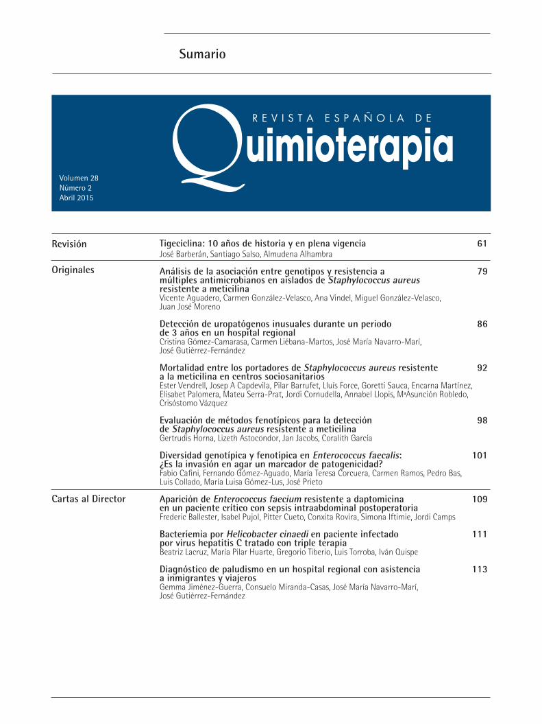


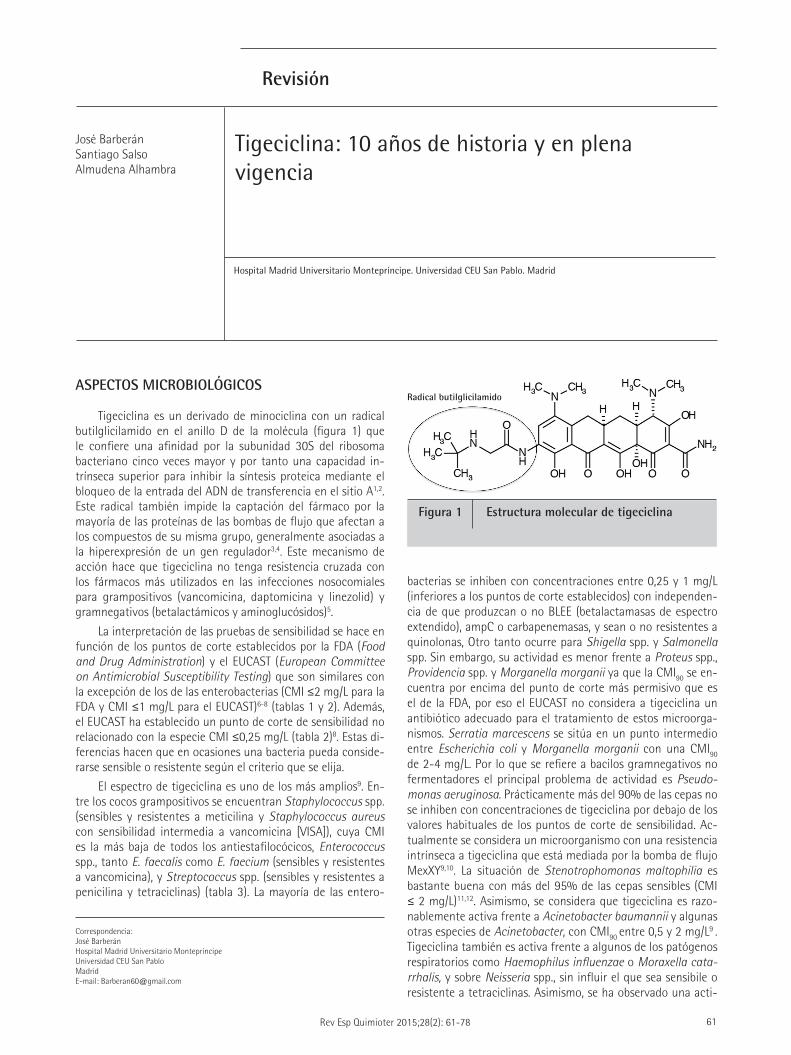


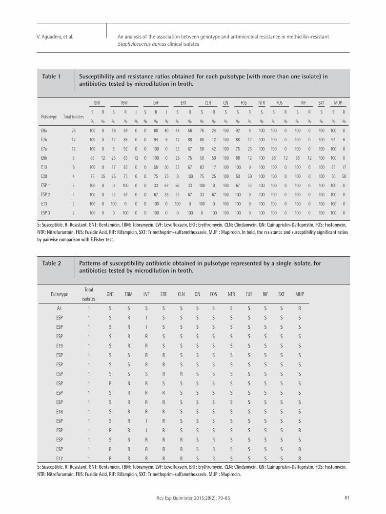
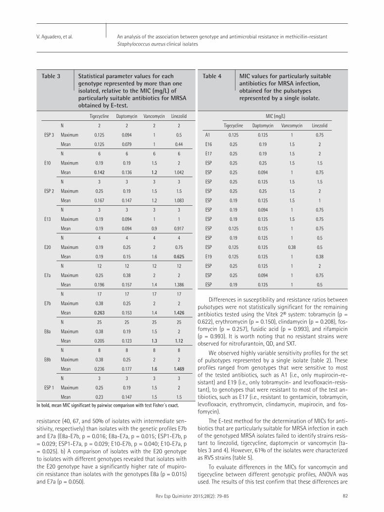


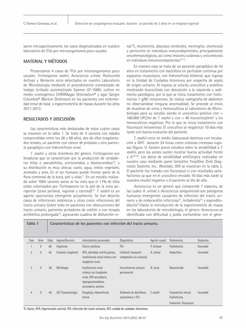
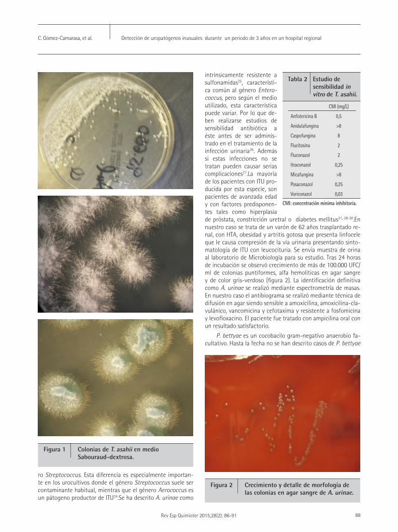


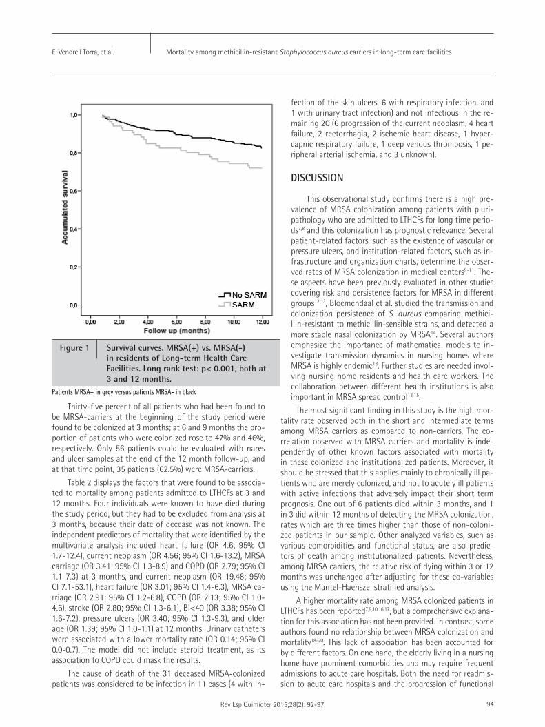


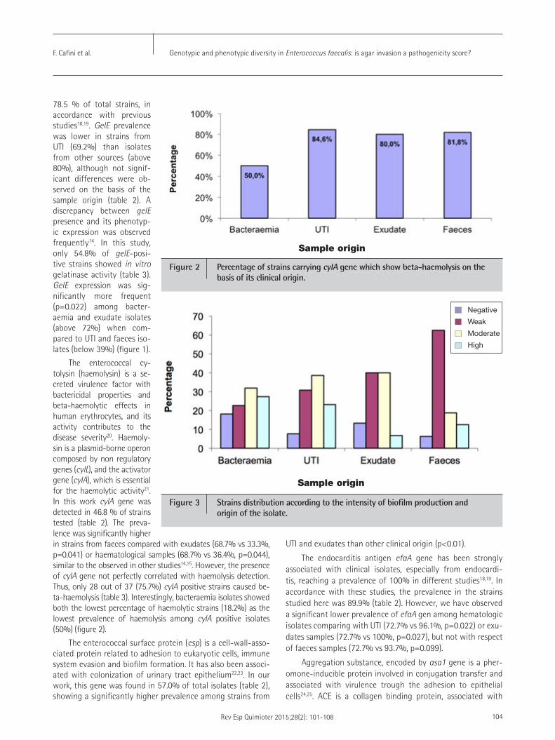


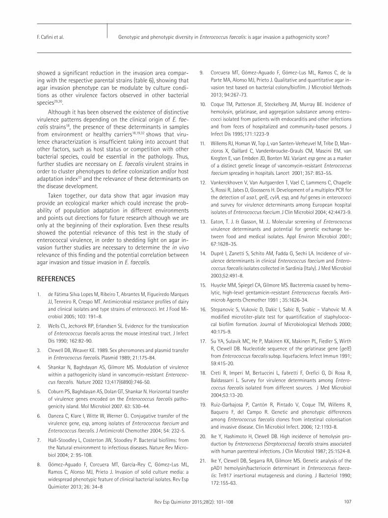


![[Lanove-CCF] Sword Art Online Volumen 02 Completo.pdf](https://static.documents.pub/doc/80x56/577cd6fc1a28ab9e789dc1de/lanove-ccf-sword-art-online-volumen-02-completopdf.jpg)


