ilateral spontaneous chylothorax after severeomiting in children
ntonio Lucas Lima Rodriguesa,∗, Mariana Tresoldi das Neves Romanelib,elso Dario Ramosb, Andrea de Melo Alexandre Fragab,icardo Mendes Pereirab, Simone Appenzellerb, Roberto Marinib,ntonia Teresinha Tresoldib
Hospital de Clínicas da Universidade Estadual de Campinas (Unicamp), Campinas, SP, BrazilFaculdade de Ciências Médicas da Universidade Estadual de Campinas (Unicamp), Campinas, SP, Brazil
eceived 11 January 2016; accepted 24 March 2016vailable online 20 August 2016
ResumoObjetivo: Relatar o caso de uma crianca com quilotórax bilateral devido a etiologia poucofrequente: lesão do ducto torácico após quadro de vômitos excessivos.Descricão do caso: Menina, sete anos, apresentava edema facial crônico iniciado após quadrode hiperemese. À avaliacão, também apresentava derrame pleural bilateral, com líquido quiloso
Chylothorax is defined as lymph accumulation in the pleuralspace, caused by injury to the thoracic duct and is a rarecause of pleural effusion in children.1,2 It can lead to sig-nificant respiratory morbidity and has an extensive list ofcauses, with great diagnostic difficulty.1,2 This study aims toreport the case of a child with spontaneous bilateral chy-lothorax.
Case description
Seven-year-old white female patient, referred due to sus-pected diagnosis of systemic lupus erythematosus. She hada five-month history of sudden-onset vomiting and self-limited abdominal bloating after ingestion of large amountsof chocolate; subsequently, she started to show insidiousand permanent chronic swelling of face. Three months aftersymptom onset and extensive evaluation of allergies, shewas submitted to a chest and abdomen computed tomo-graphy, which showed abdominal lymphadenomegaly andbilateral pleural effusion. Chest drainage was performed inanother service and the presence of milky pleural fluid was
reported. She also underwent laboratory evaluation at theoriginal service and most results were within normal val-ues (including whole blood count, renal function, C3, C4,rheumatoid factor, anti-Sm, anti-Ro, anti-La, anti-ds-DNA),
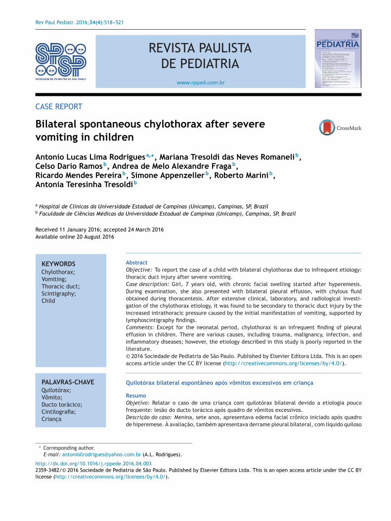
Figure 1 Chest radiography images of the patient in the posteroaeration is observed bilaterally, with pleuropulmonary opacity to tconsultation, the radiography shows no alterations.
asib
xcept for a positive antinuclear antibody, at a titration of:640, nuclear speckled pattern.
At the first outpatient visit in our service, the patientnderwent a new chest radiography (Fig. 1A), which showedecurrence of bilateral pleural effusion. A thoracentesis waserformed on the right, of which milky white fluid showedhe presence of 1.120mm3 of leukocytes (96% lymphocytes,% neutrophils, 1% plasma cells); 710mm3 of red blood cells;.7g/dL of protein; 87mg/dL of Glucose; 2.855mg/dL ofriglycerides. The child was hospitalized, kept in fastingnd started parenteral nutrition therapy. After 21 days with-ut reduction in the chylothorax volume, bilateral thoracicrainage was performed and 450mL of chylous secretion wasemoved from the right and 300mL from the left side. Therains were maintained in water seal, with a marked reduc-ion in eyelid edema. Three days after the draining she wastarted on a low-fat diet. The drains were removed after 25ays.
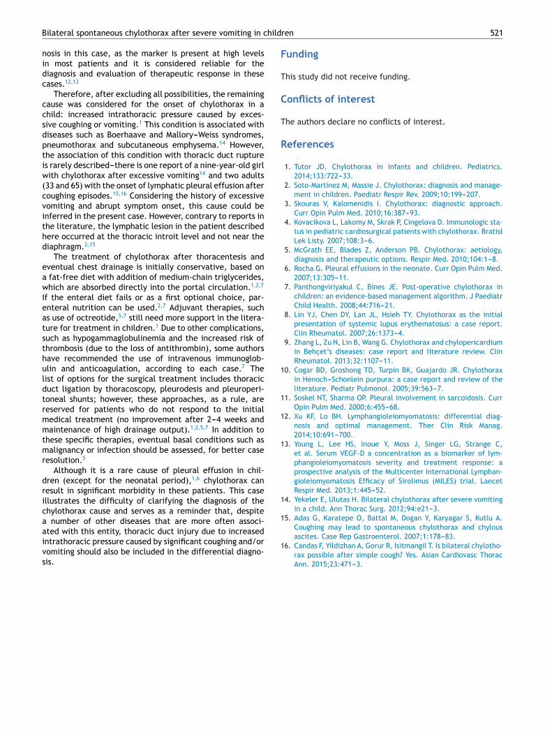
Serum levels of vascular-endothelial growth factor-DVEGF-D), a marker which, at high levels, is useful forhe diagnosis of lymphangioleiomyomatosis, was requestednd the result of 125pg/mL, a little above the refer-nce value (31---86pg/mL), ruled out that possibility. Shenderwent a lymphoscintigraphy (Fig. 2) with intradermal
nterior view; A---at patient admission, costophrenic sinus oblit-he right; B---six months after discharge, during an outpatient
dministration of the radiopharmaceutical in the instep andubsequent uptake of the radiotracer images that showedts extravasation in the topography of the thoracic introitilaterally, compatible with thoracic duct lesions, secondary
Figure 2 SPECT/CT image of the cervicothoracic and abdom-inal regions obtained from the lymphoscintigraphy assessmentwith intradermal administration of dextran-99mTc on the dor-sum of the feet; the images obtained 18hours after start of theexamination show focal area of radiotracer retention/leakagein the lymph ducts located in the topography of the thoracici
towtntwa
D
TirwreC
ibt
wioToLpflncdfw
tpspltm(
aAetlanacnccrttithmatoPlpnbo
ow
ntroit, bilaterally, more accentuated to the right.
o increased intracavitary pressure caused by vomiting thatccurred at the beginning of the clinical picture. The patientas discharged after 53 days of hospital stay, with outpa-
ient follow-up, during which she was allowed to resume aormal diet without lipid restrictions. She remains asymp-omatic at the follow-up (control chest X-ray in Fig. 1B),ithout alterations in the laboratory tests, including evalu-tion by a rheumatologist.
iscussion
he clinical picture of a patient with chylothorax isnsidious and develops as fluid accumulates in the pleu-al cavity; asymptomatic at first, it then progresses
1,2
ith cough and dyspnea ; fever and pleuritic pain areare findings.2,3 On physical examination, there is unilat-ral or bilateral dullness and decreased breath sounds.3
omplications of a chylothorax with chronic evolution
lclT
Rodrigues AL et al.
nclude protein-calorie malnutrition, immune deficiencyy lymphocyte and immunoglobulin depletion1,4 and elec-rolyte disorders.5
The diagnosis, based on clinical suspicion in patientsith suggestive clinical picture and compatible radiolog-
cal findings (pleural effusion in the plain chest X-rayr ultrasound), is attained through the thoracocentesis.1,3
he fluid removed from the pleural cavity is white,dorless, milky white,2,3 but can be as serosanguinous.3
aboratory evaluation showed triglyceride levels in the sam-le above of 110mg/dL and cholesterol ratio of pleuraluid over serum<1.0.1,3 Usually, cellularity is predomi-antly comprised of lymphocytes (>50%), with a proteinontent between 2 and 3mg/dL5 and low levels of lactateehydrogenase.3,5 If the diagnostic doubt persists, the pre-erred method is chylomicron analysis in the biological fluid,ith a positive result.1,3
After diagnostic confirmation, one can use other testso help in the investigation, such as computed tomogra-hy and/or magnetic resonance imaging, as well as morepecific tests to evaluate the lymphatic system, such as lym-hangiography and lymphoscintigraphy.1,2 According to theocation of the thoracic duct rupture, while also consideringhe anatomical variations, unilateral collection (most com-only on the right) can be detected or, more rarely, bilateral
in one-sixth of cases).5
The causes of chylothorax in children are diverse, varyingccording to age and the thoracic duct lesion mechanism.
review published in 2014 reports more than 35 possibletiologies.1 Among these are congenital malformations ofhe lymphatic system, such as pulmonary lymphangioma,ymphangiectasia and thoracic duct atresia1,2,6; chylothoraxssociated with genetic syndromes, such as Down, Noo-an and Turner syndrome,6 among others1,6; after headnd neck and thoracic surgical procedures (in up to 6% ofardiac surgeries)1,7; after other iatrogenic events in theeonatal period, such as birth trauma and superior venaava thrombosis due to central venous catheterization5,6;hylothorax after closed thoracic trauma1; and chylotho-ax associated with cancer, such as neurogenic neoplasia,eratomas, sarcomas and especially lymphomas, in whichhe lymph accumulation in the pleural space may be thenitial manifestation,1,2 in addition to granulomatous infec-ions such as tuberculosis.1 The patient in this case did notave findings that were consistent with congenital malfor-ations, had not suffered trauma or surgery, whereas cancer
nd infections were ruled out. Other possible causes forhe development of chylothorax are the rheumatologicalnes, the initial reason why our patient came to the service.ossible triggers that have been described are systemicupus erythematosus,8 Behcet’s disease,9 Henoch---Schönleinurpura10 and sarcoidosis.11 Likewise, the patient showedo clinical and laboratory criteria for these conditionsefore or during the follow-up, which were then ruledut.
Another condition ruled out in this case was lymphangi-leiomyomatosis. It is a rare disease that can be associatedith the tuberous sclerosis complex, is characterized as
ow-grade metastasizing neoplasm, which leads to insidiousystic changes in the lung parenchyma and also affects theymph vessels and lymph nodes and leads to chylothorax.12
he VEGF-D measurement was used to rule out this diag-
hild
F
T
C
T
R
1
1
1
1
1
1
Bilateral spontaneous chylothorax after severe vomiting in c
nosis in this case, as the marker is present at high levelsin most patients and it is considered reliable for thediagnosis and evaluation of therapeutic response in thesecases.12,13
Therefore, after excluding all possibilities, the remainingcause was considered for the onset of chylothorax in achild: increased intrathoracic pressure caused by exces-sive coughing or vomiting.1 This condition is associated withdiseases such as Boerhaave and Mallory---Weiss syndromes,pneumothorax and subcutaneous emphysema.14 However,the association of this condition with thoracic duct ruptureis rarely described---there is one report of a nine-year-old girlwith chylothorax after excessive vomiting14 and two adults(33 and 65) with the onset of lymphatic pleural effusion aftercoughing episodes.15,16 Considering the history of excessivevomiting and abrupt symptom onset, this cause could beinferred in the present case. However, contrary to reports inthe literature, the lymphatic lesion in the patient describedhere occurred at the thoracic introit level and not near thediaphragm.2,15
The treatment of chylothorax after thoracentesis andeventual chest drainage is initially conservative, based ona fat-free diet with addition of medium-chain triglycerides,which are absorbed directly into the portal circulation.1,2,7
If the enteral diet fails or as a first optional choice, par-enteral nutrition can be used.2,7 Adjuvant therapies, suchas use of octreotide,5,7 still need more support in the litera-ture for treatment in children.1 Due to other complications,such as hypogammaglobulinemia and the increased risk ofthrombosis (due to the loss of antithrombin), some authorshave recommended the use of intravenous immunoglob-ulin and anticoagulation, according to each case.7 Thelist of options for the surgical treatment includes thoracicduct ligation by thoracoscopy, pleurodesis and pleuroperi-toneal shunts; however, these approaches, as a rule, arereserved for patients who do not respond to the initialmedical treatment (no improvement after 2---4 weeks andmaintenance of high drainage output).1,2,5,7 In addition tothese specific therapies, eventual basal conditions such asmalignancy or infection should be assessed, for better caseresolution.5
Although it is a rare cause of pleural effusion in chil-dren (except for the neonatal period),1,6 chylothorax canresult in significant morbidity in these patients. This caseillustrates the difficulty of clarifying the diagnosis of thechylothorax cause and serves as a reminder that, despitea number of other diseases that are more often associ-
ated with this entity, thoracic duct injury due to increasedintrathoracic pressure caused by significant coughing and/orvomiting should also be included in the differential diagno-sis.
1
ren 521
unding
his study did not receive funding.
onflicts of interest
he authors declare no conflicts of interest.
eferences
1. Tutor JD. Chylothorax in infants and children. Pediatrics.2014;133:722---33.
2. Soto-Martinez M, Massie J. Chylothorax: diagnosis and manage-ment in children. Paediatr Respir Rev. 2009;10:199---207.
4. Kovacikova L, Lakomy M, Skrak P, Cingelova D. Immunologic sta-tus in pediatric cardiosurgical patients with chylothorax. BratislLek Listy. 2007;108:3---6.
5. McGrath EE, Blades Z, Anderson PB. Chylothorax: aetiology,diagnosis and therapeutic options. Respir Med. 2010;104:1---8.
6. Rocha G. Pleural effusions in the neonate. Curr Opin Pulm Med.2007;13:305---11.
8. Lin YJ, Chen DY, Lan JL, Hsieh TY. Chylothorax as the initialpresentation of systemic lupus erythematosus: a case report.Clin Rheumatol. 2007;26:1373---4.
9. Zhang L, Zu N, Lin B, Wang G. Chylothorax and chylopericardiumin Behcet’s diseases: case report and literature review. ClinRheumatol. 2013;32:1107---11.
0. Cogar BD, Groshong TD, Turpin BK, Guajardo JR. Chylothoraxin Henoch---Schonlein purpura: a case report and review of theliterature. Pediatr Pulmonol. 2005;39:563---7.
2. Xu KF, Lo BH. Lymphangioleiomyomatosis: differential diag-nosis and optimal management. Ther Clin Risk Manag.2014;10:691---700.
3. Young L, Lee HS, Inoue Y, Moss J, Singer LG, Strange C,et al. Serum VEGF-D a concentration as a biomarker of lym-phangioleiomyomatosis severity and treatment response: aprospective analysis of the Multicenter International Lymphan-gioleiomyomatosis Efficacy of Sirolimus (MILES) trial. LancetRespir Med. 2013;1:445---52.
4. Yekeler E, Ulutas H. Bilateral chylothorax after severe vomitingin a child. Ann Thorac Surg. 2012;94:e21---3.
5. Adas G, Karatepe O, Battal M, Dogan Y, Karyagar S, Kutlu A.Coughing may lead to spontaneous chylothorax and chylous
ascites. Case Rep Gastroenterol. 2007;1:178---83.
6. Candas F, Yildizhan A, Gorur R, Isitmangil T. Is bilateral chylotho-rax possible after simple cough? Yes. Asian Cardiovasc ThoracAnn. 2015;23:471---3.