96
Rheumatology Potpourri Dr. Philip A. Baer Seacourses Asia CME December 2017

Rheumatology Potpourri
Dr. Philip A. Baer
Seacourses Asia CME December 2017

Copyright © 2017 by Sea Courses Inc.
All rights reserved. No part of this document may be
reproduced, copied, stored, or transmitted in any form or
by any means – graphic, electronic, or mechanical, including
photocopying, recording, or information storage and
retrieval systems without prior written permission of Sea
Courses Inc. except where permitted by law.
Sea Courses is not responsible for any speaker or
participant’s statements, materials, acts or omissions.

Learning Objectives
Diagnose and treat polymyalgia rheumatica (PMR) and giant cell arteritis (GCA) while minimizing the adverse effects of steroid therapy.
Distinguish CPPD arthritis from other forms of crystal-induced arthritis, and manage CPPD arthritis appropriately.
Recognize different muscle problems associated with statin use, particularly necrotizing autoimmune myopathy (NAM).

Case 1 History
76 y.o. woman
Controlled hypertension and angina
Usually active
2 months history of aching pain in shoulders, upper arms, thighs
Morning stiffness 1 hour
Limited in daily activities
Appetite down; weight loss 8 pounds
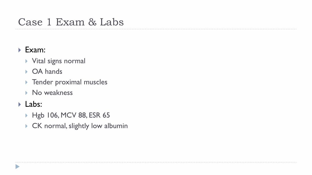
Case 1 Exam & Labs
Exam:
Vital signs normal
OA hands
Tender proximal muscles
No weakness
Labs:
Hgb 106, MCV 88, ESR 65
CK normal, slightly low albumin

Musculoskeletal Pain in Older Patients
Think polymyalgia rheumatica when
• Age >60
• Proximal muscle myalgias and stiffness without specific muscle
weakness
• High ESR
• Anemia

Polymyalgia Rheumatica (PMR)
A clinical syndrome characterized by aching and stiffness of the shoulder
and hip girdle muscles affecting older patients, associated with an
elevated ESR, lasting over 1 month and responsive to low dose steroids
First description in 1888 (Bruce)
Barber suggested the present name in 1957

PMR: Epidemiology
Incidence in Canada: Approximately 50/100,000 patients over age 50/year
Predominant age: 60 or older. Incidence increases with age (rare under 50
years old)
Predominant gender: Female > Male (2:1)


PMR: Core Inclusion Criteria

PMR: Core Exclusion Criteria
Active infection
Active cancer
Active giant cell arteritis (GCA)

PMR: Features Suggesting GCA
Abrupt-onset headache (usually temporal) and temporal tenderness
Visual disturbance, including diplopia
Jaw or tongue claudication
Prominence, beading or diminished pulse on examination of the temporal artery
Upper cranial nerve palsies
Limb claudication or other evidence of large-vessel involvement

PMR: Other Diseases to Exclude
Other inflammatory rheumatic diseases
Drug-induced myalgia
Chronic pain syndromes
Endocrine disease
Neurological conditions, e.g. Parkinson’s disease

PMR: General Principles

PMR: Lab Evaluation

PMR: Initial Steroid Dose

PMR: Initial Therapy and Follow-up
A patient-reported global improvement of ≥70% within a week of commencing steroids is consistent with PMR, with normalization of inflammatory markers in 4 weeks
A lesser response should prompt the search for an alternative condition
The diagnosis of PMR should be confirmed on further follow-up. Follow-up visits should include vigilance for mimicking conditions
Consider low-dose ASA, CV risk assessment, and osteoporosis prophylaxis during follow-up

PMR: High Dose Steroids?

PMR: NSAIDs?

PMR: Steroid Tapering

PMR: Steroid Tapering
www.rheuminfo.com

PMR: Immunosuppressants?

PMR: Biologics?

PMR: Duration of Therapy
Usually 1–3 years of treatment, although some will require small doses of steroids beyond this.
Steroids may be stopped when the patient is asymptomatic from their inflammatory symptoms.
Isolated raised ESR or CRP is not an indication for continuing steroid therapy but may require investigation and referral.
Persistent pain may arise from co-existing OA and rotator cuff tears.

PMR: Therapy of Relapses
Relapse is the recurrence of symptoms of PMR or onset of GCA, and not just unexplained raised ESR or CRP
Treatment of relapse:
Clinical features of GCA: treat as GCA (usually oral prednisone 40–60mg daily)
Clinical features of PMR: increase prednisone to previous higher dose
Single i.m. injection of methylprednisolone 120mg can also be used
Further relapses: consider introducing DMARD therapy after two relapses

Resources

www.rheuminfo.com

www.rheuminfo.com

www.rheumatology.org

ACR/EULAR PMR Algorithm

Relationship between PMR & GCA
10-15% of patients with PMR have GCA
40-60% of patients with GCA have PMR
PMR
GCA
GCA PMR

Case 2: History
A 68-year-old man presents with complaints of diffuse
muscle pain, weakness, and total body fatigue. He
reports:
• Gradual onset over past 6 months
• Morning stiffness lasting 2 to 3 hours
• Difficulty with getting out of a chair and combing his hair
• Recent onset of right-sided headache
• Recent onset of jaw pain when eating

Case 2: Exam and Labs
Proximal muscle
tenderness without
objective weakness
Tender right temporal
scalp region
Normal visual acuity
Hgb 98 g/L
ESR 85
CK 32

Case 2: Question
Based on the clinical findings, what is the most
important next step?
A. Treat now with prednisone 5 mg bid, and observe
B. Schedule a temporal artery biopsy for tomorrow morning
and use the results to determine whether prednisone will
be used
C. Start an NSAID at maximal dose
D. Treat now with prednisone at 40 to 60 mg per day and
schedule temporal artery biopsy in the next few days

Case 2: Answer
D. Treat now with prednisone at 40 to 60 mg per day and
schedule temporal artery biopsy for next week
• Sudden visual loss may occur in GCA
• Visual loss occurs in 1/3 of untreated patients
• The visual loss is usually not reversible
Nordberg E, et al. Rheum Dis Clin North Am. 1995;21:1013–1026.
For probable temporal arteritis:
TREAT NOW! BIOPSY LATER!
Biopsy as soon as possible
Hunder GC. Primer on Rheum Dis. 11th edition. 1997:294–300.
Don’t Hesitate

Elastic stain of temporal artery in GCA:
Disruption of elastic lamina and narrowing of the lumen

GCA - granulomatous inflammation and
multinucleated giant cells

DIAGNOSIS OF GCAHalo sign

GCA: Dilated Temporal Artery

GCA: Optic Atrophy

Giant Cell Aortitis
Risk of Thoracic Aortic Aneurysm in GCA: 15%

GCA: Steroid Therapy
GCA without visual symptoms:
Prednisone 20-40 mg daily for 8 weeks
Reduce by 5 mg q3-4 weeks to 10 mg daily
Then treat as PMR
GCA with possible or definite visual symptoms:
Prednisone 40-80 mg daily for 8 weeks
Reduce over 4 weeks to 20 mg daily
Then treat as uncomplicated GCA

GCA: Other Therapies
Methotrexate, azathioprine, and other immunosuppressive drugs have been used to limit dosage and duration of corticosteroid therapy.
No clear-cut data suggest that any of these drugs is superior to corticosteroid therapy.
Anti-TNF therapy with infliximab failed in GCA studies.
Anti-IL6 therapy with tocilizumab is now approved for GCA (USA)

Resources

www.rheuminfo.com

www.rheumatology.org

Case 3: History
80 y.o. woman, independent, lives alone
Occasional aches in knees, wrists
Acute swelling of right knee with pain, local warmth and redness
Prior episode in L wrist and hand
No trauma
Low grade fever, no recent infection

X-ray: Chondrocalcinosis

Aspiration: Bloody fluid

Case 3: Labs
CBC: WBC 14,000 with left shift
eGFR 55, uric acid 200
Calcium/PO4/Alkaline Phosphatase normal
Synovial fluid:
WBC 15,000
Thin, bloody fluid
Gram stain/culture negative

CPPD Disease: Crystals
Calcium
pyrophosphate
crystals
Ordinary, polarized,
and compensated
polarized light
microscopy

CPPD Disease: Classification
Chondrocalcinosis
Acute Pseudogout
Pyrophosphate Arthropathy

Chondrocalcinosis

Chondrocalcinosis: Epidemiology
Elderly – Radiographic evidence:
65-74yr 15%
75-84yr 36%
>84yr 50%
Male = Female
No geographic or racial predisposition

CPPD Disease: Clinical Features
Acute synovitis
Monoarthritis
Polyarticular: rare
Any joint: commonest in knee
: wrist, shoulder, ankle
Rapid onset of pain, stiffness and swelling (6-24 hrs)
Low-grade fever common

CPPD: Pathophysiology

CPPD Disease: Triggers

CPPD Disease: Diagnosis
Observation of calcium pyrophosphate dihydrate crystals
in synovial fluid leukocytes in a patient with acute
synovitis
CPPD crystals are
Rhomboid
Positively birefringent

Synovial Fluid Crystals

CPPD Disease: Treatment Principles
No definitive therapy for prevention
Goals in managing acute attacks
reduce symptoms
identify and treat any associated or triggering illnesses
encourage mobility as inflammation subsides

Acute CPPD Disease:
Treatment Options
Joint aspiration
NSAIDs
Colchicine (not as effective as for gout)
Steroids (not as effective as for gout)
oral
intra-articular
Analgesics

Acute CPP Crystal Arthritis:
Treatment Options

Chronic Pyrophosphate Arthropathy:
Clinical Features
Findings mimic any type of arthritis
Terminology -Pseudo RA
-Pseudo OA
-Pseudo neuropathic joint disease
Distinguishing points-Pattern of involvement
-Inflammation
-Superimposed pseudogout

Chronic Pyrophosphate Arthropathy:
Epidemiology and Distribution
Elderly
Female
Large/medium joints
Knees
Wrists, shoulders, elbows
Hips, midtarsal, MCPs (2+3)

Knee radiograph showing hypertrophic OA features. Note prominent patello-femoral involvement, typical of pyrophosphate arthropathy.

Chronic CPPD Arthropathy:
Treatment Options
Steroid injection
Colchicine
NSAID + PPI
Oral low dose steroids
Hydroxychloroquine
Methotrexate
Surgery to preserve function

Resources

www.rheuminfo.com

www.rheumatology.org

www.nejm.org June 30, 2016

Muscle Fibre: Normal Anatomy

Statin-induced Myopathy
▪ 1.5-3% of statin users in RCTs and 10-13% of participants enrolled in prospective clinical studies develop myalgias; rates of myositis lower (~0.1-0.5%) & dose-dependent
▪ Mean duration of statin therapy before onset of symptoms: 6 months
▪ Mean duration of myalgias after stopping statin therapy: 2 months


Statin-induced Myopathy: Questions
▪ What if a patient develops a myopathy after several years of taking a statin?
▪ Not likely to be statin-induced. 2/3 develop symptoms within 6 months of starting therapy, however it is still possible to develop muscle symptoms at ANY time during treatment.
▪ Are some statins more likely to cause muscle damage? Which ones?
▪ Lipophilic statins (for example, simvastatin, atorvastatin, lovastatin) are more likely to produce muscular effects than are relatively hydrophilic agents (such as pravastatin, rosuvastatin, and fluvastatin). Lipophilic compounds are more likely to penetrate into muscle tissue, enhancing the potential for myotoxic effects.


Statin-Induced Myopathy: Management
▪ Significant muscle symptoms: discontinue statin
▪ Asymptomatic but with CK>10x ULN: discontinue statin
▪ Rhabdomyolysis: no statins at any time due to risk of recurrence
▪ If patient requires a statin but muscle toxicity other than rhabdomyolysis: discontinue statin
▪ Once symptoms have resolved and the CK has returned to baseline, can try pravastatin or fluvastatin with careful monitoring

Statin-induced Myopathy: Questions
▪ When are EMG or muscle biopsy necessary in suspected statin myopathy?
▪ EMG and muscle biopsy are often done in atypical cases of statin induced myopathy. These would include those patients with persistent muscle pain after D/C of statin or with persistent, worsening weakness after D/C of statin, or persistently elevated CK long after D/C of statin (several months).
▪ Electromyography findings are commonly reported to show myopathic changes, usually in the proximal muscles, in agreement with clinical findings.
▪ Muscle pathology in statin induced myopathy is nonspecific, with necrosis, degeneration, and regeneration of fibres and phagocytic infiltration.
▪ Is Coenzyme Q10 helpful?
▪ Unclear. In one small RCT, 41 patients taking statins who had muscle pain received either coenzyme Q10 or vitamin E. After one month of treatment, 18 of 21 patients taking coenzyme Q10 reported improvement in muscle pain, compared with three of 20 taking vitamin E (P<0.001).

Statin-associated necrotizing myopathy
▪ Myopathy which persists or progresses after stopping statin
▪ Linked to autoantibodies against HMG-CoA reductase
▪ Distinct muscle biopsy findings:
▪ Macrophagocytic infiltrate engulfing necrotic muscle fibers
▪ Responds to immunosuppressant therapy

Necrotizing Autoimmune Myositis

Statin-associated necrotizing myopathy (NAM)
Necrotic muscle fibre undergoing myophagia WITHOUT inflammatory infiltrate (no lymphocytes or
neutrophils) & excessive variability of muscle fibre size. The absence of inflammation distinguishes this
entity from the idiopathic inflammatory myopathies.

Statin-associated necrotizing myopathy (NAM)

EMG findings (All Inflammatory Myopathies)
▪ Fibrillation, PSWs, CRDs at rest
▪ Increased insertional activity

Myositis: Non-medical therapies
▪ Physiotherapy and Occupational Therapy
▪ Aerobic exercise programs after the acute phase
▪ Prevent contractures
▪ May help with steroid side effects (weight gain, osteoporosis, type 2 fibre atrophy)
▪ Speech therapy
▪ Especially if concomitant dysphagia

Pharmacologic Treatment of Myositis

Myositis: Resources

Myositis: Resources

Myositis: Resources

Myositis: Resources

Statin-Associated Autoimmune Myopathy: Approach

Statin-Associated Autoimmune Myopathy: Approach

PEARLs: Rheumatology Potpourri
Think of PMR if over age 65 with proximal joint/muscle pain
Start high dose steroids immediately if GCA suspected to prevent visual loss
Think CPPD in older women with acute monoarthritis and aspirate joint if
possible for crystal analysis
Consider autoimmune statin-induced myopathy (NAM) in patients where
myopathy persists after stopping statins

Barriers to Change: Rheum. Potpourri
PMR/GCA: Difficulty distinguishing flares from OA and other comorbidities,
leading to higher than required use of steroids
CPPD: Confusion with gout and with cellulitis
Statin-induced NAM: Difficulty recognizing this syndrome given so many
patients on statins present with myalgias and high CKs

Questions ?


