Meteoritics & Planetary Science 44, Nr 11, 1695–1706 (2009)Abstract available online at http://meteoritics.org
Rietveld analysis of X-ray powder diffraction patterns as a potential tool for the identification of impact-deformed carbonate rocks
Sarah A. HUSON*, Franklin F. FOIT, A. John WATKINSON, and Michael C. POPE
School of Earth and Environmental Sciences, Washington State University, Pullman, Washington 99164, USA*Corresponding author. E-mail: [email protected]
(Received 20 March 2008; revision accepted 15 August 2009)
Abstract–Previous X-ray powder diffraction (XRD) studies revealed that shock deformed carbonatesand quartz have broader XRD patterns than those of unshocked samples. Entire XRD patterns, singlepeak profiles and Rietveld refined parameters of carbonate samples from the Sierra Madera impactcrater, west Texas, unshocked equivalent samples from 95 miles north of the crater and the MissionCanyon Formation of southwest Montana and western Wyoming were used to evaluate the use ofX-ray powder diffraction as a potential tool for distinguishing impact deformed rocks fromunshocked and tectonically deformed rocks. At Sierra Madera dolostone and limestone samples werecollected from the crater rim (lower shock intensity) and the central uplift (higher shock intensity).Unshocked equivalent dolostone samples were collected from well cores drilled outside of the impactcrater. Carbonate rocks of the Mission Canyon Formation were sampled along a transect across thetectonic front of the Sevier and Laramide orogenic belts.
Whereas calcite subjected to significant shock intensities at the Sierra Madera impact crater can bedifferentiated from tectonically deformed calcite from the Mission Canyon Formation using Rietveldrefined peak profiles, weakly shocked calcite from the crater rim appears to be indistinguishable fromthe tectonically deformed calcite. In contrast, Rietveld analysis readily distinguishes shocked SierraMadera dolomite from unshocked equivalent dolostone samples from outside the crater andtectonically deformed Mission Canyon Formation dolomite.
INTRODUCTION
The physical record of impact structures on the surface ofthe Earth dates from 3.5 Ga (Grieve 1998) and it is thought thatimpact cratering was an essential process even earlier in Earth’shistory (Ryder 2002). Putative impact structures commonly areidentified by the presence of meteorite fragments, siderophileelement anomalies, and specific shock deformational featuresincluding shatter cones, planar deformation features (PDFs) inminerals, diaplectic glass, and melting features (French 1998).Circular or nearly circular geomorphic structures lacking thesedistinctive features of suspected impact origin often are thesubject of controversy (e.g., the Silverpit structure; Stewart andAllen 2002; Underhill 2004; Reimold 2007).
Currently, there are 176 impact structures confirmed in theEarth Impact Database (www.unb.ca/passc/ImpactDatabase).Criteria used to confirm circular structures on the Earth’ssurface as meteorite craters vary with target rock type, age,bolide size, and amount of material removed by erosion.However, exposure to short-lived shock pressure is a uniquecharacteristic of the target rocks at all impact structures.
Therefore, a method of identifying minerals/rocks subjected toshock metamorphism, but lacking the usual shockdeformational features, would be extremely useful fordistinguishing suspected impact structures from non-impactgenerated structures. Past studies, X-ray diffraction (XRD) andpetrographic, often focused on both experimentally and naturallyshocked silicate minerals due to the abundance and robustnature of these minerals at the Earth’s surface (Hörz 1968;Short 1968; Hörz and Quaide 1973; Grieve et al. 1996). AnX-ray powder diffraction method for evaluating shockdeformation levels within impact craters formed in carbonatehost rocks was developed by Skála and Jakeš (1999). Abroadening (i.e., increase in peak width) and reduction inintensity of CuKα X-ray diffraction peaks in the 2θ° range56–62° was observed in impact-shocked carbonate from theKara crater (~65 km diameter) in Russia and the SteinheimBasin (3.8 km diameter) in Germany when compared todiffraction peaks of unshocked standards (Skála and Jakeš1999). Furthermore, peak broadening and loss of peak intensityincreased with increasing shock pressure (Skála and Jakeš1999). However, XRD peak broadening in carbonate samples
1696 S. A. Huson et al.
from Meteor Crater (1.2 km diameter), Arizona, and nearbyCanyon Diablo did not show the expected relationship ofincreased peak broadening with increasing shock effects fromstructure rim to structure center. These results were attributedto a non-uniform distribution of shock pressure during theimpact event (Burt and Pope 2001; Burt et al. 2005). In aneffort to resolve this discrepancy, carbonate and siliciclasticsamples from the Sierra Madera impact structure (12 kmdiameter) in Texas were analyzed using XRD techniquesadopted from Skála and Jakeš (1999). Preliminary studies ofthe Sierra Madera carbonates indicated increased peakbroadening in shatter cone samples (Huson et al. 2006a,2006b).
In the present study, XRD patterns of calcite anddolomite from the Sierra Madera impact structure arecompared to those of unshocked equivalent carbonatesamples from outside the structure, unshocked carbonatesamples from the Mississippian Mission Canyon Formationof the northern Rocky Mountains, United States, and tounshocked/undeformed mineral standards. The objective is todetermine whether XRD patterns of the shocked carbonatescan be distinguished from XRD patterns of carbonates thatwere deformed solely by terrestrial processes (e.g., burial ortectonism). This study has implications for using XRD
pattern analysis as a tool for the identification of effects ofshock metamorphic processes where conventional impactcharacteristics (i.e., shatter cones and PDFs in minerals) arelacking or poorly developed, especially as ~30% of allterrestrial impact structures formed primarily in carbonaterocks (Grieve and Robertson 1979).
GEOLOGIC SETTINGS
Sierra Madera Impact Structure
The Sierra Madera impact structure, located near FortStockton, Texas, is a well-exposed, eroded remnant of acomplex, 12 km diameter impact structure (Eggleton andShoemaker 1961; Wilshire and Howard 1968; Howard et al.1972; Wilshire et al. 1972). The central uplift is composed ofsteeply dipping, folded, and faulted Upper Permian strata(Fig. 1). Lower Cretaceous rocks occur along the easternmargin of the central uplift. The Permian Hess Formation, theoldest stratigraphic unit exposed in the central uplift, wasdisplaced vertically at least 790 m during the excavation andmodification stage of structure development (Wilshire et al.1972). The crater rim at Sierra Madera is composed of erodedLower Cretaceous carbonate and siliciclastic rocks with
Fig. 1. a) Geologic map of Sierra Madera (modified from Wilshire et al. 1972). 1 = Kwf 1, 2 = Kwf 2, 3 = Kwf 3, 4 = Kwf 4, 5 = Kwf 5, 6 =Pt, 7 = Pg B1, 8 = Mlb, 9 = Pw, 10 = Pv, 11 = Ph. b) Stratigraphic column for rocks exposed at Sierra Madera.
Rietveld analysis of X-ray powder diffraction patterns 1697
exception of the southwest side where stream incision hasexposed Upper Permian strata. Rim strata are exposed ~30–60 m above the alluvium-filled ring depression and generallydip 0–5° radially, though at some locations the dip can be asgreat as 30°. The Upper Permian units at Sierra Madera weredeposited on the southeast side of the Delaware Basin, amajor depositional basin during the late Permian. The stratarecord a gradual in-filling and restriction of the basin. LowerCretaceous rocks were deposited in a fluvial/deltaic toshallow water marine setting (Hill 1996). Post-impact erosionhas removed an estimated 600 m of material from the craterrim (Wilshire et al. 1972). Past studies at Sierra Maderadocumented shock deformation features including shattercones in siltstone and fine-grained sandstone, PDFs in quartz,grain fracturing, impact-induced brecciation, and deformedcarbonate minerals (Dietz 1960; Eggleton and Shoemaker1961; Howard and Offield 1968; Wilshire et al. 1972; Husonet al. 2005).
Unshocked Equivalent Dolostone Samples
Ninety-five miles north of the Sierra Madera impactstructure, the equivalent Queen and Yates Formations weredeposited on the northern edge of the Delaware Basin duringthe late Permian. These formations were deposited in a
shallow shelf and later evaporitic environment as theDelaware Basin gradually became restricted (Hill 1996).
Mission Canyon Formation
The Lower Mississippian (Late Osagean-MiddleMeramecian) Mission Canyon Formation in westernWyoming and southwestern Montana records shallow watercarbonate deposition along a broad carbonate shelf thatdeepened westward during the Antler Orogeny (LateDevonian to Early Mississippian; Fig. 2; Sando 1976; Smithand Dorobek 1993). This unit was subsequently altered byLate Mississippian karstification and dolomitization anddocuments deep to shallow burial from west to east (Sando1988; Smith and Dorobek 1993). Additional deformation ofPaleozoic-Mesozoic strata of southwestern Montana andwestern Wyoming occurred during the Cretaceous-PaleogeneSevier and Laramide orogenies (Snoke 1993).
EXPERIMENTAL
Sample Collection and Preparation
Fine-grained dolostone and limestone samples werecollected from the central uplift and eroded crater rim of the
Fig. 2. a) Location of Mission Canyon Formation samples. 1 = Shoshone Canyon (SH), 2 = Sheep Mtn (SM), 3 = Benbow Mine (BM), 4 =UPR, 5 = Church Buttes (CB). Gray arrow indicates increasing burial depth and deformation. b) General stratigraphic column of the MissionCanyon Formation.
1698 S. A. Huson et al.
Sierra Madera impact structure (Fig. 1; Table 1). Unshockedsamples of the dolomitic Queen and Yates Formations,equivalent to the Upper Word and Gilliam Limestone at SierraMadera, were collected from the Austin Core Facility inAustin, Texas. The cores were located in the North Ward-Estes Oil Field in Ward County, Texas, 95 miles north of theSierra Madera impact structure. The authors were not able toobtain unshocked calcite-rich samples from outside the craterstructure.
Fine-grained samples of dolostone and limestone werecollected also from the Mission Canyon Formation of theMadison Limestone Group of southwest Montana andwestern Wyoming (Fig. 2; Table 1). Three optically clearsamples of quartz, Iceland spar, and dolomite fromWashington State University’s School of Earth and
Environmental Sciences (WSU SEES) mineral collectionserved as unshocked standards for comparison. All sampleswere ground under isopropyl alcohol using a mortar andpestle and the powders were passed through a 27 µm meshsieve.
Sample Compositional Characterization
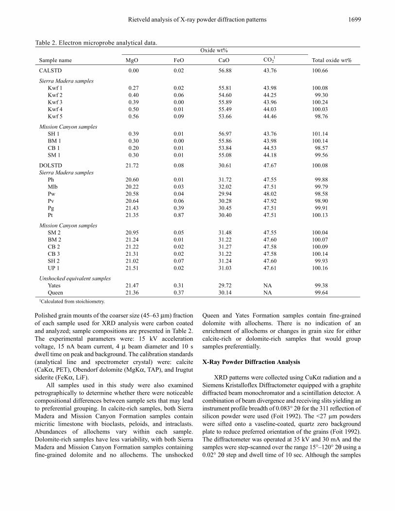
Since compositional differences can lead to XRD peakposition and breadth variability (Fiquet et al. 1994), themagnesium, iron, and calcium content of all calcite anddolomite samples were analyzed using a Cameca electronmicroprobe and a JEOL 8500 F Field Emission electronmicroprobe in the WSU SEES GeoAnalytical Laboratory. Noother elements were observed in more than trace amounts.
1F = fractures; Fc = fractures filled with coarse calcite; B = brecciation; SC = shatter cones.2MS = mudstone; WS = wackestone; WS-PS = wackestone-packstone; Mono = monomict impact generated breccia; DS = dolostone; LS = limestone.3Contains visible sulfur.
Rietveld analysis of X-ray powder diffraction patterns 1699
Polished grain mounts of the coarser size (45–63 µm) fractionof each sample used for XRD analysis were carbon coatedand analyzed; sample compositions are presented in Table 2.The experimental parameters were: 15 kV accelerationvoltage, 15 nA beam current, 4 µ beam diameter and 10 sdwell time on peak and background. The calibration standards(analytical line and spectrometer crystal) were: calcite(CaKα, PET), Obendorf dolomite (MgKα, TAP), and Irugtutsiderite (FeKα, LiF).
All samples used in this study were also examinedpetrographically to determine whether there were noticeablecompositional differences between sample sets that may leadto preferential grouping. In calcite-rich samples, both SierraMadera and Mission Canyon Formation samples containmicritic limestone with bioclasts, peloids, and intraclasts.Abundances of allochems vary within each sample.Dolomite-rich samples have less variability, with both SierraMadera and Mission Canyon Formation samples containingfine-grained dolomite and no allochems. The unshocked
Queen and Yates Formation samples contain fine-graineddolomite with allochems. There is no indication of anenrichment of allochems or changes in grain size for eithercalcite-rich or dolomite-rich samples that would groupsamples preferentially.
X-Ray Powder Diffraction Analysis
XRD patterns were collected using CuKα radiation and aSiemens Kristalloflex Diffractometer equipped with a graphitediffracted beam monochromator and a scintillation detector. Acombination of beam divergence and receiving slits yielding aninstrument profile breadth of 0.083° 2θ for the 311 reflection ofsilicon powder were used (Foit 1992). The <27 µm powderswere sifted onto a vaseline-coated, quartz zero backgroundplate to reduce preferred orientation of the grains (Foit 1992).The diffractometer was operated at 35 kV and 30 mA and thesamples were step-scanned over the range 15°–120° 2θ using a0.02° 2θ step and dwell time of 10 sec. Although the samples
Unshocked equivalent samplesYates 21.47 0.31 29.72 NA 99.38Queen 21.36 0.37 30.14 NA 99.64
1Calculated from stoichiometry.
1700 S. A. Huson et al.
were scanned from 15°–120° 2θ, only the portion of the scancovering 55–69° 2θ is shown in Fig. 3 to highlight peakbroadening differences. XRD powder patterns for all sampleswere compared to dolomite or calcite standard patterns.
Diffraction Peak Profile Analysis
XRD peak broadening due to shock deformation wasanalyzed using both single peak profiling and Rietveldrefinement of peak shape parameters, both of which werecarried out using Jade 8.0 software (Materials DataCorporation). With single peak profiling, the peak full widthhalf maximum (FWHM) values are not appreciably affectedby small variations in sample composition, therefore they arepotentially useful for distinguishing weakly from moreintensely shocked samples (Fig. 4). Peak broadening can alsobe analyzed by Rietveld crystal structure refinement (Young1993) of the peak shape parameters (Skála and Jakeš 1999).XRD peak FWHM values are a function of the diffractionangle theta and three least squares refineable parameters U, V,and W (Caglioti 1958).
(FWHM)2 = UTan2 theta + Vtan theta + W
The values of FWHM, which are related to the amount ofshock-induced deformation, were calculated over the range15°–120° 2θ. Single peak and Rietveld refined FWHMs werethen fitted with a fourth order polynomial (Fig. 5).
RESULTS
Diffraction Patterns: Limestone
XRD patterns of calcite-rich Mission Canyon Formationsamples are compared with calcite-rich Sierra Maderasamples (Fig. 3a). Samples obtained from the central uplift ofSierra Madera display more peak broadening than those fromthe crater rim with an almost complete disappearance ofpeaks at 58° and 63° in sample Kwf 4. Although XRD peaksof Mission Canyon Formation sample SH 1 (Fig. 3a) arebroader than crater rim samples from Sierra Madera (Kwf 1,Kwf 2, and Kwf 3), in general, calcite-rich Sierra Maderasamples show more peak broadening than calcite-richMission Canyon Formation samples. Some Sierra Maderaand Mission Canyon samples contain extra peaks. SamplesKwf 2, Kwf 4, Kwf 5 and SM 1 contain minor quartz peakswhile samples SH 1 and CB 1 contain minor anhydrite peaks(Fig. 3).
Diffraction Patterns: Dolomite
Of the 14 unshocked samples collected from the AustinCore Facility only two, one from the Queen Formation andone from the Yates Formation, are dolomite. The remaining12 samples contained dolomite with significant amounts of
quartz and anhydrite and are therefore not compared to theshocked samples from Sierra Madera. XRD patterns ofdolomite-rich Sierra Madera samples are compared with twounshocked equivalent samples of the Queen and YatesFormations (Fig. 3.3B). Shocked samples from Sierra Maderahave more peak broadening than the unshocked Queen andYates samples. For example, note broadening between sampleYates/Gil and Pw in the 60° and 67.5° regions.
Samples from Sierra Madera show an increase in peakbroadening towards the central uplift of the impact structure.For some samples (Pw, Ph, and Pv) peak broadening is greatenough to result in almost a complete disappearance of thethree peaks in the 64° to 66° 2θ range.
For Mission Canyon Formation samples, patterns ofsamples from locations close to the Antler Orogenic fronthave less peak broadening than those from farther away. Forexample, the peaks for sample UP 1 (location 4; Fig. 3.2A)are sharper than those for sample SM 2 (location 2);especially note the three peaks in the 64° to 66° region. Ingeneral, dolomitic Sierra Madera samples have considerablymore peak broadening than Mission Canyon Formation andunshocked Queen and Yates samples.
Single Peak Profiling—Full Width Half Maximum(FWHM)
The FWHM curves (Fig. 4a, b) that deviate most fromthe standard curves (CALSTD and DOLSTD) are those thatexperienced higher shock (shatter cones, monomict impactbreccias and samples from the crater rim). UnshockedQueen and Yates samples plot close to the standard curve.Also the majority of the Mission Canyon Formation samplestend to plot between the scans of the standards and those ofthe Sierra Madera samples (with the exception of BM 2and SH 1; Figs. 4a, 4b). However, some samples (CB 1and Mlb 1, for example) have a convex curve instead of theexpected concave curve as seen in the calcite and dolomitestandards.
Rietveld Refinement of Peak Shape Parameters
FWHM curves (Fig. 5a) for calcite-rich Mission CanyonFormation samples SH 1 and BM 1 overlap calcite-richSierra Madera crater rim samples Kwf 3 and Kwf 1/Kwf 2,respectively. FWHM values obtained from Rietveld crystalstructure refinements increase with shock intensity for allcalcite-rich Sierra Madera samples. FWHM values ofshocked dolomite from the central uplift of Sierra Maderaare distinctly higher than dolomite from unshocked Queenand Yates samples as well as samples from the tectonicallydeformed dolomite from the Mission Canyon Formation(Fig. 5b) and follow a pattern of progressive broadeningfrom crater rim to central uplift. Sierra Madera samples withshock features (shatter cones, monomict breccia) have
Rietveld analysis of X-ray powder diffraction patterns 1701
Fig. 3. X-ray powder diffraction patterns of calcite and dolomite samples from the Sierra Madera impact structure, unshocked equivalent rocks,and Lower Mississippian Mission Canyon Formation. a) Calcite-rich samples compared with the calcite standard (CALSTD). b) Dolomite-rich samples compared with the dolomite standard (DOLSTD). All scans were arranged from least (bottom of chart) to most (top of chart)broadened. See Table 1 for sample descriptions.
1702 S. A. Huson et al.
uniformly higher FWHM values than those without shockfeatures.
DISCUSSION
All XRD peak patterns deviate from those of the calciteand dolomite standards. This is particularly true for calciteand dolomite from the central uplift of Sierra Madera. Severalfactors contribute to XRD peak profile variability includingMg content (Gavish and Friedman 1973), grinding time ofpowder (Burns and Bredig 1956; Gavish and Friedman 1973),grain size of powder (Klug and Alexander 1974; Langfordet al. 2000), and phase transformations (Burns and Bredig1956; Merrill and Bassett 1975; Fiquet et al. 1994). The abovementioned factors were considered when preparing samplesfor this study. The Mg, Fe, and Ca contents of all carbonate
samples were measured (Table 2) and while all samplesdeviate slightly from the calcite and dolomite stoichiometricvalues, there is no correlation between composition andvariability in XRD peak profiles or Rietveld refined FWHMcurves. The grain size of the powder used was <27 µm as alarger crystallite size can lead to inaccurate and impreciseintensity measurements (Bish and Reynolds 1989). Phasetransformations (e.g., calcite to aragonite due to grinding;Burns and Bredig 1956) were not a factor as XRD powderpatterns for samples from Sierra Madera show no peaksother than those of dolomite, calcite, and in a few cases,quartz and anhydrite (Fig. 3). Quartz is a common mineral innearshore carbonate rocks. The Mission Canyon Formationcontains anhydrite and other evaporitic minerals due toevaporitic tidal flat depositional environments (Dorobek et al.1993).
Fig. 4. Single peak profiling using FWHM values for Sierra Madera, unshocked equivalent samples, and Mission Canyon Formation samples.a) Calcite values compared with a calcite standard, CALSTD. b) Dolomite values compared with a dolomite standard, DOLSTD.
Rietveld analysis of X-ray powder diffraction patterns 1703
Single peak profile patterns of calcite and dolomite fromall samples while useful at lower 2θ values, are complex andindicate little relationship to shock intensity at higher 2θvalues. This is because weaker high angle reflections arepoorly estimated by the profile shape function. High anglereflections from shocked material may disappear from thepattern since these reflections are often low intensity andshocking decreases the degree of the crystallinity (Skála andJakeš 1999). While single peak profiling may be useful atlower 2θ values, at higher values, especially above ~80° 2θ,the method is unreliable.
More useful for this study was the Rietveld refinementof peak shape parameters. Rietveld refinement of peak shapeparameters yields better resolution of overlapping diffractionpeaks and a more precise measure of the 2θ angulardependence of peak FWHM, thus, providing a bettermeasure of the degree of shock deformation (Skála and Jakeš1999).
Rietveld results for the mildly shocked calcite samplesfrom the crater rim overlap with those of Mission CanyonFormation calcite samples, whereas those of the central upliftare distinguishable from Mission Canyon Formation samples.
Fig. 5. Rietveld refinement analysis for Sierra Madera, unshocked equivalent samples, and Mission Canyon Formation samples. a) Calcitevalues compared with a calcite standard, CALSTD. b) Dolomite values compared with a dolomite standard, DOLSTD.
1704 S. A. Huson et al.
Dolomite samples from Sierra Madera also show increasingRietveld refined FWHM values as the central uplift isapproached reflecting increasing deformation of the dolomitecrystal structure with increasing shock pressure (Skála andJakeš 1999). Noticeably, Sierra Madera samples plot wellabove unshocked equivalent dolomite-rich samples fromoutside the crater. Therefore, XRD with Rietveld refinementanalysis indicates that it may not always be possible todistinguish calcite-rich samples subjected to terrestrialtectonic processes from those mildly shocked in an impactevent. However, with exposure to increasing shock pressureswithin the central uplift of the structure, dolomitic impactshocked rocks are readily distinguished from dolomiticunshocked rocks from outside of the structure and otherterrestrially deformed dolomitic rocks.
Interestingly, Mission Canyon Formation samples do notindicate a systematic variation in Rietveld-refined FWHMswith distance from the Sevier and Laramide orogenic fronts.With Mission Canyon Formation samples, due todeformation associated with increasing compression andburial depth, peak broadening was expected to increase assample locations approach the tectonic front of the Sevierand Laramide Orogenies (Fig. 2). The ideal and expectedorder for sample locations, from least to most peakbroadening, is Sheep Mountain (SM), Benbow Mine (BM),Shoshone Canyon (SH) to Church Buttes (CB). However,observed XRD patterns for Mission Canyon Formationsamples indicate that peak breadth increases in the order SM1 to CB 1 to BM 1 to SH 1. The fronts of these orogens maybe more complicated, with deformation unequally distributedalong thrust fronts or out of phase thrusting may haveoccurred. Similarly, tectonic forebulge versus foreland basinevents overprinted burial deformation of the Antler orogeny,increasing the complexity of the deformational history in thearea. Additionally, secondary alteration of the MissionCanyon Formation associated with burial andrecrystallization (Smith and Dorobek 1993) may also haveaffected XRD parameters; however, precipitation ofsecondary dolomite, one potential cause of the unexpectedorder, can be ruled out as it should result in peak profilesmore similar to those of the standard.
Hydrothermal and Secondary Alteration in SierraMadera
Hydrothermal and secondary alteration in rocks mayaffect XRD peak patterns due to modification of the originalrock material. Evidence of fluorite crystals occurring in somepolymict breccia outcrops (Wilshire et al. 1972) indicatehydrothermal alteration occurred as a post-impact process atSierra Madera. However, it is not uncommon for impactcraters to interact with the hydrosphere either as a disruptionof the local water table or on a larger scale such as an oceanbasin (Osinski et al. 2001). Rocks at Sierra Madera were not
deformed after the impact event (Wilshire et al. 1972).Therefore, if hydrothermal alteration occurred at SierraMadera, it was directly related to the impact event and not dueto a subsequent deformational process.
Interestingly, XRD samples from smaller impactstructures that do not develop post-impact hydrothermalsystems, like Meteor Crater, show broadening. In the case ofMeteor Crater, XRD samples show peak broadening but notin the expected pattern, i.e., decrease in broadening fromcrater center to crater rim. The results have been attributed toa pre-impact structural joint pattern in the target rocks (Burtand Pope 2001; Burt et al. 2005). Hydrothermal alterationmay be a factor in XRD peak broadening patterns, however, itis not the cause of peak broadening in individual samples.
Shock Pressures and XRD Broadening
Using the shock classification scheme for impactmetamorphosed sandstones developed by Kieffer (1971) andmodified by Osinski (2007), we can assign shock pressurevalues to CaCO3 cemented sandstones at Sierra Madera andapply those values to the adjacent carbonate units. Impactmetamorphosed sandstones at Sierra Madera give a shockpressure range from 5.5–20 GPa based on the presence of“toasted” quartz, a “jigsaw” interlocking texture betweenquartz grains, and multiple sets of planar deformation features(PDFs) within quartz grains. The presence of well-developedshatter cones in porous sandstone indicates a shock pressurerange of 3–10 GPa and corresponds to a post-shocktemperature range of 350−>1000° C (Osinski 2007).
Sandstone beds within the Gilliam Limestone have thegreatest amount of deformational features and, therefore, onewould assume XRD peak patterns from the adjacentlimestone would be the broadest of all Sierra Madera samples.However, the broadest XRD peak patterns come from theHess Formation located within the center of the central uplift(Fig. 3). While the adjacent quartz-rich Cathedral MountainFormation and sandstone beds within the Word Formationcontain shock features (multiple sets of PDFs and thegeneration of quartz microbreccia), these units are matrix-supported, not grain-supported like the sandstone beds withinthe Gilliam Limestone. Without the interaction betweenquartz grains, higher shock pressure indicators are not presentwithin the Cathedral Mountain Formation and WordFormation. Therefore, shock features within these two quartz-rich units do not accurately indicate the highest shockpressures to which these rocks or the adjacent limestoneswere exposed. Nevertheless, the presence of shatter coneswithin the Hess, Cathedral Mountain, and Word Formationsindicate rocks from the central uplift of Sierra Madera wereexposed to shock pressures in the range of 3–10 GPa;however the actual pressure range of formation is probablynarrower (French 1998; Baratoux and Melosh 2003; Wielandet al. 2006; Osinski 2007).
Rietveld analysis of X-ray powder diffraction patterns 1705
CONCLUSIONS
Rietveld refined parameters (especially XRD peakFWHM) potentially can be used to distinguish impact-shocked rocks from unshocked equivalent rocks from outsidethe structure. However, this study suggests that whencomparing shocked rocks to those exposed to mild tectonicdeformation, shock pressures must reach at least 3–10 GPa tomake this distinction, as only samples from the central upliftof Sierra Madera have significantly higher FWHM values(peak broadening) than the tectonically deformed MissionCanyon Formation samples. Similarly, it is not known how asample exposed to intense tectonic deformation compares to ashocked sample. The Mission Canyon Formation was neverexposed to intense forces associated with direct convergentboundary contact. Instead the formation was only associatedwith the distal edge of two orogenies, the Sevier andLaramide. If the XRD patterns of less intensely deformedMission Canyon Formation calcites and dolomites areindistinguishable from those exposed to lower shock levelsalong the crater rim of Sierra Madera, it is conceivable thatminerals severely deformed by tectonic processes will also beindistinguishable from higher shocked rocks from the centraluplift of an impact structure. Therefore, a study of morehighly shocked rocks from a different impact structure andsimilar rock types from a more intense tectonic setting shouldbe carried out to further determine the usefulness of Rietveldanalysis and X-ray diffraction as a tool for thecharacterization of impact deformed rocks.
Acknowledgments—The authors would like to thank DavidKatz of the University of Miami for providing Mission CanyonFormation samples and James Donnelly at the Austin CoreFacility in Austin, Texas for allowing SAH access to collectunshocked carbonate samples from cores located north ofSierra Madera. Additional thanks goes to Glenn Lang and theLyda Family who provided access to the Sierra Madera impactstructure. This manuscript benefited from constructive reviewsby E. Buchner, R. Skála, P. Buchanan and W. U. Reimold. Thiswork was supported by NASA Grant NNX06AE69G andfunding provided by the Barringer Crater Company.
Editorial Handling—Dr. W. U. Reimold
REFERENCES
Baratoux D. and Melosh H. J. 2003. The formation of shatter conesby shock wave interference during impacting. Earth andPlanetary Science Letters 216:43–54.
Bish D. L. and Reynolds R. C. 1989. Sample preparation for X-raydiffraction. In Modern powder diffraction, edited by Bish D. L.and Post J. E. Washington D. C.: Mineralogical Society ofAmerica. pp. 73–99.
Burns J. H. and Bredig M. A. 1956. Transformation of calcite toaragonite by grinding. Journal of Chemical Physics 25:1281.
Burt J. B. and Pope M. C. 2001. Shock-induced effects of calcite
crystals within the Kaibab Limestone at Meteor Crater, Arizona(abstract). Geological Society of America Abstracts withPrograms 33:383.
Burt J. B., Pope M. C., and Watkinson A. J. 2005. Petrographic, X-ray diffraction, and electron spin resonance analysis of deformedcalcite: Meteor Crater, Arizona. Meteoritics & Planetary Science40:296–305.
Caglioti G., Paoletti A., and Ricci F. P. 1958. Choice of collimatorsfor a crystal spectrometer for neutron diffraction. NuclearInstruments and Methods 3:223–228.
Dietz R. S. 1960. Meteorite impact suggested by shatter cones inrock. Science 131:1781–1784.
Dorobek S. L., Smith T. M., and Whitsitt P. M. 1993. Microfabricsand geochemistry of meteoritically altered dolomite in Devonianand Mississippian carbonates, Montana and Idaho. In Carbonatemicrofabrics, edited by Rezak R. and Lavoie D. L. New York:Springer-Verlag. pp. 205–225.
Earth Impact Database. http://www.unb.ca/passc/ImpactDatabase.Accessed June 15, 2008.
Eggleton R. E. and Shoemaker E. M. 1961. Breccia at Sierra Madera.USGS Professional Paper 424-D. pp. D151–D153.
Fiquet G., Guyot F., and Itié J. 1994. High-pressure X-ray diffractionstudy of carbonates: MgCO3, CaMg(CO3)2, and CaCO3.American Mineralogist 79:15–23.
Foit F. F. Jr. 1992. X-ray and optical data for a vanadium-rich dravitefrom Silver Knob, Mariposa County, California, USA. PowderDiffraction 7:236–238.
French B. M. 1998. Traces of catastrophe: A handbook of shock-metamorphic effects in terrestrial meteorite impact structures.Houston: Lunar and Planetary Institute. 120 p.
Gavish E. and Friedman G. M. 1973. Quantitative analysis of calciteand Mg-calcite by X-ray diffraction: Effect of grinding on peakheight and peak area. Sedimentology 20:437–444.
Grieve R. A. F. 1998. Extraterrestrial impact on Earth: The evidenceand the consequences. In Meteorites; flux with time and impacteffects, edited by Grady M. M., Hutchison R., McCall G. J. H.,and Rothery D. A. London: Geological Society of LondonSpecial Publication 140. pp. 105–131.
Grieve R. A. F., Langenhorst F., and Stöffler D. 1996. Shockmetamorphism of quartz and experiment: II. Significance ingeoscience. Meteoritics & Planetary Science 31:6–35.
Grieve R. A. F. and Robertson P. B. 1979. The terrestrial crateringrecord. 1, Current status of observations. Icarus 38:212–229.
Hill C. A. 1996. Geology of the Delaware Basin, Guadalupe, Apache,and Glass Mountains, New Mexico and West Texas: SEMP,Permian Basin Section, Publ. 96–39. 480 p.
Hörz F. 1968. Statistical measurements of deformation structures andrefractive indices in experimentally shock loaded quartz. InShock metamorphism of natural materials, edited by French B.M. and Short N. M. Baltimore: Mono Book Corp. pp. 243–253.
Hörz F. and Quaide W. L. 1973. Debye-Scherrer investigations ofexperimentally shocked studies. The Moon 6:45–82.
Howard K. A. and Offield T. W. 1968. Shatter cones at Sierra Madera,Texas. Science 162:261–265.
Howard K. A., Offield T. W., and Wilshire H. G. 1972. Structure ofSierra Madera, Texas, as a guide to central peaks of lunar craters.Geological Society of America Bulletin 83:2795–2808.
Huson S. A., Pope M. C., Watkinson A. J., and Foit F. F. Jr. 2005.Possible planar elements in zircon as an indicator of peak impactpressures from the Sierra Madera impact crater, West Texas(abstract #2048). 36th Lunar and Planetary Science Conference.CD-ROM.
Huson S. A., Foit F. F. Jr., and Pope M. C. 2006a. X-ray diffractionstudy at Sierra Madera impact structure, West Texas (abstract).Geological Society of America Abstracts with Programs 38:81.
1706 S. A. Huson et al.
Huson S. A., Foit F. F. Jr., Watkinson A. J., and Pope M. C. 2006b.X-ray diffraction powder patterns and thin section observationsfrom the Sierra Madera impact structure (abstract #2377). 37thLunar and Planetary Science Conference. CD-ROM.
Kieffer S. W. 1971. Shock metamorphism of the Coconino Sandstoneat Meteor Crater, Arizona. Journal of Geophysical Research 76:5449–5473.
Klug H. P. and Alexander L. E. 1974. X-ray diffraction procedures,2nd ed., New York: John Wiley and Sons. 966 p.
Langford J. I., Louër D., and Scardi P. 2000. Effect of a crystallitesize distribution of X-ray diffraction line profiles and whole-powder-pattern fitting. Journal of Applied Crystallography 33:964–974.
Merrill L. and Basset W. A. 1975. The crystal structure of CaCO3(II),a high-pressure metastable phase of calcium carbonate. ActaCrystallographica B31:343–349.
Osinski G. R. 2007. Impact metamorphism of CaCO3-bearingsandstones at the Haughton structure, Canada. Meteoritics &Planetary Science 42:1945–1960.
Osinski G. R., Spray J. G., and Lee P. 2001. Impact-inducedhydrothermal activity within the Haughton impact structure,arctic Canada: Generation of a transient, warm, wet oasis.Meteoritics & Planetary Science 36:731–745.
Reimold W. U. 2007. The impact crater bandwagon (some problemswith the terrestrial impact cratering record). Meteoritics &Planetary Science 42:1467–1472.
Ryder G. 2002. Mass flux in the ancient Earth-Moon system andbenign implications for the origin of life on Earth. Journal ofGeophysical Research 107:6-1–6-13.
Sando W. J. 1976. Mississippian history of the Northern RockyMountains Region. Journal of Research United States GeologicSurvey 4:317–338.
Sando W. J. 1988. Madison Limestone (Mississippian) paleokarst: A
geologic synthesis. In Paleokarst edited by James N. P. andChoquette P. W. New York: Springer-Verlag. pp. 256–277.
Short N. M. 1968. Nuclear-explosion-induced microdeformation inrocks: An aid to recognition of meteorite impact structures. InShock metamorphism of natural materials, edited by French B.M. and Short N. M. Baltimore: Mono Book Corp. pp. 185–210.
Skála R. and Jakeš P. 1999. Shock-induced effects in natural calcite-rich targets revealed by X-ray powder diffraction. GeologicalSociety of America Special Paper 339. pp. 205–214.
Smith T. M. and Dorobek S. L. 1993. Alteration of early-formeddolomite during shallow to deep burial: Mississippian MissionCanyon Formation, central to southwestern Montana. GeologicalSociety of America Bulletin 105:1389–1399.
Snoke A. W. 1993. Geologic history of Wyoming within the tectonicframework of the North American Cordillera. Memoir-GeologicSurvey of Wyoming 5:2–56.
Stewart S. A. and Allen P. J. 2002. A 20-km-diameter multi-ringedimpact structure in the North Sea. Nature 418:520–523.
Underhill J. R. 2004. Earth science: An alternative origin for the“Silverpit crater.” Nature 428:280.
Wieland F., Reimold W. U., and Gibson R. L. 2006. Newobservations on shatter cones in the Vredefort impact structure,South Africa, and evaluation of current hypotheses for shattercone formation. Meteoritics & Planetary Science 41:1737–1759.
Wilshire H. G. and Howard K. A. 1968. Structural pattern in centraluplifts of cryptoexplosion structures as typified by SierraMadera. Science 162:258–261.
Wilshire H. G., Offield T. W., Howard K. A., and Cummings D. 1972.Geology of the Sierra Madera Cryptoexplosion Structure, PecosCounty, Texas. USGS Professional Paper 599-H. pp. 1–49.
Young R. A. 1993. The Rietveld method. Oxford: Oxford UniversityPress. 298 p.