Rigid and Deformable Vasculature-to-Image Registration : a Hierarchical Approach Julien Jomier and Stephen R. Aylward Computer-Aided Diagnosis and Display Lab The University of North Carolina at Chapel Hill, Department of Radiology 27510 Chapel Hill, USA {jomier, aylward}@unc.edu Abstract. Several recent studies demonstrate the potential of using tubular structures such as vessels as a basis for image registration. In this paper, we present a novel technique for the deformable registration of tubular structures. Our approach aligns tubular models, e.g. vessels of an organ, with an image by combining both rigid and non-rigid transfor- mations in a hierarchical manner. The physical structure and properties of the vessels are taken into account to drive the registration process. Our model-to-image registration method shows sub-voxel accuracy as well as robustness to noise and a convergence time of less than one minute. 1 Introduction Most popular registration methods such as mutual information and related tech- niques register one image to another and use, in most cases, a rigid or an affine transformation [7]. Non-rigid versions of these methods also exist [9]. Aylward et al. [1] have demonstrated the rigid registration of vessels with an image. However its extension to deformable registration has not been shown. A variety of deformation field estimation methods exist. Fluid based regis- tration approaches [5] handle arbitrary deformations but do not take advantage of the object’s geometry in images. On the other hand, model-to-model regis- tration techniques that fully exploit the geometric correspondences have been developed. Finite element modeling also shows excellent results [4] by deform- ing a mesh given image forces. Our technique differs from these approaches by (1) combining both rigid and deformable transformations in a hierarchical man- ner, (2) combining geometry and intensity information and (3) persisting as an instance of model-to-image registration. Our technique takes advantage of the typical tree structure of blood vessels and uses branch points to constrain the deformation field. We perform three distinct steps to achieve final registration of the model with the image: global rigid transformation, piece-wise rigid registration and deformable registration. The first stage deals with the global rigid body registration and has been shown to have sub-voxel accuracy, handle large initial mis-registrations and converge in 2-10 seconds [1]. Such rigid registration is a preliminary and necessary stage in
Transcript
Rigid and Deformable Vasculature-to-ImageRegistration : a Hierarchical Approach
Julien Jomier and Stephen R. Aylward
Computer-Aided Diagnosis and Display LabThe University of North Carolina at Chapel Hill, Department of Radiology
27510 Chapel Hill, USA{jomier, aylward}@unc.edu
Abstract. Several recent studies demonstrate the potential of usingtubular structures such as vessels as a basis for image registration. Inthis paper, we present a novel technique for the deformable registrationof tubular structures. Our approach aligns tubular models, e.g. vessels ofan organ, with an image by combining both rigid and non-rigid transfor-mations in a hierarchical manner. The physical structure and propertiesof the vessels are taken into account to drive the registration process. Ourmodel-to-image registration method shows sub-voxel accuracy as well asrobustness to noise and a convergence time of less than one minute.
1 Introduction
Most popular registration methods such as mutual information and related tech-niques register one image to another and use, in most cases, a rigid or an affinetransformation [7]. Non-rigid versions of these methods also exist [9]. Aylward etal. [1] have demonstrated the rigid registration of vessels with an image. Howeverits extension to deformable registration has not been shown.
A variety of deformation field estimation methods exist. Fluid based regis-tration approaches [5] handle arbitrary deformations but do not take advantageof the object’s geometry in images. On the other hand, model-to-model regis-tration techniques that fully exploit the geometric correspondences have beendeveloped. Finite element modeling also shows excellent results [4] by deform-ing a mesh given image forces. Our technique differs from these approaches by(1) combining both rigid and deformable transformations in a hierarchical man-ner, (2) combining geometry and intensity information and (3) persisting as aninstance of model-to-image registration.
Our technique takes advantage of the typical tree structure of blood vesselsand uses branch points to constrain the deformation field. We perform threedistinct steps to achieve final registration of the model with the image: globalrigid transformation, piece-wise rigid registration and deformable registration.The first stage deals with the global rigid body registration and has been shownto have sub-voxel accuracy, handle large initial mis-registrations and converge in2-10 seconds [1]. Such rigid registration is a preliminary and necessary stage in
2 Julien Jomier and Stephen R. Aylward
order to be “close enough” to the deformed structure. The second stage uses thetree structure inherent in vascular network to perform a piece-wise rigid align-ment. First, the root of the tree is aligned and then its children are registered, inorder, from root to leaves. Branch points and physical parameters of the tubularstructure have to be known to approximately constrain this task. Hierarchicallocal deformation is the concern of the third stage.
2 Method
Blood vessels in the human body are organized as a tree structure. For instance,in the liver, portal and hepatic vessels define two distinct trees; in the brain,vessels are divided into several trees, among them, right and left cerebral group.Formally, a tree is composed of at least one root, but vasculature trees canhave multiple roots and can contain cycles. Our technique relies on this treeconfiguration to perform a global to local registration. First, a 3-dimensionalmodel of the vasculature is formed using a ridge traversal technique [2]. Eachextracted blood vessel is represented as a centerline with an associated radiusat each point on the line. Next, we initiate our deformable registration strategyby solving for a global rigid transform.
2.1 Global rigid registration
Our rigid registration method maps a 3-dimensional vascular model into thetarget image using a tube-to-image technique developed by Aylward et al. [1].This algorithm relies on blood vessels to have high intensity values in the targetimage. For each sample point of the model, the intensity is computed in thetarget image at a scale proportional to the radius of the vessel at that point.The sum of these intensities is the value of the match metric and the parametersof the transformation are optimized to maximize this metric. A unique additionalproperty of this method is that it limits vessel to inducing registration updates intheir normal directions. Furthermore, the iterative updates of the rigid transformare adjusted for the orientation bias of the vessels. The second step consists ofa piece-wise rigid registration from root to leaves.
2.2 Piece-wise rigid transformation via propagation
A rigid registration is applied to each vessel in a hierarchical manner. As in ourglobal rigid registration step, we align the model to match high intensity valuesin the target image. First the root of the tree is registered with the image using arigid body transformation. Second, the branches of the tree are registered rigidlywith the image one branch at a time using the parent-child hierarchy with anchorpoints at the branch points. That is during this step we solve for the rotation ateach branch point using the parent-child hierarchy.
The magnitude of the rotation is given by the displacement vector v com-puted along each branch individually (sub-branches do not contribute). The
Rigid and Deformable Vasculature-to-Image Registration 3
evaluation of the rotation is done using a linear weighted factor λ(i) along thechild tube so that points close to the branch contribute more to the rotation. Theimage gradient is computed only at centerline points x at a scale σ proportionalto the radius of the tube at that point. N represents the number of centerlinepoints that compose the vessel. For each centerline point i the image gradient isprojected onto its normal plane ni = (n1,n2)i.
v =1N
N∑
i=1
λ(i)∇x(σ) · ni (1)
To translate a branch, the elastic property of the parent has to be taken intoaccount. Specifically, the translation vector v of the child is projected onto thetangent direction t of its parent at the specified branch point x and the amountof translation T allowed is constrained by the elasticity γ of the parent tubemultiplied by the initial distance d between the two consecutive points near thebranch.
T = max(v · t, γd− |x− x0|) · t (2)
Once the branch point is moved, the points of the parent are updated to prop-agate the translation along the tube. We repeat the process until convergencebefore going to the next step: the deformable registration.
2.3 Deformable registration
Non-rigid registration is also driven by derivative information from the targetimage. Our approach uses the image gradient computed at a scale proportionalto the radius of the tube and projected onto the normal of the tube. Due to thepotential complexity of the elastic deformations, i.e. folding, shrinking, expan-sion, etc., we add constraints to the registration process.
The first constraint is the elasticity coefficient γ of the tube which limits themovement of points along a tube. This is the same as 2.1 but now everypointalong a tube may move.
The second constraint is the rigidity coefficient which defines the bendingfactor of the tube. There are several ways to define such a coefficient. We definerigidity as the maximum angle between the initial tangent t0 at rest and theactual tangent t. The rigidity coefficient can be different for each point alongthe structure or can be constant. Intuitively, the rigidity of a vessel is propor-tional to its radius and depends on its material and physical properties. In ourimplementation we choose to keep the rigidity constant and use a non-uniformsampling rate to accommodate the rigidity coefficient as the radius changes.
An iterative optimization process uses these two coefficients and the projectedgradient at each point along a centerline to fit each centerline to the data. Thiscontinues until the full hierarchy has been fit to the data.
4 Julien Jomier and Stephen R. Aylward
3 Results
In order to evaluate the accuracy of our registration algorithm we comparedregistered blood vessels with vessels that had been extracted from the targetimage. It is important to note that the extracted target image vessels are notused in the registration process, but only for validation purposes.
3.1 Simulated Data
First, we tested our algorithm on simulated data to perform noise sensitivitymeasurements. We created an artificial tree composed of a main trunk and twobranches. The main trunk is a straight tube composed of 50 points while thebranches are 20 points long. The three tubes have a constant radius of 2mm.Next, we deformed the tree and created a binary image of the deformed treesuch that high intensity pixels fall inside the tubes. Finally, the image is blurredby a Gaussian filter (σ = 5) since blood vessel’s cross section have a Gaussianprofile in clinical data. Figure 1 shows a slice of the synthetic image.
Fig. 1. Simulated image, with no noise(left) and with a noise range [0,255](right), usedto test the robustness to noise of our algorithm.
Figure 2 shows the three consecutive steps of the algorithm, after rigid regis-tration(left), after piece-wise rigid transformations(middle) and after non-rigidregistration(left). Before and after the registration process we compute the cu-mulated measures of the percentile of points inside a given distance from thecenterline using the closest point metric. Results are shown in Figure 3.
Next, we quantify the robustness to noise of our algorithm by adding uniformadditive noise to the simulated image (Figure 1-right). Table 4 presents theresults of the registration given different ranges of noise level. The accuracy ofthe registration process shows a fall-off of 3.5% only even when the noise pansthe same range as the image. The summation of derivatives calculated usinggaussians with standard deviation proportional to the size of the object is veryrobust to noise [6].
Rigid and Deformable Vasculature-to-Image Registration 5
Fig. 2. Simulated tubes used to test the robustness of our algorithm. Original sets oftubes(left), After semi-rigid registration(middle) and after non-rigid registration(right).Only the light grey tubes are moving, they are being registered with the deformedimage. The dark vessels are only being shown to illustrate “truth”
0 1 2 3 4 5 6 7 8 9 100
10
20
30
40
50
60
70
80
90
100
Distance from centerline (voxel)
Per
cent
age
of c
ente
rline
poi
nts
0 1 2 3 4 5 6 7 8 9 100
10
20
30
40
50
60
70
80
90
100
Distance from centerline (voxel)
Per
cent
age
of c
ente
rline
poi
nts
Fig. 3. Cumulative graphs representing the percentile of vascular points within a cer-tain distance from the centerline of the true vasculature point, before registration(left)and after rigid and non-rigid transformations(right)
3.2 Pre-post-surgery Brain Data
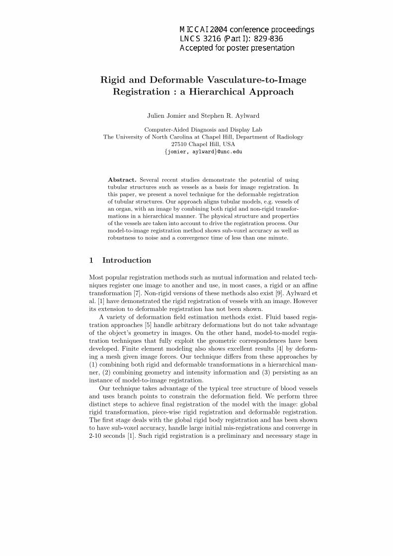
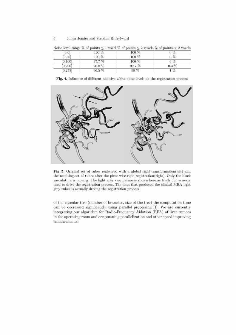
We applied our algorithm on pre- and post-surgery brain Time-of-flight MRAdata in which an arteriovenous malformation (AVM) had been embolized. Thedata volume sizes 256x256x104 and has been isotropically resampled to a spacingof 0.87. Approximately 100 vessels were extracted with an average of 150 pointsper vessel. An initial global rigid registration was performed using a samplefactor of 10, i.e. approximately 15 points per vessel are used for registration.Fig.5-left shows the result. Next we applied 40 piece-wise rigid transformationiterations per branch, Fig.5-right. Fig.6 shows the final registration using bothpiece-wise rigid and non-rigid transformations.
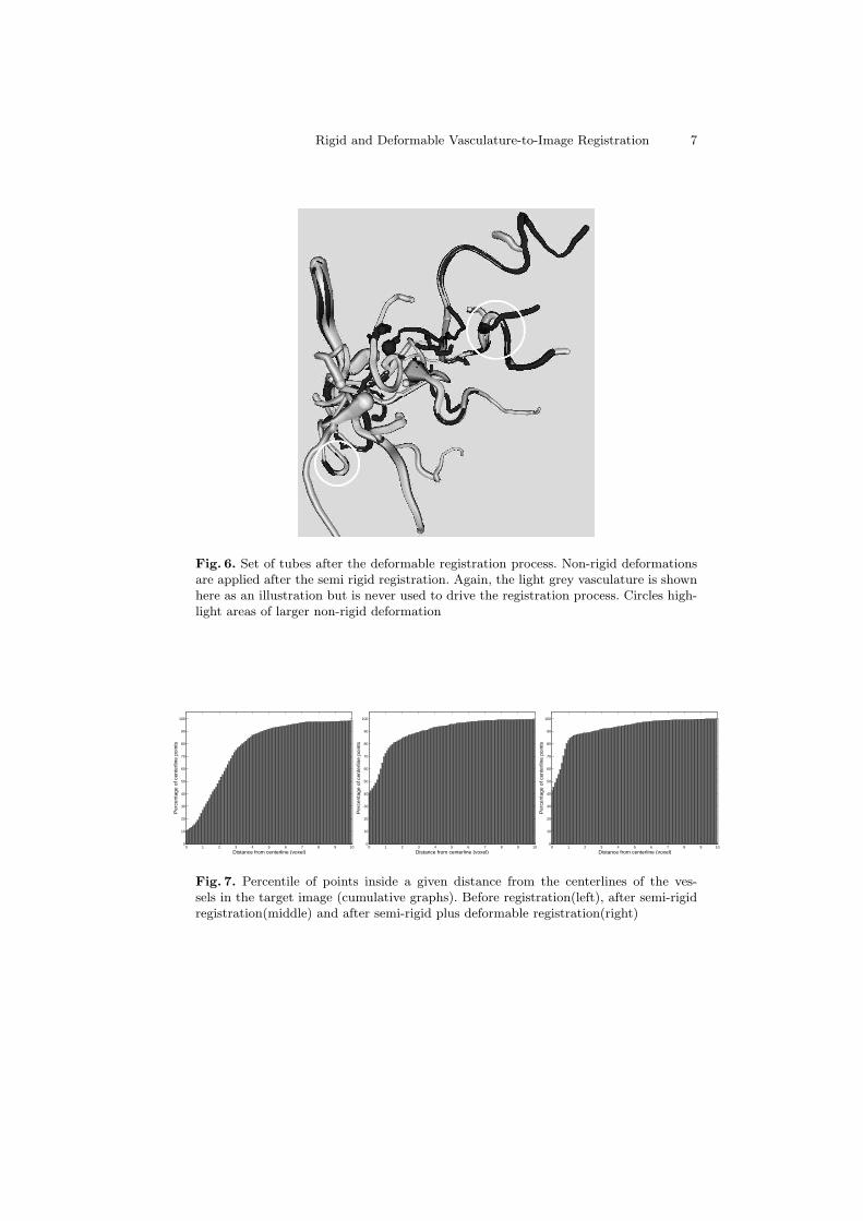
After each stage of the registration process we compute the percentile ofcenterline points inside a given distance from the centerlines of the vessels inthe target image. Figure 7 shows the results. Both stages of the deformableregistration requires less than 10 seconds to converge on a standard desktop PCPentium 4 (2.4GHz) without any parallelization. Depending on the complexity
6 Julien Jomier and Stephen R. Aylward
Noise level range % of points ≤ 1 voxel % of points ≤ 2 voxels % of points > 2 voxels
[0,0] 100 % 100 % 0 %
[0,50] 100 % 100 % 0 %
[0,100] 97.7 % 100 % 0 %
[0,200] 96.8 % 99.7 % 0.3 %
[0,255] 96.5 % 99 % 1 %
Fig. 4. Influence of different additive white noise levels on the registration process
Fig. 5. Original set of tubes registered with a global rigid transformation(left) andthe resulting set of tubes after the piece-wise rigid registration(right). Only the blackvasculature is moving. The light grey vasculature is shown here as truth but is neverused to drive the registration process. The data that produced the clinical MRA lightgrey tubes is actually driving the registration process
of the vascular tree (number of branches, size of the tree) the computation timecan be decreased significantly using parallel processing [1]. We are currentlyintegrating our algorithm for Radio-Frequency Ablation (RFA) of liver tumorsin the operating room and are pursuing parallelization and other speed improvingenhancements.
Rigid and Deformable Vasculature-to-Image Registration 7
Fig. 6. Set of tubes after the deformable registration process. Non-rigid deformationsare applied after the semi rigid registration. Again, the light grey vasculature is shownhere as an illustration but is never used to drive the registration process. Circles high-light areas of larger non-rigid deformation
0 1 2 3 4 5 6 7 8 9 100
10
20
30
40
50
60
70
80
90
100
Distance from centerline (voxel)
Per
cent
age
of c
ente
rline
poi
nts
0 1 2 3 4 5 6 7 8 9 100
10
20
30
40
50
60
70
80
90
100
Distance from centerline (voxel)
Per
cent
age
of c
ente
rline
poi
nts
0 1 2 3 4 5 6 7 8 9 100
10
20
30
40
50
60
70
80
90
100
Distance from centerline (voxel)
Per
cent
age
of c
ente
rline
poi
nts
Fig. 7. Percentile of points inside a given distance from the centerlines of the ves-sels in the target image (cumulative graphs). Before registration(left), after semi-rigidregistration(middle) and after semi-rigid plus deformable registration(right)
8 Julien Jomier and Stephen R. Aylward
4 Discussion and Conclusions
We have developed a novel model to image registration technique that uses bothrigid and deformable transformations in a hierarchical manner. The model, a setof blood vessels, is registered with the image by exploiting the parent-child hier-archy present in the vasculature. Furthermore, elasticity and rigidity coefficientof the vessels are taken into account during the registration process to constrainthe deformation field. Our algorithm shows high accuracy and robustness tonoise on simulated data and operate with ≈ 87% of centerline points within 2voxels on pre-to-post AVM embolization MRA registration.
References
1. Aylward, S., Jomier, J., Weeks, S., Bullitt, E.: Registration of Vascular ImagesInternational Journal of Computer Vision, November 2003, pages 15
2. Aylward, S., Bullitt, E.: Initialization, Noise, Singularities, and Scale in Height-Ridge Traversal for Tubular Object Centerline Extraction IEEE Transactions onMedical Imaging, Feb, 2002, Pages 61-75
3. Danielsson, P.E.: Euclidean distance mapping Computer Graphics and Image Pro-cessing, 14, 1980, pp. 227-248.
4. Ferrant, M., Warfield, S., Guttmann, C., Mulkern, R., Jolesz, F., Kikinis, R.: 3DImage Matching Using a Finite Element Based Elastic Deformation Model MICCAI1999,pp 202-209
5. Lorenzen, P. J., Joshi, S. C.: High-Dimensional Multi-modal Image Registration.WBIR 2003: 234-243
6. Lindeberg, T.: Linear Spatio-Temporal Scale-Space. Scale-Space 1997: 113-1277. Maes, F., Collignon, A., Vandermeulen, D., Marchal, G., Suetens, P.: Multimodality
image registration by maximization of mutual information IEEE Transactions onMedical Imaging, vol. 16, no. 2, pp. 187-198, April 1997.
8. Maintz, J.B.A., Viergever, M.A.: A Survey of medical image registration. In U.Spetzger, H.S. Stiehl, J.M. Gilsbach (Eds.), Navigated Brain Surgery (pp. 117-136).Aachen: Verlag Mainz.
9. Rueckert, D., Clarkson, M. J., Hill,D. L. G., Hawkes,D. J.: Non-Rigid RegistrationUsing Higher-Order Mutual Information. Proc. SPIE Medical Imaging 2000: ImageProcessing, pp. 438-447
This work is funded in part by the Whitaker Foundation (Aylward-RG-01-0341)and the National Library of Medicine’s Insight Toolkit (ITK: Aylward-N01-LM-0-3501)