European Health Risk Assessment Network on Electromagnetic Fields Exposure Risk analysis of human exposure to electromagnetic fields (revised) Deliverable Report D2 of EHFRAN project Due date of submission February 2010 Actual date of submission July 2010 Revised version submitted October 2012 Start date of project February 2009 Duration 42 months Draft prepared by Zenon Sienkiewicz, HPA, UK Joachim Schüz and Aslak Harbo Poulsen, DCS, Denmark Elizabeth Cardis, CREAL, Spain A PROJECT FUNDED BY Executive Agency for Health and Consumers Framework of the Programme of Community Action in The Field of Health 2008-2013
Transcript
European Health Risk Assessment Network on Electromagnetic Fields Exposure
Risk analysis of human exposure to electromagnetic fields (revised)
Deliverable Report D2 of EHFRAN project
Due date of submission February 2010
Actual date of submission July 2010 Revised version submitted October 2012
Start date of project February 2009
Duration 42 months
Draft prepared by
Zenon Sienkiewicz, HPA, UK Joachim Schüz and Aslak Harbo Poulsen, DCS, Denmark
Elizabeth Cardis, CREAL, Spain
A PROJECT FUNDED BY Executive Agency for Health and
Consumers Framework of the Programme of Community Action in The Field of Health
Europe is facing the burden of environmental exposures to many new
physical and chemical agents, some of which may be potentially detrimental to public health. Among these agents, electromagnetic fields
(EMF) are one of the most diffuse and ubiquitous, especially as many new technologies and novel applications based on high frequency fields are
being developed and commercialized.
Research on the possible health and biological effects of EMF is being carried out by many centres in Europe, North America, Japan and other
countries. These activities are supported to various extents by public and private funding bodies at both the national and international levels. The
extent and diversity of these activities, encompassing many areas of medical and biological research, as well as the latest developments in
physics and engineering, make it particularly difficult to provide relevant, authoritative and timely input for the development of public health
policies. Furthermore, it is possible that specific assessments for one situation can be misinterpreted or inappropriately applied to other
exposures or conditions.
In order to help to meet the needs of public health policy makers in these
areas, the European Commission (EC) funded the European Health Risk Assessment Network on Electromagnetic Fields Exposure (EFHRAN)
project. EFHRAN has the specific aim of establishing a wide-ranging network of recognised experts in relevant disciplines that interact and co-
operate to perform a health risk assessment of exposure to EMF across the frequency spectrum. The network consists of a co-ordinator and a
number of associated participants from universities and research centres in seven European countries, as well as 17 collaborating partners from a
further ten countries, which include the World Health Organization (WHO) and three stakeholder associations.
1.1 Objectives and structure of report
EFHRAN is the first project to produce a risk assessment network on EMF
and health issues. In doing so, EFHRAN will provide the EC and EU with a means by which these bodies may react to the present health concerns of
exposure to EMF with full understanding of the scientific issues. EFHRAN is also expected to provide input for future risk management steps, and the
structure of the project is designed with sufficient flexibility to allow for the development of updated assessments in the future.
EFHRAN builds upon the expertise and experience gained by a previous
European Co-ordination Action, entitled “Effects of the Exposure to Electromagnetic Fields: from Science to Public Health and Safer
Workplace” (EMF-NET). This was financed under the 6th Framework
3
Programme by the European Commission. Briefly, the main aims of EMF-
NET were to collate the results of ongoing research into the effects of EMF that were funded by the European Commission or under other national
and international actions, and to provide advice for the development of policy by the European Union and other stakeholders. In addition, it
provided observations on existing research projects in terms of current priorities, gaps in knowledge, results, and emerging technology, as a
means of generating judicious and policy-relevant information concerning the health implications of exposure to EMF. Such information was intended
to facilitate the development of policy options covering public health and consumer protection, health and safety at work, European
competitiveness, and environmental issues. (Complete details of EMF-NET and its many reports and deliverables are available online at
http://web.jrc.ec.europa.eu/emf-net/).
EFHRAN was specifically designed to achieve the following strategic objectives:
Monitor and search for evidence of health risks related to EMF
exposure
Characterize and, where appropriate, quantify potential health risk posed by EMF exposure
Enhance the EC's ability to respond rapidly to health issues and
concerns related to EMF using scientifically sound advice and analyses
Improve the compilation of knowledge and its dissemination on
issues related to EMF and health.
In order to achieve these objectives, the activities of EFHRAN have been divided into five specific objectives: risk analysis and hazard identification;
exposure assessment; dose assessment; risk characterisation; and risk management. These objectives have been further divided into nine work
packages (WP). This report represents the main output and deliverable of WP 4.
This report considers and reviews the latest published research exploring
the possible effects of EMF on humans in order to identify any potential health concerns. Both epidemiological and experimental studies are
considered, for cancer and non-cancer endpoints with separate analyses made for low, intermediate and high frequencies. For the purposes of this
document, low frequencies are defined as time-varying EMF with frequencies of up to 300 Hz; intermediate frequencies as EMF of 300 Hz to
100 kHz; and high frequencies as EMF with frequencies between 100 kHz
Many studies have been published over the last 30 years or so on the biological and health effects of exposure to low, intermediate, and high
frequency electromagnetic fields. It was not feasible to evaluate all the studies on an individual basis for the purposes of this report. Therefore a
number of recent reviews were consulted to establish the current consensus opinion regarding evidence of a variety of health effects. The
reviews included were the 39 reports resulting from EMF-NET (published between 2004 and 2009) and two reports from the Scientific Committee
on Emerging and Newly Identified Health Risks (SCENIHR, 2007, 2009a). These provided a starting point for the health risk analysis. The
monograph on extremely low frequency fields (ELF) (WHO, 2007) and the epidemiological review on health effects of radiofrequency exposure from
the International Commission for Non-Ionizing Radiation Protection (ICNIRP) Standing Committee on Epidemiology (Ahlbom et al, 2004;
ICNIRP, 2009) were also reviewed. More recent studies not available to either SCENIHR or EMF-NET and published after August 2008 were
evaluated separately, and their results incorporated into the consensus opinion. In this way, it was possible to construct an updated health risk
assessment.
In order to evaluate the strength of evidence for adverse effects arising as a consequence of exposure to EMF, EMF-NET used a very simple, yet
powerful, four point classification system that itself was based on the system used by the International Agency for Research on Cancer (IARC)
to report on the carcinogenic risk to humans of a wide range of chemicals and physical agents, including static and extremely low frequency electric
and magnetic fields (IARC, 2002). EFHRAN decided to adopt the same classification system to evaluate the strength of evidence for any
particular effect. The four classifications and criteria for inclusion into any particular category are shown in Table 1.
Clearly, a classification of sufficient evidence requires there to have been
a large amount of high quality research producing a consistent outcome; independent replication is also considered a key element. Similarly,
evidence suggesting a lack of effects indicates that several studies have reported the absence of field-related effects using a range of appropriate
models and relevant exposure conditions.
In May 2011, after the publication of the first draft of the present paper, a group of scientists met at IARC in Lyon, France to assess the
carcinogenicity of electromagnetic fields with frequencies between 30 kHz to 300 GHz (Baan et al, 2011). After examining the relevant evidence
from human, animal and cellular studies, the fields were classified as “possibly carcinogenic to humans (Group 2B)”. This means that a causal
link between RF fields and an increased risk of cancer is considered to be
credible, but some combination of chance, bias or confounding in the data
5
cannot be ruled out with an acceptable degree of confidence. One
consequence of this evaluation is the need for additional epidemiological and experimental studies to help resolve these uncertainties; WHO
recently updated their research agenda for RF fields (van Deventer et al, 2011) which lists high priority and other research needs.
Classification Necessary inclusion criteria
• when a positive relationship is observed
between the exposure and the effect investigated
• when the effect is replicated in several studies by independent investigators or under different
protocols, and when there is a consistent exposure-response relationship
• when confounding factors could be ruled out with reasonable confidence
Sufficient evidence
Limited evidence • when the evidence of the effect is restricted to a few studies, or when there are unsolved
questions regarding the adequacy of the design, conduct or interpretation of the study
• when confounding factors could not be ruled out in the studies with reasonable confidence
Inadequate evidence
• when the studies are of insufficient quality,
consistency or statistical power to permit a conclusion
Evidence suggesting
a lack of effects
• when no effects are reported in several studies
by independent investigators under different protocols involving at least two species or two
cell types and a sufficient range of field intensities
Table 1. The four point system used in this report to classify the strength of evidence for any particular effect; a similar system was
used by EMF-NET.
6
Because of the assessment by the Working Group, it was decided to
update the present report to include more recent studies on the effects of exposure to high frequency fields that were available to the Working
Group and published after the original report from EHFRAN, and to see whether inclusion of these data necessitated a revision of the original
strength of evidence classification for high frequency fields.
2 Low frequencies (up to 300 Hz)
For more than a century, exposure to extremely low frequency (ELF) electric and magnetic fields has been ubiquitous, related to the
production, transmission, distribution and use of electric currents. Research into the possible adverse health effects of such exposures
intensified in the late 1970s, with epidemiological and experimental studies focusing mainly on cancer, neurodegenerative diseases,
cardiovascular diseases, reproductive effects, and non-specific symptoms affecting well-being. On the exposure side, research has focussed on
residential exposures, for instance people living close to power lines, on occupational exposures such as for electricians, and on the use of electric
household appliances. While some studies estimated exposure in a crude way, using simply the distance between the residence and the nearest
power line, using broad job titles to categorize occupational exposure, or
asking study participants about past use of electric appliances, assessment methods have been refined over the years and comprehensive
stationary or personal measurements as well as detailed job-exposure-matrices based on work activities have been developed. In addition to
studies on health effects (WHO, 2007), many measurement surveys have been conducted to better understand the distribution of exposure in time
and space and the relative contribution of various exposure sources to an individual’s total exposure. For all European countries where measurement
data are available (described in EFHRAN Deliverable D4: Report on the level of exposure in the European Union), it appears that average
exposure over 24 hours is usually well below 0.1 microtesla (µT), and the proportion of the general population exposed to average ELF magnetic
fields above 0.2 µT is small, i.e., between 1-5%; average exposures to magnetic fields exceeding 1 µT are exceptional but may occur in
residences just beneath high-voltage power lines or with transformers in the basement, or in the context of certain occupations, e.g., among
electric welders, electricians, electric power engineers, or locomotive engineers.
2.1 Current consensus opinion
Although numerous studies have been completed in this field, the
evidence remains ambiguous. The major reasons for this are that study results are inconsistent and many studies have suffered from
methodological shortcomings. It is therefore important to continuously
7
review the body of evidence. This has recently been done by three
international organisations; namely, the World Health Organization (WHO, 2007), the EMF-NET project of the European Union (EMF-NET, 2009), and
the Scientific Committee on Emerging and Newly Identified Health Risks (SCENIHR) of the European Commission (SCENIHR, 2009a). Overall,
these risk analyses are in line with assessments carried out by national authorities responsible for radiation protection issues. A comparison of
these risk assessments demonstrates few disparities, hence, the 2009 report of SCENIHR is used to illustrate the current consensus opinion
(SCENIHR, 2009a). The SCENIHR report included scientific evidence published up to the end of 2008.
SCENIHR reported limited evidence for an association between exposure
to ELF magnetic fields and the risk of childhood leukaemia (SCENIHR, 2009a). This was based on a classification performed by the International
Agency for Research on Cancer in 2001, ranking ELF magnetic fields as possibly carcinogenic to humans (Group 2B) (IARC, 2002). The
classifications then as today were based on the facts that epidemiological studies showed a consistent association between magnetic fields above
approximately 0.3/0.4 µT and a doubling in risk for childhood leukaemia, although chance, bias and confounding could not be ruled out as an
explanation with reasonable confidence, but that experimental studies or mechanistic modelling provided little support for, or explanation of, these
findings.
Since the assessment in 2001, further epidemiological studies were conducted. However, these did not provide further insight, instead being
consistent with the previous assessment (Schüz and Ahlbom, 2008). New experimental studies did not strengthen the biological plausibility of the
observed association either (SCENIHR, 2009a). SCENIHR has noted that overall little targeted research has been done to reconcile the disparity
between epidemiological and mechanistic data and suggested that ELF magnetic fields and childhood leukaemia should be a high priority research
area (SCENIHR, 2009b). For cancers other than childhood leukaemia there was either inadequate evidence or some evidence against an association
(SCENIHR, 2009a).
SCENIHR further reported that some recent studies support previous notions that the risk of Alzheimer’s disease may be linked to exposure to
ELF magnetic fields (SCENIHR, 2009a). While the majority of studies has been done in relation to occupational exposures, the first study on
residential exposures was conducted in Switzerland and this suggested an increased risk of Alzheimer’s disease among people living close to high-
voltage power lines (Huss et al, 2009). Based on these findings, SCENIHR classified ELF magnetic fields and Alzheimer’s disease as a high priority for
further research (SCENIHR, 2009b). For other neurodegenerative diseases
the evidence appears to be weaker. The possible association between
8
occupational exposure to ELF magnetic fields and the risk of amyotrophic
lateral sclerosis is discussed in detail in the WHO risk assessment (WHO, 2007). However, the evidence was classified as inadequate, mainly due to
possible confounding by electric shocks or chemical exposures at the respective work places, and since then no new influential studies have
been published. For Parkinson’s disease and multiple sclerosis there are fewer studies, but they show no consistent indications of an increased
risk. For cardiovascular diseases more recent studies suggest an absence of any association (SCENIHR, 2009a).
Lastly, SCENIHR (2009a) concluded that there is no consistent
relationship between exposure to ELF fields and a variety of self-reported symptoms, such as skin irritations, headache, sleep problems,
concentration difficulties, or fatigue.
2.2 More recent studies
2.2.1 Epidemiology
With regard to the childhood leukaemia findings, results of a recent pooled
analysis of studies from Germany, Italy, Japan, Tasmania and UK (Kheifets et al, 2010) are consistent with those of the previous pooled
analyses by Ahlbom et al (2000) and Greenland et al (2000), with an odds ratio (OR) of about 2 for magnetic fields above approximately 0.4 µT.
There was some indication of a possible exposure-response relationship, but overall this analysis did not alter the previous IARC assessment that
magnetic fields are possibly carcinogenic to humans.
In order to gain further insights into this association, new pilot activities
have been started in an attempt to identify cohorts of children with increased ELF magnetic field exposure in order to reduce the impact of
participation bias that has affected previous case-control studies; these activities aim at identifying residences with transformers leading to higher
exposures in children (Ilonen et al, 2008) or suggest how to use existing birth cohort studies in this context (Greenland and Kheifets, 2009). A
recent methodological study explains why further studies applying the simple distance-to-power-line metric are unlikely to provide new insights
(Maslanyj et al, 2009). The hypothesis that ELF magnetic field exposure is related to a poorer survival after childhood leukaemia, suggesting that ELF
magnetic fields promote the growth of leukemic cells resulting in a recurrence of the disease, continues to be followed up. Indeed, poorer
survival has been observed in the hypothesis-generating study in the USA (Foliart et al, 2006); this was broadly confirmed in a subsequent study
from Germany (Svendsen et al, 2007), but since both studies included very small numbers of exposed children no firm conclusions could be
made. An ongoing project on this issue is expected to provide further insight as for this purpose, whereby data on cases enrolled in previous
case-control studies from the US, the UK, Canada, Germany, Japan, New
9
Zealand and the Nordic countries (Ahlbom et al, 2000) will be pooled and
health status of these cases followed up.
Mezei et al (2008) conducted a meta-analysis of 13 studies on residential exposure to ELF magnetic fields and the risk of brain tumours in children
and observed a statistically non-significant 70% increased effect estimate at exposures above 0.3/0.4 µT; In a subsequent meta-analysis of the
original data, no association was observed between level of ELF magnetic fields and risk of brain tumours (Kheifets et al., 2010b).
A recent US case-control study of occupational exposures and risk of brain
tumours in adults did not show an association (Coble et al, 2009). This was consistent with findings of a recent meta-analysis pooling more than
20 studies (Kheifets et al, 2008). It was concluded that while a small increase of 10% was observed in the summary risk estimate, the more
recent and methodologically improved studies showed weaker associations than the earlier studies, providing little evidence for an association.
Yenugadhati et al (2009) explored associations between various occupations and the risk of lung cancer in a Canadian case-control study
and discuss a possible role of exposure to EMF for some of their findings; however, due to this rather indirect approach the evidence remains
unchanged.
All recent studies on neurodegenerative diseases had already been included in the SCENIHR report (SCENIHR, 2009a) and no new studies
have appeared in the meantime. Another US study on cardiovascular disease confirmed previous findings of no association (Cooper et al,
2009).
2.2.2 Experimental studies
Very few recent studies have investigated the effects of low frequency fields on volunteers. Overall, these studies only provide very limited
additional information, and they do not substantially alter the previous health risk assessment.
Bellieni et al (2008) investigated the possible effects of fields generated
by electric motors in incubators on autonomic function in newborn babies. Transient changes in the total power and spectral components of heart
rate variability (HRV) were noted when the motors were running. Confirmatory studies are required to determine the significance of this
observation. Lednev et al (2008) reported that heart rate variability in adults was affected by exposure to very weak fields (above 2 µT); the
direction of change was dependent on the frequency used.
Cook et al (2009) reported changes in alpha activity measured over the occipital-parietal regions of the brain after acute exposure of volunteers to
10
two weak pulsed magnetic field sequences (+/- 200 µT peak). The
direction of change depended on the specific sequence used.
Albert et al (2009) found no evidence that exposure of male and female volunteers to a 60 Hz magnetic field at 200 µT for 4 h exposure could
cause either DNA damage in peripheral blood leukocytes as assayed using the alkaline comet assay, or increased incidence of micronuclei. Two
independent studies have examined the effects of occupational exposure to magnetic fields by examining peripheral blood of exposed workers. At
best, these provide only weak evidence for a field-related effect on natural killer cell activity (Gobba et al, 2009) and antioxidant activity (Sharifian et
al, 2009). Previously, Dasdag et al (2002) investigated effects of magnetic fields on haematology and immunology in welders. Despite measuring
differences between exposed and control subjects in haematocrit levels and in T lymphocyte surface antigens, these changes were not considered
to be clinically significant.
Lastly, Skomro et al (2009) found that repeated, acute exposures to low frequency magnetic fields at 3 or 4 µT had no consistent effect on the
content of calcium, magnesium and fluoride ions in saliva.
2.3 Summary and Conclusions
The strength of evidence for each health outcome is summarised in Table 2. This has been derived from the previous evaluations of EMF-NET (2009)
and SCENIHR (2009a) synthesized, where considered relevant, with the more recent data described in the present evaluation.
For none of the diseases is there sufficient evidence for a causal
association between exposure to low frequency fields and the risk of the respective disease.
There is limited evidence for an association between magnetic fields and
the risk of leukaemia in children. This evaluation reflects the current state of knowledge that epidemiological studies have shown an association
between residential exposures to power frequency magnetic fields at above approximately 0.3/0.4 µT and a two-fold risk of childhood
leukaemia with some degree of consistency, but the observed association alone is not sufficient to conclude a causal relationship due to the
following three reasons:
i) there is no known mechanistic explanation for the observed association and none of the hypotheses put forward are convincingly supported by the
data;
ii) overall, experimental studies do not provide evidence that low frequency magnetic fields are carcinogenic;
11
iii) a combination of chance, bias and confounding may well have
produced a spurious association in the epidemiological studies.
It is unlikely that further epidemiological studies of the same design as that used previously will provide any new insights. New concepts to
identify cohorts of children with higher exposures may turn out to be promising. If the hypothesis of a poorer survival of children with
leukaemia is confirmed by other studies, this will increase the biological plausibility of a causal association. Conversely, further methodological
work investigating the impact of possible biases in the childhood leukaemia studies may shift the evidence in the opposite direction.
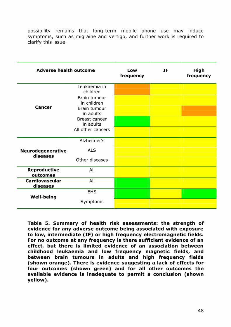
Outcome Strength of evidence
Cancer outcomes
Leukaemia in children Limited
Brain tumours in children Inadequate Brain tumours in adults Inadequate
Breast cancer in adults Lack of effect Other cancer (children or adults) Inadequate
Neurodegenerative diseases
Alzheimer’s disease Inadequate
Amyotrophic lateral sclerosis (ALS) Inadequate Other neurodegenerative diseases Inadequate
Reproductive outcomes
All outcomes Inadequate
Cardiovascular diseases
All diseases Lack of effect
Well-being
Electrical hypersensitivity (EHS) Lack of effect Symptoms Inadequate
Table 2. The strength of evidence for any health outcome being
associated with exposure to low frequency magnetic fields as suggested by EMF-NET (2009) and SCENIHR (2009a) and modified
by the results of more recent research.
12
There is inadequate evidence with respect to several diseases, however. For Alzheimer’s disease the evidence is suggestive; however compared to
the data on childhood leukaemia, there are far fewer epidemiological studies and the results are less consistent. Since recent, methodologically
superior studies are suggestive of an association, there is ample justification to demand further studies into this topic. The situation is
similar for childhood brain tumours, where awaited results of an ongoing pooled analysis may make a re-evaluation necessary. Amyotrophic lateral
sclerosis is a third outcome for which there is some indication of an elevated risk, but the data are not consistent enough to support a
classification of limited evidence.
For brain tumours in adults, it appears that more recent studies tend to suggest a lack of an association, but due to positive findings in some
studies the classification of inadequate evidence remains more appropriate.
For all other cancers, other neurodegenerative diseases and for subjective
symptoms, the classification of inadequate evidence displays rather the lack of data. However, due to the weak biological plausibility there
appears to be no emerging demand to conduct further studies. There is lack of evidence for any association between exposure to low
frequency magnetic fields and breast cancer or cardiovascular disease. For breast cancer, no new studies have been published, but as there were
already a large number of studies available at the time of the previous evaluations, this assessment is considered robust. For cardiovascular
disease, one new study was published that confirmed the absence of any association.
There is continuing public debate about whether non-specific symptoms
may be caused by exposure to low frequency fields, and whether some individuals show increased sensitivity to exposure, commonly termed
electrical hypersensitivity (EHS). As this is a long-lasting discussion characterised by a series of failures to demonstrate the existence of EHS,
the overall evaluation suggests a lack of any effect. Given the uncertainty regarding the role played by EMF in the aetiology of this condition, the
World Health Organization (WHO) has proposed that EHS should be better termed Idiopathic Environmental Intolerance with attribution to EMF.
3 Intermediate frequencies (300 Hz – 100 kHz)
Exposure to intermediate frequency (IF) fields has in the past largely been restricted to long-range radio, welding devices, cathode-ray based
monitors and magnetic resonance imaging (MRI). However, sources and exposures to these fields are now increasing due to the development of
13
new and emerging technologies, such as anti-theft devices, badge readers
and induction hobs and hotplates; compact fluorescent lights also produce fields in the IF range. However, explicit data on the possible health effects
of IF fields remain limited.
3.1 Current consensus opinion
For the purposes of risk assessment, IF fields have only been considered
as a separate entity relatively recently. Largely depending on the definition of their frequency range, IF fields have been considered in
various reviews and monographs alongside either low or high frequency fields. IF fields can induce electric fields and currents in the human body,
much as is seen with low frequency fields, but they can also induce heating effects in the body as seen in high frequency field exposures.
Assessments of possible hazards at intermediate frequencies are based
primarily on extrapolation from data on exposure to higher and lower frequencies (SCENIHR, 2007, 2009a).
Very little useful epidemiological data are available. The existing evidence
largely comes from older studies that tended to used job title as a surrogate for exposure. Groups studied include users of visual display
units (VDUs) associated with personal computers and radio and telegraph operators. Outcomes studied included cancer as well as effects on the eye,
the cardiovascular system and the reproductive system. Although no particular risks were indentified, the quality of existing studies is limited,
There have been some animal studies exploring the effects of IF fields
from VDUs, particularly on reproduction and development. This older literature has been well reviewed previously. There are fewer studies on
humans, although some studies have investigated effects of IF fields on skin. With the demise of cathode-ray based monitors, more recent work
exploring health risks associated with computer use in humans has concentrated on ergonomic issues (and is not considered here). Despite
some limited evidence from animal studies that have reported field-dependent effects on reproduction and development, there is no
consistent or conclusive evidence of field-dependent adverse effects.
Overall, SCENIHR (2009a) concluded that there was insufficient data for a health risk assessment, so the overall evaluation for all health endpoints
has to be considered to be inadequate. Since then, an IARC Working Group has classified electromagnetic fields with a frequency range of
30 kHz to 300 GHz as being “possibly carcinogenic to humans”. This decision was informed by studies using fields above 100 kHz fields, and is
discussed in Section 4.
14
3.2 More recent studies
3.2.1 Epidemiology
No recent epidemiological studies investigating risks of IF fields have been published.
3.2.2 Experimental studies
No recent volunteer studies investigating IF fields have appeared.
3.3 Summary and Conclusions
Interest in the potential of IF fields to cause adverse effects has been
sporadic at best and no recent research appears to have investigated exposures associated with new or emerging technologies. The available
evidence is insufficient to conclude about whether or not an association may exist between exposure and the risk of any disease.
Given the lack of recent data, it is not possible to revise the existing
classification, and therefore the strength of evidence for all outcomes remains as inadequate (Table 3). Given that occupational exposures to
these frequencies are increasing, it would be useful if well targeted studies could be performed as a priority to address this lack of research.
Outcome Strength of evidence
All outcomes Inadequate
Table 3. The strength of evidence for any health outcome being associated with exposure to intermediate frequency fields as
suggested by EMF-NET (2009) and SCENIHR (2009a); there is a lack of more recent research.
4 High frequencies (100 kHz – 300 GHz)
Research into the possible effects of exposure to low level radiofrequency
(RF) fields has increased over the last decade or so following the widespread increase in mobile phone usage and the roll out of base
station networks. More recently, concerns have been raised about DECT cordless phones, and interest in the potential health effects of wireless
LANs and Wi-Fi has followed the introduction of these applications into schools, homes and workplaces. However, the effects of RF fields
15
associated with commonly occurring sources in the environment, such as
broadcasting, radar, and microwave communication links have been considered for many years before that, and an extensive effects literature
had been generated. ICNIRP have reviewed much of these data (Ahlbom et al, 2004, 2009; ICNIRP 2009; van Rongen et al, 2009; Swerdlow et al,
2011).
4.1 Current consensus opinions
Early epidemiological investigations centred on a variety of occupational
groups with the potential for high exposures to RF fields, such as radar technicians, and radio and telegraph operators, with interest focussed on
brain tumour and leukaemia risks. In general, results from these studies were inconsistent, and no conclusion could be drawn, due to the generally
small size and/or methodological limitations of many of these studies as
well as very limited exposure assessment. Other studies investigated risks to people living near radio or TV transmitters. These studies did not
demonstrate the existence of any risk; again, results have been inconsistent, but were dependent on very crude measures of exposure
such as using distance from broadcasting masts (Baan et al, 2011).
Very few volunteer studies have been undertaken, but a range of in vivo and in vitro studies have indicated that consistent effects are only
observed for exposures that increase whole body or localised tissue temperatures by about a degree or more. Such thermal responses remain
a cornerstone of existing guidelines limiting human exposures to RF fields (e.g. ICNIRP, 1998). Effects of RF fields in the absence of overt heating
have been reported, but they remain controversial, and the mechanism whereby such effects may be caused remains elusive.
More recent studies investigating the health risks of RF fields have been
summarised and reviewed by EMF-NET, and by SCENIHR (2007, 2009a). These studies concentrated on cancer risks from the use of mobile
phones, but other endpoints and sources have been considered; attention is also starting to be given to new and emerging technologies, such as
ultra wide band signals.
SCENIHR (2009a) reviewed the evidence from the various national studies and pooled analyses from parts of the Interphone study: severe concerns
were raised about reporting biases that may exist in these data. Nonetheless, it was concluded that this evidence, combined with the
results of animal and cellular studies, indicated that exposure to RF fields was unlikely to lead to an increase in brain cancer or parotid gland
tumours in humans. However, it was noted that since the widespread duration of exposure of humans to the fields from mobile phones was
shorter than the induction time of some cancers, further studies were required to identify whether exposure periods in excess of ten years may
pose some cancer risk. Regarding shorter periods of exposure, it was
16
concluded that mobile phone use for less than ten years was not
associated with increased cancer incidence. In addition, SCENIHR (2009a) concluded that two well-conducted case-control studies investigating the
association between the fields from broadcast transmitters and childhood leukaemia provided no evidence for such an association.
On non-cancer outcomes, it was concluded that the available scientific
evidence failed to provide support for an effect of RF fields on self-reported symptoms. Although an association between RF exposure and
single symptoms was indicated in a few cross-sectional studies, there was a lack of consistency in these findings, and several provocation studies
indicated a lack of effect on well-being using handset or base stations signals (SCENIHR, 2009a). Furthermore, a number of studies reporting on
sensitivity to RF exposure have demonstrated that neither self-diagnosed cases nor healthy controls could reliably detect the presence of either GSM
or UMTS signals. The possibility that nocebo effects may play a role in symptom formation was highlighted.
Regarding effects of RF fields on the brain and nervous system, several
studies using volunteers have reported no consistent effects on various behaviours or cognitive functions, although sporadic effects were noted in
some studies. A large number of studies have reported that exposure has no detectable effect on either the auditory or visual systems. Some, but
not all studies have reported effects on sleep and sleep encephalogram (EEG) patterns, and others have reported an effect on specific EEG
components during exposure. However, SCENIHR questioned the relevance of these subtle changes to health, and noted that no interaction
mechanism could be identified.
Epidemiological studies investigating the effects of RF fields on adverse pregnancy outcomes are limited mainly to occupational exposures among
physiotherapists (SCENIHR, 2007). Despite some positive findings, no consistent adverse outcome has been reported, but the available results
do not allow any definite conclusions to be drawn due to the limited statistical power and potential recall bias in the data. Including more
recent data did not change this conclusion (SCENIHR, 2009a).
Studies investigating effects of RF fields on fertility or sperm quality in men also have failed to provide consistent evidence of adverse effects.
These have investigated occupational exposures in the Norwegian Navy and in those attending infertility clinics. However these studies suffer from
a number of weaknesses, including self reporting of endpoints, and a lack of measurement of RF fields in the occupational studies, and confounding
due to lifestyle differences in the clinic studies, making them inadequate for the purposes of risk assessment.
17
In May 2011, IARC convened a multinational Working Group of 30
scientists to assess the carcinogenicity of RF fields (Baan et al, 2011. Based on a critical evaluation of the peer-reviewed epidemiological and
experimental evidence that had been published or was in press at the time of the meeting (a number of these studies were not available to either
EMF-NET or SCENHIR in 2009), the IARC Working Group classified RF fields as “possibly carcinogenic to humans (Group 2B), based on limited
evidence in humans for the carcinogenicity of RF fields1, limited evidence of carcinogenicity in animals and weak mechanistic evidence relevant to
RF induced cancer in humans.
The epidemiological evaluation was based on positive associations between glioma and acoustic neuroma and exposure to RF fields from
wireless telephones. The evidence to draw conclusions for other types of cancer was judged inadequate.
A few members of the Working Group, however, considered that all the
evidence in humans was inadequate for a number of reasons, including inconsistencies between the two major case-control studies, and an
absence of an increase in brain cancer incidence rates in time-trend data; another consideration was the Danish cohort study of mobile phone users
which has not observed any increase in rates of glioma or acoustic neuroma.
The complete assessment will be published as Volume 102 of the IARC
Monograph series. Other agents previously evaluated by IARC as Group 2B include extremely low frequency magnetic fields, carbon tetrachloride,
chloroform and coffee.
Shortly after the IARC Monographs evaluation, the International Commission for Non-Ionizing Radiation Protection (ICNIRP) Standing
Committee on Epidemiology published a review (Swerdlow et al, 2011), which did not include some of the more recent papers reviewed by the
IARC Working Group. It concluded that, although there remains some uncertainty, the trend in the accumulating evidence is increasingly against
the hypothesis that mobile phone use can cause brain tumours in adults. Results from current epidemiological, biological and animal studies, and
brain tumour incidence trends, suggest that within about 10-15 years after first use of mobile phones there is unlikely to be a material increase
1The definitions used by IARC are similar, but not identical, to those used in this report and elsewhere by EFHRAN (Table 1). IARC define limited evidence of carcinogenicity as “a positive association has been observed between exposure to the agent and cancer for which a causal interpretation is considered by the Working Group to be credible, but chance, bias or confounding could not be ruled out with reasonable confidence”. Whereas inadequate evidence of carcinogenicity is defined as “the available studies are of insufficient quality, consistency or statistical power to permit a conclusion regarding the presence or absence of a causal association between exposure and cancer, or no data on cancer in humans are available”.
18
in the risk of brain tumours in adults. Data for childhood tumours and for
periods beyond 15 years are currently lacking.
4.2 Other recent studies
4.2.1 Epidemiology
Since the current report was first issued, a number of epidemiological
studies have been published, some investigating the risks of short-term mobile phone use by adults on brain cancer and other tumours of the
head, others investigating effects on child development and behaviour. As yet, there are sparse data on the health risks associated with long-term
mobile phone use.
Brain tumour case-control studies – adults Hardell et al (2011) studied the association between use of mobile and
cordless phones and brain tumours by pooling data from two previous case-control studies on patients with malignant brain tumours diagnosed
during 1997-2003. The risk appeared to increase with latency period and cumulative hours of use for both mobile and cordless phones. The highest
risk was found for astrocytoma: the OR for mobile phone use was 2.7, (95% CI 1.9-3.7) and that for cordless phone use was 1.8 (95% CI 1.2-
2.9) for use 10 years or more in the past. Risks were not higher for tumours in the temporal lobe than overall (Hardell et al., 2011). The risk
for astrocytoma appeared to be highest in the group with first use of a wireless phone before the age of 20. The similarity of ORs for mobile and
cordless phones is unexpected given the difference in average output power of these phone types.
Results of the Interphone international analyses of glioma, meningioma and acoustic neuroma have been published (the Interphone Study Group,
2010, 2011). Analyses included 2708 glioma, 2409 meningioma and 1105 acoustic neuroma cases and their matched controls. A reduced OR related
to ever having been a regular mobile phone user was seen for glioma (OR 0.81, 95% CI 0.70, 0.94), meningioma (OR 0.79; 95% CI 0.68, 0.91) and
acoustic neuroma (OR 0.85, 95% CI 0.69-1.04), possibly reflecting participation bias or other methodological limitations. No elevated OR was
observed 10 or more years after first phone use (glioma: OR 0.98, 95% CI 0.76, 1.26; meningioma: OR 0.83, 95% CI 0.61, 1.14; acoustic
neuroma: OR 0.76, 95% CI 0.52–1.11). Odds ratios were below 1.0 for all deciles of lifetime number of phone calls and nine deciles of cumulative
call time. There was no trend of increasing ORs with increasing cumulative call time or cumulative number of calls but higher odds ratios were seen
for all tumour types in the highest decile of recalled cumulative call time, 1640 hours or longer: 1.40 (95% CI 1.03, 1.89) for glioma, 1.15 (95% CI
0.81, 1.62) for meningioma, and 1.32 (95% CI 0.88–1.97) for acoustic neuroma. With censoring at 5 years before the reference date in an
attempt to take into account the slow growth and possible long diagnostic
19
delay for this disease, the OR for acoustic neuroma in the highest decile of
cumulative call time was 2.79 (1.51–5.16). There were, however, implausible values of reported use in this group.
Odds ratios in the highest decile of cumulative call time tended to be
greater in the temporal lobe than in other lobes of the brain for glioma (but not meningioma), but the confidence intervals around the lobe-
specific estimates were wide. Odds ratios in the highest decile were also greater in subjects who reported usual phone use on the same side of the
head as their tumour than on the opposite side for glioma, meningioma and acoustic neuroma. For glioma, ipsilateral ORs were almost always
greater than contralateral ORs. There was a trend towards a stronger effect of ipsilateral use relative to contralateral use with increasing
cumulative number of calls as well as with increasing cumulative call time (except for the lowest exposure category where the ipsilateral to
contralateral ratio was highest).
The Interphone Study Group concluded that no increase in risk of glioma, meningioma or acoustic neuroma was observed overall in association with
use of mobile phones. It noted, however, that there were suggestions of an increased risk of glioma at the highest exposure levels, but biases and
errors prevent a causal interpretation. The Group concluded that possible effects of long-term heavy use of mobile phones require further
investigation.
Further analyses of subgroups of Interphone countries have been conducted to further explore the observed associations, taking into
account the localized nature of RF energy absorption in the brain when using a mobile phone (Larjavaara et al, 2011; Cardis et al, 2011a,b).
A case-only analysis of data from seven Interphone countries (Denmark,
Finland, Germany, Italy, Norway, Sweden, UK-South) was conducted to evaluate whether gliomas occur preferentially in the areas of the brain
having the highest exposure to RF fields, based on estimated distance from the centre of their tumour to a hypothetical phone axis (Larjavaara
et al, 2011). No difference was found between tumours with a centre within 5 cm of the phone line and tumours with a centre further than 5
cm, either in terms of ever having used a mobile phone regularly or duration of phone use. Complementary case-specular analyses were also
conducted, in which the distance from the centre of the tumour to the phone axis was compared between the cases and their “specular” controls
(for each case, the location of the specular tumour was obtained as a mirror image in 2 dimensions - within the same brain hemisphere - of the
location of the original tumour). In these analyses, an OR of 2.00 (95% CI 0.68, 5.85) was observed among long term users (10 years or more)
based on small numbers of cases.
20
Cardis and colleagues investigated the main parameters thought to
influence absorption of RF energy in the brain from mobile phone use (Cardis et al, 2011a). This was based on information from the Interphone
questionnaire, network operators, and laboratory measurements and from software-modified phones issued to a subset of study participants. An
algorithm was developed to evaluate the total cumulative RF energy (in joules per kilogram), or dose, absorbed at a particular location in the
brain. The main determinants of absorbed energy were the communication system and frequency band, location in the brain and the
amount and duration of mobile phone use. Results of epidemiological analyses of total cumulative RF energy are therefore potentially subject to
recall biases, like those of the more traditional analyses based only on amount and duration of use. Though there was substantial agreement
between categorisation of subjects by cumulative absorbed energy and cumulative call time (the exposure variable used in the main Interphone
analyses and in many other epidemiological studies), misclassification appeared non-negligible, particularly at higher frequency bands.
The above algorithm was applied to Interphone Study subjects in five
countries (Australia, Canada, France, Israel and New Zealand) (Cardis et al, 2011b). An increased risk of glioma was seen in individuals at the
highest quintile of absorbed dose, though reduced risks were seen in the four lower quintiles. When risk was examined as a function of absorbed
dose received in different time windows before diagnosis, an increasing trend was observed with increasing absorbed dose for exposures 7 years
or more in the past. Due to small numbers of subjects, it was not possible to use the same time windows 5-9 and 10+ years as in the INTERPHONE
study. Complementary case-case analyses (in which laterality of phone use was not considered to avoid a possible laterality recall bias), also
indicated an increased risk in the most exposed region of the brain, based on small numbers of subjects, compared with other areas among long-
term users. Patterns of risk for meningioma in relation to absorbed dose were similar, although increases in risk were much smaller than for
glioma, and not statistically significant. These results may suggest an increased risk of glioma in the most exposed area of the brain among
long-term and heavy users of mobile phones. However, the exposure algorithm still relies heavily on the bias-susceptible questionnaire data,
and as pointed out by the authors, there are uncertainties associated with tumour centre localisation, estimation of absorbed dose, and sample size.
These results require replication in an independent and preferably improved setting before they could be taken to indicate a cause-effect
relationship.
The reasons for the differences in the results of the two studies of independent subsets of Interphone countries are unclear. However, there
are differences in the detail of exposure assessment – for the case-case
analyses based only on location of the tumour, Cardis et al (2011b)
21
defined the most exposed area of the brain from analyses of results of
experiments on the spatial distribution of the specific energy absorption rate (SAR) on phantoms for over 100 phone models (Cardis et al, 2008),
while Larjavaara et al (2011) calculated distance from the centre of the tumour to a hypothetical phone axis. Different approaches were also used
to define the centre of the tumour in both studies. Analyses are underway to compare the two approaches and further evaluate risk as a function of
tumour location and absorbed dose to the tumour.
Brain tumour case-control studies – children and adolescents A multicenter case–control study of brain tumours in young people (aged
7-19 years) was conducted in Denmark, Sweden, Norway, and Switzerland including all cases diagnosed between 2004 and 2008 (Aydin
et al, 2011). Analyses included 352 cases and 646 population controls matched by age, sex, and geographical region. The OR for ever having
been a regular mobile phone use was 1.36; 95% CI = 0.92 to 2.02). There was no association with duration or amount of use. In a small
subset of study participants for whom operator recorded data were available, brain tumour risk was related to the time elapsed since the
mobile phone subscription was started but not to amount of use. The subjects in this study were young (the median age at diagnosis was 13
years) and the study included very few long-term or heavy users.
Cohort studies The cohort of all subscription holders in Denmark until end of 1995 has
been updated for all Danes aged 30 or older and born after 1925 (Frei et al 2011). The new analysis had 3.8 million person years of follow-up, and
included detailed socioeconomic indicators. Because of the comprehensive health and population registers in Denmark, there was only 2% loss to
follow-up and virtually complete case ascertainment providing 356 exposed glioma cases. There was no suggestion of an increased risk of
glioma in subscription holder overall or after 10 or more years of subscription (men: IRR 1.06, 95%CI: 0.85-1.26, n=117, women: IRR:
1.04, 95%CI: 0.56-1.95, n=10), nor was there any increase among men with 13+ years of subscribing (n=37). The study, however, relies on
having a subscription registered to a named individual before the end of 1995 for exposure. The reference population therefore includes any
corporate paid subscriptions not registered to an individual as well as any prepaid cards without a registered user. Also persons only having a
subscription after 1995 will be in the reference population. Although the reference group is therefore never completely unexposed, the low
percentage of misclassified persons in the reference category before 1996 together with adjustment for calendar period, and analysis by duration of
use, ensures that the exposed group will always include more users than the reference group. The crude exposure assessment does however mean
that the study cannot address effects that are small or restricted to small
user-segments such as very heavy users.
22
While this is a very large cohort study, and is very useful for surveillance
of multiple endpoints, the study has a number of limitations. There is potential for substantial misclassification (Schüz and Johansen, 2007;
Ahlbom et al, 2007). Indeed, the cohort is based on the fact of having a personal subscription at any time between 1982 and 1995 – there is no
information about the actual identity of the user or the amount of use; however, subscribers were estimated to be 4 times more likely to be
regular users of mobile phones than non-subscribers.
Brain tumour – time trend analyses Several recent studies have analysed time trends in brain tumours in
relation to mobile telephone use in different countries. Deltour et al
(2009) studied trends in incidence rates of brain tumours between 1998 and 2003 in the Nordic countries (Denmark, Finland, Norway, Sweden)
and found no clear change overall, with rates were either stable, decreasing, or continuing a gradual increase that started before the
introduction of mobile phones. Time trends appeared to be similarly unaffected in the United States up to 2006/2007 (Inskip et al 2010,
Kohler et al 2011), and in the United Kingdom up to 2007 (de Vocht et al 2011), though increases in rates for tumours of the temporal lobe were
observed in men and women in the later study, along with decreases in rate of tumours in the parietal lobe, cerebrum and cerebellum in men.
A more recent analysis of the Nordic countries data (Deltour et al, 2012),
analyzing trends in men and women aged 20 to 79 years during 1979–2008 also found no clear trend in the incidence of glioma. The authors
further conducted simulations using different scenarios of risk in relation to time since beginning of phone use. Results indicated that current time
trends could rule out relative risks of the order of 2.0 in relation to ever having used a mobile phone up to 15 years in the past, of 1.5 for ever use
up to 10 years in the past and 1.2 for ever use less than five years in the past. Heavy mobile phone use is, however, a relatively recent
phenomenon. Based on information about amount of phone use among Nordic Interphone participants, the authors could also rule out that heavy
use (of the order of 1600 hours lifetime) could double the risk of glioma in a time period of 5 years. Current trends however cannot rule out lower
risks or risks of the order of 2 related to heavy use 10 years or more in the past.
Similar analyses were conducted in the USA, based on age specific
incidence rates of glioma over the period 1992-2008 (Little et al, 2011). This study, which examined different scenarios of risk with varying latency
periods in relation to start of mobile phone use could rule out ORs of the order of 1.5 related with ever using a mobile phone 10 years in the past,
but concluded that the incidence trends could be consistent with predicted glioma rates based on the small proportion of highly exposed people in
the Interphone study.
23
While trend analyses of incidence rates are a very helpful surveillance tool
and can provide bounds on the magnitude of a potential risk associated with widespread population exposure (Deltour et al 2012; Little et al
2012), they provide at present limited information on potential risks of brain tumours associated with mobile phones. Indeed, though mobile
phone use started already in the late 1980s and has become very prevalent in many countries since the mid-1990s, increased periods of
mobile phone use is still a relatively recent phenomenon (the median monthly use reported in Interphone controls interviewed between 2000
and 2004 was of the order of 2 hours, while it is not unusual today to see persons who use mobile phones an hour or more per day) and hence its
potential impact on cancer trends is likely not to be appreciable yet, if excess risk only manifests more than a decade after phone use begins and
if, as suggested by the Interphone study, mobile phone use only affects a small proportion of cases, in the most heavily exposed areas of the brain,
or a subset of brain tumours. Clearly, however, continued monitoring of trends is needed and may be a very important tool in the future.
Leukaemia and childhood cancers Recent epidemiological studies based on RF field strength predictions for
each participant provide little evidence for an association between RF fields and childhood leukaemia risk, and weaken findings from earlier
reports on leukaemia clusters around radio and television broadcast transmitters (Schüz and Ahlbom, 2008). Ha et al (2007) conducted a
case-control study in South Korea, with a correction of the main results table in a reply to a letter by Schüz et al (2008). The study involved 1,928
childhood leukaemia cases and RF exposure was calculated using a field prediction program. Although there was an excess of leukaemias in the 2
km circles of the transmitters (a relative risk estimate of 2.15, 95% CI 1.00-4.67), no association was seen between childhood leukaemia risk
and the predicted field strengths (0.83, 95% CI 0.63-1.08 for the highest quartile of exposure); in the intermediate categories, relative risks were
often statistically significantly decreased.
A large, case control study by Elliott et al (2010) examined whether
proximity to a mobile phone base station during pregnancy raised the risk of developing cancer in children aged 0-4 years. The study identified 1397
children in the UK national cancer registry 1999-2001 with leukaemia, non-Hodgkin’s lymphoma or a tumour in the brain or CNS, and it
compared each of these with four matched controls. Consistent with earlier studies investigating the childhood leukaemia risk and predicted
field strengths from broadcast transmitters, this study found no evidence of an association between the risk of early childhood cancers and
proximity to base stations during pregnancy. Although distance from a base station is not necessarily a good exposure metric, no associations
were seen using modelled estimates of exposure either.
24
Effects on the brain and nervous system
Schüz et al (2009) conducted a large nationwide cohort study in Denmark of 420,095 persons whose first mobile phone subscription was between
1982 and 1995, who were followed through 2003 for hospital contacts for a diagnosis of a CNS disorder. Effect estimates were increased by 10–20%
for migraine and vertigo. No associations were seen for amyotrophic lateral sclerosis, multiple sclerosis or epilepsy in women. Effect estimates
decreased by 30–40% were observed for dementia (Alzheimer disease, vascular and other dementia), Parkinson’s disease and epilepsy among
men. The excesses of migraine and vertigo deserve further attention. An interplay of a healthy cohort effect and reversed causation bias due to
prodromal symptoms impedes detection of a possible association with dementia and Parkinson’s disease.
A Swedish cross-sectional study (Söderqvist et al, 2009a, 2009a)
investigated the effects of mobile phone use on the integrity of the blood-brain barrier (BBB) by measuring serum levels of S100B, a putative
marker of leakage of the BBB, and transthyretin, a marker of altered function of the blood-cerebrospinal fluid barrier. From a pool of 1000
randomly selected adults, 314 subjects provided blood-samples and answered a questionnaire on their use of mobile and DECT phones.
Overall, the study found no association between S100B levels and use of wireless phones (either mobile or cordless). Among the many analyses
performed, only a significant positive association with years since first use of a UMTS phone among men was reported (Söderqvist et al, 2009a).
In an exploratory analysis of the same dataset, Söderqvist et al (2009b)
observed a positive correlation of serum transthyretin levels with time since first use of wireless phones. The effect was, however, largely
restricted to users of analogue mobile phones; in women the overall estimates where closer to zero but the standardized beta coefficient for
analogue phones was also elevated though not significantly so. For users of UMTS phones, statistically significant beta coefficients was seen for
both men and women, but in opposite directions.
In addition, an analysis was conducted of the short-term effects of exposure (Söderqvist et al, 2009b, 2009c). Among subjects who had
made calls on the day of the blood sample, there were indications of a weak negative correlation between S100B levels and time since last DECT
call, but no such association was seen for use of mobile phones. In women only there was a negative correlation of transthyretin levels with time
since last use of wireless phones, largely restricted to the use of mobile phones. The authors conceded that additional information was required
before conclusions could be drawn. While these results are interesting, the study was small, had low participation, and especially for the short-term
effects, uncontrolled confounding, such as stress and oestrogen level, may
have impacted the results.
25
Söderqvist et al (2009c) conducted a provocation study of 41 volunteers
to further investigate short-term effects of exposure to a 30 min GSM 890 MHz signal (SAR of 1.0 W/kg). The levels of S100B and transthyretin were
measured twice before and twice after exposure. S100B levels where unaltered by exposure, whereas for transthyretin the median levels upon
arrival in the lab and 60 minutes after exposure (median 0.234 and 0.235 g/l respectively) were significantly higher than at the two intermediate
measurements (median 0.230 g/l). It is possible that the elevated levels at the beginning of the study were caused by stress and that the last
measurement reflected exposure-induced leakage. However, a similar U-shaped association was observed in 22 subjects who had not been
exposed to a GSM signal, suggesting other factors as likely causes.
Child development and behaviour The Danish National Birth Cohort consists of nearly 100,000 mothers who
were pregnant during 1996 to 2002. Participants completed four phone interviews over the first 27 months after conception and one when the
child turned 7 years of age which included questions on past and present use of mobile phones of the mother and child. A previous investigation of
a subset of this cohort (n=12,796) had reported increased scores for behavioural problems at age seven in children of mothers using mobile
phones during pregnancy (Divan et al, 2008).
A larger, follow-up study (n=28,745), based on a different subset of the cohort and including a range of additional confounders, also reported
increased ORs, though closer to unity than in the original publication (Divan et al, 2010). The highest adjusted OR was 1.5 (95% CI 1.3-1.7) in
children with both pre- and postnatal exposure compared to 1.9 (95% CI 1.5-2.3) in the original analysis (Divan et al, 2008). The authors pointed
out that the association was not limited to early users of the technology but computational studies indicated that exposure of the fetus would have
been very low, far below exposure guideline values (Dimbylow, 2007; Dimbylow et al, 2009; Wiart et al, 2008) making it unlikely that exposure
could have induced direct effects (although Hocking (2009) has suggested that effects may be due to altered melatonin levels). Despite finding
significantly elevated ORs, as in the original publication, this does not prove causality as both exposure and outcome assessment were fairly
crude, plus the participation at the seven year questionnaire was only around 60-65%, and residual confounding is likely.
A possible effect on behaviour was reported by Thomas et al (2010a)
using a version of the Strengths and Difficulties Questionnaire. Compared to subjects in the lowest exposure quartile, those in the highest exposure
quartile exhibited an increased prevalence of conduct problems (usually characterised as aggressive and destructive activities) for both
adolescents and children; the other three categories of behaviours
assessed were not significantly altered for either group. Overall, an
26
association between exposure and total behavioural problems was seen
for the adolescents (OR 2.2; 95% CI 1.1-4.5) but not for the children (OR 1.3; CI 0.7-2.6). However, the authors urged that these results must be
treated with caution, particularly since the behavioural measures were only assessed once, and they recommended further study. This study was
performed as part of the German MobilEe project (see below).
Vrijheid et al (2010) investigated early behavioural development of children from mothers who had used a mobile phone during pregnancy.
Mothers (n = 587) completed questions about mobile phone use in week 32 of pregnancy, and children were tested at 14 months of age using the
Bayley Scales of Infant Development. Only small differences were found between the offspring of mobile phone users and non-users, which the
authors attributed to possible confounding. No trend was found with amount of mobile phone use within users.
Divan et al (2011) investigated the same issue in 41,000 infants from the
Danish National Birth Cohort, based on development milestones from questionnaires conducted at 6 and 18 months of age and on retrospective
exposure data collected at age 7. No differences in milestone delays were observed between children of mothers with or without mobile phone use
during pregnancy.
Cognitive performance The Mobile Radiofrequency Phone Exposed Users' Study (MoRPhEUS) is an
Australian cohort of secondary school students (n=317; participation rate = 66%) aged 12-13 years from representative schools around Melbourne.
Participants completed an Interphone-derived questionnaire on mobile phone habits and a battery of five computerised cognitive tests that
measured working memory and reaction time, and the Stroop word-colour test.
In a cross sectional analysis, Abramson et al (2009) analyzed the outcome
scores with multiple linear regression models using total number of calls per week, total number of text messages per week, as well as years since
first use as exposure metrics against a range of covariates including socio-economic status and adjusting for clustering by school. Overall, increasing
mobile usage was associated with faster but less accurate responding in higher level cognitive tasks, but since this was also seen in relation to
increasing use of text messages, the authors suggested that these changes may reflect behaviours learned through frequent use of a mobile
phone, and were unlikely to be due to exposure to the RF field.
Approximately one year after the initial tests, the MoRPHEUS children were retested, with 232 participants providing complete data. Thomas et
al (2010b) examined the correlation between each performance score and
numbers of calls or text messages at base-line, and changes in the
27
numbers of calls or texts between base-line and follow-up. In general,
response times were reduced between first and second testing. A higher number of self-reported calls at baseline was associated with a smaller
decrease in response times in the two-back task and the one card learning task but for the two-back task there was also a significant effect for
number of text messages, suggesting that exposure to RF fields per se was not the cause of the observed effect. Furthermore, subjects with an
increase in number of calls from base-line to follow-up had an increased response time in the simple reaction time task and a reduced response
time in the two-back task.
Overall, while some changes in cognitive behaviour were seen, there was no clear direction in the observed effects, exposure assessment had to
rely on self-reported data, and many tests were performed making chance is a possible explanation for the observed differences. Also students with
low use at base-line were more likely to have increased their usage over time, which is why the authors suggested that regression towards the
mean was a likely explanation of the observed effects.
Symptoms and increased sensitivity Many laboratory studies have investigated the acute effects of short-term
exposure to RF fields associated with mobile phones on symptoms in adults (see below). Recently, several epidemiological studies have begun
investigating whether such symptoms or a reduction in health quality are associated with long-term, real-life exposures of children, adolescents or
adults.
The MobilEe project is a population-based cross-sectional study consisting of 1,498 children (aged 8-12 years) and 1,524 adolescents (aged 13-17
years) from four towns and cities in southern Germany. Personal exposures to GSM signals (both uplink and downlink), DECT cordless
phones and WLANs (but not TV bands or FM radio) were individually measured for 24 h using a compact dosimeter2 placed on the upper arm;
exposure over waking hours was summed and expressed as mean percentage of the ICNIRP (1998) reference level for public exposure.
Acute symptoms were recorded in a diary three times during the field measurement day, and chronic symptoms during the last sixth months
were assessed by computer-assisted personal interview, as were mental health and behavioural problems.
Reporting on personal dosimetry, Thomas et al (2008) found differences in
exposure over the day and between children and adolescents, but exposures overall were less than 1% of the ICNIRP reference level.
2 The merits and shortcomings of the two dosimeters (exposimeters) that have been used in personal measurement studies are discussed by Röösli et al (2010a).
28
Investigating acute symptoms, Heinrich et al (2010) reported no
consistent associations with measured field values in either children or adolescents. Fatigue was the most commonly-reported symptom,
especially in the evening, and the prevalence of symptoms was generally higher in adolescents than in children. Three significant differences were
seen, for example, adolescents in the highest quartile of exposure in the afternoon exhibited significantly higher levels of irritation in the evening
(OR 1.79; 95% CI 1.23-2.61) but these findings were attributed to chance.
Heinrich et al (2011) investigated the impact on chronic well-being.
Fatigue was the most reported symptom in both children and adolescents, but no significant associations were observed with any of the outcomes
assessed. A significantly elevated estimate for irritation seen in adolescents with self-reported daily use of either a mobile phone (OR:
1.48, 95% CI: 1.13-1.93) or DECT phone (OR: 1.30; 95% CI: 1.02-1.64) was attributed to reporting bias, since this association was not seen in the
measured data, and reliable recollection of past use is very difficult.
Kühnlein et al (2009) analysed the MobilEe data on children for chronic symptoms (including headache, sleeping problems and fatigue) using
logistic regression models adjusted for potential confounders. The exposure data were categorized into low and high groups using standard
and nonparametric function methods. No significant differences were seen between the categorized exposures and any of the symptoms considered.
Also within the MobilEe project, Milde-Busch et al (2010) investigated the
association between use of a range of electronic devices, including mobile phones, game consoles and computers/internet, and different types of
headache in a sample of 1025 adolescents. Headache was measured by questionnaire and device use was assessed using computer-assisted
personal interviews. No consistent associations between headache and use of any of the devices were seen after adjustment for socio-
demographic variables.
QUEBEB is a large, cross-sectional study investigating symptoms due mobile phone base stations among adults in Germany. In the first phase
of the study (Blettner et al, 2010), a sample of 30,047 participants (aged 14-69 years), selected from a panel of 73,000 households used for
nationwide health surveys, completed a postal questionnaire that included questions about 38 symptoms and health complaints. Participants also
answered whether they were worried about health effects of base stations and if their health was adversely affected by them. It was found that
nearly 19% of participants were concerned about health effects from base stations, and about 10% attributed adverse consequences from field
exposure. The summary health score of people calculated to be living
within 500 m of a base station was slightly higher than that of those living
29
further away, perhaps suggesting a weak effect, but in absolute terms,
this difference was less than many of those obtained for other variables in this analysis, especially gender.
In the second phase of the QUEBEB study (Berg-Beckhoff et al, 2010),
five standardised health questionnaires were completed by 1326 participants in urban areas and RF fields were measured using an
Antennessa dosimeter. The fields were measured during the day for five minutes in each of four locations on the participant’s bed. Combined
exposures were calculated for three base station downlink frequencies, and for all RF fields excluding the corresponding uplink frequencies: a
person was considered to have been exposed when the field exceeded 0.1 V m-1 and not exposed below that value. All field measurements were
far below guideline values, and neither measure of exposure was associated with a significant change of the scores in any of the
questionnaires. However, sleep disturbances and health complaints were related to the belief that health is seriously affected by mobile phone base
stations.
The Qualifex project is an ongoing, prospective cohort study that is investigating whether RF field exposure under real-life conditions can
cause symptoms or impair health-related quality of life (Röösli et al, 2008). Participants are drawn from the urban and suburban areas of Basel
in Switzerland. A exposure prediction model has been developed that can calculate long-term average, personal RF field exposure with reasonable
accuracy to measured field values, both indoors and outdoors (Frei et al, 2009, 2010; Bürgi et al, 2010). About 1500 persons are taking part in a
written questionnaire study about exposure to RF fields and health status; effects on sleep quality are being further examined in a subset of these
participants. Results of these studies have yet to be published.
Using the Qualifex data, Mohler et al (2010) investigated the association of various aspects of self-reported sleep quality with personal and
environmental exposure to RF fields in 1212 randomly selected Swiss citizens (from 4000 invited). Exposure was assessed from self-reported
use of mobile and DECT phones and a validated address-based GIS model of far field sources, and operator data on use of mobile phones was
obtained for a subset of participants (n=470). The study, found some indication for a nocebo effect and information bias but there was no
evidence of an association of subjective sleep parameters with predicted RF field exposure. While it had quite comprehensive exposure assessment,
the study was limited by being cross-sectional in design, with low participation.
Landgrebe et al (2009) assessed the occurrence and severity of tinnitus in
people who self-report hypersensitivity to EMF and matched controls who
did not report such sensitivity. It was found that tinnitus was reported
30
significantly more often in hypersensitive subjects (n = 69) compared to
controls (n = 80) but there were no differences between the groups in tinnitus duration or severity. In addition, the risk of tinnitus was not
associated with mobile phone use, which is consisted with the results of an earlier study by Davidson and Lutman (2007).
Lastly, two cross-sectional studies indicate differences between those who
perceive themselves as sensitive to signals from mobile phones alone and those with sensitivity to electrical equipment in general. Differences were
observed in symptom severity and prevalence, in general health status, and in self-reported personality traits such as anxiety, depression,
exhaustion and stress (Rubin et al, 2008; Johansson et al, 2010). Such differences could be of importance in the management of these groups of
patients to ensure delivery of appropriate medical treatment.
Röösli et al (2010b) conducted a systematic review of 17 studies (consisting of five randomized trials and 12 epidemiological or field
intervention studies) on health effects from mobile phone base stations published until March 2009. Within these studies, 14 papers investigated
self-reported non-specific symptoms, and Röösli and colleagues noted that the studies with the most primitive exposure assessment were also the
ones most likely to report positive associations with symptoms. The authors concluded that the evidence suggesting a lack of any association
between base station exposure and acute symptoms could be considered strong, as it was based on randomized blinded laboratory trials, whereas
the evidence concerning health effects of long-term exposure was insufficient, especially for children and adolescents.
4.2.2 Experimental studies
Laboratory studies have continued to investigate the effects of exposure of volunteers to the signals associated with mobile phones. Recent well-
performed studies have found that these signals appear to be without significant effect on cognitive function, although some studies report
subtle effects on the electrical activity of the brain. Very few experimental studies have been conducted using children, and it is still not clear
whether children are more sensitive to RF fields than adults. Overall, all the results of the recent studies are consistent with the evaluations
previously reached by EMF-NET (2009) and SCENIHR (2009a) and generally add confidence to these health risk assessments.
Sensory-related functions
Consistent with many earlier results, most recent experimental studies have showed that short-term exposure to mobile phone signals does not
appear to have any measurable effect on auditory function or the early processing of auditory information.
31
As part of the European project investigating effects of EMF on hearing
(EMFnEAR), Parazzini et al (2009) reported that 20 min exposure to UMTS signals from a modified handset producing an SAR of 0.07 W kg-1 in the
region of the cochlear did not have any consistent effect on auditory function as measured using a battery of tests, including hearing threshold
levels, distortion product otoacoustic emissions, and event-related potentials (ERPs) recorded while performing an auditory oddball task. A
similar lack of effects was observed when a patch antenna system was used to deliver UMTS signals at 1.75 W kg-1 for 20 min (Parazzini et al,
2010).
Kwon et al (2010a) found that short-term exposure to a GSM signal (902 MHz pulsed at 217 Hz, SAR of 0.8 W kg-1) did not engender any
changes in the amplitudes, latencies or interwave intervals of the main components (waves I, III and IV) of the auditory brainstem response
(ABR) in 17 healthy volunteers. In addition, Kwon et al (2009) investigated the effects of short-term exposure to GSM signals (peak SAR
of 1.2 W kg-1) on a component of the EEG associated with early auditory discrimination processing, called mismatch negativity (MMN). MMN
produced in response to a specific change in tone during a series of standard auditory stimuli was measured in 17 healthy volunteers.
Compared to sham exposure, no changes in MMN were observed during exposure. Further, Kwon et al (2010b) used the same paradigm and study
design to investigate the effects of GSM signals on 17 school-age children. As with adults, short-term exposure to a GSM signal (peak SAR of
1.2 W kg-1) had no significant effect on MMN nor on other components of the EEG associated with sensory encoding and attention shifting.
Bak et al (2010) measured ERPs to auditory tones before, during and after
RF field exposure in 15 healthy volunteers with normal hearing. The field produced by a GSM 935 MHz handset during an active call was used to
provide exposure; the handset was turned off for sham exposure. The maximum SAR was taken from the manufacturers’ website as being
0.81 W kg-1, but no dosimetry was performed3. There were no significant effects on P300 latencies, but P300 amplitudes were significantly
decreased during exposure.
Colletti et al (2011) reported that acute exposure to the fields from a mobile phone resulted in significant changes in auditory-evoked compound
action potentials (CAP) recorded from the cochlear nerve in seven patients with Ménière's disease. During surgery, a phone during an active call was
3 While the use of a mobile phone as an exposure source in human experiments may appear to offer some advantages over custom-built exposure systems, these studies should perform dosimetric assessments and not rely exclusively on manufacturers’ estimation of local SAR in the head: exposures from handsets depend on a complex interplay of many factors that include the ergonomics of the setup, morphology and anatomy, and on the mobile network.
32
placed above an opening in the skull for 5 min; the maximum SAR was
stated to be 0.82 W kg-1 but no dosimetry was performed on the nervous tissues. All patients showed significant decreases in amplitude and
significant increases in latency of the CAP during and for around 5 min after exposure, but analysis of auditory evoked brainstem potentials did
not show any significant differences. In addition, postoperative hearing thresholds were not affected in any of the patients. The unusual design of
the experiment, including the craniotomy, limit the usefulness of this study for risk assessment.
Electroencephalography studies
Previously SCENIHR (2009a) concluded that there was some evidence that exposure to RF fields may influence brain activity as measured in EEG
studies. Several studies have investigated this possibility further.
Using an antenna-based exposure system, Henrikis et al (2008a, 2008b) investigated the effects on the power of the EEG from short-term,
intermittent exposure (1 min on, 1 min off) to 450 MHz fields pulse-modulated at different frequencies: the SAR averaged over 1 g of tissue
was estimated at about 0.3 W kg-1. In the first study, 13 subjects were exposed at modulations of 7, 14 or 21 Hz (Henrikis et al, 2008a). Overall,
significant increases in EEG power in the alpha and beta (but not theta) frequency bands were seen using 14 and 21 Hz; no significant effects
were seen using 7 Hz. Only the changes in the alpha band persisted beyond the first 30 s of exposure. Differences between individuals in
responsiveness to exposure were striking, with three subjects in particular showing very large effects. These differences in sensitivity were explored
further in a second study (Henrikis et al, 2008b). This found that between 13 and 31% of subjects tested showed a significant increase in beta power
of the EEG in response to exposure to microwaves modulated at between 7 and 217 Hz.
Hountala et al (2008) explored the effects of exposure to (unmodulated)
900 or 1800 MHz on volunteers while performing an auditory memory task. In this task, a tone indicated that a list of digits was to be presented
which had to be recalled. A significant effect on the EEG 500 ms prior to the tone was reported, as well as sex-related differences that depended
on the frequency of the field. Spectral power coherence was used to analyse the EEG, which was considered useful to reveal very small effects.
De Tommaso et al (2009) explored the effects on the EEG of acute
exposure on the left hand side of the head to pulsed 900 MHz fields of 10 volunteers using the paradigm of contingent negative variation (CNV).
Subjects were presented with a warning tone followed 3 s later by a second tone when they had to press a response button. Compared to the
results with a phone turned off, both exposure (maximum local SAR of
0.5 W kg-1) and sham exposure (where the RF signal was sent to an
33
internal load) resulted in decreases in initial CNV amplitude, and greater
habituation to the warning tone. It was suggested that both the RF fields and the low frequency magnetic fields produced by the phone battery had
exerted the same effect and reduced arousal and expectation of the warning tone, although the low number of volunteers investigated, and
possible laterality effects, were acknowledged.
In an extension of an earlier study showing field-related effects on the eyes-closed, resting EEG in young subjects, Vecchio et al (2010)
investigated the effect of age. Compared to young subjects, elderly subjects showed significant increases in inter-hemispheric synchronization
of frontal and temporal alpha rhythms following exposure to a GSM signal (45 min at 0.5 W kg-1).
Croft and colleagues have investigated the effects of exposure to 2G and
3G signals on age-related changes on electrophysiology and behaviour in 41 adolescents (aged 13 to 15 years), 40 young adults (aged 19-40
years) and 20 older adults (aged 55 to 70 years). Using two phones held in a head-worn cradle, subjects were exposed for 50 min to either a GSM
894 MHz signal at 0.7 W kg-1 (peak), a W-CDMA 1900 MHz signal at 1.7 W kg-1 (peak) or they were sham exposed.
Croft et al (2010) reported that the W-CDMA signal had no effect on the
resting alpha-band EEG power for any age group. However, a significant increase in alpha power (by about 10%) was seen for young adults with
GSM exposure (but not for the other age groups). This increase was not confined to the areas closest to the antenna, but was seen equally across
the scalp, suggesting both direct and remote changes had occurred. None of the groups could detect the presence of either signal. The effect in
young adults replicated an earlier study from the same laboratory, although the lack of effects in the other groups was surprising given the
closeness of the age groups investigated.
Using tasks tailored to individual ability, Leung et al (2011) reported that performance of an auditory oddball detection task was not affected by
exposure to either signal, but the amplitude of the auditory N1 component of the ERP was significantly increased during GSM exposure for all groups.
Also, accuracy was reduced during performance of an N-back task during W-CDMA exposure, particularly in adolescents; and a significant delay in
event-related changes in alpha-band EEG was seen during exposure to both signals, when all age groups were combined.
Overall, it was concluded that adolescents did not show increased
sensitivity to the effects of RF fields on resting EEG, and that these changes in young adults were restricted to GSM signals, possibly due to
their pulsed nature. In addition, the functional significance of the reported
changes in ERPs are unclear since these responses were not necessarily
34
accompanied by changes in behaviour. Nevertheless, while these studies
provide some support for a subtle effect on brain electrophysiology, the effect of age on responsiveness follows no discernible pattern.
Stefanics et al (2008) reported that exposure of 29 subjects to UMTS
signals for 20 min had no significant effect on the latency or amplitude of the major ERP components measured while performing an auditory
oddball task. In addition, no effects were seen on attentional mechanisms, as measured by analysis of early evoked gamma activity. The average
SAR in the head was estimated to be less than 2 W kg-1.
Using a non-linear method of EEG analysis, Carrubba et al (2010) detected ERPs in the EEGs of 18 out of 20 volunteers exposed to electric
field pulses (0.7 ms, 0.3 Hz, at 100 V m-1). Time averaging analysis, however, did not detect these potentials.
Sleep
Previous studies that have examined the effects of RF fields on sleep behaviour and sleep EEG in volunteers have provided some evidence to
suggest field-related responses may occur (for example, see studies by Regel et al, 2007; Hung et al, 2007). A few recent studies have examined
these possibilities further.
Danker-Hopfe et al (2010) reported that attitudes to base stations, but
not their emitted fields, may have a significant negative impact on sleep
quality. In this study, participants (aged 18 to 81 years) were drawn from the residents of 10 rural villages in Germany where no mobile phone
signals were available. Sleep was assessed over 10 nights during which there was either real or sham exposure to GSM signals at 900 and 1800
MHz from an experimental base station in test mode (so the signal would not be registered by a mobile phone). Participants and the experimenters
were unaware of the exposure conditions Outdoor measurements confirmed only weak fields were present from other RF sources, and DECT
phones in homes were replaced by land-lines during the study. No differences were found between the real and sham exposure in either
subjective measures of sleep quality (recorded in 365 residents) or in objective measures of sleep (measured in 335 residents using a portable
polygraph to record frontal EEG and eye movements). However, it was found that sleep quality was significantly worse during sham exposure in
participants who had moderate or severe concerns about the possible health risks posed by base stations.
Danker-Hopfe et al (2011) investigated the effects of continuous night-
time exposure to GSM 900 or UMTS 1966 MHz handset signals on sleep in a laboratory. Over a period of 20 weeks, 30 male subjects were exposed
using a head-worn antenna to either signal at 2 W kg-1 for 3 nights. Sleep was monitored using EEG during each 8 h exposure period. Some
parameters appeared to show field-related changes (particularly using
35
GSM signals) but after applying correction factors for multiple testing,
none of the 177 sleep parameters recorded during exposure to either signal was significantly different compared to sham exposure.
Lowden et al (2011) measured EEG throughout sleep in 48 volunteers
exposed to a GSM 884 MHz signal for 3 h prior to sleep; 23 of the volunteers reported symptoms, such as having headaches, vertigo or pain
in the head, in relation to mobile phone use. Exposures were made using a micro-patch antenna that was held against the left hand side of the
head in an appropriate headset. The time-averaged SAR in the head was estimated at 1.4 W kg-1 and the signals was constructed to mimic a typical
phone call with alternating periods of discontinuous transmission (DTX) and non-DTX modes. ECG, was also measured as were eye movements,
muscle activity and movements of the legs. Compared to sham exposure, neither the time from sleep onset until final waking (sleep period time)
nor the total sleep time was affected by exposure, and there were no effects on subjective measures of sleep. However, slow-wave sleep was
slightly, but significantly, reduced in length (by 8.3 min) and was delayed in onset. Stage 2 sleep was also significantly prolonged (by about 8 min).
EEG recordings showed no changes during slow wave sleep, but significant increases were seen in alpha (and delta and theta) power during the first
hour of stage 2 sleep and in theta power during the second hour of stage 2 sleep. No differences were seen between volunteers with and without
self-reported sensitivity to mobile phone signals. While this was a large and well-conducted study, and reported effects on alpha power broadly
consistent with other EEG studies, the exposure setup did not produce a distribution of SAR in the tissues of the head that duplicated that
produced by any single phone, making direct comparisons to mobile phone use less obvious.
Cerebral blood flow and glucose metabolism
A few studies have investigated whether exposure to mobile phone signals may affect brain activity in healthy volunteers by measuring regional
cerebral blood flow (rCBF) or glucose metabolism mostly using positron emission tomography (PET). The results of these studies are mixed and
inconsistent, which may reflect methodological differences between studies, or a high inter-individual variability and the use of relatively small
numbers of subjects in each study. Although there are some intriguing results, there is as yet no compelling evidence of robust field-dependent
changes in any particular area of the cortex.
Huber et al (2005) reported exposure of 16 male volunteers for 30 min at 1 W kg-1 to a handset-like GSM signal increased rCBF in the dorsolateral
prefrontal cortex, the premotor cortex and the somatosensory cortex in the exposed hemisphere of the brain. Blood flow was measured 10 min
after exposure ceased. The highest SAR was not coincident with the rCBF
peak. No significant changes were seen with a base station-like signal
36
(which had a different signal structure from the handset-like signal). The
exposure system consisted of a pair of patch antennas mounted on either side of the head which resulted in a less localised RF energy deposition in
a brain hemisphere compared to that from a typical mobile phone.
Aalto et al (2006) measured rCBF during exposure of 12 male volunteers to GSM 900 fields at 0.743 W kg-1 from a modified mobile phone.
Volunteers performed a simple working memory task during exposure in order to reduce random variation in rCBF, and they were exposed and
sham exposed (each for 51 min) in a single session, in a blinded, counterbalanced order. A significant decrease in rCBF was observed in the
posterior inferior temporal lobe, in the hemisphere beneath the antenna, where the maximum SAR would have occurred. However, an increase in
rCBF was also seen at several locations in the frontal lobes in both hemispheres. Exposure also had no effect on task performance.
After correction for multiple comparisons, and compared to sham
exposure, Mizuno et al (2009) reported no significant differences in rCBF in 9 volunteers either during or after exposure for 30 min to a W-
CDMA/3G 1950 MHz signal at 2 W kg-1. In particular, no changes in blood flow were seen in the prefrontal or inferior temporal cortices.
Functional near-infrared spectroscopy (fNIRS) is a safe, non-invasive
technique to measure neuronal activity by measuring blood oxygenated and deoxygenated haemoglobin concentration using an array of optical
fibres on the scalp. Curcio et al (2009) used fNIRS to measure heamodynamic changes in 11 female volunteers during and after exposure
to a basic GSM 902 MHz signal from a mobile phone at 0.5 W kg-1 for 40 min. Compared to sham exposure, a significant and progressive increase
in deoxygenation was seen in the frontal lobes of both sides of the brain, that continued to increase after the field was turned off. Thermal
mechanisms were rejected as likely explanations these results.
Volkow et al (2011) measured brain glucose metabolism in 47 healthy volunteers as a surrogate for short-term, cumulative neuronal activity.
Identical GSM 838 MHz phones were secured on either side of the head, and brain glucose metabolism was measured using PET following injection
of (18F)fluorodeoxyglucose (18FDG), once with the right hand side phone turned on and receiving a call (but with the speaker muted) and once with
both phones turned off. Exposures lasted 50 min: 20 min before injection of 18FDG and for 30 min afterwards, during which subjects remained
quietly at rest. The local SAR in the brain during the experiment was not quantified, but the maximum SAR of the particular phone model used was
quoted at 0.901 W kg-1. It was found that there were no significant changes in overall activity across the whole brain, but glucose metabolism
was increased in the right orbitofrontal cortex and in the right superior
temporal gyrus, areas considered to have had the greatest absorption of
37
RF energy in this experiment. A positive correlation was seen between the
increases in glucose metabolism and the estimated electric field from the antenna: this may have been fortuitous as the real internal electric fields
in the brain would have been modified by the overlying tissues. Also it surprising that no area of decreased metabolism was found, since there
were local increases in metabolism and no differences in overall activity. The volunteers should have been blind to the status of the phones, but it
is possible that they could have detected that a phone had been on as it would have become warm during use, and both this knowledge and heat
may have had some influence on the outcome: a double-blind replication with detailed assessment of the local SAR in the brain is required.
Cognitive effects
Results of recent studies investigating possible effects of mobile phone signals on behaviour and cognition are largely consistent with earlier well
performed studies, and there is little evidence that short-term exposures can have strong effects on attention, memory or executive functions. Little
is known about the consequences of long-term mobile phone use on human cognitive function in either children or adults.
In a double-blind study using around 160 volunteers, Cinel et al (2008)
found that exposure to either modulated or unmodulated 888 MHz (average SAR of 1.4 W kg-1) for about 40 min was without detectable
effect on tests of short-term memory, vigilance and attention (a significant result in one test was attributed to chance). Some previous
studies had suggested that changes in performance during exposure depended on the cognitive load, but varying the task difficulty here did not
have any effect. There were also no effects attributable to laterality of exposure.
Luria et al (2009) reported that the performance of a spatial working
memory task could be transiently affected in 48 right-handed, male volunteers exposed to pulsed 890 MHz fields for 1 h. The task required the
subjects to make a response with either the left or right hand, while being exposed on one side of the head using a pair modified handsets.
Compared to other conditions, the average reaction times of right-hand responses under left–side exposure conditions were significantly longer
during the first two blocks of trials (each lasting about 5 min) but these changes became insignificant after allowing for multiple comparisons.
There were no field-related effects on accuracy of responding.
However, results obtained by Hareuveny et al (2011) suggest that heating from the battery or other factors associated with mobile phone use may
have an effect on the outcome of cognitive function tests. In this study, 29 male volunteers were exposed on their right or left side to pulsed 890
MHz fields from a mobile phone that had been modified by the addition of
an external antenna; this reduced the local SAR in the head to negligible
38
values. Under these conditions, a transient change in performance of the
spatial working memory task was again obtained that was similar to that in the previous study (Luria et al, 2009): during the first of three blocks of
trials, the average reaction time of right-hand responses under left-side exposure showed a trend for longer reaction times compared to right-side
exposures. Also an increase in reaction time was seen in left hand responses with left hand exposure during the second block of trials; this
response was not seen in the previous study.
Furubayashi et al (2009) exposed 54 female volunteers (11 of whom reported symptoms to mobile phone signals) to 2.14 GHz from a W-CDMA
base station at 10 V m-1 for 30 min. Whole body average SAR was calculated to be 1.5 mW kg-1. Compared to sham-exposure and exposure
to audible noise, continuous or intermittent (field turned on/off every 5 min) exposure had no significant effect on the performance of a precued
choice reaction time task, and there were no effects on skin temperature, cutaneous blood flow or heart rate.
After applying corrections for multiple testing, Eltiti et al (2009) reported
that short-term exposure of self-reporting sensitive and control subjects to GSM or UMTS base stations signals did not have any effect on attention
and working memory, nor on blood pressure, heart rate and skin conduction. Differences were noted between groups, however, with
sensitive subjects having higher mean levels of skin conductance during performance of the cognitive tests than control subjects. All subjects were
exposed at 10 mW m-2 for 50 min to either a mixed 900/1800 MHz signal or a 2020 MHz signal in a screened semi-anechoic chamber.
In an extension of the previous study by Eltiti and colleagues, and using
the same design, Wallace et al (2011) reported that exposure to 420 MHz TETRA4 base station signals at 10 mW m-2 for 50 min also had no effect on
short-term or working memory in either control or sensitive subjects. In addition, physiological responses measured during performance of the
cognitive tasks were not changed in either group: skin conductance showed greater variability in the sensitive group compared to controls but
this difference did not reach statistical significance The whole body SAR was estimated at 271 μW kg-1 and was considered representative of
TETRA signals.
Riddervold et al (2010) investigated the effects of TETRA handset signals
on cognitive performance (and symptoms) in 53 male volunteers. A
4 Terrestrial Trunked Radio Telecommunication (TETRA) is a digital communications system that is being used by the police and other emergency services as a replacement for their analogue radios. It operates in the VHF and UHF frequency bands, often around 400 MHz. Fields from TETRA handsets include pulse modulation at 17.6 Hz. There are anecdotal reports that some users of TETRA have complained of headaches and effects on memory
39
pulsed 420 MHz signal was generated for 45 min using a commercial
TETRA handset running in a 1 min sequence to emulate a high exposure scenario while an exposure antenna was mounted on the left side of the
face to produce an SAR of 2 W kg-1 in the head. No significant field-dependent effects were found on either simple or choice reaction time, or
on performance of the Trail Making B test, which examines a combination of perceptual, executive and motor functions. (Results of the study
relating to symptoms are described below.)
Sauter et al (2011) investigated the effects of GSM 900 and WCDMA/3G UMTS 1966 MHz handset signals on cognitive function in 30 healthy men.
Subjects were exposed continuously from 10:45 to 18:00 to either field using a head-worn antenna; the local SAR in the head was 2 W kg-1. A
battery of four tasks assessing attention, vigilance and working memory was conducted twice each day, beginning at 12:00 and 17:00. In total,
each subject was exposed to either field or sham exposed, 3 times over a 20 week period, with each testing day being separated by two weeks.
After applying corrections for multiple testing, no field-dependent effects on cognitive function were seen, although a time of day effect was seen in
a divided attention task with UMTS, and in a working memory task with GSM signals; in both, subjects had significantly decreased reaction times
in the afternoon. Sauter and colleagues emphasized the necessity to control for time of day to avoid spurious results.
Symptoms and increased sensitivity
Investigations into the provocation of subjective symptoms by RF fields and the possibility that some individuals may show increased
responsiveness (EHS) have continued (see Rubin et al (2010) for a review).
Consistent with earlier provocation studies with GSM signals, Nam et al
(2009) reported that CDMA signals could not be detected by either non-sensitive individuals or those reporting EHS, nor did these signals have
any significant effect on subjective symptoms, such as headache or dizziness. In this study, volunteers were exposed for 30 min using a
modified handset to pulsed 835 MHz at a local peak in the brain of about 1.2 W kg-1. A significant difference between EHS and non-sensitive groups
was noted, but this was attributed to a bias between the groups in reporting the presence or absence of the field. Kwon et al (2008)
investigated whether a pulsed 902 MHz field from a modified handset (SAR of 1.2 W kg-1) could be perceived in 82 volunteers, six of which
reported some sensitivity to mobile phone signals. In a series of double-blind trials, all subjects were unable to discriminate between real and
sham exposure at levels significantly better than chance or to determine whether the field changed during a trial (from off to on, or from on to off).
Two additional volunteers who achieved a very high rate of correct
40
performance in detecting the field could not replicate this result in further
trials one month later, suggesting a chance phenomenon.
As part of the cognitive study mentioned above, Furubayashi et al (2009) found that both females who self-report symptoms to mobile phone
signals and those who do not were unable to reliably detect the presence of a W-CDMA signal. However, significant differences in mood states
were seen between these two groups: for example, those reporting symptoms had higher levels of anxiety, fatigue and confusion, and they
experienced more discomfort during testing irrespective of the exposure conditions.
Examining the potential role of heavy metal ions in EHS, Ghezel-Ahmadi
et al (2010) found that overall levels of lead, mercury and cadmium in the blood were not different between 132 patients reporting EHS and 101
non-sensitive subjects; higher levels of cadmium were found in controls but these were attributed in part to the increased numbers of smokers in
the controls compared to the patients.
Some regular users of TETRA handsets have reported symptoms such as nausea, fatigue and headache which they have attributed to their use of
this radio. Two randomized, double-blind studies investigated the effects of short-term exposure to the signals from TETRA handsets and another
has investigated the effects of base station signals.
Riddervold et al (2010) used a computer-based questionnaire to assess symptoms prevalence in 53 healthy male volunteers during exposure for
45 min to a TETRA signal in a controlled-climate chamber that minimised adventitious exposure from other electromagnetic fields. A pulsed
420 MHz signal was generated using a commercial TETRA handset running in a 1 min sequence to emulate a high exposure scenario while an
exposure antenna was mounted on the left side of the face to produce an SAR of 2 W kg-1 in the head. Compared to sham exposure, it was found
that exposure had no significant effect on the self-reported perception of any of the symptoms. In addition, the volunteers could not reliably
perceive the presence of the field. (The study also investigated effects on cognitive performance; these results are described above.)
In a second study, Nieto-Hernandez et al (2011) examined the effects of
exposure to TETRA-like signals for 50 min on subjective mood rating and on a range of symptoms, including headache and nausea, in users of
TETRA reporting increased sensitivity to RF fields and non-sensitive users. Exposures were performed using a handset held next to the head, and this
produced either pulsed (at 17.6 Hz) or unmodulated/continuous wave (CW) fields at 385 MHz; the maximum SAR close to the handset antenna
was 1.3 W kg-1, while minor leakage of the signal during sham exposure
resulted in an mean SAR of around 0.0002 W kg-1. Unexpectedly and
41
paradoxically, the sensation of itching showed a significant decrease in the
sensitive subjects during exposure to the CW, but not with the pulsed field; no explanation could be offered for this result. All other measured
outcomes were not significantly affected by either exposure, and subjects were unable to detect the presence of the field.
Wallace at al (2010) investigated whether short-term exposure to TETRA
base station signals had any impact on well-being or physiology in both self-reported sensitive and control subjects. Under double-blind
conditions, neither group showed any significant differences on any variable, nor could either group reliably detect the presence of the signal
(although a few subjects from both groups were able to correctly judge their exposure status). In contrast, under open provocation conditions,
sensitive subjects did report feeling worse with increased levels of anxiety, arousal and tension as well as experiencing more severe symptoms during
exposure to TETRA signals compared with sham exposure. The whole body SAR used in this study was estimated at 271 μW kg-1. Wallace and
colleagues suggested that it was not the base station signal that caused symptoms, but the knowledge of that exposure.
4.3 Summary and Conclusions
The strength of evidence for each health outcome is summarised in Table 4. These have been derived from the previous evaluations of EMF-NET
(2009) and SCENIHR (2009a) coupled with the more recent data described in the present evaluation. For none of these outcomes is there
sufficient evidence of a causal association between exposure and disease.
Although no increase in risk was observed overall or by time since first use in association with use of mobile phones in the Interphone study, an
increased risk in glioma and acoustic neuroma has been reported among heaviest users. The increase was greater for reported use on the side of
the head where the tumour developed and, for glioma, in the temporal lobe, but biases and errors prevent a causal interpretation.
Analyses of data in five countries in the Interphone study, in which a
detailed algorithm was developed to estimate the amount of RF energy absorbed at the location of the tumour, found an exposure-related
increase in the risk of glioma among longest term users. In analyses by tumour location, an increased risk of glioma was also found in this study
among long term users in the most exposed areas of the brain. In a case-specular analysis of data from seven other countries in the Interphone
study a non-significantly increased risk was also observed in the most exposed area among long term users, while there was no indication of an
association in a case-case analysis of the same data. Both these studies, however, are subject to small numbers of subjects and uncertainties
regarding tumour location.
42
An increase risk of malignant brain tumours in long-term or heavy uses of mobile and cordless phones has been suggested in a Swedish study.
However, these findings appear inconsistent with recently reported time trends of glioma incidence rates from the Nordic countries and elsewhere,
as described below.
A recent update of a Danish cohort study of mobile phone subscribers did not show any association with any type of brain tumour. No increased risk
of glioma was seen in long-term subscribers of 13 or more years, based on small numbers of cases. There was the potential for exposure
misclassification, as there was no information about the identity of the user or the amount of use, and as the reference population included
corporate subscribers and persons who started using mobile phones after 1995. Effect of heavy use could therefore not be investigated, and results
of the cohort are not inconsistent with the findings of the Interphone study.
Time trend studies from several countries have not reported any
consistent increase in brain tumours since the introduction of mobile phones; this is particularly so for middle-aged men who were the first
population group widely using mobile phones. Such studies, though they provide valuable surveillance tools, provide at present limited information
on potential risks of brain tumours associated with mobile phones. Indeed, though mobile phone use had started already in the late 1980s and has
become very prevalent in many countries since the mid-1990s, heavy mobile phone use is still a relatively recent phenomenon, and hence its
potential impact on cancer trends may not to be appreciable yet. This may be of particular relevance if, as suggested by the results of the Interphone
study, increased risks are limited to a small proportion of uses, or to the most heavily exposed areas of the brain. The main value of incidence time
trends is in providing consistency checks of risk estimates derived from the analytical studies: there is no inconsistency with the finding of no
association from the Danish cohort study, as well as no inconsistency with a modest risk increase in only the heaviest users, as seen in the
Interphone study.
Overall, the strength of evidence regarding adult brain tumours is considered to be best described as being limited. However, this
classification is subject to uncertainty because the evidence for an increased risk of brain tumours is restricted to two large-scale case-
control studies, and there are unresolved questions relating to possible biases and errors inherent to retrospective epidemiological studies.
Further, the time-trend analyses are also not compatible with a large increase in brain tumour incidence in relation to mobile phone use,
43
This revision updates the existing consensus opinion of EMF-NET (2009)
and SCENHIR (2009a) but is consistent with the more recent assessment performed by the IARC Working Group (Bann et al, 2011) regarding the
carcinogenicity of RF fields.
Consistent with an earlier paper, a South Korean study using predictors of RF field intensity provides little evidence for an association between
exposure from broadcast transmitters and the risk of childhood leukaemia. A study from the UK found no association between proximity to a mobile
phone base station during pregnancy and early childhood leukaemia risk. Therefore the evidence regarding effects from low level, whole body
exposures associated with base stations and broadcast transmitters is weak, rather suggesting a lack of effect based on few but large studies.
Whether the higher but more localised exposures from mobile phones themselves could contribute to an increased risk of leukaemia in children
and adolescents remains to be determined; therefore the overall evidence is considered inadequate (Table 4).
Provocation and cross-sectional studies have not indicated the existence
of field-related symptoms or of hypersensitivity to EMF, and some point to nocebo effects in the development of symptoms. The evidence also
indicates that both healthy people and those report themselves to be sensitive to RF fields are unable to consciously detect the presence of RF
fields. However, a nationwide cohort study of mobile phone users in Demark reported an increase in migraine and vertigo, and basic
differences in physiology and psychology have been suggested between those who report EHS and those who do not. Overall, this suggests that
there is evidence suggesting a lack of effect regarding hypersensitivity, but the classification regarding symptoms should be considered
inadequate, and further studies are necessary to perform an improved health risk assessment.
SCENHIR (2007, 2009a) considered that the available epidemiological
evidence regarding adverse pregnancy outcomes and cardiovascular disease did not allow any definite conclusions. In the absence of new data,
the classification for both outcomes therefore remains as inadequate.
At present, there is also inadequate evidence regarding the possibility of an association between long-term RF field exposure and increased risks of
dementia and Parkinson’s disease. A growing number of laboratory studies indicate that the fields associated with mobile phones do not have any
detectable effect on sensory function and the early processing of information, or a significant influence on any tested cognitive function.
This suggests that acute exposures up to guideline values are without
significant risk. However, there is also evidence that exposure to specific
modulated fields, including those from mobile phones, may have subtle
44
effects on the spontaneous EEG and increase the power of the alpha
frequency band, but only in some individuals. It is possible that localised increases in brain metabolism may also occur following exposure from
mobile phones. The mechanisms behind this increased responsiveness are not clear at present, and the consequences for health of these subtle
changes remain to be determined. Contrary to earlier suggestions, one study investigating age-related differences in potential sensitivity to RF
fields does not suggest that adolescents are more sensitive than adults.
The few recent studies investigating the possibility of field-dependent effects on sleep have failed to provide any evidence that RF fields have
any negative impact on sleep quality, but it has been suggested that sleep may be affected in people with more than moderate concerns about the
possible health risks posed by base stations. Effects on EEG during sleep are more equivocal, but one large and well-conducted study suggested
exposure before sleep could increase alpha band power during subsequent sleep.
Outcome Strength of evidence
Cancer outcomes
Leukaemia in children Inadequate Brain tumours in children Inadequate
Brain tumours in adults Limited
Breast cancer in adults Inadequate Other cancer (children or adults) Inadequate