LARGE-SCALE BIOLOGY ARTICLE RNA Sequencing of Laser-Capture Microdissected Compartments of the Maize Kernel Identifies Regulatory Modules Associated with Endosperm Cell Differentiation OPEN Junpeng Zhan, a,1 Dhiraj Thakare, a,1 Chuang Ma, a,2 Alan Lloyd, b Neesha M. Nixon, b Angela M. Arakaki, b William J. Burnett, b Kyle O. Logan, b Dongfang Wang, a,3 Xiangfeng Wang, a,4 Gary N. Drews, b and Ramin Yadegari a,5 a School of Plant Sciences, University of Arizona, Tucson, Arizona 85721 b Department of Biology, University of Utah, Salt Lake City, Utah 84112 ORCID IDs: 0000-0001-7353-7608 (J.Z.); 0000-0002-4870-5064 (D.T.); 0000-0002-0975-2984 (R.Y.) Endosperm is an absorptive structure that supports embryo development or seedling germination in angiosperms. The endosperm of cereals is a main source of food, feed, and industrial raw materials worldwide. However, the genetic networks that regulate endosperm cell differentiation remain largely unclear. As a first step toward characterizing these networks, we profiled the mRNAs in five major cell types of the differentiating endosperm and in the embryo and four maternal compartments of the maize (Zea mays) kernel. Comparisons of these mRNA populations revealed the diverged gene expression programs between filial and maternal compartments and an unexpected close correlation between embryo and the aleurone layer of endosperm. Gene coexpression network analysis identified coexpression modules associated with single or multiple kernel compartments including modules for the endosperm cell types, some of which showed enrichment of previously identified temporally activated and/or imprinted genes. Detailed analyses of a coexpression module highly correlated with the basal endosperm transfer layer (BETL) identified a regulatory module activated by MRP-1, a regulator of BETL differentiation and function. These results provide a high-resolution atlas of gene activity in the compartments of the maize kernel and help to uncover the regulatory modules associated with the differentiation of the major endosperm cell types. INTRODUCTION Seed development is initiated by double fertilization of the haploid egg cell and the dikaryotic central cell to produce two filial structures, a diploid embryo and a triploid endosperm, re- spectively (Faure, 2001; Hamamura et al., 2012). Endosperm func- tions as an absorptive structure that supports embryo development or seedling germination in angiosperms (Lopes and Larkins, 1993). Recent evidence also indicates that endosperm plays a critical role in regulation of seed development through interaction with the embryo and the seed coat (Berger et al., 2006; Lafon-Placette and Köhler, 2014). The endosperm of cereal grains occupies a large portion of the mature seed, holds large amounts of proteins and carbohydrates required for seedling development, and is an important source of food, feed, and renewable industrial raw materials (Lopes and Larkins, 1993; Olsen, 2001, 2004; Sabelli and Larkins, 2009; FAO, 2012). In most flowering plants, endosperm development begins with the formation of a coenocyte, as the fertilized central cell undergoes multiple rounds of nuclear divisions without cytokinesis. The mul- tinucleated coenocyte then undergoes cellularization and cell dif- ferentiation (Olsen, 2004; Sabelli and Larkins, 2009). In dicots, the endosperm is mostly absorbed by the developing embryo shortly after cellularization. By contrast, in monocots, and particularly in cereals, the endosperm enlarges signi ficantly after cellularization through many rounds of cell division accompanied by cell enlarge- ment and organelle proliferation. Consequently, the cereal endo- sperm acquires a high storage capacity of carbohydrates and proteins prepared for mobilization upon seedling germination (Lopes and Larkins, 1993; Sreenivasulu and Wobus, 2013). The acquisition of endosperm storage capacity is enabled in part through the activity of specialized cell types or compartments that mediate uptake of nutrients from the maternal structures and their storage in the inner compartments of the endosperm. Therefore, elucidating how cell differentiation is regulated during endosperm development is central to understanding endosperm structure and function. Because of its relatively large size and economic importance, the maize (Zea mays) endosperm represents an excellent model system to study early regulatory processes that regulate regional and cellular differentiation events. The initial coenocytic phase of endosperm growth in maize occurs during the first 2 d after polli- nation (DAP), and this is followed by a period of cellularization during 3 to 4 DAP. Following cellularization, the endosperm cells 1 These authors contributed equally to this work. 2 Current address: College of Life Sciences, Northwest A&F University, Yangling, Shaanxi 712100, China. 3 Current address: Department of Biology, Spelman College, Atlanta, GA 30314. 4 Current address: Department of Plant Genetics and Breeding, China Agricultural University, Beijing 100193, China. 5 Address correspondence to [email protected]. The author responsible for distribution of materials integral to the findings presented in this article in accordance with the policy described in the Instructions for Authors (www.plantcell.org) is: Ramin Yadegari (yadegari@ email.arizona.edu). OPEN Articles can be viewed without a subscription. www.plantcell.org/cgi/doi/10.1105/tpc.114.135657 The Plant Cell, Vol. 27: 513–531, March 2015, www.plantcell.org ã 2015 American Society of Plant Biologists. All rights reserved.
Transcript
LARGE-SCALE BIOLOGY ARTICLE
RNA Sequencing of Laser-Capture MicrodissectedCompartments of the Maize Kernel Identifies RegulatoryModules Associated with Endosperm Cell DifferentiationOPEN
Junpeng Zhan,a,1 Dhiraj Thakare,a,1 Chuang Ma,a,2 Alan Lloyd,b Neesha M. Nixon,b Angela M. Arakaki,b
William J. Burnett,b Kyle O. Logan,b DongfangWang,a,3 XiangfengWang,a,4 Gary N. Drews,b and Ramin Yadegaria,5
a School of Plant Sciences, University of Arizona, Tucson, Arizona 85721bDepartment of Biology, University of Utah, Salt Lake City, Utah 84112
Endosperm is an absorptive structure that supports embryo development or seedling germination in angiosperms. Theendosperm of cereals is a main source of food, feed, and industrial raw materials worldwide. However, the genetic networksthat regulate endosperm cell differentiation remain largely unclear. As a first step toward characterizing these networks, weprofiled the mRNAs in five major cell types of the differentiating endosperm and in the embryo and four maternal compartmentsof the maize (Zea mays) kernel. Comparisons of these mRNA populations revealed the diverged gene expression programsbetween filial and maternal compartments and an unexpected close correlation between embryo and the aleurone layer ofendosperm. Gene coexpression network analysis identified coexpression modules associated with single or multiple kernelcompartments including modules for the endosperm cell types, some of which showed enrichment of previously identifiedtemporally activated and/or imprinted genes. Detailed analyses of a coexpression module highly correlated with the basalendosperm transfer layer (BETL) identified a regulatory module activated by MRP-1, a regulator of BETL differentiation andfunction. These results provide a high-resolution atlas of gene activity in the compartments of the maize kernel and help touncover the regulatory modules associated with the differentiation of the major endosperm cell types.
INTRODUCTION
Seed development is initiated by double fertilization of thehaploid egg cell and the dikaryotic central cell to produce twofilial structures, a diploid embryo and a triploid endosperm, re-spectively (Faure, 2001; Hamamura et al., 2012). Endosperm func-tions as an absorptive structure that supports embryo developmentor seedling germination in angiosperms (Lopes and Larkins, 1993).Recent evidence also indicates that endosperm plays a critical rolein regulation of seed development through interaction with theembryo and the seed coat (Berger et al., 2006; Lafon-Placette andKöhler, 2014). The endosperm of cereal grains occupies a largeportion of the mature seed, holds large amounts of proteins andcarbohydrates required for seedling development, and is animportant source of food, feed, and renewable industrial raw
materials (Lopes and Larkins, 1993; Olsen, 2001, 2004; Sabelliand Larkins, 2009; FAO, 2012).In most flowering plants, endosperm development begins with
the formation of a coenocyte, as the fertilized central cell undergoesmultiple rounds of nuclear divisions without cytokinesis. The mul-tinucleated coenocyte then undergoes cellularization and cell dif-ferentiation (Olsen, 2004; Sabelli and Larkins, 2009). In dicots, theendosperm is mostly absorbed by the developing embryo shortlyafter cellularization. By contrast, in monocots, and particularly incereals, the endosperm enlarges significantly after cellularizationthrough many rounds of cell division accompanied by cell enlarge-ment and organelle proliferation. Consequently, the cereal endo-sperm acquires a high storage capacity of carbohydrates andproteins prepared for mobilization upon seedling germination (Lopesand Larkins, 1993; Sreenivasulu and Wobus, 2013). The acquisitionof endosperm storage capacity is enabled in part through the activityof specialized cell types or compartments that mediate uptake ofnutrients from the maternal structures and their storage in the innercompartments of the endosperm. Therefore, elucidating how celldifferentiation is regulated during endosperm development is centralto understanding endosperm structure and function.Because of its relatively large size and economic importance,
the maize (Zea mays) endosperm represents an excellent modelsystem to study early regulatory processes that regulate regionaland cellular differentiation events. The initial coenocytic phase ofendosperm growth in maize occurs during the first 2 d after polli-nation (DAP), and this is followed by a period of cellularizationduring 3 to 4 DAP. Following cellularization, the endosperm cells
1 These authors contributed equally to this work.2 Current address: College of Life Sciences, Northwest A&F University,Yangling, Shaanxi 712100, China.3 Current address: Department of Biology, Spelman College, Atlanta, GA30314.4 Current address: Department of Plant Genetics and Breeding, ChinaAgricultural University, Beijing 100193, China.5 Address correspondence to [email protected] author responsible for distribution of materials integral to the findingspresented in this article in accordance with the policy described in theInstructions for Authors (www.plantcell.org) is: Ramin Yadegari ([email protected]).OPENArticles can be viewed without a subscription.www.plantcell.org/cgi/doi/10.1105/tpc.114.135657
The Plant Cell, Vol. 27: 513–531, March 2015, www.plantcell.org ã 2015 American Society of Plant Biologists. All rights reserved.
undergo two major phases of mitotic proliferation, an early phasethat lasts until 8 to 12 DAP in the central region, and a late phasethat continues until 20 to 25 DAP in the outer endosperm layers.Starting at ;8 to 10 DAP, the central portion of endosperm cellsgradually switches from mitosis to endoreduplication and be-comes filled with starch and storage proteins (Brink and Cooper,1947; Olsen, 2001, 2004; Sabelli and Larkins, 2009; Becraft andGutierrez-Marcos, 2012; Olsen and Becraft, 2013; Leroux et al.,2014).
Differentiation of maize endosperm cells occurs primarily at 4to 6 DAP (following endosperm cellularization and before theinitiation of mitotic proliferation), resulting in four main cell types,including the starchy endosperm (SE), the aleurone (AL), theembryo-surrounding region (ESR), and the basal endospermtransfer layer (BETL) (Olsen, 2001; Becraft and Gutierrez-Marcos,2012; Leroux et al., 2014). The SE is the cell type that accumulatesstarch and storage proteins. The SE itself contains at least threesubregions, including the central starchy endosperm (CSE), theconducting zone (CZ), and the subaleurone (Becraft, 2001; Olsen,2001, 2004; Sabelli and Larkins, 2009). The AL is a single peripherallayer of cells that produces hydrolytic enzymes to mobilize thestorage products in the SE when activated during seed germina-tion. The ESR is believed to act as a physical barrier and mes-senger between endosperm and embryo (Olsen, 2004). The BETLis a transfer cell layer that transports nutrients from the maternaltissue into the inner endosperm cells, including the developing SE,in order to enable starch and protein synthesis (Sabelli and Larkins,2009; Becraft and Gutierrez-Marcos, 2012). Recent studies haveidentified many genes expressed specifically in the BETL, includingmultiple genes encoding cysteine-rich proteins that are thought toact as antimicrobial or intercellular signal molecules (Tailor et al.,1997; Marshall et al., 2011), and MRP-1 (Myb-Related Protein-1),a MYB-related (MYBR) transcription factor previously shown toactivate a number of these genes in the BETL (Gómez et al., 2002,2009; Gutiérrez-Marcos et al., 2004). Moreover, ectopic expressionof MRP-1 in the AL has been shown to produce a transient BETL-like structure (Gómez et al., 2009). Additional recent efforts haveenabled genome-wide identification of gene expression duringnearly all stages of endosperm development (Sekhon et al., 2013;Chen et al., 2014; Li et al., 2014). However, little is known about thegene regulatory networks (GRNs) that regulate the differentiationand determine the function of the individual cell types or com-partments of the endosperm in maize.
Here, we used a coupled laser-capture microdissection (LCM)and RNA sequencing (RNA-Seq) strategy to comprehensivelyprofile the mRNA populations present in each of the main celltypes of the maize endosperm, as well as the embryo and fourmaternal compartments of the kernel at 8 DAP. We identifiedmRNAs that specifically accumulate in each of the capturedcompartments. Also, using an unbiased network analysis tool, wedetected modules of coexpressed genes that are either pre-dominantly expressed in a single compartment or expressed inmultiple compartments, including several endosperm-correlatedmodules that are enriched for temporally upregulated genes and/orimprinted genes that we previously identified. By focusing on theanalysis of genes in a BETL-correlated coexpression module, weidentified and experimentally validated a regulatory module of theBETL GRN that is activated by MRP-1.
RESULTS
Capture and Analysis of mRNA Populations of Filial andMaternal Compartments of 8-DAP Kernel
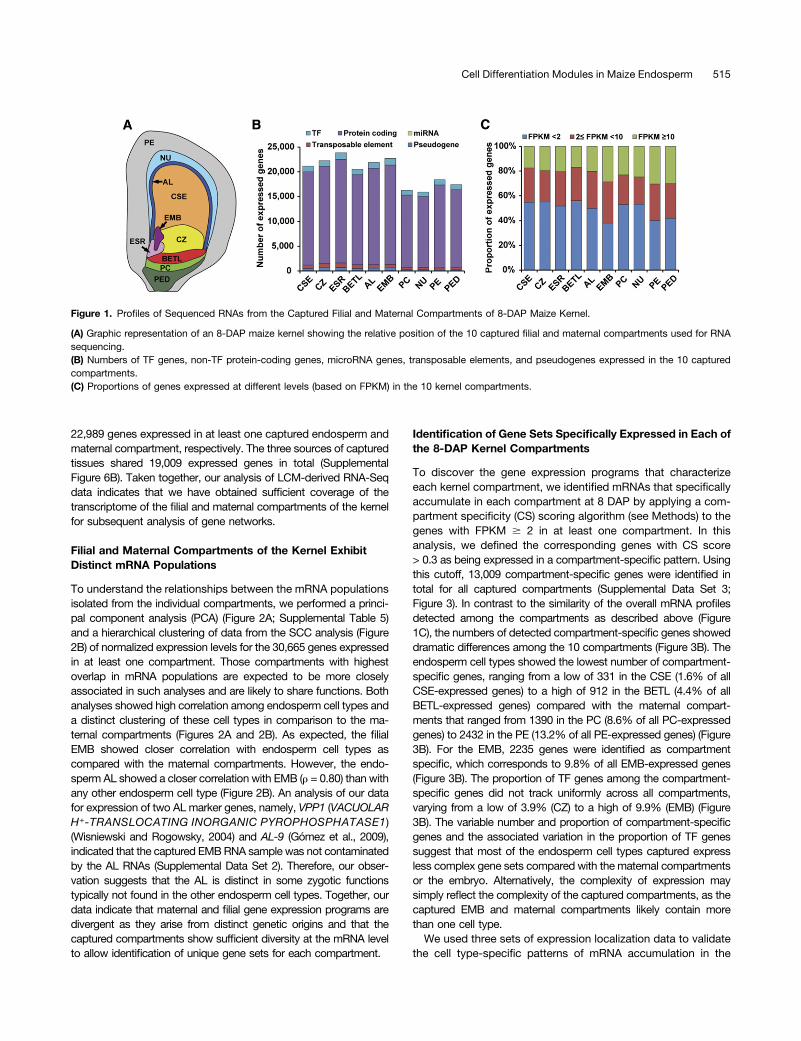
To identify the genes active in each of the endosperm cell types,we used LCM to isolate and profile mRNA populations of fiveendosperm compartments (cell types) of maize inbred line B73at 8 DAP. The compartments analyzed included AL, BETL, ESR,and two subregions of SE, the CSE and the CZ. To compareendosperm gene expression programs with embryonic and mater-nal programs, we also captured the embryo (EMB), nucellus (NU),placento-chalazal region (PC), pericarp (PE), and the vascular regionof the pedicel (PED) at 8 DAP (Figure 1A; Supplemental Figures 1 to3 and Supplemental Table 1). We selected this time point because itfollows differentiation of the main cell types of the endosperm, whichoccurs at 4 to 6 DAP, and precedes developmental programs as-sociated with endosperm function, including the activation of stor-age product synthesis and deposition program, which initiates at;8 to 10 DAP (Becraft, 2001; Olsen, 2001, 2004; Sabelli andLarkins, 2009; Becraft and Gutierrez-Marcos, 2012; Olsen andBecraft, 2013; Leroux et al., 2014). Total RNA extracted from bi-ological triplicates for the endosperm compartments and embryo,and single replicates of maternal compartments (22 samples) werereverse-transcribed to cDNA using oligo(dT) and random primers,amplified, and paired-end sequenced using an Illumina HiSequation2000 platform.The resulting reads were quality checked and mapped to the
maize reference genome (B73 RefGen_v3). Of the resultingmapped reads (8.9 to 34.7 million, 52.3 to 89.7% of total reads),3.2 to 11.3 million (22.1 to 39.5%) were mapped to exonicsequences (Supplemental Table 2). The exonic reads were nor-malized using Cufflinks (Trapnell et al., 2012) and reported as frag-ments per kilobase of transcript per million mapped reads (FPKM). Agene was considered expressed in a given sample if the lowerboundary of its FPKM 95% confidence interval (FPKM_conf_lo) wasgreater than zero (Hansey et al., 2012). Based on this criterion,29,369 genes were identified as expressed in at least one of the 22samples (Supplemental Data Set 1). Using pairwise Spearmancorrelation coefficient (SCC) analysis, the triplicate FPKM valuesfrom each of the endosperm compartments and the embryo wereshown to be highly correlated (r = 0.87 to 0.91; SupplementalFigure 4). Accordingly, we pooled each triplicate set of exonic readsand renormalized the data using Cufflinks. Using the same cutoff asindicated above, we detected 30,665 genes expressed in at leastone of the 10 compartments (Supplemental Data Set 2 andSupplemental Figure 5), 10,725 genes expressed in all 10 com-partments (Supplemental Figure 6A), and between 15,910 and23,853 (NU and ESR, respectively) expressed in individual com-partments (Figure 1B; Supplemental Table 3). In all cases, theproportion of transcription factor (TF) genes detected as expressedtracked closely with the total number of expressed genes (be-tween ;5.3 and 6.0% for CZ and PED, respectively; Figure 1B;Supplemental Table 3). The proportions of high-expressing(FPKM $ 10), medium-expressing (2 # FPKM < 10), and low-expressing (FPKM < 2) genes were relatively similar in all com-partments (Figure 1C; Supplemental Table 4). Collectively, 22,703genes were detected as expressed in EMB, and 28,078 and
22,989 genes expressed in at least one captured endosperm andmaternal compartment, respectively. The three sources of capturedtissues shared 19,009 expressed genes in total (SupplementalFigure 6B). Taken together, our analysis of LCM-derived RNA-Seqdata indicates that we have obtained sufficient coverage of thetranscriptome of the filial and maternal compartments of the kernelfor subsequent analysis of gene networks.
Filial and Maternal Compartments of the Kernel ExhibitDistinct mRNA Populations
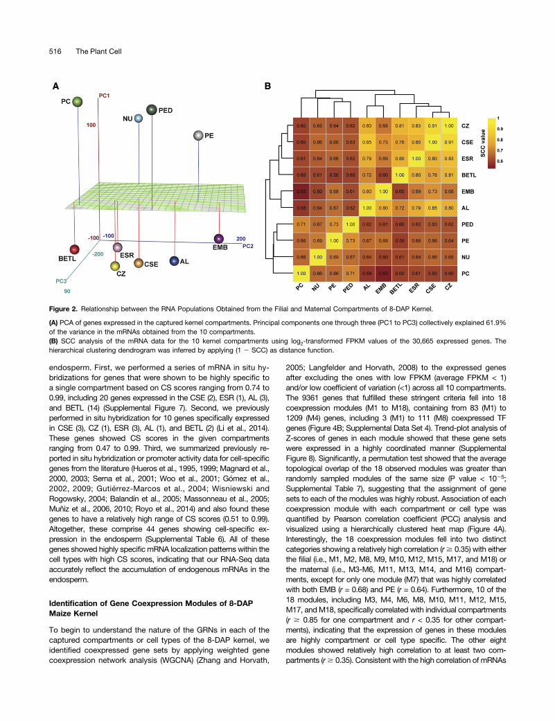
To understand the relationships between the mRNA populationsisolated from the individual compartments, we performed a princi-pal component analysis (PCA) (Figure 2A; Supplemental Table 5)and a hierarchical clustering of data from the SCC analysis (Figure2B) of normalized expression levels for the 30,665 genes expressedin at least one compartment. Those compartments with highestoverlap in mRNA populations are expected to be more closelyassociated in such analyses and are likely to share functions. Bothanalyses showed high correlation among endosperm cell types anda distinct clustering of these cell types in comparison to the ma-ternal compartments (Figures 2A and 2B). As expected, the filialEMB showed closer correlation with endosperm cell types ascompared with the maternal compartments. However, the endo-sperm AL showed a closer correlation with EMB (r = 0.80) than withany other endosperm cell type (Figure 2B). An analysis of our datafor expression of two AL marker genes, namely, VPP1 (VACUOLARH+-TRANSLOCATING INORGANIC PYROPHOSPHATASE1)(Wisniewski and Rogowsky, 2004) and AL-9 (Gómez et al., 2009),indicated that the captured EMBRNA sample was not contaminatedby the AL RNAs (Supplemental Data Set 2). Therefore, our obser-vation suggests that the AL is distinct in some zygotic functionstypically not found in the other endosperm cell types. Together, ourdata indicate that maternal and filial gene expression programs aredivergent as they arise from distinct genetic origins and that thecaptured compartments show sufficient diversity at the mRNA levelto allow identification of unique gene sets for each compartment.
Identification of Gene Sets Specifically Expressed in Each ofthe 8-DAP Kernel Compartments
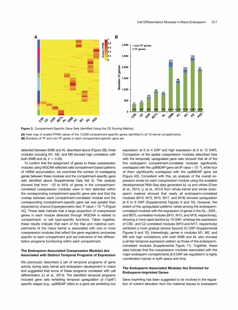
To discover the gene expression programs that characterizeeach kernel compartment, we identified mRNAs that specificallyaccumulate in each compartment at 8 DAP by applying a com-partment specificity (CS) scoring algorithm (see Methods) to thegenes with FPKM $ 2 in at least one compartment. In thisanalysis, we defined the corresponding genes with CS score> 0.3 as being expressed in a compartment-specific pattern. Usingthis cutoff, 13,009 compartment-specific genes were identified intotal for all captured compartments (Supplemental Data Set 3;Figure 3). In contrast to the similarity of the overall mRNA profilesdetected among the compartments as described above (Figure1C), the numbers of detected compartment-specific genes showeddramatic differences among the 10 compartments (Figure 3B). Theendosperm cell types showed the lowest number of compartment-specific genes, ranging from a low of 331 in the CSE (1.6% of allCSE-expressed genes) to a high of 912 in the BETL (4.4% of allBETL-expressed genes) compared with the maternal compart-ments that ranged from 1390 in the PC (8.6% of all PC-expressedgenes) to 2432 in the PE (13.2% of all PE-expressed genes) (Figure3B). For the EMB, 2235 genes were identified as compartmentspecific, which corresponds to 9.8% of all EMB-expressed genes(Figure 3B). The proportion of TF genes among the compartment-specific genes did not track uniformly across all compartments,varying from a low of 3.9% (CZ) to a high of 9.9% (EMB) (Figure3B). The variable number and proportion of compartment-specificgenes and the associated variation in the proportion of TF genessuggest that most of the endosperm cell types captured expressless complex gene sets compared with the maternal compartmentsor the embryo. Alternatively, the complexity of expression maysimply reflect the complexity of the captured compartments, as thecaptured EMB and maternal compartments likely contain morethan one cell type.We used three sets of expression localization data to validate
the cell type-specific patterns of mRNA accumulation in the
Figure 1. Profiles of Sequenced RNAs from the Captured Filial and Maternal Compartments of 8-DAP Maize Kernel.
(A) Graphic representation of an 8-DAP maize kernel showing the relative position of the 10 captured filial and maternal compartments used for RNAsequencing.(B) Numbers of TF genes, non-TF protein-coding genes, microRNA genes, transposable elements, and pseudogenes expressed in the 10 capturedcompartments.(C) Proportions of genes expressed at different levels (based on FPKM) in the 10 kernel compartments.
Cell Differentiation Modules in Maize Endosperm 515
endosperm. First, we performed a series of mRNA in situ hy-bridizations for genes that were shown to be highly specific toa single compartment based on CS scores ranging from 0.74 to0.99, including 20 genes expressed in the CSE (2), ESR (1), AL (3),and BETL (14) (Supplemental Figure 7). Second, we previouslyperformed in situ hybridization for 10 genes specifically expressedin CSE (3), CZ (1), ESR (3), AL (1), and BETL (2) (Li et al., 2014).These genes showed CS scores in the given compartmentsranging from 0.47 to 0.99. Third, we summarized previously re-ported in situ hybridization or promoter activity data for cell-specificgenes from the literature (Hueros et al., 1995, 1999; Magnard et al.,2000, 2003; Serna et al., 2001; Woo et al., 2001; Gómez et al.,2002, 2009; Gutiérrez-Marcos et al., 2004; Wisniewski andRogowsky, 2004; Balandín et al., 2005; Massonneau et al., 2005;Muñiz et al., 2006, 2010; Royo et al., 2014) and also found thesegenes to have a relatively high range of CS scores (0.51 to 0.99).Altogether, these comprise 44 genes showing cell-specific ex-pression in the endosperm (Supplemental Table 6). All of thesegenes showed highly specificmRNA localization patterns within thecell types with high CS scores, indicating that our RNA-Seq dataaccurately reflect the accumulation of endogenous mRNAs in theendosperm.
Identification of Gene Coexpression Modules of 8-DAPMaize Kernel
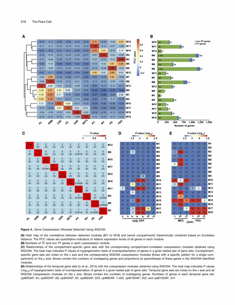
To begin to understand the nature of the GRNs in each of thecaptured compartments or cell types of the 8-DAP kernel, weidentified coexpressed gene sets by applying weighted genecoexpression network analysis (WGCNA) (Zhang and Horvath,
2005; Langfelder and Horvath, 2008) to the expressed genesafter excluding the ones with low FPKM (average FPKM < 1)and/or low coefficient of variation (<1) across all 10 compartments.The 9361 genes that fulfilled these stringent criteria fell into 18coexpression modules (M1 to M18), containing from 83 (M1) to1209 (M4) genes, including 3 (M1) to 111 (M8) coexpressed TFgenes (Figure 4B; Supplemental Data Set 4). Trend-plot analysis ofZ-scores of genes in each module showed that these gene setswere expressed in a highly coordinated manner (SupplementalFigure 8). Significantly, a permutation test showed that the averagetopological overlap of the 18 observed modules was greater thanrandomly sampled modules of the same size (P value < 1025;Supplemental Table 7), suggesting that the assignment of genesets to each of the modules was highly robust. Association of eachcoexpression module with each compartment or cell type wasquantified by Pearson correlation coefficient (PCC) analysis andvisualized using a hierarchically clustered heat map (Figure 4A).Interestingly, the 18 coexpression modules fell into two distinctcategories showing a relatively high correlation (r$ 0.35) with eitherthe filial (i.e., M1, M2, M8, M9, M10, M12, M15, M17, and M18) orthe maternal (i.e., M3-M6, M11, M13, M14, and M16) compart-ments, except for only one module (M7) that was highly correlatedwith both EMB (r = 0.68) and PE (r = 0.64). Furthermore, 10 of the18 modules, including M3, M4, M6, M8, M10, M11, M12, M15,M17, andM18, specifically correlated with individual compartments(r $ 0.85 for one compartment and r < 0.35 for other compart-ments), indicating that the expression of genes in these modulesare highly compartment or cell type specific. The other eightmodules showed relatively high correlation to at least two com-partments (r$ 0.35). Consistent with the high correlation of mRNAs
Figure 2. Relationship between the RNA Populations Obtained from the Filial and Maternal Compartments of 8-DAP Kernel.
(A) PCA of genes expressed in the captured kernel compartments. Principal components one through three (PC1 to PC3) collectively explained 61.9%of the variance in the mRNAs obtained from the 10 compartments.(B) SCC analysis of the mRNA data for the 10 kernel compartments using log2-transformed FPKM values of the 30,665 expressed genes. Thehierarchical clustering dendrogram was inferred by applying (1 2 SCC) as distance function.
detected between EMB and AL described above (Figure 2B), threemodules including M1, M2, and M9 showed high correlation withboth EMB and AL (r $ 0.35).
To confirm that the assignment of genes to these coexpressionmodules usingWGCNA reflected valid compartment-based patternsof mRNA accumulation, we examined the number of overlappinggenes between these modules and the compartment-specific genesets identified above (Supplemental Data Set 3). This analysisshowed that from ;52 to 94% of genes in the compartment-correlated coexpression modules were in fact detected withinthe corresponding compartment-specific gene sets and that theoverlap between each compartment-correlated module and thecorresponding compartment-specific gene set was greater thanexpected by chance (hypergeometric test, P value < 1025) (Figure4C). These data indicate that a large proportion of coexpressedgenes in each module detected through WGCNA is related tocompartment- or cell type-specific functions. Taken together,these results indicate that each of the filial and maternal com-partments of the maize kernel is associated with one or morecoexpression modules that reflect the gene regulatory processesspecific to each compartment and are indicators of the differen-tiation programs functioning within each compartment.
The Endosperm-Associated Coexpression Modules AreAssociated with Distinct Temporal Programs of Expression
We previously described a set of temporal programs of geneactivity during early kernel and endosperm development in maizeand suggested that some of these programs correlated with celldifferentiation (Li et al., 2014). The identified temporal programsincluded gene sets exhibiting temporal upregulation at (“up@”)specific stages (e.g., up@6DAP refers to a gene set exhibiting low
expression at 0 to 4 DAP and high expression at 6 to 12 DAP).Comparison of the spatial coexpression modules described herewith the temporally upregulated gene sets showed that all of thefive endosperm compartment-correlated modules significantlyoverlapped with the up@6DAP gene set (P value < 1025), while fourof them significantly overlapped with the up@8DAP gene set(Figure 4D). Consistent with this, an analysis of the overall ex-pression levels for each coexpression module using the availabledevelopmental RNA-Seq data generated by us and others (Chenet al., 2014; Li et al., 2014) from whole kernel and whole endo-sperm material showed that nearly all endosperm-correlatedmodules (M10, M12, M15, M17, and M18) showed upregulationat 6 to 8 DAP (Supplemental Figures 9 and 10). However, theextent of the upregulated patterns varied among the endosperm-correlated modules with the expression of genes in the AL-, ESR-,and BETL-correlated modules (M12, M15, and M18, respectively),showing a more rapid decline by 10 DAP, whereas the expressionof CSE- and CZ-correlated modules (M10 and M17, respectively)exhibited a more gradual decline beyond 22 DAP (SupplementalFigures 9 and 10). Interestingly, genes in modules M1, M2, andM9 with high correlations with both EMB and AL also showeda similar temporal expression pattern as those of the endosperm-correlated modules (Supplemental Figure 11). Together, thesedata indicate that the coexpression modules associated with themajor endosperm compartments at 8 DAP are regulated in a highlycoordinated manner in both space and time.
The Endosperm-Associated Modules Are Enriched forEndosperm-Imprinted Genes
Gene imprinting has been suggested to be involved in the regula-tion of nutrient allocation from the maternal tissues to endosperm
Figure 3. Compartment-Specific Gene Sets Identified Using the CS Scoring Method.
(A) Heat map of scaled FPKM values of the 13,009 compartment-specific genes identified in all 10 kernel compartments.(B) Numbers of TF and non-TF genes in each compartment-specific gene set.
Cell Differentiation Modules in Maize Endosperm 517
Figure 4. Gene Coexpression Modules Detected Using WGCNA.
(A) Heat map of the correlations between detected modules (M1 to M18) and kernel compartments hierarchically clustered based on Euclideandistance. The PCC values are quantitative indicators of relative expression levels of all genes in each module.(B) Numbers of TF and non-TF genes in each coexpression module.(C) Relationships of the compartment-specific gene sets with the corresponding compartment-correlated coexpression modules obtained usingWGCNA. The heat map indicates P values of hypergeometric tests of overrepresentation of genes in a given tested pair of gene sets. Compartment-specific gene sets are noted on the x axis and the corresponding WGCNA coexpression modules (those with a specific pattern for a single com-partment) on the y axis. Boxes contain the numbers of overlapping genes and proportions (in parentheses) of these genes in the WGCNA-identifiedmodules.(D) Relationships of the temporal gene sets (Li et al., 2014) with the coexpression modules obtained using WGCNA. The heat map indicates P values(-log10) of hypergeometric tests of overrepresentation of genes in a given tested pair of gene sets. Temporal gene sets are noted on the x axis and allWGCNA coexpression modules on the y axis. Boxes contain the numbers of overlapping genes. Numbers of genes in each temporal gene set:up@2DAP, 54; up@3DAP, 68; up@4DAP, 92; up@6DAP, 523; up@8DAP, 1,402; up@10DAP, 552; and up@12DAP, 241.
518 The Plant Cell
(Haig and Westoby, 1989; Moore and Haig, 1991; Costa et al.,2012). To investigate the relationship between the spatial programsof gene expression and gene imprinting, we examined the overlapbetween the WGCNA-generated coexpression modules with theimprinted genes that we previously identified in developing endo-sperm (Xin et al., 2013). This analysis showed that a subset ofgenes in the CZ- and BETL-correlated coexpression modules (M17and M18, respectively) significantly overlapped with a subset ofthe previously described paternally expressed gene sets (PEGs); theCSE-correlated module M10 and the modules associated with thematernal compartments (M3, M5, M6, M11, M13, M14, and M16)showed significant overlap with the maternally expressed gene sets(MEGs); and a subset of the ESR-correlated coexpression moduleexhibited significant overlap with both the previously describedPEGs and MEGs (hypergeometric P value < 1025; Figure 4E). Insupport of these data, a similar pattern of overlaps was observedwhen we applied less stringent criteria to identify genes with al-lele-biased expression patterns using the same set of normalizedRNA-Seq data (Supplemental Figure 12). Interestingly, many ofthe imprinted/allele-biased genes assigned by WGCNA to theendosperm-associated coexpression modules were TF genes frommultiple families (Supplemental Table 8). These results indicate anextensive interplay between epigenetic programs that regulate al-lelic expression and the transcriptional regulatory programs in-volved in the cellular differentiation and function of endosperm.
Biological Processes Enriched in Coexpression Modules ofthe Filial Compartments of the Kernel
The identified coexpression modules (Figure 4A) are likely associ-ated with specific biological processes or pathways involved in thedevelopment or function of each compartment. To identify themajor biological processes associated with the filial coexpressionmodules, we used Blast2GO (Conesa et al., 2005; Conesa andGötz, 2008; Götz et al., 2008) to identify the processes that weresignificantly enriched (false discovery rate < 0.05) in the modulesthat showed high correlation with endosperm compartments and/or the EMB. These included modules M1, M2, M8, M9, M10, M17,M12, M15, and M18. As expected, the CSE-correlated M10 wasshown to be enriched for “starch biosynthetic process” and “gly-cogen biosynthetic process” (Supplemental Figure 13), with theformer Gene Ontology (GO) category including the Shrunken-2(Sh2), Brittle-2 (Bt2), STARCH-BRANCHING ENZYME1 (SBE1),and Waxy1 genes, which all have well characterized functions instarch biosynthesis (Shure et al., 1983; Giroux et al., 1994; Blauthet al., 2002). Additionally, close inspection of genes in M10 revealedthat this module also contained other starch synthesis-relatedgenes without any current GO annotation, including the Sh1,Sugary1 (Su1), and STARCH SYNTHASE1 (SS1) genes (Choureyand Nelson, 1979; James et al., 1995; Commuri and Keeling, 2001).
In the case of zein-related genes, the mRNAs for only fourgenes encoding the 15-kD b-zein, the 16-kD g-zein, the 27-kDg-zein, and the 18-kD d-zein were detected in our analysis(FPKM_conf_lo > 0) in at least one cell type (Supplemental DataSet 2). Interestingly, all four zein genes, as well as the Floury-1gene, which encodes an endoplasmic reticulum membraneprotein involved in the targeted localization of an 22-kD a-zein inprotein body formation (Holding et al., 2007), were containedwithin the M10 module (Supplemental Data Set 4). This obser-vation correlates well with previous reports that the formation ofzein-containing protein bodies start as small accretions con-sisting primarily of b- and g-zeins (Woo et al., 2001), suggestingthat the M10 module contains the key early genes necessary forstorage protein body biogenesis. The M10 module also includedthe TF genes Opaque-2 (O2) and PBF (PROLAMIN-BOX BIND-ING FACTOR), with the relatively high M10 module membership(MM) scores of 0.93 and 0.97, respectively (Supplemental DataSet 4). Furthermore, visualization of M10 using VisANT (Hu et al.,2004) showed that these two TFs are among the most highlyconnected intramodular hubs of this module (SupplementalFigure 14A). O2 and PBF have previously been shown to regu-late storage program gene expression in maize endosperm(Schmidt et al., 1990, 1992; Marzábal et al., 2008). For example,the 15-kD b-zein and the 27-kD g-zein have been shown to beregulated by O2 and PBF, respectively (Cord Neto et al., 1995;Marzábal et al., 2008). Therefore, the M10 coexpression modulelikely includes a number of direct gene targets of both TFs.For the CZ-correlated M17 module, key functional over-
representations included “glycolysis,” “response to hydrogenperoxide,” “response to cadmium ion,” and “response to heat”(Supplemental Figure 13). This module showed a modest correla-tion with the CSE (r = 0.27; Figure 4A), suggesting that the CZ mayexpress an overlapping set of genes that function similarly to thosedetected in the CSE. Conversely, the captured CSE cells are ex-pected to have contained a portion of the CZ cells (SupplementalFigure 1) as the latter likely extend basally and centrally into thecaptured CSE region (Cooper, 1951; Charlton et al., 1995; Becraft,2001). This may explain the modest level of correlation of M10 withCZ (r = 0.29; Figure 4A).The AL-correlatedmoduleM12was enriched for “single-organism
process” (Supplemental Figure 13), and the two previously de-scribed markers of the aleurone, VPP1 (Wisniewski and Rogowsky,2004) and AL-9 (Gómez et al., 2009), were assigned within thismodule with high MM values (0.91 and 0.85, respectively).Likely due to the greater structural complexity of the EMB
compared with the captured endosperm compartments, the EMB-specific module M8 was enriched for more diverse GO categoriesin comparison to the endosperm-specific modules. The four bi-ological processes that were most significantly enriched for thismodule included “regulation of transcription, DNA-templated,”
Figure 4. (continued).
(E) Relationships of the imprinted gene sets (Xin et al., 2013) with the coexpression modules obtained using WGCNA. The heat map indicates P values(-log10) of hypergeometric tests of overrepresentation of genes in a given tested pair of gene sets. Imprinted gene sets are noted on the x axis and allWGCNA coexpression modules on the y axis. Boxes contain the numbers of overlapping genes. Numbers of genes in each imprinted gene set: 7-DAPMEG, 37; 10-DAP MEG, 185; 15-DAP MEG, 15; 7-DAP PEG, 80; 10-DAP PEG, 50; and 15-DAP PEG, 48.
Cell Differentiation Modules in Maize Endosperm 519
“floral organ development,” “phyllome development,” and “re-sponse to hormone.” Modules M1, M2, and M9 showed relativelyhigh correlation with both the AL and the EMB (r $ 0.35). Amongthese, M9 also showed a slightly lower correlation with CSE (r =0.33). Among these modules, M2 contained the largest number(477) of genes. Similar to M8, this module was also enriched formany GO categories, most of which were biological processesrelated to DNA replication and mitotic cell division (e.g., “DNAreplication,” “DNA repair,” “mitotic spindle assembly checkpoint,”and “microtubule-based movement,” etc.). Similarly, M9 (contain-ing 194 genes) was enriched for “cell cycle process,” “chromosomesegregation,” “regulation of DNA replication,” and “single-organismorganelle organization,” etc. Furthermore, M1, M2, and M9 sharedenrichment for “nucleosome assembly” and “DNA duplex un-winding,” which are also biological processes involved in DNAreplication and cell division (Supplemental Figure 13). These GOenrichments suggest that the EMB and AL, and to some degree theCSE, are programmed to undergo extensive mitotic cell pro-liferation at 8 DAP via the coordinated expression of a relativelyextensive gene network.
Consistent with the presumptive function of the ESR in mediatingendosperm-embryo interaction and expressing antimicrobial prod-ucts (Sabelli and Larkins, 2009), the ESR-specific module M15 wasenriched for “cell-cell signaling involved in cell fate commitment”(Supplemental Figure 13). This module also included many genesisolated previously by virtue of their highly specific ESR expressionpattern, including ESR-1, ESR-2, ESR-3, ESR-6, ESR-6B, and AE-3(Schel et al., 1984; Opsahl-Ferstad et al., 1997; Bonello et al., 2000;Balandín et al., 2005; Sosso et al., 2010). In our analysis, many of thesame genes, including ESR-1, ESR-2, ESR-3, and ESR-6, werepositioned within the intramodular hubs with high MM (e.g., 0.99) toM15 (Supplemental Figure 14C and Supplemental Data Set 4).
As expected from the BETL’s reported role as mediator ofsugar and metabolite uptake into the endosperm through itsinteraction with the underlying maternal placenta-chalazal region(Sabelli and Larkins, 2009), the BETL-correlated module M18 wasfound to be enriched for “transmembrane transport,” “ion trans-port,” and “sucrose transport” functions (Supplemental Figure 13).The M18 module contained nearly all of the previously identifiedBETL-expressed genes (Hueros et al., 1995, 1999; Cheng et al.,1996; Doan et al., 1996; Serna et al., 2001; Magnard et al., 2003;Gutiérrez-Marcos et al., 2004; Massonneau et al., 2005; Gruis et al.,2006; Muñiz et al., 2006, 2010; Brugière et al., 2008; Gómez et al.,2009), including BETL-1, 3, 4, 9, and 10; BAP-1A, 1B, 2, 3A, and3B; TCRR-1 and 2; and INCW2 (CELL WALL INVERTASE2),EBE-2 (EMBRYO SAC/BASAL ENDOSPERM-LAYER/EMBRYO-SURROUNDING REGION-2), IPT-2 (ISOPENTENYL TRANS-FERASE-2), and CC-8 (CORN CYSTATIN-8) (Supplemental DataSet 4). In addition, this module contained 11 of the 13 MEG genes(with MEG-4 and MEG-14 being the two exceptions) that havebeen identified in the B73 genome (Supplemental Data Set 4), in-cluding MEG-1, which is a BETL-specific gene that has beenshown to be important for the development and differentiation ofthe BETL (Costa et al., 2012). The BETL-, BAP-, and MEG-typegenes encode small, secreted, cysteine-rich proteins that havebeen suggested to protect the embryo from maternally transmittedpathogens (Tailor et al., 1997) and to serve as signaling moleculesthat coordinate the supply of nutrients to the embryo during kernel
development (Marshall et al., 2011), while the TCRR genes encodetype-A response regulators (Muñiz et al., 2006, 2010). Significantly,all of these genes showed high MM (>0.90) to module M18, andmany of them were among the top-scoring hubs (SupplementalFigure 14E and Supplemental Data Set 4). Furthermore, the BETL-specific TF gene MRP-1, previously shown to be involved in reg-ulation of BETL differentiation (Gómez et al., 2002, 2009), was alsodetected in M18 with a high MM score (1.00; Supplemental DataSet 4). This is consistent with its role as an activator of many BETL-specific genes, including BETL-1, BETL-9, BETL-10, BAP-2,MEG-1,TCRR-1, and TCRR-2 (Gómez et al., 2002, 2009; Gutiérrez-Marcoset al., 2004; Muñiz et al., 2006, 2010). Therefore, these data suggestthat MRP-1 acts as a major regulator of a subset of genes in theM18 coexpression module. Together, our results suggest that theWGCNA-identified coexpression modules can be used as startingpoints for identification of GRNs functioning in each endospermcompartment.
De Novo Identification of cis-Motifs Associated with theEndosperm Coexpression Modules
As a first step toward identification of the endosperm GRNs, weused MEME software (Bailey and Elkan, 1994) to detect putativecis-regulatory elements in upstream gene sequences from eachof the five endosperm coexpression modules identified usingWGCNA, including M10, M12, M15, M17, and M18 (Figure 4A).We searched for 10- to 12-bp sequence motifs overrepresentedwithin 21 to +0.5 kb (relative to transcription start site) of genesin each module and further identified motifs that were signifi-cantly similar (q-value < 0.05) to the known plant cis-motifsavailable in the JASPAR CORE database using TOMTOM program(Gupta et al., 2007). We found that most of the detected motifswere shared among the majority of the modules as exemplified bythe motifs that contained exclusively CG- or AT-rich sequences(Supplemental Table 9) with significant similarity to the reportedbinding sites of ABI4 (a maize AP2-EREBP protein) and SOC1 (anArabidopsis thaliana MIKC-type MADS box protein), respectively(Riechmann et al., 1996; Niu et al., 2002). Coincidently, four of thefive endosperm modules (M10, M12, M15, and M17) included atleast one MIKC-type MADS box gene, while all five modules in-cluded at least one AP2-EREBP gene based on the current maizegenome annotation (Supplemental Data Set 4), indicating that theMIKC-type MADS box and AP2-EREBP families may play broadregulatory roles in maize endosperm development.In contrast, a small number of motifs were detected specifically
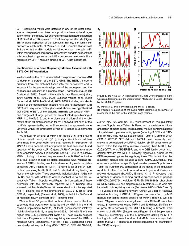
in single modules. In one instance, the Motif 10 that was enriched inthe CSE-correlated module M10 showed significant similarity toa cis-motif that has been shown to bind a bZIP TF in snapdragon(Antirrhinum majus) (Supplemental Table 9) (Martínez-García et al.,1998). Accordingly, two bZIP TF genes, bZIP46 and the storageprogram regulator O2, were detected within M10 (SupplementalData Set 4). These results suggest that a subset of genes in moduleM10 may be regulated by the bZIP genes in the same module. Ina second case, Motifs 3, 6, and 8 that were detected in upstreamsequences of M18 genes each contained a repeated GATA se-quence (Figure 5A) similar to a sequence previously shown to beinvolved in binding and activation of target genes by MRP-1(Baranowskij et al., 1994; Barrero et al., 2006), whereas no
GATA-containing motifs were detected in any of the other endo-sperm coexpression modules. In support of a transcriptional regu-latory role for the motifs, our analysis indicated a biased distributionof Motifs 3, 6, and 8 upstream to the transcription start site (Figure5B). A close inspection of the submotifs, namely, the variant se-quences of each motif, of Motifs 3, 6, and 8 revealed that at least148 genes in the M18 module contained one or more submotifswithin their upstream sequences. Collectively, our data suggest thata large subset of genes in the M18 coexpression module is likelyregulated by MRP-1 through binding at GATA-rich sequences.
Identification of a Gene Regulatory Module Associated withBETL Cell Differentiation
We focused on the BETL-associated coexpression module M18to decipher a portion of the BETL GRN. The BETL transportsnutrients from the maternal tissue into the endosperm and isimportant for the proper development of the endosperm and theendosperm’s capacity as a storage organ (Thompson et al., 2001;Costa et al., 2012). Based on the available data (Baranowskij et al.,1994; Gómez et al., 2002, 2009; Gutiérrez-Marcos et al., 2004;Barrero et al., 2006; Muñiz et al., 2006, 2010) including our identi-fication of the coexpression module M18 and its association withGATA-rich sequence motifs (discussed above), we hypothesizedthat a GRN for BETL differentiation is minimally composed of MRP-1and a large set of target genes that are activated upon binding ofMRP-1 to Motifs 3, 6, and 8. A close examination of all the sub-motifs of the 10 motifs enriched for M18 showed that each of themotifs is represented by two to 21 submotifs that appeared one to90 times within the promoters of the M18 genes (SupplementalTable 10).
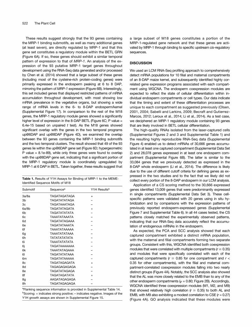
We tested for binding of MRP-1 to Motifs 3, 6, and 8 usingdirected yeast one-hybrid (Y1H) assays. We introduced twoconstructs into yeast cells, one that resulted in expression ofMRP-1 and a second that comprised the test sequence fusedupstream of the yeast AUR1-C gene. AUR1-C confers resistanceto aureobasidin A (AbA) (Heidler and Radding, 1995). In this assay,MRP-1 binding to the test sequence results in AUR1-C activationand, thus, growth of cells on plates containing AbA, whereas ab-sence of MRP-1 binding results in absence of growth on platescontaining AbA. Testing for MRP-1 binding to all of the 24 sub-motifs comprising Motifs 3, 6, and 8 indicated strong binding tofour of the submotifs. These submotifs included Motifs 3a/8a, 6a/8b, 8c, and 8f, with Motifs 3a and 6a identical to 8a and 8b, re-spectively (Table 1; Supplemental Figure 15). Comparison of thesesubmotifs with the previously reported MRP-1 binding sitesshowed that Motifs 6a/8b and 8c were identical to the reportedMRP-1 binding site in the promoters of BETL-1 (Motif IV) andBETL-2, respectively (Barrero et al., 2006), while Motifs 3a/8a and8f represent newly identified MRP-1 binding sites.
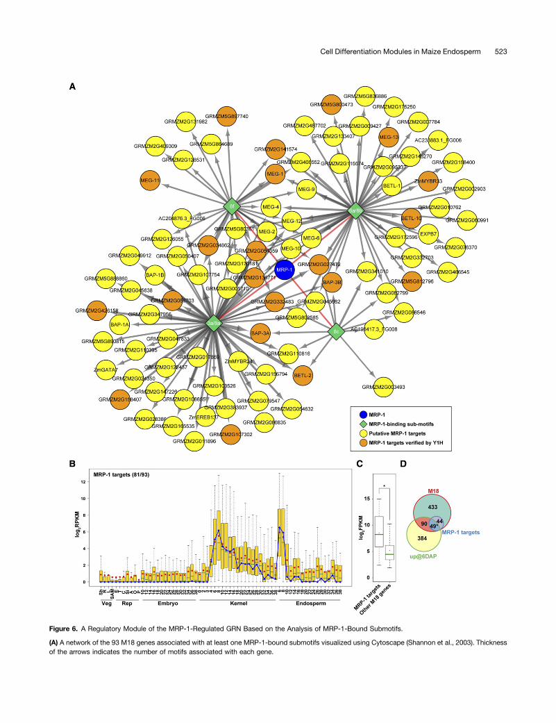
We identified 93 genes that contain at least one of the foursubmotifs that were shown to be bound by MRP-1 in the Y1Hassays (Supplemental Table 11). All the 93 genes showed relativelyhigh MM values to M18, ranging from 0.62 to 1.00, with 78 of themhigher than 0.95 (Supplemental Table 11). These results suggestthat these 93 genes constitute a regulatory module of the MRP-1-regulated GRN. Significantly, 7 of the 14 BETL-expressed genesdescribed previously, includingMEG-1, BETL-1, BETL-10, BAP-1A,
BAP-2, BAP-3A, and BAP-3B, were present in this regulatorymodule (Supplemental Table 11). Based on the available functionalannotation of maize genes, this regulatory module contained at least17 cysteine-rich protein-coding genes (including 3 BETL-, 4 BAP-,and 10 MEG-type genes; Supplemental Table 11), among whichBETL-1, BETL-2, BETL-10, and MEG-1 have previously beenshown to be regulated by MRP-1. Notably, six TF genes were de-tected within this regulatory module, including three MYBR-, twoC2C2-GATA, one AP2-EREBP, and one DBB family genes, sug-gesting strongly that MRP-1 indirectly regulates a subset of theBETL-expressed genes by regulating these TFs. In addition, thisregulatory module also included a gene (GRMZM2G406552) thatencodes a putative nonspecific lipid transfer protein (SupplementalTable 11). Furthermore, comparison of the genes in the regulatorymodule to the GenBank nonredundant (nr) and Swissprotprotein databases (BLASTX, E-value < 1026) revealed thata number of genes encoding putative transporters of peptide(GRMZM2G156794), calcium (GRMZM5G836886), phosphate(GRMZM2G466545), andmagnesium (GRMZM2G054632) were alsoincluded in this regulatory module (Supplemental Data Sets 5 and 6).To validate this putative network further, we used Y1H assays
to test for binding of MRP-1 to 22 gene promoters from the M18module containing motifs positive for MRP-1 binding. We alsotested 19 gene promoters lacking these motifs. Of the 41 promoterstested, 31 were shown to bind MRP-1 and 10 did not. Significantly,all promoters containing the MRP-1 binding submotifs wereshown to bind MRP-1 (Supplemental Figure 16 and SupplementalTable 12). Interestingly, 7 of the 19 promoters lacking the MRP-1binding submotifs were found to bind MRP-1 in our assays, indi-cating that MRP-1 binds to additional sequences not identified inthe MEME analysis.
Figure 5. De Novo GATA-Rich Sequence Motifs Overrepresented in theUpstream Sequences of the Coexpression Module M18 Genes Identifiedby the MEME Program.
(A) Motifs 3, 6, and 8 enriched among the M18 genes.(B) Position frequencies of the same motifs determined as number ofmotifs per 50-bp bins in the upstream gene regions.
Cell Differentiation Modules in Maize Endosperm 521
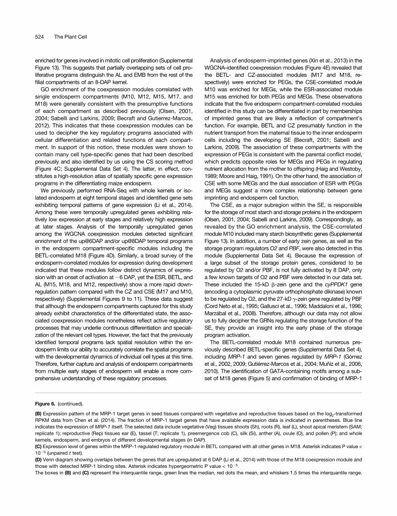
These results suggest strongly that the 93 genes containingthe MRP-1 binding submotifs, as well as many additional genes(at least seven), are directly regulated by MRP-1 and that thisgene set constitutes a regulatory module within the BETL GRN(Figure 6A). If so, these genes should exhibit a similar temporalpattern of expression to that of MRP-1. An analysis of the ex-pression of the 93 putative MRP-1 target genes throughoutdevelopment using the RNA-Seq data generated and/or processedby Chen et al. (2014) showed that a large subset of these genes(including most of the cysteine-rich protein-coding genes) wereprimarily expressed in the endosperm peaking at 6 to 8 DAP,mirroring the pattern ofMRP-1 expression (Figure 6B). Interestingly,this set included genes that displayed restricted patterns of mRNAaccumulation throughout development, with most showing lowmRNA prevalence in the vegetative organs, but showing a widerange of mRNA levels in the 6- to 8-DAP endosperm/kernel(Supplemental Figure 17). In comparison to the rest of the M18genes, the MRP-1 regulatory module genes showed a significantlyhigher level of expression in the 8-DAP BETL (Figure 6C; P value =6.4e-15 based on unpaired t test). As the M18 genes showedsignificant overlap with the genes in the two temporal programsup@6DAP and up@8DAP (Figure 4D), we examined the overlapbetween the 93 genes containing the MRP-1 binding submotifsand the two temporal clusters. The result showed that 49 of the 93genes lie within the up@6DAP gene set (Figure 6D; hypergeometricP value = 8.1e-89), while only three genes were found to overlapwith the up@8DAP gene set, indicating that a significant portion ofthe MRP-1 regulatory module is coordinately upregulated byMRP-1 at 6 DAP in BETL. Taken together, these results suggest that
a large subset of M18 genes constitutes a portion of theMRP-1-regulated gene network and that these genes are acti-vated by MRP-1 through binding to specific upstream cis-regulatorysequences.
DISCUSSION
We used an LCM RNA-Seq profiling approach to comprehensivelydetect mRNA populations for 10 filial and maternal compartmentsof an 8-DAP maize kernel, and subsequently identified highly cor-related gene expression programs associated with each compart-ment using WGCNA. The endosperm coexpression modules areexpected to reflect the state of cellular differentiation within in-dividual endosperm compartments or cell types. Our data indicatethat the timing and extent of these differentiation processes areunique to each compartment as suggested previously (Olsen,2001, 2004; Sabelli and Larkins, 2009; Becraft and Gutierrez-Marcos, 2012; Leroux et al., 2014; Li et al., 2014). As a test case,we deciphered an MRP-1 regulatory module containing 93 genesthat are likely involved in BETL cellular differentiation.The high-quality RNAs isolated from the laser-captured cells
(Supplemental Figures 2 and 3 and Supplemental Table 1) andthe resulting highly reproducible RNA-Seq data (SupplementalFigure 4) enabled us to detect mRNAs of 30,666 genes accumu-lated in at least one captured compartment (Supplemental Data Set2) and 28,078 genes expressed in at least one endosperm com-partment (Supplemental Figure 6B). The latter is similar to the33,084 genes that we previously detected as expressed in the8-DAP whole endosperm (Li et al., 2014). The difference is likelydue to the use of different cutoff criteria for defining genes as ex-pressed in the two studies and to the fact that we likely did notcollect every portion of the 8-DAP endosperm in our LCM analysis.Application of a CS scoring method to the 30,666 expressed
genes identified 13,009 genes that were predominantly expressedin single compartments (Supplemental Data Set 3). These cell-specific patterns were validated with 20 genes using in situ hy-bridization and by comparisons with the expression patterns ofpreviously reported endosperm-expressed genes (SupplementalFigure 7 and Supplemental Table 6). In all 44 cases tested, the CSpatterns closely matched the experimentally observed patterns,indicating that our RNA-Seq data accurately reflect the accumu-lation of endogenous mRNAs in the endosperm.As expected, the PCA and SCC analysis showed that each
captured compartment exhibited a distinct mRNA population,with the maternal and filial compartments forming two separategroups. Consistent with this, WGCNA identified both coexpressionmodules that were correlated with multiple compartments (r$ 0.35)and modules that were specifically correlated with each of thecaptured compartments (r $ 0.85 for one compartment and r <0.35 for other compartments), with the filial and maternal com-partment-correlated coexpression modules falling into two nearlydistinct groups (Figure 4A). Notably, the SCC analysis also showedthat the AL was more closely related to the EMB than to any of theother endosperm compartments (r = 0.80; Figure 2B). Accordingly,WGCNA identified three coexpression modules (M1, M2, and M9)that showed relatively high correlation (r $ 0.35) to both AL andEMB, with M9 also exhibiting a modest correlation to CSE (r = 0.27)(Figure 4A). GO analysis indicated that these modules were
Table 1. Results of Y1H Assays for Binding of MRP-1 to the MEME-Identified Sequence Motifs of M18
Submotif Sequencea Y1H Resultsb
3a/8a TAGATAGATAGA +3b TAGATATATAGA –
3c TAGATAAATAGA –
6a/8b TAGATATAGATA +6b TAGATATATATA –
6c TAAATATAAATA –
6d TAGATATAGAAA –
6e TAGATATAAATA –
6f TAAATATAAAAA –
6g TAAATATATAAA –
6h TATATATATATA –
6i TAAATATATATA –
6j TAGATAAAAAAA –
6k TAAATATAGAAA –
6l TAGATATATAAA –
6m TAGATATAAAAA –
8c TAGATAGAGATA +8d TAGAGAGAGAGA –
8e TAGATATAGAGA –
8f TAGATAGATATA +8g AAGATAGAGAGA –
8h TAGATAGAGAGA –
aFlanking sequence information is provided in Supplemental Table 14.bScoring: + indicates strong positive; – indicates negative. Images of theY1H growth assays are shown in Supplemental Figure 15.
Figure 6. A Regulatory Module of the MRP-1-Regulated GRN Based on the Analysis of MRP-1-Bound Submotifs.
(A) A network of the 93 M18 genes associated with at least one MRP-1-bound submotifs visualized using Cytoscape (Shannon et al., 2003). Thicknessof the arrows indicates the number of motifs associated with each gene.
Cell Differentiation Modules in Maize Endosperm 523
enriched for genes involved in mitotic cell proliferation (SupplementalFigure 13). This suggests that partially overlapping sets of cell pro-liferative programs distinguish the AL and EMB from the rest of thefilial compartments of an 8-DAP kernel.
GO enrichment of the coexpression modules correlated withsingle endosperm compartments (M10, M12, M15, M17, andM18) were generally consistent with the presumptive functionsof each compartment as described previously (Olsen, 2001,2004; Sabelli and Larkins, 2009; Becraft and Gutierrez-Marcos,2012). This indicates that these coexpression modules can beused to decipher the key regulatory programs associated withcellular differentiation and related functions of each compart-ment. In support of this notion, these modules were shown tocontain many cell type-specific genes that had been describedpreviously and also identified by us using the CS scoring method(Figure 4C; Supplemental Data Set 4). The latter, in effect, con-stitutes a high-resolution atlas of spatially specific gene expressionprograms in the differentiating maize endosperm.
We previously performed RNA-Seq with whole kernels or iso-lated endosperm at eight temporal stages and identified gene setsexhibiting temporal patterns of gene expression (Li et al., 2014).Among these were temporally upregulated genes exhibiting rela-tively low expression at early stages and relatively high expressionat later stages. Analysis of the temporally upregulated genesamong the WGCNA coexpression modules detected significantenrichment of the up@6DAP and/or up@8DAP temporal programsin the endosperm compartment-specific modules including theBETL-correlated M18 (Figure 4D). Similarly, a broad survey of theendosperm-correlated modules for expression during developmentindicated that these modules follow distinct dynamics of expres-sion with an onset of activation at;6 DAP, yet the ESR, BETL, andAL (M15, M18, and M12, respectively) show a more rapid down-regulation pattern compared with the CZ and CSE (M17 and M10,respectively) (Supplemental Figures 9 to 11). These data suggestthat although the endosperm compartments captured for this studyalready exhibit characteristics of the differentiated state, the asso-ciated coexpression modules nonetheless reflect active regulatoryprocesses that may underlie continuous differentiation and speciali-zation of the relevant cell types. However, the fact that the previouslyidentified temporal programs lack spatial resolution within the en-dosperm limits our ability to accurately correlate the spatial programswith the developmental dynamics of individual cell types at this time.Therefore, further capture and analysis of endosperm compartmentsfrom multiple early stages of endosperm will enable a more com-prehensive understanding of these regulatory processes.
Analysis of endosperm-imprinted genes (Xin et al., 2013) in theWGCNA-identified coexpression modules (Figure 4E) revealed thatthe BETL- and CZ-associated modules (M17 and M18, re-spectively) were enriched for PEGs, the CSE-correlated moduleM10 was enriched for MEGs, while the ESR-associated moduleM15 was enriched for both PEGs and MEGs. These observationsindicate that the five endosperm compartment-correlated modulesidentified in this study can be differentiated in part by membershipsof imprinted genes that are likely a reflection of compartment’sfunction. For example, BETL and CZ presumably function in thenutrient transport from the maternal tissue to the inner endospermcells including the developing SE (Becraft, 2001; Sabelli andLarkins, 2009). The association of these compartments with theexpression of PEGs is consistent with the parental conflict model,which predicts opposite roles for MEGs and PEGs in regulatingnutrient allocation from the mother to offspring (Haig and Westoby,1989; Moore and Haig, 1991). On the other hand, the association ofCSE with some MEGs and the dual association of ESR with PEGsand MEGs suggest a more complex relationship between geneimprinting and endosperm cell function.The CSE, as a major subregion within the SE, is responsible
for the storage of most starch and storage proteins in the endosperm(Olsen, 2001, 2004; Sabelli and Larkins, 2009). Correspondingly, asrevealed by the GO enrichment analysis, the CSE-correlatedmodule M10 included many starch biosynthetic genes (SupplementalFigure 13). In addition, a number of early zein genes, as well as thestorage program regulators O2 and PBF, were also detected in thismodule (Supplemental Data Set 4). Because the expression ofa large subset of the storage protein genes, considered to beregulated by O2 and/or PBF, is not fully activated by 8 DAP, onlya few known targets of O2 and PBF were detected in our data set.These included the 15-kD b-zein gene and the cyPPDK1 gene(encoding a cytoplasmic pyruvate orthophosphate dikinase) knownto be regulated by O2, and the 27-kD g-zein gene regulated by PBF(Cord Neto et al., 1995; Gallusci et al., 1996; Maddaloni et al., 1996;Marzábal et al., 2008). Therefore, although our data may not allowus to fully decipher the GRNs regulating the storage function of theSE, they provide an insight into the early phase of the storageprogram activation.The BETL-correlated module M18 contained numerous pre-
viously described BETL-specific genes (Supplemental Data Set 4),including MRP-1 and seven genes regulated by MRP-1 (Gómezet al., 2002, 2009; Gutiérrez-Marcos et al., 2004; Muñiz et al., 2006,2010). The identification of GATA-containing motifs among a sub-set of M18 genes (Figure 5) and confirmation of binding of MRP-1
Figure 6. (continued).
(B) Expression pattern of the MRP-1 target genes in seed tissues compared with vegetative and reproductive tissues based on the log2-transformedRPKM data from Chen et al. (2014). The fraction of MRP-1 target genes that have available expression data is indicated in parentheses. Blue lineindicates the expression of MRP-1 itself. The selected data include vegetative (Veg) tissues shoots (Sh), roots (R), leaf (L), shoot apical meristem (SAM;replicate 1); reproductive (Rep) tissues ear (E), tassel (T; replicate 1), preemergence cob (C), silk (Si), anther (A), ovule (O), and pollen (P); and wholekernels, endosperm, and embryos of different developmental stages (in DAP).(C) Expression level of genes within the MRP-1-regulated regulatory module in BETL compared with all other genes in M18. Asterisk indicates P value <1025 (unpaired t test).(D) Venn diagram showing overlaps between the genes that are upregulated at 6 DAP (Li et al., 2014) with those of the M18 coexpression module andthose with detected MRP-1 binding sites. Asterisk indicates hypergeometric P value < 1025.The boxes in (B) and (C) represent the interquantile range, green lines the median, red dots the mean, and whiskers 1.5 times the interquantile range.
to these sequences using Y1H assays (Supplemental Table 11)allowed us to propose a 93-gene, direct-target regulatory modulefor MRP-1 (Figure 6). Functional annotation of the 93-gene regu-latory module indicates a diverse array of gene functions activatedby MRP-1, including putative signaling and nutrient transport(Supplemental Table 11). The larger M18 gene set also exhibitsa wide range of putative functions including those expected fora transfer cell layer (Supplemental Figure 13). These gene functionsare likely sufficient to support BETL differentiation, as a recentstudy showed that the ectopic expression of MRP-1 via an aleu-rone-specific gene promoter was capable of inducing differentia-tion of an ectopic BETL in the aleurone albeit in a transient manner(Gómez et al., 2009). On the other hand, ectopic activation of thesegenes may not produce viable cells outside the endosperm con-text, as attempts with overexpressing MRP-1 in maize usingubiquitously expressing promoters produced no transformants(Gómez et al., 2009). Further understanding of the MRP-1-regulatednetwork and the associated gene functions will likely requirecharacterization of complete or partial loss-of-function mutants.
The gene set activated by MRP-1 is likely significantly largerthan the 93-gene set discussed above for two reasons. First, ourY1H assays showed activation of seven genes that lack motifsidentified in our MEME analysis (Supplemental Figure 16 andSupplemental Table 12), indicating that MRP-1 binds to additionalsequences not identified in our MEME analysis and that M18contains additional MRP-1-regulated genes. Second, MRP-1 likelydirectly regulates six TFs, including MYBR24, MYBR33, GATA7,GATA33, and EREB137, and a DBB family TF (Supplemental Table11). Each of these TFs may activate a gene set within M18. Addi-tional protein-DNA interaction studies, such as electrophoreticmobility shift assays, and transient directed-expression assayswould be necessary to further characterize the interaction betweenMRP-1 and the full spectrum of its direct targets. Thus, the entireregulatory module activated by MRP-1 (i.e., both directly and in-directly) probably encompasses a much larger proportion of theM18 module than what we report here. Studies devoted to identi-fying the full spectrum of MRP-1 binding sites and to the identifi-cation of the target genes activated by the TFs activated by MRP-1are in progress to fully characterize this module.
Furthermore, it is notable that in addition to MRP-1 itself, M18contains at least 48 coexpressed TF genes including six MYBRgenes (Supplemental Data Set 4). The latter may regulate over-lapping gene sets with MRP-1, possibly through binding to theidentified GATA-containing motifs. Limitations of the de novo cis-motif detection approaches utilized here and an absence of anextensive cis-motif-TF database for plants have precluded identi-fication of additional TF targets in M18. Therefore, further ap-proaches including directed Y1H assays for specific TF-targetinteractions in combination with transient expression studies of theTFs and analysis of any available TF gene mutants will be requiredto determine the full extent of the BETL GRN.
In summary, our data set provides a high-resolution atlas ofgene expression in differentiating endosperm compartmentsand maternal compartments of an early maize kernel. This dataset provides insights into the functions of the endosperm celltypes and into the coexpressed gene sets that establish the dif-ferentiated states and functions of these cell types. Furthermore, asexemplified by our initial analysis of the MRP-1 regulatory module,
this data set can be used as a starting point to dissect the modulesregulating endosperm cell differentiation. The analyses providedhere constitutes a significant step toward the identification of GRNsthat regulate maize endosperm cell differentiation and determine itsfunction.
METHODS
Plant Materials and Growth
Plants of the reference maize (Zea mays) genotype, B73, were grownunder greenhouse conditions (16-h day) at the University of Arizona duringApril to July 2012 and self-pollinated to obtain 8-DAP kernels (Li et al., 2014)for LCM. The kernels for morphological analysis shown in SupplementalFigure 1 were collected during October and November 2013. Kernelcompartments were delineated for capture using tissue sections ob-tained from Farmer’s fixed (see below) or from paraformaldehyde-fixedmaterial that was further stained with Toluidine Blue as describedpreviously (Drews, 1998). The tissue sections for each biological replicateof a given compartment were captured from multiple kernels of a single ear(Supplemental Table 1).
LCM, RNA Isolation, and cDNA Synthesis and Amplification
Kernels were harvested from plants mid-day and cut at the pedicel,punctured through the pericarp, vacuum infiltrated with cold Farmer’sfixative (ethanol:glacial acetic acid, 3:1) (Kerk et al., 2003), and stored incold fixative overnight. Fixed kernels were then dehydrated in a gradedethanol series, cleared in n-butanol, embedded in Paraplast X-tra (Mc-Cormick Scientific Leica) using microwave (Takahashi et al., 2010), cut to10-mm sections, and mounted on PEN-coated slides. Shortly beforecapture, sections were deparaffinized in Xylenes and air-dried (Takahashiet al., 2010). Individual cell types or kernel compartments were captureddirectly into an aliquot of Arcturus PicoPure RNA extraction buffer (Ap-plied Biosystems) using a Leica LMD6500 laser microdissection system(Leica Microsystems). RNA was extracted following the manufacturer’ssuggested protocol (Arcturus PicoPure kit), checked for quality, DNasetreated using TURBO DNase (Life Technologies), and further purifiedusing the Arcturus PicoPure columns (Applied Biosystems). For each ofthe 22 samples (Supplemental Table 1), 10 ng purified RNA was used forcDNA synthesis and amplification. cDNA synthesis with oligo(dT) andrandom primers and cDNA amplifications were performed using anOvation RNA-Seq System V2 kit (Nugen Technologies) following themanufacturer’s protocols with minor modifications. The quality and profileof the RNA and amplified cDNA samples were checked on an Agilent 2100Bioanalyzer (Agilent Technologies) using an Agilent RNA 6000 Pico Kit(Agilent Technologies) and an Agilent High Sensitivity DNA Kit (AgilentTechnologies), respectively.
RNA-Seq Library Construction and Sequencing
Construction and sequencing of RNA-Seq libraries were performed at theUniversity of Arizona Genetics Core. Using an Illumina TruSeq DNAsample preparation kit (v2; Illumina), nearly 1 µg amplified cDNA for eachof the 22 samples was used to generate multiplexed RNA-Seq libraries(mean size 350 to 380 bp, including 120-bp adapters) by following themanufacturer’s suggested procedures. The samples were sequenced inbatches on four flow cell lanes of an Illumina HiSequation 2000 platformusing a TruSeq SBS kit (v3) to produce 2 3 100-nucleotide paired-endreads. Two of the four lanes each contained libraries for a single replicateof the captured endosperm compartments (AL, BETL, ESR, CSE, and CZ);the third lane contained libraries for a single replicate of the endospermcompartments plus the library for a single replicate of the maternal
Cell Differentiation Modules in Maize Endosperm 525
compartment PC; and the fourth lane contained libraries for EMB (threereplicates) and the other three maternal compartments (NU, PE, and PED,one replicate each). After the raw reads were generated, adapter se-quences were trimmed using the Trimmomatic program (Bolger et al.,2014). Quality of the trimmed reads was checked using the FastQCprogram (Andrews, 2010).
Reads Mapping and Analysis
RNA-Seq reads were aligned to the maize reference genome version 3(B73 RefGen_v3) (Hubbard et al., 2002) using TopHat v2.0.9 (Trapnellet al., 2009). Intron length was set to 30 to 8000 nucleotides, whilemaximum number of mismatches per read was set to 3. To eliminate theeffect of reads mapping in intergenic and/or repeated genomic regions onthe estimation of effective library size that may be caused by the cDNAamplification method, reads mapped to exonic regions were extractedusing the intersect function of BEDTools v2.17.0 (Quinlan and Hall, 2010)and were provided as input to Cufflinks v2.1.1 (Trapnell et al., 2010) fornormalization and estimation of gene expression level. The multi-mappedreads correction and fragment bias correction options of Cufflinks wereused. Gene expression levels were reported as FPKM (Mortazavi et al.,2008). The upper and lower bound FPKM values (FPKM_conf_hi andFPKM_conf_lo, respectively) for the 95% confidence interval of each genewere also provided by Cufflinks. A gene was defined as expressed ina sample if the FPKM_conf_lo was greater than zero. Information re-garding maize genome annotation used in these analyses was obtainedfrom Ensembl Plants (plants.ensembl.org, release 19).
SCC analysis (Zar, 1972; Hollander et al., 2013) was used to quantifythe reproducibility of data between the triplicates of endosperm com-partments and EMB. SCCs was calculated from log2-transformed FPKMvalues [i.e., log2 (FPKM + 1)] of the expressed genes. Based on the highcorrelation of gene expression profiles among the replicated samples,exonic reads were merged to create a union of each triplicate, and FPKMswere recalculated for the six merged samples. With the resulting FPKMdata (log2-transformed) of the expressed genes (FPKM_conf_lo > 0 afterrenormalization), PCA and SCC analyses were used to compare geneexpression profiles among all 10 compartments. The prcomp and cor.testfunctions in R were used for PCA and SCC analysis, respectively.
Identification of Compartment-Specific Gene Sets
The genes specifically expressed in each kernel compartment wereidentified using a CS scoring algorithm that compares the expression levelof a gene in a given compartment with its maximal expression level in theother nine compartments (Ma and Wang, 2012; Ma et al., 2014). Fora given gene i, its expression values in 10 compartments are denoted asEVi ¼ ðEi
1;Ei2;E
i3;.;Ei
10Þ, and the CS score of this gene in compartment j
is defined as:CSði; jÞ ¼ 12max Ei
kEij
, where 1# k#10, k� j. Thus, CS scoresrange from 0 to 1, and the higher the CS score of a gene for a com-partment, the more likely the gene is specifically expressed in thatcompartment.
In Situ Hybridization Localization of mRNAs
Staged kernels were obtained from B73 plants grown at the University ofUtah. Kernels were harvested, fixed, processed, and hybridized to probesas previously described (Li et al., 2014). Primers used to generate theprobe clones are listed in Supplemental Table 13.
Identification of Coexpression Modules
The R package WGCNA (Zhang and Horvath, 2005; Langfelder andHorvath, 2008) was used to identify modules of highly correlated genesbased on the FPKM data. Genes with low FPKM (mean FPKM < 1 for 10
compartments) or low coefficient of variation of FPKM (CV < 1 among 10compartments) were filtered out. Using the FPKM values of the remaining9361 genes, a matrix of pairwise SCCs between all pairs of genes wascreated and transformed into a matrix of connection strengths (an ad-jacency matrix) by raising the correlation matrix to the power
b ¼ 12
�connection strength ¼
�1þcorrelation
2
�b�. The power b was in-
terpreted as a soft threshold of the correlation matrix. The resultingadjacency matrix was then converted to a topological overlap (TO) matrixby the TOMsimilarity algorithm. Genes were hierarchically clusteredbased on TO similarity. The Dynamic Tree Cut algorithm was used to cutthe hierarchal clustering tree, andmodules were defined as branches fromthe tree cutting. Modules with fewer than 30 genes were merged into theirclosest larger neighbor module. Eachmodule was summarized by the firstprincipal component of the scaled module expression profiles (referred toasmodule eigengene [ME]). MM (also known asmodule eigengene-basedconnectivity kME) of a gene to a given module was calculated as PCCbetween the expression levels (FPKMs, in 10 compartments) of the geneand the ME of the module using the signedKME algorithm. Finally, geneswere reassigned using the moduleMergeUsingKME algorithm to ensureeach gene possesses the highest MM in its own assigned module.Module-compartment associations were quantified by PCC analysiswhere each module was represented by its ME, and each compartmentwas represented with a numeric vector with “1” for the compartment ofinterest, and “0” for all other compartments.
Computational Validation of Module Robustness
Average TO for each identified coexpression module was calculated andcompared with the average TO of modules of the same size generated byrandomly assigning the 9361 tested genes to 18 modules; 100,000permutations of randomly sampled modules were tested. The observedmodules were considered robust if the average TOs were significantlyhigher than the randomly generated modules (P value < 1025).
Visualization of Hub Genes
Genes with highest degree of connectivity within a module are referred toas intramodular hub genes (Langfelder and Horvath, 2008). The top 200connections (based on topological overlap) among the top 100 genes ineach module ranked by kME was visualized by VisANT (Hu et al., 2004).
Gene Annotation and Functional Enrichment Analysis
Locus names and functional annotation of maize genes were obtainedfrom Ensembl Plants. The recently annotatedMEG family members (Xionget al., 2014) were also incorporated. Annotation of TF family memberswere based on information from Plant Transcription Factor Database v3.0(Jin et al., 2014) and GrassTFDB of GRASSIUS (Gray et al., 2009; Yilmazet al., 2009). Annotation of zein genes in the B73 genome was based onthe information as summarized by Chen et al. (2014). Putative functions ofgenes of interest were identified and inspected manually. cDNA se-quences of the longest isoform of each gene were obtained from EnsemblPlants and used for homology searches against the NCBI nr and Swis-sprot protein databases, respectively, using the BLASTX program. Onlythe top five hits for each gene (E-value < 1026) were considered in ouranalysis of putative gene functions.
GO term enrichment analyses of the WGCNA-identified coexpressionmodules were performed using amodified Fisher’s exact test in Blast2GOsoftware (false discovery rate < 0.05). GO annotations for maize geneswere obtained fromGramene (gramene.org, release 40). For eachmodule,only protein-coding genes (i.e., transposable elements, microRNA genes,and pseudogenes excluded) were subject to GO enrichment analysis,with the most specific biological processes reported.
MEME program was used to identify de novo motifs in the promoterregions of genes in each coexpression module. We defined the promoterregions as 1 kb upstream and 500 bp downstream of transcription startsites and obtained the genomic sequences using a customized Perlscript. For each module, 10 motifs (Motifs 1 through 10) were reported byMEME, and the ones with E-value higher than 1026 were excluded manually.Using the TOMTOMmotif comparison tool, the resulting motifs were alignedwith motifs in the JASPAR CORE Plantae database (Mathelier et al., 2014) toidentify significantly similar known cis-motifs (q-value < 0.05). A customizedPerl script was used to identify the genes that contain at least one of the foursubmotifs shown to be bound by MRP-1 in the Y1H assays.
Y1H Assays
The Matchmaker Gold kit from Clontech for Y1H assays was used tovalidate MRP-1-target sequence interactions following the manu-facturer’s procedures with modifications, including the use of the yeastCUP1 promoter (Etcheverry, 1990) in place of the yeast ADH promoter todriveMRP-1 expression in yeast cells. The plasmid containing theMRP-1coding region driven by the CUP1 promoter was generated as follows.The CUP1 promoter was provided in plasmid pMB465 by Marcus Babst.A NotI/SacI fragment containing the CUP1 promoter fragment wassubcloned into yeast vector pRS425 to make pCUP425. TheMRP-1 openreading frame was generated by PCR using primers MRP1-FEco(59-GGCCGAATTCAATCCCAACTTCAACAGTGTG-39) and MRP1-RBam(59-GGCCGGATCCTCGGTTATATATCTGGCTCTCC-39). The resulting PCRproduct was cloned into pGADT7 (Clontech) using the EcoRI and BamHIrestriction sites introduced during PCR. The resulting plasmid was calledMRP1/pGADT7. Using primers NoAD-FNot (59-GATCGCGGCCGCATG-GAGTACCCATACGACG-39) and PGAD-R2Sal (59-GATCGTCGACGGAA-TATGTTCATAGGGTAG-39), fragments containing an HA tag, the MRP-1open reading frame, and the ADH1 terminator were amplified. The resultingPCR product was cloned into pCUP425 through the use of the NotI and SalIrestriction sites introduced during PCR. The final plasmid was called MRP1/pCUP425. The identical plasmid lacking the MRP-1 coding region was calledpCUP425.
The motif:AUR1-C constructs included 147 bp of sequence upstreamof the translational start codon of TCRR-1. The motif-promoter fragmentswere generated byPCRamplification using the primers listed in SupplementalTable 14. The promoter:AUR1-C constructs included 257 to 1187 bp ofsequence upstream of the translational start codon. The promoter fragmentswere generated byPCRamplification using the primers listed in SupplementalTable 15. The resulting PCR products were cloned into pAbAi (Clontech)through the use of unique 59 and 39 restriction sites introduced during PCR(Supplemental Tables 14 and 15). The resulting plasmids then were thenintegrated into the yeast genome of strain Y1HGold following the manu-facturer’s recommended protocol (Clontech).
We introduced theMRP1/pCUP425 plasmid into each of the promoter:AUR1-C and motif:AUR1-C yeast strains; these were called +MRP-1strains in the Y1H figures. To generate control strains, we also introducedpCUP425 into eachof thepromoter:AUR1-C yeast strains andmotif:AUR1-C;these were called -MRP-1 (or empty vector) strains in the Y1H figures. Yeasttransformations were performed using the lithium acetate procedure.
Yeast growth assays were performed as follows. Equal numbers ofcells from single colonies were spotted in a 1:5 dilution series (;625 cells/spot,;125 cells/spot,;25 cells/spot, and;5 cells/spot) onto plates. Theplate media was deficient in leucine and contained 750 to 2000 ng/mLAbA (variation in the concentration of AbA used was due to manufacturerbatch variability) and 0 or 0.1 mM CuSO4. As a growth control, each cellsuspension was also spotted onto plates lacking AbA. This procedurewas followed for both the +MRP1 and 2MRP strains. The plates con-taining the spotted yeast were incubated at 30°C for ;48 h and then
images of the yeast cells were captured. This procedure was performedwith two colonies per strain for each experiment, and each experimentwas performed twice on separate days with independently transformedstrains and independently prepared plates (i.e., four independent coloniestested per strain). In all cases, all four colonies exhibited very similargrowth patterns.
Yeast growth was scored as follows. Cell growth fell into three generalcategories. In the first category, the +MRP-1 and 2MRP-1 strains bothgrew well on plates lacking AbA but failed to grow on plates containingAbA. This growth pattern indicated no binding of MRP-1 to the testsequence and these strains were scored as negative. In the secondcategory, the 2MRP-1 strains grew well on plates lacking AbA but failedto grow on plates containing AbA, and the +MRP-1 strains grew equallywell (or nearly so) on plates lacking and containing AbA. This growthpattern indicated strong binding of MRP-1 to the test sequence and thesestrains were scored as positive. In the third category, growth of the +MRP-1strains was reduced on plates containing AbA relative to plates lacking AbAbut this growth was significantly greater than that of the –MRP-1 strains onplates containingAbA. This growthpattern suggestedweakbinding ofMRP-1to the test sequence and these strains were scored as weak positives.
Accession Numbers
Sequence data for the genes cited in this article can be found in theGramene database or GenBank/EMBL libraries under the following accessionnumbers: 15-kD b-zein, GRMZM2G086294; 16-kD g-zein, GRMZM2G060429;18-kD d-zein, GRMZM2G100018; 27-kD g-zein, GRMZM2G138727; AL-9,GRMZM2G091054;BAP-1A, GRMZM2G008271;BAP-1B, GRMZM2G008403;BAP-2, GRMZM2G152655; BAP-3A, GRMZM2G133382; BAP-3B,GRMZM2G133370; BETL-1, GRMZM2G082785; BETL-3, GRMZM2G175976;BETL-4, GRMZM2G073290; BETL-9, GRMZM2G087413; BETL-10,GRMZM2G091445; Bt2, GRMZM2G068506; cyPPDK1, GRMZM2G306345;EBE-2, GRMZM2G167733; ESR-1, GRMZM2G046086; ESR-2,GRMZM2G315601; ESR-3, GRMZM2G140302; ESR-6, GRMZM2G048353;ESR-6B, GRMZM2G437040; Floury-1, GRMZM2G094532; INCW2,GRMZM2G119689; IPT-2, GRMZM2G084462; MEG-1, GRMZM2G354335;MEG-2, GRMZM2G502035; MEG-3, GRMZM2G344323; MEG-4,GRMZM2G137959; MEG-6, GRMZM2G094054; MEG-7, GRMZM2G116212;MEG-8, GRMZM2G123153; MEG-9, GRMZM2G088896; MEG-10,GRMZM2G086827;MEG-11, GRMZM2G181051;MEG-12, GRMZM2G175896;MEG-13, GRMZM2G175912; MEG-14, GRMZM2G145466; MRP-1,GRMZM2G111306; O2, GRMZM2G015534; PBF, GRMZM2G146283; Sh1,GRMZM2G089713; Sh2, GRMZM2G429899; SS1, GRMZM2G129451; Su1,GRMZM2G138060; TCRR-1, GRMZM2G016145; TCRR-2, GRMZM2G090264;VPP1, GRMZM2G069095; Waxy1, GRMZM2G024993; bZIP46,GRMZM2G037910;EREB137, GRMZM2G028386;GATA33, GRMZM2G048850;GATA7, GRMZM2G118214; MYBR24, GRMZM2G049695; and MYBR33,GRMZM2G422083. The data reported in this article have been deposited inthe Gene Expression Omnibus database (www.ncbi.nlm.nih.gov/geo; acces-sion number GSE62778).
Supplemental Data
Supplemental Figure 1. Structure of an 8-DAP B73 maize kernel.
Supplemental Figure 2. Representative images of kernel compart-ments that were marked and collected for the LCM RNA-Seq analysesreported in this study.
Supplemental Figure 3. Representative quality assessments of LCM-derived RNAs used in sequencing.
Supplemental Figure 4. Reproducibility of RNA-Seq reads for eachtriplicate of the filial compartments.
Supplemental Figure 5. Distribution profile of RNA-Seq reads alongthe length of the gene models.
Cell Differentiation Modules in Maize Endosperm 527
Supplemental Figure 6. Profiles of sequenced RNAs from thecaptured kernel compartments.
Supplemental Figure 7. Validation of the compartment-specificexpression patterns using in situ hybridization localization of the mRNAs.
Supplemental Figure 8. Z-score plots showing expression profiles ofall genes in the WGCNA-generated coexpression modules M1-M18.
Supplemental Figure 9. Expression pattern of genes in the endo-sperm-associated modules identified by WGCNA based on previouslyreported RNA-Seq data.
Supplemental Figure 10. Expression pattern of genes in the endo-sperm-associated modules identified by WGCNA based on RNA-Seqdata from Chen et al. (2014).
Supplemental Figure 11. Expression pattern of genes in the WGCNAcoexpression modules (M1, M2, and M9) associated with both AL andEMB (in comparison to the other two EMB-associated modules M7and M8) based on RNA-Seq data from Chen et al. (2014).
Supplemental Figure 12. Relationships of new sets of allele-biasedgenes with the coexpression modules obtained using WGCNA.
Supplemental Figure 13. Enrichment of biological processes in thefilial compartment-correlated modules.
Supplemental Figure 14. Visualization of the five endosperm compart-ment-correlated coexpression modules using the VisANT program.
Supplemental Figure 15. Yeast one-hybrid assays for binding ofMRP-1 to the sequence motifs listed in Table 1.
Supplemental Figure 16. Yeast one-hybrid assays for binding ofMRP-1 to the promoters of the genes listed in Supplemental Table 12.
Supplemental Figure 17. Expression pattern of the 93 genes con-taining MRP-1 binding submotifs based on RNA-Seq data from Chenet al. (2014).
Supplemental Table 1. The origins, quality, and quantities of RNAsisolated using laser-capture microdissection.
Supplemental Table 2. Summary statistics of RNA-Seq reads andmapping.
Supplemental Table 3. Number of genes expressed in each of the tencompartments.
Supplemental Table 4. Number of genes expressed at different FPKMlevels in the ten compartments.
Supplemental Table 5. Summary of principal component analysis ofthe ten compartments.
Supplemental Table 6. Summary of the available in situ hybridizationand promoter analysis data for endosperm cell-specific genes.
Supplemental Table 7. Robustness of WGCNA-generated coexpres-sion modules based on a permutation test of average topological overlap.
Supplemental Table 8. Allele-biased genes enriched in the endo-sperm-associated coexpression modules M10, M15, M17, and M18.
Supplemental Table 9. Motif analysis of the endosperm compart-ment-correlated modules M10, M12, M15, M17, and M18.
Supplemental Table 10. Occurrences of submotifs of each motifenriched in upstream sequences of M18 genes as identified by MEME.
Supplemental Table 11. Putative functions of the 93 M18 genes thatcontain at least one MRP-1 binding submotifs (Motifs 3a/8a, 6a/8b, 8c,and 8f).
Supplemental Table 12. Results of Y1H assays for binding of MRP-1to the promoters of genes within M18.
Supplemental Table 13. Sequences of the primers used to generateclones for the in situ hybridization probes.
Supplemental Table 14. Sequences of the primers used to generatethe constructs to test the submotifs in Y1H assays.
Supplemental Table 15. Sequences of the primers used to generatethe constructs to test the promoters in Y1H assays.
Supplemental Data Set 1. Normalized expression levels in FPKM forthe 29,369 expressed genes expressed in at least one of the 22sequenced samples.
Supplemental Data Set 2. Normalized expression levels in FPKM forthe 30,665 genes expressed in at least one of the ten compartmentsafter merge of replicates.
Supplemental Data Set 3. Compartment specificity scores for the13,009 compartment-specific genes.
Supplemental Data Set 4. Module assignment and module member-ships for the 9361 genes selected for WGCNA.
Supplemental Data Set 5. BLASTX search of the 93 M18 genes thatcontain at least one MRP-1 binding submotifs against the NCBI nrdatabase (E-value < 1026).
Supplemental Data Set 6. BLASTX search of the 93 M18 genes thatcontain at least one MRP-1 binding submotifs against the Swissprotdatabase (E-value < 1026).
ACKNOWLEDGMENTS
We thank John Harada, Julie Pelletier, and Bob Goldberg for suggestionsand advice on improving the protocols for LCM, RNA isolation, andcDNA amplification. J.Z. was supported by a graduate fellowship fromthe China Scholarship Council. This work was supported by NationalScience Foundation Grant IOS-0923880 (to G.N.D. and R.Y.).
AUTHOR CONTRIBUTIONS
D.T., D.W., G.N.D., and R.Y. designed the project. D.T., A.L., N.M.N.,A.M.A., W.J.B., and K.O.L. performed research. J.Z., C.M., and X.W.conducted bioinformatics analysis. J.Z., D.T., C.M., X.W., G.N.D., andR.Y. analyzed the data. J.Z., D.T., G.N.D., and R.Y. wrote the article.
Received December 18, 2014; revised January 28, 2015; acceptedFebruary 26, 2015; published March 17, 2015.
REFERENCES
Andrews, S. (2010). FastQC: A quality control tool for high-throughputsequence data. (http://www.bioinformatics.babraham.ac.uk/projects/fastqc).
Bailey, T.L. and Elkan, C. (1994). Fitting a Mixture Model by Expec-tation Maximization to Discover Motifs in Bipolymers. (San Diego,CA: University of California).
Balandín, M., Royo, J., Gómez, E., Muniz, L.M., Molina, A., andHueros, G. (2005). A protective role for the embryo surroundingregion of the maize endosperm, as evidenced by the character-isation of ZmESR-6, a defensin gene specifically expressed in thisregion. Plant Mol. Biol. 58: 269–282.
Baranowskij, N., Frohberg, C., Prat, S., and Willmitzer, L. (1994). Anovel DNA binding protein with homology to Myb oncoproteinscontaining only one repeat can function as a transcriptional acti-vator. EMBO J. 13: 5383–5392.
Barrero, C., Muñiz, L.M., Gómez, E., Hueros, G., and Royo, J.(2006). Molecular dissection of the interaction between the tran-scriptional activator ZmMRP-1 and the promoter of BETL-1. PlantMol. Biol. 62: 655–668.
Becraft, P.W. (2001). Cell fate specification in the cereal endosperm.Semin. Cell Dev. Biol. 12: 387–394.
Becraft, P.W., and Gutierrez-Marcos, J. (2012). Endosperm de-velopment: dynamic processes and cellular innovations underlyingsibling altruism. Dev. Biol. 1: 579–593.
Berger, F., Grini, P.E., and Schnittger, A. (2006). Endosperm: anintegrator of seed growth and development. Curr. Opin. Plant Biol.9: 664–670.
Blauth, S.L., Kim, K.-N., Klucinec, J., Shannon, J.C., Thompson, D., andGuiltinan, M. (2002). Identification of Mutator insertional mutants of starch-branching enzyme 1 (sbe1) in Zea mays L. Plant Mol. Biol. 48: 287–297.
Bolger, A.M., Lohse, M., and Usadel, B. (2014). Trimmomatic:a flexible trimmer for Illumina sequence data. Bioinformatics 30:2114–2120.
Bonello, J.F., Opsahl-Ferstad, H.G., Perez, P., Dumas, C., andRogowsky, P.M. (2000). Esr genes show different levels of expression inthe same region of maize endosperm. Gene 246: 219–227.
Brink, R.A., and Cooper, D.C. (1947). The endosperm in seed de-velopment. Bot. Rev. 13: 479–541.
Brugière, N., Humbert, S., Rizzo, N., Bohn, J., and Habben, J.E.(2008). A member of the maize isopentenyl transferase gene family,Zea mays isopentenyl transferase 2 (ZmIPT2), encodes a cytokininbiosynthetic enzyme expressed during kernel development. Cyto-kinin biosynthesis in maize. Plant Mol. Biol. 67: 215–229.
Charlton, W.L., Keen, C.L., Merriman, C., Lynch, P., Greenland,A.J., and Dickinson, H.G. (1995). Endosperm development in Zeamays; implication of gametic imprinting and paternal excess inregulation of transfer layer development. Development 121: 3089–3097.
Chen, J., Zeng, B., Zhang, M., Xie, S., Wang, G., Hauck, A., and Lai,J. (2014). Dynamic transcriptome landscape of maize embryo andendosperm development. Plant Physiol. 166: 252–264.
Cheng, W.-H., Taliercio, E.W., and Chourey, P.S. (1996). The Miniature1seed locus of maize encodes a cell wall invertase required for normaldevelopment of endosperm and maternal cells in the pedicel. Plant Cell 8:971–983.
Chourey, P.S., and Nelson, O.E. (1979). Interallelic complementationat the sh locus in maize at the enzyme level. Genetics 91: 317–325.
Commuri, P.D., and Keeling, P.L. (2001). Chain-length specificitiesof maize starch synthase I enzyme: studies of glucan affinity andcatalytic properties. Plant J. 25: 475–486.
Conesa, A., and Götz, S. (2008). Blast2GO: A comprehensive suite forfunctional analysis in plant genomics. Int. J. Plant Genomics 2008:619832.
Conesa, A., Götz, S., García-Gómez, J.M., Terol, J., Talón, M., andRobles, M. (2005). Blast2GO: a universal tool for annotation, visualizationand analysis in functional genomics research. Bioinformatics 21: 3674–3676.
Cooper, D.C. (1951). Caryopsis development following matings betweendiploid and tetraploid strains of Zea mays. Am. J. Bot. 38: 702–708.
Cord Neto, G., Yunes, J.A., da Silva, M.J., Vettore, A.L., Arruda, P.,and Leite, A. (1995). The involvement of Opaque 2 on beta-pro-lamin gene regulation in maize and Coix suggests a more generalrole for this transcriptional activator. Plant Mol. Biol. 27: 1015–1029.
Costa, L.M., Yuan, J., Rouster, J., Paul, W., Dickinson, H., andGutierrez-Marcos, J.F. (2012). Maternal control of nutrient allocation inplant seeds by genomic imprinting. Curr. Biol. 22: 160–165.
Doan, D.N., Linnestad, C., and Olsen, O.-A. (1996). Isolation ofmolecular markers from the barley endosperm coenocyte and thesurrounding nucellus cell layers. Plant Mol. Biol. 31: 877–886.
Drews, G.N. (1998). In situ hybridization. Methods Mol. Biol. 82: 353–371.Etcheverry, T. (1990). Induced expression using yeast copper met-
docrep/015/i2490e/i2490e00.htm).Faure, J.-E. (2001). Double fertilization in flowering plants: discovery,
study methods and mechanisms. C. R. Acad. Sci. III 324: 551–558.Gallusci, P., Varotto, S., Matsuoko, M., Maddaloni, M., and Thompson,
R.D. (1996). Regulation of cytosolic pyruvate, orthophosphatedikinase expression in developing maize endosperm. Plant Mol. Biol.31: 45–55.
Giroux, M.J., Boyer, C., Feix, G., and Hannah, L.C. (1994). Co-ordinated transcriptional regulation of storage product genes in themaize endosperm. Plant Physiol. 106: 713–722.
Gómez, E., Royo, J., Guo, Y., Thompson, R., and Hueros, G. (2002).Establishment of cereal endosperm expression domains: identifi-cation and properties of a maize transfer cell-specific transcriptionfactor, ZmMRP-1. Plant Cell 14: 599–610.
Gómez, E., Royo, J., Muñiz, L.M., Sellam, O., Paul, W., Gerentes,D., Barrero, C., López, M., Perez, P., and Hueros, G. (2009). Themaize transcription factor myb-related protein-1 is a key regulatorof the differentiation of transfer cells. Plant Cell 21: 2022–2035.
Götz, S., García-Gómez, J.M., Terol, J., Williams, T.D., Nagaraj,S.H., Nueda, M.J., Robles, M., Talón, M., Dopazo, J., and Conesa,A. (2008). High-throughput functional annotation and data mining withthe Blast2GO suite. Nucleic Acids Res. 36: 3420–3435.
Gray, J., et al. (2009). A recommendation for naming transcriptionfactor proteins in the grasses. Plant Physiol. 149: 4–6.
Gruis, D.F., Guo, H., Selinger, D., Tian, Q., and Olsen, O.-A. (2006).Surface position, not signaling from surrounding maternal tissues,specifies aleurone epidermal cell fate in maize. Plant Physiol. 141:898–909.
Gupta, S., Stamatoyannopoulos, J.A., Bailey, T.L., and Noble, W.S.(2007). Quantifying similarity between motifs. Genome Biol. 8: R24.
Gutiérrez-Marcos, J.F., Costa, L.M., Biderre-Petit, C., Khbaya, B.,O’Sullivan, D.M., Wormald, M., Perez, P., and Dickinson, H.G.(2004). maternally expressed gene1 Is a novel maize endospermtransfer cell-specific gene with a maternal parent-of-origin patternof expression. Plant Cell 16: 1288–1301.
Haig, D., and Westoby, M. (1989). Parent-specific gene expressionand the triploid endosperm. Am. Nat. 134: 147–155.
Hamamura, Y., Nagahara, S., and Higashiyama, T. (2012). Doublefertilization on the move. Curr. Opin. Plant Biol. 15: 70–77.
Hansey, C.N., Vaillancourt, B., Sekhon, R.S., de Leon, N., Kaeppler,S.M., and Buell, C.R. (2012). Maize (Zea mays L.) genome diversity asrevealed by RNA-sequencing. PLoS ONE 7: e33071.
Heidler, S.A., and Radding, J.A. (1995). The AUR1 gene in Saccharomycescerevisiae encodes dominant resistance to the antifungal agent aur-eobasidin A (LY295337). Antimicrob. Agents Chemother. 39: 2765–2769.
Holding, D.R., Otegui, M.S., Li, B., Meeley, R.B., Dam, T., Hunter,B.G., Jung, R., and Larkins, B.A. (2007). The maize floury1 geneencodes a novel endoplasmic reticulum protein involved in zeinprotein body formation. Plant Cell 19: 2569–2582.
Hollander, M., and Wolfe, D.A. Chicken, E. (2013). NonparametricStatistical Methods. (New York: John Wiley & Sons).
Hu, Z., Mellor, J., DeLisi, C. (2004). Analyzing networks with VisANT.In Current Protocols in Bioinformatics, Andreas D. Baxevanis, ed(New York: John Wiley & Sons), pp. 8.8.1–8.8.24.
Hubbard, T., et al. (2002). The Ensembl genome database project.Nucleic Acids Res. 30: 38–41.
Hueros, G., Royo, J., Maitz, M., Salamini, F., and Thompson, R.D.(1999). Evidence for factors regulating transfer cell-specific ex-pression in maize endosperm. Plant Mol. Biol. 41: 403–414.
Cell Differentiation Modules in Maize Endosperm 529
Hueros, G., Varotto, S., Salamini, F., and Thompson, R.D. (1995).Molecular characterization of BET1, a gene expressed in the en-dosperm transfer cells of maize. Plant Cell 7: 747–757.
James, M.G., Robertson, D.S., and Myers, A.M. (1995). Character-ization of the maize gene sugary1, a determinant of starch com-position in kernels. Plant Cell 7: 417–429.
Jin, J., Zhang, H., Kong, L., Gao, G., and Luo, J. (2014). PlantTFDB3.0: a portal for the functional and evolutionary study of planttranscription factors. Nucleic Acids Res. 42: D1182–D1187.
Kerk, N.M., Ceserani, T., Tausta, S.L., Sussex, I.M., and Nelson,T.M. (2003). Laser capture microdissection of cells from plant tissues.Plant Physiol. 132: 27–35.
Lafon-Placette, C., and Köhler, C. (2014). Embryo and endosperm,partners in seed development. Curr. Opin. Plant Biol. 17: 64–69.
Langfelder, P., and Horvath, S. (2008). WGCNA: an R packagefor weighted correlation network analysis. BMC Bioinformatics9: 559.
Leroux, B.M., Goodyke, A.J., Schumacher, K.I., Abbott, C.P., Clore,A.M., Yadegari, R., Larkins, B.A., and Dannenhoffer, J.M. (2014). Maizeearly endosperm growth and development: from fertilization through celltype differentiation. Am. J. Bot. 101: 1259–1274.
Li, G., et al. (2014). Temporal patterns of gene expression in de-veloping maize endosperm identified through transcriptome se-quencing. Proc. Natl. Acad. Sci. USA 111: 7582–7587.
Lopes, M.A., and Larkins, B.A. (1993). Endosperm origin, de-velopment, and function. Plant Cell 5: 1383–1399.
Ma, C., and Wang, X. (2012). Application of the Gini correlation co-efficient to infer regulatory relationships in transcriptome analysis.Plant Physiol. 160: 192–203.
Ma, C., Xin, M., Feldmann, K.A., and Wang, X. (2014). Machinelearning-based differential network analysis: a study of stress-responsivetranscriptomes in Arabidopsis. Plant Cell 26: 520–537.
Maddaloni, M., Donini, G., Balconi, C., Rizzi, E., Gallusci, P.,Forlani, F., Lohmer, S., Thompson, R., Salamini, F., and Motto,M. (1996). The transcriptional activator Opaque-2 controls the ex-pression of a cytosolic form of pyruvate orthophosphate dikinase-1in maize endosperms. Mol. Gen. Genet. 250: 647–654.
Magnard, J.-L., Le Deunff, E., Domenech, J., Rogowsky, P.M.,Testillano, P.S., Rougier, M., Risueño, M.C., Vergne, P., andDumas, C. (2000). Genes normally expressed in the endosperm areexpressed at early stages of microspore embryogenesis in maize.Plant Mol. Biol. 44: 559–574.
Magnard, J.L., Lehouque, G., Massonneau, A., Frangne, N.,Heckel, T., Gutierrez-Marcos, J.F., Perez, P., Dumas, C., andRogowsky, P.M. (2003). ZmEBE genes show a novel, continuousexpression pattern in the central cell before fertilization and inspecific domains of the resulting endosperm after fertilization. PlantMol. Biol. 53: 821–836.
Marshall, E., Costa, L.M., and Gutierrez-Marcos, J. (2011). Cyste-ine-rich peptides (CRPs) mediate diverse aspects of cell-cell com-munication in plant reproduction and development. J. Exp. Bot. 62:1677–1686.
Martínez-García, J.F., Moyano, E., Alcocer, M.J., and Martin, C.(1998). Two bZIP proteins from Antirrhinum flowers preferentiallybind a hybrid C-box/G-box motif and help to define a new sub-family of bZIP transcription factors. Plant J. 13: 489–505.
Marzábal, P., Gas, E., Fontanet, P., Vicente-Carbajosa, J., Torrent,M., and Ludevid, M.D. (2008). The maize Dof protein PBF activatestranscription of gamma-zein during maize seed development. PlantMol. Biol. 67: 441–454.
Massonneau, A., Condamine, P., Wisniewski, J.-P., Zivy, M., andRogowsky, P.M. (2005). Maize cystatins respond to developmentalcues, cold stress and drought. Biochim. Biophys. Acta 1729: 186–199.
Mathelier, A., et al. (2014) JASPAR 2014: an extensively expandedand updated open-access database of transcription factor bindingprofiles. Nucleic Acids Res. 42: D142–D147.
Moore, T., and Haig, D. (1991). Genomic imprinting in mammaliandevelopment: a parental tug-of-war. Trends Genet. 7: 45–49.
Mortazavi, A., Williams, B.A., McCue, K., Schaeffer, L., and Wold,B. (2008). Mapping and quantifying mammalian transcriptomes byRNA-Seq. Nat. Methods 5: 621–628.
Muñiz, L.M., Royo, J., Gómez, E., Baudot, G., Paul, W., and Hueros,G. (2010). Atypical response regulators expressed in the maizeendosperm transfer cells link canonical two component systemsand seed biology. BMC Plant Biol. 10: 84.
Muñiz, L.M., Royo, J., Gómez, E., Barrero, C., Bergareche, D., andHueros, G. (2006). The maize transfer cell-specific type-A responseregulator ZmTCRR-1 appears to be involved in intercellular signal-ling. Plant J. 48: 17–27.
Niu, X., Helentjaris, T., and Bate, N.J. (2002). Maize ABI4 bindscoupling element1 in abscisic acid and sugar response genes. PlantCell 14: 2565–2575.
Olsen, O.-A. (2004). Nuclear endosperm development in cereals andArabidopsis thaliana. Plant Cell 16 (Suppl): S214–S227.
Olsen, O.-A., and Becraft, P.W. (2013). Endosperm development. InSeed Genomics, P.W. Becraft, ed (New York: John Wiley & Sons),pp. 43–62.
Opsahl-Ferstad, H.G., Le Deunff, E., Dumas, C., and Rogowsky,P.M. (1997). ZmEsr, a novel endosperm-specific gene expressed ina restricted region around the maize embryo. Plant J. 12: 235–246.
Quinlan, A.R., and Hall, I.M. (2010). BEDTools: a flexible suite ofutilities for comparing genomic features. Bioinformatics 26: 841–842.
Riechmann, J.L., Wang, M., and Meyerowitz, E.M. (1996). DNA-binding properties of Arabidopsis MADS domain homeotic proteinsAPETALA1, APETALA3, PISTILLATA and AGAMOUS. Nucleic AcidsRes. 24: 3134–3141.
Royo, J., Gómez, E., Sellam, O., Gerentes, D., Paul, W., and Hueros, G.(2014). Two maize END-1 orthologs, BETL9 and BETL9like, are tran-scribed in a non-overlapping spatial pattern on the outer surface of thedeveloping endosperm. Front. Plant Sci. 5: 180.
Sabelli, P.A., and Larkins, B.A. (2009). The development of endo-sperm in grasses. Plant Physiol. 149: 14–26.
Schel, J., Kieft, H., and Lammeren, A.v. (1984). Interactions betweenembryo and endosperm during early developmental stages of maizecaryopses (Zea mays). Can. J. Bot. 62: 2842–2853.
Schmidt, R.J., Burr, F.A., Aukerman, M.J., and Burr, B. (1990). Maizeregulatory gene opaque-2 encodes a protein with a “leucine-zipper” motifthat binds to zein DNA. Proc. Natl. Acad. Sci. USA 87: 46–50.
Schmidt, R.J., Ketudat, M., Aukerman, M.J., and Hoschek, G.(1992). Opaque-2 is a transcriptional activator that recognizesa specific target site in 22-kD zein genes. Plant Cell 4: 689–700.
Sekhon, R.S., Briskine, R., Hirsch, C.N., Myers, C.L., Springer, N.M.,Buell, C.R., de Leon, N., and Kaeppler, S.M. (2013). Maize geneatlas developed by RNA sequencing and comparative evaluation oftranscriptomes based on RNA sequencing and microarrays. PLoSONE 8: e61005.
Serna, A., Maitz, M., O’Connell, T., Santandrea, G., Thevissen, K.,Tienens, K., Hueros, G., Faleri, C., Cai, G., Lottspeich, F., andThompson, R.D. (2001). Maize endosperm secretes a novel anti-fungal protein into adjacent maternal tissue. Plant J. 25: 687–698.
Shannon, P., Markiel, A., Ozier, O., Baliga, N.S., Wang, J.T.,Ramage, D., Amin, N., Schwikowski, B., and Ideker, T. (2003).Cytoscape: a software environment for integrated models of bio-molecular interaction networks. Genome Res. 13: 2498–2504.
530 The Plant Cell
Shure, M., Wessler, S., and Fedoroff, N. (1983). Molecular identifi-cation and isolation of the Waxy locus in maize. Cell 35: 225–233.
Sosso, D., Wisniewski, J.-P., Khaled, A.-S., Hueros, G., Gerentes,D., Paul, W., and Rogowsky, P.M. (2010). The Vpp1, Esr6a, Esr6band OCL4 promoters are active in distinct domains of maize en-dosperm. Plant Sci. 179: 86–96.
Sreenivasulu, N., and Wobus, U. (2013). Seed-development pro-grams: a systems biology-based comparison between dicots andmonocots. Annu. Rev. Plant Biol. 64: 189–217.
Tailor, R.H., Acland, D.P., Attenborough, S., Cammue, B.P., Evans,I.J., Osborn, R.W., Ray, J.A., Rees, S.B., and Broekaert, W.F.(1997). A novel family of small cysteine-rich antimicrobial peptidesfrom seed of Impatiens balsamina is derived from a single precursorprotein. J. Biol. Chem. 272: 24480–24487.
Takahashi, H., Kamakura, H., Sato, Y., Shiono, K., Abiko, T.,Tsutsumi, N., Nagamura, Y., Nishizawa, N.K., and Nakazono,M. (2010). A method for obtaining high quality RNA from paraffinsections of plant tissues by laser microdissection. J. Plant Res. 123:807–813.
Thompson, R.D., Hueros, G., Becker, H., and Maitz, M. (2001).Development and functions of seed transfer cells. Plant Sci. 160:775–783.
Trapnell, C., Pachter, L., and Salzberg, S.L. (2009). TopHat: dis-covering splice junctions with RNA-Seq. Bioinformatics 25: 1105–1111.
Trapnell, C., Williams, B.A., Pertea, G., Mortazavi, A., Kwan, G.,van Baren, M.J., Salzberg, S.L., Wold, B.J., and Pachter, L.(2010). Transcript assembly and quantification by RNA-Seq revealsunannotated transcripts and isoform switching during cell differ-entiation. Nat. Biotechnol. 28: 511–515.
Trapnell, C., Roberts, A., Goff, L., Pertea, G., Kim, D., Kelley, D.R.,Pimentel, H., Salzberg, S.L., Rinn, J.L., and Pachter, L. (2012).Differential gene and transcript expression analysis of RNA-seqexperiments with TopHat and Cufflinks. Nat. Protoc. 7: 562–578.
Wisniewski, J.-P., and Rogowsky, P.M. (2004). Vacuolar H+-trans-locating inorganic pyrophosphatase (Vpp1) marks partial aleuronecell fate in cereal endosperm development. Plant Mol. Biol. 56:325–337.
Woo, Y.M., Hu, D.W.N., Larkins, B.A., and Jung, R. (2001). Ge-nomics analysis of genes expressed in maize endosperm identifiesnovel seed proteins and clarifies patterns of zein gene expression.Plant Cell 13: 2297–2317.
Xin, M., et al. (2013). Dynamic expression of imprinted genes asso-ciates with maternally controlled nutrient allocation during maizeendosperm development. Plant Cell 25: 3212–3227.
Xiong, Y., Mei, W., Kim, E.D., Mukherjee, K., Hassanein, H.,Barbazuk, W.B., Sung, S., Kolaczkowski, B., and Kang, B.H.(2014). Adaptive expansion of the maize maternally expressed gene(MEG) family involves changes in expression patterns and proteinsecondary structures of its members. BMC Plant Biol. 14: 204.
Yilmaz, A., Nishiyama, M.Y., Jr., Fuentes, B.G., Souza, G.M., Janies, D.,Gray, J., and Grotewold, E. (2009). GRASSIUS: a platform for com-parative regulatory genomics across the grasses. Plant Physiol. 149:171–180.
Zar, J.H. (1972). Significance testing of the Spearman rank correlationcoefficient. J. Am. Stat. Assoc. 67: 578–580.
Zhang, B., and Horvath, S. (2005). A general framework for weightedgene co-expression network analysis. Stat. Appl. Genet. Mol. Biol.4: e17.
Cell Differentiation Modules in Maize Endosperm 531
DOI 10.1105/tpc.114.135657; originally published online March 17, 2015; 2015;27;513-531Plant Cell
J. Burnett, Kyle O. Logan, Dongfang Wang, Xiangfeng Wang, Gary N. Drews and Ramin YadegariJunpeng Zhan, Dhiraj Thakare, Chuang Ma, Alan Lloyd, Neesha M. Nixon, Angela M. Arakaki, William
Regulatory Modules Associated with Endosperm Cell DifferentiationRNA Sequencing of Laser-Capture Microdissected Compartments of the Maize Kernel Identifies
This information is current as of June 23, 2015
Supplemental Data http://www.plantcell.org/content/suppl/2015/03/10/tpc.114.135657.DC1.html