Characterization of brain neurons that expressenzymes mediating neurosteroid biosynthesisRoberto C. Agıs-Balboa*, Graziano Pinna, Adrian Zhubi, Ekrem Maloku, Marin Veldic, Erminio Costa †,and Alessandro Guidotti
Department of Psychiatry, Psychiatric Institute, University of Illinois, 1601 Taylor Street, Chicago, IL 60612
Contributed by Erminio Costa, July 31, 2006
Allopregnanolone (ALLO) and tetrahydrodeoxycorticosterone
(THDOC) are potent positive allosteric modulators of GABA action
at GABAA receptors. ALLO and THDOC are synthesized in the brain
from progesterone or deoxycorticosterone, respectively, by the
sequential action of two enzymes: 5-reductase (5-R) type I and
3-hydroxysteroid dehydrogenase (3-HSD). This study evaluates
5-R type I and 3-HSD mRNA expression level in mouse brain by
using in situ hybridization combined with glutamic acid decarbox-
The neurosteroids 3-hydroxy-5-pregnan-20-one [allopreg-nanolone (ALLO)] and 3,21-dihydroxy-5-pregnan-20-one
[tetrahydrodeoxycorticosterone (THDOC)] are potent positiveallosteric modulators of GABA action at GABA A receptors(1–6). These neurosteroids can be synthesized in the brain fromprogesterone (7) or deoxycorticosterone (8, 9), respectively, bythe sequential action of two enzymes, 5-reductase (5-R) typeI and 3-hydroxysteroid dehydrogenase (3-HSD) (10).
Two types (IandII)of 5-Rs, which convert progesterone into5-dihydroprogesterone (5-DHP) or convert deoxycorticoste-
rone into 5-dihydrodeoxycorticosterone (5-DHDOC), havebeen identified in tissues of rodents and humans (11). Whereas5-R typeI and II are abundantly expressed in several peripheraltissues, 5-R type I is the most abundant 5-R molecular formdetected in the adult brains of rats, mice, and humans (11–17).The human brain expresses four types of 3-HSD, which, underdifferent optimal conditions, either catalyze the reduction of 5-DHP into ALLO or reverse this reaction (18). So far, onlyone 3-HSD isoform has been identified in the rat or mousebrain (19–22).The mRNA sequences of 5-RtypeI(88%) and3-HSD (89%) are highly homologous in mouse (5-R type IGeneBank accession number (GBAN) NM 175283.3; 3-HSD,GBA N A Y7 30 28 3. 1) a nd r ats ( 5-R t yp e I , G BA NNM 017070.3; 3-HSD, GBAN NM 138547.1).
In various mouse and rat brain regions, the rank order of 5-Rtype I and 3-HSD expression matches the nonuniform distri-bution of ALLO and 5-DHP (7, 14). A down-regulation of brain 5-DHP and ALLO can be induced by environmentalfactors (i.e., social isolation) (23–26) and may be mediated by adecrease of brain 5-R type I expression (14). Taken together,this information suggests that 5-R type I and 3-HSD may actin concert to control ALLO or THDOC and 5-DHP or5-DHDOC biosynthesis.
Neuronal cultures from neonatal or embr yonic rats synthesize ALLO and express 5-R type I (27–29). However, in the adultrat brain, 5-R type I immunoreactivity is ex pressed primarily byglial cells that also express S100 (30, 31). Hence, it is not clear
whether 5-R type I is selectively expressed in the neurons of theadult rodent brain and whether it coexists with 3-HSD.
Because GABA A receptors are expressed in neuronal popu-lations of several brain regions, it is important to establish
whether ALLO, which positively modulates GABAergic signaltransduction, is released from GABAergic axon terminals di-rectly on GABA A receptors or if it reaches GABA A receptors bydiffusion from contiguous synapses, perhaps via a local paracrinemechanism. This article deals with our attempts to clarify theseissues by using mouse brain sections and in situ histochemistrytechnologies to compare the subcellular expression of 5-R typeI and 3-HSD in various brain regions. We also have combinedin situ antisense hybridization and immunohistochemistry label-ing with specific markers to verify the brain-region distribution
of neurons coexpressing 5-R type I and 3-HSD with confocalfluorescence microscopy.
Results
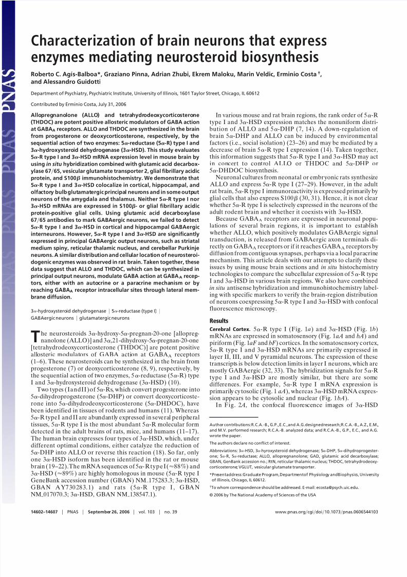
Cerebral Cortex. 5-R type I (Fig. 1 a) and 3-HSD (Fig. 1 b)mRNAs are expressed in somatosensory (Fig. 1 aA and bA) andpiriform (Fig. 1 aF and bF ) cortices. In the somatosensory c ortex,5-R type I and 3-HSD mRNAs are primarily expressed inlayer II, III, and V pyramidal neurons. The expression of thesetranscripts is below detection limits in layer I neurons, which aremostly GABAergic (32, 33). The hybridization signals for 5-Rtype I and 3-HSD are mostly similar, but there are somedifferences. For example, 5-R type I mRNA expression isprimarily cy tosolic (Fig. 1 aA), whereas 3-HSD mRNA expres-sion appears to be cytosolic and nuclear (Fig. 1 bA).
In Fig. 2 A, the confocal fluorescence images of 3-HSD
mRNA and 5-R type I protein show that, very frequently(95%), both enzymes are coexpressed in the same neurons.
Fig. 2 B shows that vesicular glutamate transporter (VGLUT)1mRNA, a glutamatergic neuronal marker, and 5-R type Iprotein colocalize. In contrast, we could not detect a colocal-ization of 5-R type I or 3-HSD with glutamic acid decarboxy-lase (GAD)6765 (Fig. 2 D), a marker for GABAergic neurons.Finally, 5-R type I mRNA fails to colocalize with S100, aspecific glial cell marker (Table 1).
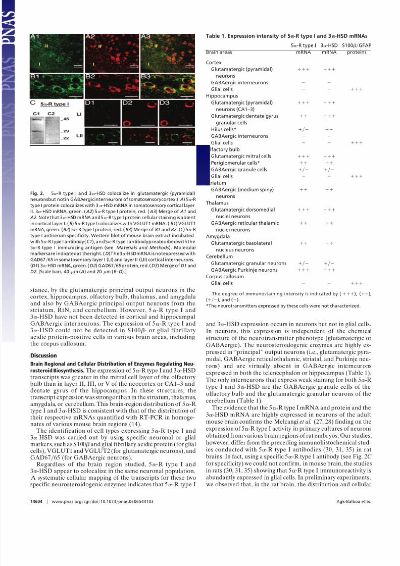
Hippocampus. The detection signals for 5-R type I and 3-HSDmRNAs are reported in Fig. 1 aB and bB, respectively. Thehybridization signal of both expressed transcripts in CA1 andCA2 is very intense, but, in CA3, the signal is faint. The cells of dentate gyrus and hilus also show a less-intense staining thanthat detectedin CA1and CA2, especially in thecase of 5-R typeI (Fig. 1 aB). The stratum oriens, stratum radiatum, and layerlacunosum moleculare, and the molecular layers of the dentategyrus show faint hybridization signal. However, a few cells arelabeled for 5-R type I mRNA in the stratum radiatum and, verylikely, these are interneurons. A coexistence of 5-R type I or3-HSD mRNAs with GAD6765 immunoreactivity was notdetected (Table 1). In the hippocampus, as in the cortex, the5-R type I signal fails to colocalize with S100 (Fig. 3C). As in
the cortex, in CA1 pyramidal neurons, the 5-R type I in situhybridization signal consistently colocalizes with the VGLUT2immunostaining (Fig. 3 A).
The VGLUT2 antibodies predominantly label glutamatergicneuron axon terminals (34). However, under the conditions of double in situ hybridization and immunohistochemistry shown inFig. 3 A, the cell bodies are also labeled. This labeling is elimi-nated when the VGLUT2 antiserum is preabsorbed with theimmunizing peptide (Fig. 3 B).
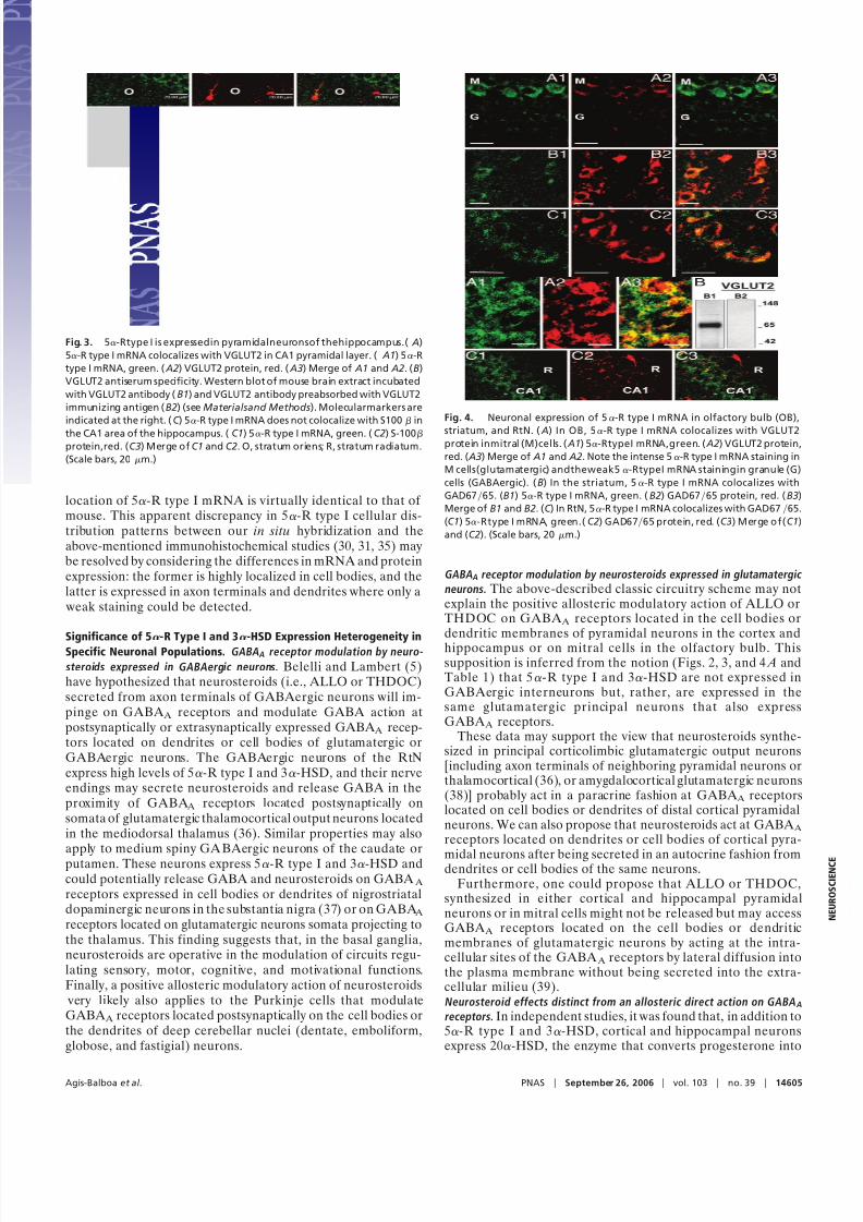
Olfactory Bulb. The olfactory bulb is characterized by the expres-sion of intense 5-R type I (Fig. 1 aC) and 3-HSD (Fig. 1 bC)hybridization signals, primarily expressed in the mitral andperiglomerular cells. The granule GABAergic neurons, express
faint staining for both 5-R type I and 3-HSD (Fig. 4 A). In themitral cell layer, 5-R type I mRNA colocalizes with VGLUT1mRNA (data not shown) or with VGLUT2 proteins (Fig. 4 A).However, we failed to observe the colocalization of 5-R type ImRNA with glial fibrillary acidic protein, a specific glial cellmarker (Table 1).
Striatum. The striatum includes numerous neurons that expressstrong 5-R type I (Fig. 1 aE) and 3-HSD (Fig. 1 bE) staining.Confocal fluorescence images show that 5-R type I mRNAcolocalizes with GAD6765 in striatal neurons (Fig. 4 B).
Thalamus. Sparse 5-R type I (Fig. 1 aG) and 3-HSD mRNA(Fig. 1 bG) positive neurons are expressed in various thalamicnuclei. An intense hybridization signal is detected in neurons of the reticular thalamic nucleus (RtN), where 5-R type I iscolocalized with GAD6765 (Fig. 4C). In contrast, in the
ventromedial thalamic nucleus, 5-R type I colocalizes withVGLUT1 mRNA and VGLUT2 proteins (Table 1).
Amygdala. The central and basolateral amygdaloid nuclei arecharacterized by sparse neurons exhibiting a faint staining foreither 5-R type I (Fig. 1 aF ) or 3-HSD mRNAs (Fig. 1 bF ).Strong staining is expressed in the basolateral anterior amygda-loid nucleus (BLA), whereas, in the central amygdaloid nucleus,staining is weak. The somata of glutamatergic principal neuronexpressed in the BLA show a colocalization of 5-R type ImRNA with VGLUT2 (Table 1).
Cerebellum. The staining of 5-R type I and 3-HSD mRNA isselectively localized in the somata of Purkinje neurons (Fig. 1 aDand bD). A fainter staining is also present in thegranule cell layerbut virtually absent in the molecular layer. As expected, 5-RtypeI mRNA is colocalized withGAD6765 proteins in Purkinjecells (Table 1).
Overview of 5-R Type I and 3-HSD mRNA Expression in the Mouse
Brain. Table 1 shows that, in the mouse brain, 5-R type I and3-HSD are expressed in principal output neurons, for in-
Fig. 1. Mouse brain distribution of 5-R type I (a) and 3-HSD (b) mRNAs. (aA and bA) Somatosensory cortex, coronal section at bregma 1.10 mm (49). I to
V identifythe corticallayers.(Fig.1 aB and bB) Hippocampus,coronal sectionat bregma 2.46mm (49). CA,cornusammonis; DG,dentategyrus; O,stratumoriens;
R, stratum radiatum; LM, layer lacunosum moleculare; M, molecular layer dentate gyrus; H, hilus. ( aC and bC ) Olfactory bulb, coronal section at bregma 3.92
mm (49). M, mitral; PG, periglomerular; G, granular cell layers. ( aD and bD) Cerebellum. The Purkinje cell layer is indicated by arrowheads (Œ). G, granule cell
layer; M, molecularlayer.(aE and bE ) Striatalmediumspiny neurons,coronal section as in A. Shown is unstainedstriatal fibertract(stf). (aF and bF ) Piriformcortexand amygdala, coronal section at bregma 1.58 mm (49). BLA, basolateral anterior amygdaloid nucleus; BM, basomedial amygdaloid nucleus; Ce, central
amygdaloid nucleus; Pir, piriform cortex. (aG and bG) Thalamic nuclei, coronal section as in F . VPL, ventral posterolateral thalamic nucleus; VPM, ventral
posteromedial thalamic nucleus. [Scale bars, 50 m (aA and bA) and 100 m (aB and bB–aG and bG).]
Agıs-Balboa et al. PNAS September 26, 2006 vol. 103 no. 39 14603
8/3/2019 Roberto C. Agis-Balboa et al- Characterization of brain neurons that express enzymes mediating neurosteroid biosy…
stance, by the glutamatergic principal output neurons in the
cortex, hippocampus, olfactory bulb, thalamus, and amygdalaand also by GABAergic principal output neurons from thestriatum, RtN, and cerebellum. However, 5-R type I and3-HSD have not been detected in cortical and hippocampalGABAergic interneurons. The expression of 5-R type I and3-HSD could not be detected in S100- or glial fibrillaryacidic protein-positive cells in various brain areas, includingthe corpus callosum.
Discussion
Brain Regional and Cellular Distribution of Enzymes Regulating Neu-
rosteroid Biosynthesis. The expression of 5-R type I and 3-HSDtranscripts was greater in the mitral cell layer of the olfactorybulb than in layer II, III, or V of the neocortex or CA1–3 anddentate gyrus of the hippocampus. In these structures, the
transcript expression was stronger than in the striatum, thalamus,amygdala, or cerebellum. This brain-region distribution of 5-Rtype I and 3-HSD is consistent with that of the distribution of their respective mRNAs quantified with RT-PCR in homoge-nates of various mouse brain regions (14).
The identification of cell types expressing 5-R type I and3-HSD was carried out by using specific neuronal or glialmarkers, such as S100 and glial fibrillary acidic protein (for glialcells), VGLUT1 and VGLUT2 (for glutamatergic neurons), andGAD6765 (for GABAergic neurons).
Regardless of the brain region studied, 5-R type I and3-HSD appear to colocalize in the same neuronal population.
A systematic cellular mapping of the transcripts for these twospecific neurosteroidogenic enzymes indicates that 5-R type I
and 3-HSD expression occurs in neurons but not in glial cells.In neurons, this expression is independent of the chemicalstructure of the neurotransmitter phenotype (glutamatergic orGABAergic). The neurosteroidogenic enzymes are highly ex-pressed in ‘‘principal’’ output neurons (i.e., glutamatergic pyra-midal, GABAergic reticulothalamic, striatal, and Purkinje neu-rons) and are virtually absent in GABAergic interneuronsexpressed in both the telencephalon or hippocampus (Table 1).The only interneurons that express weak staining for both 5-Rtype I and 3-HSD are the GABAergic granule cells of the
olfactory bulb and the glutamatergic granular neurons of thecerebellum (Table 1).The evidence that the 5-R type I mRNA and protein and the
3-HSD mRNA are highly expressed in neurons of the adultmouse brain confirms the Melcangi et al. (27, 28) finding on theexpression of 5-R type I activity in primary cultures of neuronsobtained from various brain regions of rat embr yos. Our studies,however, differ from the preceding immunohistochemical stud-ies conducted with 5-R type I antibodies (30, 31, 35) in ratbrains. In fact, using a specific 5-R type I antibody (see Fig. 2Cfor specificity) we could not confirm, in mouse brain, the studiesin rats (30, 31, 35) showing that 5-R type I immunoreactivity isabundantly expressed in glial cells. In preliminary experiments,
we observed that, in the rat brain, the distribution and cellular
Fig. 2. 5-R type I and 3-HSD colocalize in glutamatergic (pyramidal)
neuronsbut notin GABAergicinterneurons of somatosensorycortex.( A) 5-Rtype I protein colocalizes with 3-HSD mRNA in somatosensory cortical layer
II. 3-HSD mRNA, green. ( A2) 5-R type I protein, red. ( A3) Merge of A1 and
A2. Notethat 3-HSD mRNA and5-R type I protein cellular staining is absent
in cortical layer I. (B) 5-R type I colocalizes with VGLUT1 mRNA. (B1) VGLUT1
mRNA, green. (B2) 5-R type I protein, red. (B3) Merge of B1 and B2. (C ) 5-R
type I antiserum specificity. Western blot of mouse brain extract incubated
with 5-R type I antibody( C1),and5-R type I antibodypreabsorbedwiththe
5-R type I immunizing antigen (see Materials and Methods). Molecular
markersare indicatedat theright. (D)The3-HSDmRNA is notexpressed with
GAD6765 in somatosensory layer I (LI) and layer II (LII) cortical interneurons.
(D1) 3-HSD mRNA, green.(D2) GAD6765protein,red.(D3) Merge of D1 and
D2. [Scale bars, 40 m ( A) and 20 m (B–D).]
Table 1. Expression intensity of 5-R type I and 3-HSD mRNAs
Brain areas
5-R type I
mRNA
3-HSD
mRNA
S100GFAP
proteins
Cortex
Glutamatergic (pyramidal)
neurons
GABAergic interneurons
Glial cells
Hippocampus
Glutamatergic (pyramidal)
neurons (CA1–3)
Glutamatergic dentate gyrus
granular cells
Hilus cells*
GABAergic interneurons
Glial cells
Olfactory bulb
Glutamatergic mitral cells
Periglomerular cells*
GABAergic granule cells
Glial cells
Striatum
GABAergic (medium spiny)
neurons
Thalamus
Glutamatergic dorsomedial
nuclei neurons
GABAergic reticular thalamic
nuclei neurons
Amygdala
Glutamatergic basolateral
nucleus neurons
Cerebellum
Glutamatergic granular neurons
GABAergic Purkinje neurons
Corpus callosum
Glial cells
The degree of immunostaining intensity is indicated by (), (),
(), and ().
*The neurotransmitters expressed by these cells were not characterized.
14604 www.pnas.orgcgidoi10.1073pnas.0606544103 Agıs-Balboa et al.
8/3/2019 Roberto C. Agis-Balboa et al- Characterization of brain neurons that express enzymes mediating neurosteroid biosy…
location of 5-R type I mRNA is virtually identical to that of mouse. This apparent discrepancy in 5-R type I cellular dis-tribution patterns between our in situ hybridization and theabove-mentioned immunohistochemical studies (30, 31, 35) maybe resolved by considering the differences in mRNA and proteinexpression: the former is highly localized in cell bodies, and thelatter is expressed in axon terminals and dendrites where only a
weak staining could be detected.
Significance of 5-R Type I and 3-HSD Expression Heterogeneity inSpecific Neuronal Populations. GABAA receptor modulation by neuro-
steroids expressed in GABAergic neurons. Belelli and Lambert (5)have hypothesized that neurosteroids (i.e., ALLO or THDOC)secreted from axon terminals of GABAergic neurons will im-pinge on GABA A receptors and modulate GABA action atpostsynaptically or extrasynaptically expressed GABA A recep-tors located on dendrites or cell bodies of glutamatergic orGABAergic neurons. The GABAergic neurons of the RtNexpress high levels of 5-R type I and 3-HSD, and their nerveendings may secrete neurosteroids and release GABA in theproximity of GABA A receptors located postsynaptically onsomata of glutamatergic thalamocortical output neurons locatedin the mediodorsal thalamus (36). Similar properties may alsoapply to medium spiny GA BAergic neurons of the caudate orputamen. These neurons express 5-R type I and 3-HSD andcould potentially release GABA and neurosteroids on GABA A
receptors expressed in cell bodies or dendrites of nigrostriataldopaminergic neurons in the substantia nigra (37) or on GABA A
receptors located on glutamatergic neurons somata projecting tothe thalamus. This finding suggests that, in the basal ganglia,neurosteroids are operative in the modulation of circuits regu-lating sensory, motor, cognitive, and motivational functions.Finally, a positive allosteric modulatory action of neurosteroids
very likely also applies to the Purkinje cells that modulateGABA A receptors located postsynaptically on the cell bodies orthe dendrites of deep cerebellar nuclei (dentate, emboliform,globose, and fastigial) neurons.
GABAA receptor modulation by neurosteroids expressed in glutamatergic
neurons. The above-described classic circuitry scheme may notexplain the positive allosteric modulatory action of ALLO orTHDOC on GABA A receptors located in the cell bodies ordendritic membranes of pyramidal neurons in the cortex andhippocampus or on mitral cells in the olfactory bulb. Thissupposition is inferred from the notion (Figs. 2, 3, and 4 A andTable 1) that 5-R type I and 3-HSD are not expressed inGABAergic interneurons but, rather, are expressed in thesame glutamatergic principal neurons that also expressGABA A receptors.
These data may support the view that neurosteroids synthe-sized in principal corticolimbic glutamatergic output neurons[including axon terminals of neighboring pyramidal neurons orthalamocortical (36), or amygdalocortical glutamatergic neurons(38)] probably act in a paracrine fashion at GABA A receptorslocated on cell bodies or dendrites of distal cortical pyramidalneurons. We can also propose that neurosteroids act at GABA A
receptors located on dendrites or cell bodies of cortical pyra-
midal neurons after being secreted in an autocrine fashion fromdendrites or cell bodies of the same neurons.
Furthermore, one could propose that ALLO or THDOC,synthesized in either cortical and hippocampal pyramidalneurons or in mitral cells might not be released but may accessGABA A receptors located on the cell bodies or dendriticmembranes of glutamatergic neurons by acting at the intra-cellular sites of the GABA A receptors by lateral diffusion intothe plasma membrane without being secreted into the extra-cellular milieu (39).Neurosteroid effects distinct from an allosteric direct action on GABAA
receptors. In independent studies, it was found that, in addition to5-R type I and 3-HSD, cortical and hippocampal neuronsexpress 20-HSD, the enzyme that converts progesterone into
Fig. 3. 5-Rtype I is expressedin pyramidalneuronsof thehippocampus.( A)
5-R type I mRNA colocalizes with VGLUT2 in CA1 pyramidal layer. ( A1) 5-R
type I mRNA, green. ( A2) VGLUT2 protein, red. ( A3) Merge of A1 and A2. (B)
VGLUT2 antiserum specificity. Western blot of mouse brain extract incubated
with VGLUT2 antibody (B1) and VGLUT2 antibody preabsorbed with VGLUT2
immunizing antigen (B2) (see Materialsand Methods). Molecularmarkers are
indicated at the right. (C ) 5-R type I mRNA does not colocalize with S100 in
the CA1 area of the hippocampus. (C1) 5-R type I mRNA, green. (C2) S-100
protein,red. (C3) Merge of C1 and C2. O, stratum oriens; R, stratum radiatum.
(Scale bars, 20 m.)
Fig. 4. Neuronal expression of 5-R type I mRNA in olfactory bulb (OB),
striatum, and RtN. ( A) In OB, 5-R type I mRNA colocalizes with VGLUT2protein inmitral (M)cells. ( A1) 5-RtypeI mRNA,green. ( A2) VGLUT2 protein,
red. ( A3) Merge of A1 and A2. Note the intense 5-R type I mRNA staining in
M cells(glutamatergic) andtheweak5-RtypeI mRNA stainingin granule (G)
cells (GABAergic). (B) In the striatum, 5-R type I mRNA colocalizes with
GAD6765. (B1) 5-R type I mRNA, green. (B2) GAD6765 protein, red. (B3)
Merge of B1 and B2. (C ) In RtN, 5-R type I mRNA colocalizes with GAD6765.
the inactive 20-hydroxyprogesterone or converts ALLO into20-hydroxyallopregnanolone (40). Cortical and hippocampalneurons also express 3-HSD, the enzyme that converts preg-nanolone into progesterone (41). Thus, glutamatergic corticaland hippocampal neurons and, also, very likely, the olfactorymitral neurons, may synthesize both progesterone and 5-DHP,
which may be involved in the regulation of intracellular proges-terone-receptor function.
In this context, it is noteworthy to mention that neurosteroids
have been reported to (i) influence prefrontal cortex structureand thalamocortical connectivity (42), (ii) increase cerebellargranule-cell neurogenesis (43), (iii) increase the proliferation of neuroprogenitor cells expressed in the rat hippocampus (44),and (iv) increase the proliferation of human embryonic neuralstem cells expressed in the cerebral cortex (44). It is probablethat a receptor mechanism different from a direct modulation of GABA A receptor signal transduction may be operative in theseactions. We can speculate that the microtubular-associatedprotein 2 (MAP2) is a possible putative intracellular receptor forneurosteroids (i.e., ALLO) (45).
Conclusion
A decrease of brain neurosteroid availability has been associated with psychiatric conditions, including anxiety, aggression, pre-
menstrual dysphoria, and cognitive and mood disorders. Anti-depressants (fluoxetine and other selective serotonin-reuptakeinhibitors, ‘‘SSRIs’’) and antipsychotics (clozapine) may exerttheir beneficial effects, at least in part, by increasing the brainlevels of neurosteroids (26, 46–48).
Although the neurosteroids that act as positive allostericmodulators of GABA action at GABA A receptors were initiallyconsidered endocrine messengers that are indiscriminately syn-thesized and secreted from glial cells in all brain regions, thepresent histochemical data suggest that there is a cellular andmolecular basis to imply that neurosteroids are synthesized inneurons and act locally at GABA A receptors expressed onspecific corticolimbic circuitries. Understanding the contribu-tion of local brain neurosteroid biosynthesis to the pathogenesisof neurological and psychiatric disorders may become a stimulus
to develop new psychoactive drugs that act selectively by nor-malizing neurosteroid action on GABAergic neurotransmission.
Materials and Methods
Animals and Tissue Preparation. Adult male Swiss–Webster mice(Harlan, Indianapolis, IN), 25–30 g in body weight, were perfused
with 0.9% NaCl and 4% paraformaldehyde. The brains were (i)postfixed for 72h in 4% paraformaldehyde and (ii) embedded in30% sucrose in 0.15 M PBS, pH 7.4, at 4°C. All experiments wereconducted in groups of three to five animals. For each condition,(antisense probes or antibodies) four to six sections were processedfor each animal. All animal procedures were approved by theUniversity of Illinois Animal Care Committee.
mRNA in Situ Hybridization. Antisense probe design. To visualize 5-R
type I mRNA, free-floating 16-to 20-m coronal sections (49)wereincubated for 72 h at 42°C with a mixture of 50 pmolml of threeantisense oligonucleotide probes: R1 (nt 910–933), R2 (nt 989–1,012), and R3 (nt 1180–1203) (GBAN NM
175283). To visualize
3-HSD mRNA, adjacent sections were incubated with two anti-sense oligonucleotide probes, H1 (nt 526–549) and H2 (nt 804–827) (GBAN AY730283.1). To visualize VGLUT1 mRNA, we useda hybridization technique with antisense oligonucleotide probescomplementary to bases 626–649 (V1) and 1499–1522 (V2) of themouse VGLUT1 cDNA (GBAN NM 182993). The oligonucleotide3 terminals were labeled with digoxigenin by using the Oligonu-cleotide Digoxigenin Tailing kit (Roche Diagnostics, Indianapolis,IN). The in situ hybridization protocol followed a variation of theprocedure described by Rodriguez et al. (32) for the avidin–biotin–
peroxidase complex (ABC; Vector Laboratories, Burlingame, CA)method and by Pesold et al. (50, 51) and Veldic et al. (52) forconfocal immunofluorescence.Antisense probes: specificity tests. The antisense probe sequences didnot match any other known mRNA sequences, as determined bymultiple genome-wide BLAST c omparisons. However, becausegenomic sequences that have not been reported as part of theintronexon structures of transcribed genes can be transcribed(53), we performed separate in situ hybridization studies with the
three 5-R type I antisense probes, the two 3-HSD antisenseprobes, and the t wo VGLUT1 antisense probes to establishspecificity. In coronal prefrontal cortex slices, the distribution of neurons stained with the 5-R type 1 probe R1 is virtuallyidentical to the distribution of neurons stained with probes R2and R3. The distribution of neurons stained with the 3-HSDprobe H1 is also virtually identical to the distribution of neuronsdetected with H2. The same virtually identical distribution wasalso found with the VGLUT1 probes V1 and V2. Oligoprobespecificity was tested by using digoxigenin-labeled scrambleroligonucleotides for 5-R type I, 3-HSD, and VGLUT1. Asexpected, specific neuronal staining was not detected.
Double in Situ Hybridization and Immunohistochemistry. Double in situ hybridization and immunohistochemistry were performed by
following a variation of the procedure described by Pesold etal. (50,51) and Veldic et al. (52). After the in situ hybridization procedure was terminated, the following antibodies were used: (i) rabbitanti-GAD6765 (diluted 1:2,000; Chemicon, Temecula, CA), (ii)rabbit anti-VGLUT2 (diluted 1:500; Synaptic Systems, Gottingen,Germany), (iii) rabbit anti-GFAP (diluted 1:250; Chemicon), (iv)rabbit anti-S-100 (diluted 1:5,000; Swant, Bellinzona, Switzer-land), and (iv) rabbit anti-rat 5-R type I (diluted 1:100; Acris
Antibodies, Hiddenhausen, Germany). After the double in situ hybridization and immunohistochem-
istry procedures, the slices were incubated with Cy5-labeled goatanti-rabbit IgG (diluted 1:1,000; Amersham Biosciences, Pisca-taway, NJ) or Cy5-labeled goat anti-guinea pig IgG (diluted1:1,000; Abcam, Cambridge, MA) to produce red fluorescentstaining or Cy2-labeled streptavidin (diluted 1:1,000; Amersham
Biosciences) to produce green f luorescent staining, as indicatedin the figure legends. The number of cells in which green and redfluorescence colocalize compared with the number of cells thatexpress only green or only red fluorescence was quantified withconfocal microscopy (Leica, Bannockburn, IL) at a magnifica-tion of 40 in a counting box of 100 100 20 m.
Western Blot Analysis to Assess 5-R Type I and VGLUT2 Antibody
Specificity. Mouse brain extracts (10mg per200l of SDS loadingbuffer) were electrophorized on 10–20% SDSPAGE gel andblotted onto a nitrocellulose membrane (Amersham Bio-sciences). For the study of 5-R type I antiserum specificity, themembranes were incubated w ith either anti-5-R type I (diluted1:2,500; Acris Antibodies) or anti-5-R type I (diluted 1:2,500)preabsorbed for 12 h at 4°C with a 1 mM solution of the
immunizing peptide (V-V-F-A-L-F-T-L-S-T-L-T-R-A-K-Q-H-H-Q-W-Y) in 0.005 M NaPHO4 buffer (pH 7.2), 0.2 M NaCl, and5% BSA. Two immunoreactive bands, one of 45 kDa and oneof 29 kDa, are recognized by the nonpreabsorbed antiserum(Fig. 2C1). The immunoreactive bands disappear when thepreabsorbed antiserum is used (Fig. 2C2). The size of 5-R typeI in mice is 29 kDa, according to the GenBank accession no.
AAH94503; however, as indicated by Russell and Wilson (11),the hydrophobic amino acid content of 5-R type I (37%) mayexplain the aberrant electrophoretic mobilities in SDDPAGEthat have been reported for the 5-R isoenzymes. The preab-sorbed antiserum failed to immunoreact w ith mouse brain slices.
The membranes were also incubated with anti-VGLUT2 anti-serum (diluted 1:4,000; Synaptic Systems) or anti-VGLUT2 anti-
14606 www.pnas.orgcgidoi10.1073pnas.0606544103 Agıs-Balboa et al.
8/3/2019 Roberto C. Agis-Balboa et al- Characterization of brain neurons that express enzymes mediating neurosteroid biosy…
serum (diluted 1:4,000) preabsorbed for 12 h at 4°C with 20 l of solution, 1 mg per 1 ml of the immunizing control peptide (aminoacids 510–582 of rat VGLUT2DNPI; Synaptic Systems) in blottingbuffer (3% nonfat dry milk0.1% Tween-20). Only one immuno-reactive band of 65 kDa is recognized by the nonpreabsorbedantiserum (Fig. 3 B1). This band completely disappears in thepreabsorbed antiserum (Fig. 3 B2). The preabsorbed antiserumfailed to immunoreact with mouse brain slices.
Digital Photomicrography. DAB (3-3-diaminobenzidine tetrahy-drochloride) (Sigma, St. Louis, MO) staining images were
captured by Axiovision 3.1 (Zeiss) and confocal immunofluo-rescence by a confocal microscope (Leica Microsystems, Ban-nockburn, IL). The final composites were processed by usingPhotoshop (Adobe Systems, Mountain View, CA) and Power-point (Microsoft, Redmond, WA).
We thank Dr. A. L. Morrow (University of North Carolina, Chapel Hill,NC) and Dr. H. Mohler (University of Zurich, Switzerland) for theconstructive criticism and suggestions in the preparation of the manu-
script. This work was supported by National Institute of Mental HealthGrant R01-M4 56890 (to A.G.).
1. Mienville JM, Vicini S (1989) Brain Res 489:190–194.2. Puia G, Santi MR, Vicini S, Pritchett DB, Purdy RH, Paul SM, Seeburg PH,
Costa E (1990) Neuron 4:759–765.3. Baulieu EE (1998) Psychoneuroendocrinology 23:963–987.4. Puia G, Mienville JM, Matsumoto K, Takahata H, Watanabe H, Costa E,
JJ (2006) Neuroscience 138:821–829.7. Cheney DL, Uzunov D, Costa E, Guidotti A (1995) J Neurosci 15:4641–4650.8. PurdyRH,MorrowAL, MoorePH, Jr,PaulSM (1991) Proc Natl Acad SciUSA.
88:4553–4557.9. Khisti RT, Boyd KN, Kumar S, Morrow AL (2005) Brain Res 1049:104–111.
10. Karavolas HJ, Hodges DR (1991) in Neurosteroids and Brain Function eds
Costa E, Paul SM (Thieme, New York), pp 135–145.11. Russell DW, Wilson JD (1994) Annu Rev Biochem 63:25–61.12. Melcangi RC, Poletti A, Cavarretta I, Celotti F, Colciago A, Magnaghi V, Motta
M, Negri-Cesi P, Martini L (1998) J Steroid Biochem Molec Biol 65:295–299.13. Thigpen AE, Silver RI, Guileyardo JM, Casey ML, McConnell JD, Russell DW
(1993) J Clin Invest 92:903–910.14. Dong E, Matsumoto K, Uzunova V, Sugaya I, Takahata H,Nomura H, Watanabe
H, Costa E, Guidotti A (2001) Proc Natl Acad Sci USA 98:2849–2854.15. Steckelbroeck S, Watzka M, Reichelt R, Hans VH, Stoffel-Wagner B, Heidrich
86:1324–1331.16. Stoffel-Wagner B (2003) Ann N Y Acad Sci 1007:64–78.17. Torres JM, Ortega E (2003) FASEB J 1428–1433.18. Penning TM, Jin Y, Steckelbroeck S, Lanis nik Rizner T, Lewis M (2004) Mol
Cell Endocrinol 215:63–72.19. Hara A, Inoue Y, Nakagawa M, Naganeo F, Sawada H (1988) J Biochem
(Tokyo) 103:1027–1034.
20. Pawlowski JE, Huizinga M, Penning TM (1991) J Biol Chem 266:8820–8825.21. Hoog SS, Pawlowski JE, Alzari PM, Penning TM, Lewis M (1994) Proc Natl
Acad Sci USA 91:2517–2521.22. Penning TM, Jin Y, Heredia VV, Lewis M (2003) J Steroid Biochem Mol Biol
85:247–255.23. Matsumoto K, Uzunova V, Pinna G, Taki K, Uzunov DP, Watanabe H,
Mienville JM, Guidotti A, Costa E (1999) Neuropharmacology 38:955–963.24. Serra M, Pisu MG, Littera M, Papi G, Sanna E, Tuveri F, Usala L, Purdy RH,
Biggio G (2000) J Neurochem 75:732–740.25. Pinna G, Dong E, Matsumoto K, Costa E, Guidotti A (2003) Proc Natl Acad
Sci USA 100:2035–2040.26. Pinna G, Costa E, GuidottiA (2006) Psychopharmacology (Berlin) 186:362–372.27. Melcangi RC, Celotti F, Castano P, Martini L (1993) Endocrinology 132:1252–
1259.
28. Melcangi RC, Celotti F, Martini L (1994) Brain Res 639:202–206.29. Follesa P, Serra M, Cagetti E, Pisu MG, Porta S, Floris S, Massa F, Sanna E,
Biggio G (2000) Mol Pharmacol 57:1262–1270.30. Tsuruo Y, Miyamoto T, Yokoi H, Kitagawa K, Futaki S, Ishimura K (1996)
Brain Res 722:207–211.31. Kiyokage E, Toida K, Suzuki-Yamamoto T, Ishimura K (2005) J Comp Neurol
493:381–395.32. Rodriguez MA, Caruncho HJ, Costa E, Pesold C, Liu WS, Guidotti A (2002)
J Comp Neurol 451:279–288.33. Gabbott PL, Somogyi P (1986) Exp Brain Res 61:323–331.34. Fremeau RT, Jr, Voglmaier S, Seal RP, Edwards RH (2004) Trends Neurosci
27:98–103.35. Pelletier G, Luu-The V, Labrie F (1994) Mol Cell Neurosci 5:394–399.36. Pinault D (2004) Brain Res Brain Res Rev 46:1–31.37. Gale K, Guidotti A, Costa E (1977) Science 95:503–505.38. Gisabella B, Bolshakov VY, Benes FM (2005) Proc Natl Acad Sci USA
102:13301–13306.39. Akk G, Shu HJ, Wang C, Steinbach JH, Zorumski CF, Covey DF, Mennerick
S (2005) J Neurosci 25:11605–11613.40. Pelletier G, Luu-The V, Li S, Labrie F (2004) Brain Res Mol Brain Res
125:143–146.41. Guennoun R, Fiddes RJ, Gouezou M, Lombes M, Baulieu EE (1995) Brain Res
Mol Brain Res 30:287–300.42. Grobin AC, Gizerian S, Lieberman JA, Morrow AL (2006) Neuroscience
138:809–819.43. Keller EA, Zamparini A, Borodinsky LN, Gravielle MC, Fiszman ML (2004)
Brain Res Dev Brain Res 153:13–17.44. Wang JM, Johnston PB, Ball BG, Brinton RD (2005) J Neurosci 25:4706–4718.45. Fontaine-Lenoir V, Chambraud B, Fellous A, David S, Duchossoy Y, Baulieu
EE, Robel P (2006) Proc Natl Acad Sci USA 103:4711–4716.46. Guidotti A, Costa E (1998) Biol Psychiatry 44:865–873.47. Marx CE, VanDoren MJ, Duncan GE, Lieberman JA, Morrow AL (2003)
Neuropsychopharmacology 28:1–13.48. Barbaccia ML (2004) Crit Rev Neurobiol 16:67–74.49. Franklin KBJ, Paxinos G (1997) in The Mouse Brain in Stereotaxic Coordinates