- 57 - Imaging Science in Dentistry 2016; 46: 57-62 http://dx.doi.org/10.5624/isd.2016.46.1.57 Fusion is defined as the union of two contiguous den- tal germs occurring in any stage of odontogenesis. 1 This union may develop at the crown level (enamel) or at the crown and root level (enamel and dentin). 2 Nonetheless, the pulp chambers and the pulp canals are either unified or separated. 1 This anatomic irregularity is commonly ob- served in the primary dentition, particularly in the anteri- or region of the mouth. 3 It is usually seen in the maxillary arch, and rarely occurs in the posterior mandible. 4 The etiology of fusion is still unclear; however, it has been suggested that the pressure or physical forces that provide close contact between two adjacent dental follicles may cause the follicles to fuse before calcification. 5 The main periodontal problem in cases of fusion is the presence of deep fissures or grooves that extend subgingivally on the union between the teeth involved. These fissures may al- low the accumulation of bacterial plaque, leading to den- tal caries and periodontal diseases in this area. 1 Due to recurrent periodontal problems, the jaws may exhibit cysts, such as paradental cysts. This type of cyst has been described as an inflammatory cyst that develops on the cervical margin of a vital tooth due to an inflam- matory process in the periodontal pocket. 6 The etiology of these cysts has been proposed to involve the inflamma- tory proliferation of the epithelial cell rests of Malassez or reduced enamel epithelium. 5 An unusual morphological variation of the mandible is the retromolar canal, which permits the passage of the vascular and nerve bundles of the pulp and periodontium of mandibular molar teeth. 7 This canal branches off from the mandibular canal behind the last molar tooth and pass- es through the retromolar fossa via the retromolar fora- men. 8 However, the content of retromolar canals has not been conclusively established. 7 The lack of information on this subject may be related to complications involving local anesthesia 9 and surgical nerve damage during third molar surgery in the retromolar region. 8 Panoramic radiography plays a fundamental role in pro- viding data on the oral and maxillofacial region. 10 Never- Role of cone-beam computed tomography in the evaluation of a paradental cyst related to the fusion of a wisdom tooth with a paramolar: A rare case report Gozde Ozcan 1, * , Ahmet Ercan Sekerci 1 , Emrah Soylu 2 , Sinan Nazlim 3 , Mehmet Amuk 1 , Fatma Avci 1 1 Department of Oral and Maxillofacial Radiology, Faculty of Dentistry, Erciyes University, Kayseri, Turkey 2 Department of Oral and Maxillofacial Surgery, Faculty of Dentistry, Gaziosmanpasa University, Tokat, Turkey 3 Department of Pathology, Yozgat State Hospital, Yozgat, Turkey ABSTRACT Fusion is an abnormality of tooth development defined as the union of two developing dental germs, resulting in a single large dental structure. This irregular tooth morphology is associated with a high predisposition to dental caries and periodontal diseases. As a result of recurring inflammatory periodontal processes, disorders such as periodontal pocket, pericoronitis, and paradental cysts may develop. A rare mandibular anatomic variation is the retromolar canal, which is very significant for surgical procedures. The fusion of a paramolar and mandibular third molar associated with a paradental cyst co-occurring with the presence of a retromolar canal is rare, and the aim of the present study is to describe the evaluation of this anatomical configuration using cone-beam computed tomography. (Imaging Sci Dent 2016; 46: 57-62) KEY WORDS: Fused Teeth; Odontogenic Cyst; Cone-Beam Computed Tomography; Tooth, Supernumerary Copyright ⓒ 2016 by Korean Academy of Oral and Maxillofacial Radiology This is an Open Access article distributed under the terms of the Creative Commons Attribution Non-Commercial License ( http://creativecommons.org/licenses/by-nc/3.0) which permits unrestricted non-commercial use, distribution, and reproduction in any medium, provided the original work is properly cited. Imaging Science in Dentistry·pISSN 2233-7822 eISSN 2233-7830 Received December 4, 2015; Revised January 13, 2016; Accepted January 16, 2016 *Correspondence to : Dr. Gozde Ozcan Department of Oral and Maxillofacial Radiology, Faculty of Dentistry, Erciyes Uni- versity, 38039, Kayseri, Turkey Tel) 90-352-207-6666, Fax) 90-352-438-0657, E-mail) [email protected]
Transcript
- 57 -
Imaging Science in Dentistry 2016; 46: 57-62http://dx.doi.org/10.5624/isd.2016.46.1.57
Fusion is defined as the union of two contiguous den-tal germs occurring in any stage of odontogenesis.1 This union may develop at the crown level (enamel) or at the crown and root level (enamel and dentin).2 Nonetheless, the pulp chambers and the pulp canals are either unified or separated.1 This anatomic irregularity is commonly ob-served in the primary dentition, particularly in the anteri-or region of the mouth.3 It is usually seen in the maxillary arch, and rarely occurs in the posterior mandible.4 The etiology of fusion is still unclear; however, it has been suggested that the pressure or physical forces that provide close contact between two adjacent dental follicles may cause the follicles to fuse before calcification.5 The main periodontal problem in cases of fusion is the presence of deep fissures or grooves that extend subgingivally on the union between the teeth involved. These fissures may al-low the accumulation of bacterial plaque, leading to den-
tal caries and periodontal diseases in this area.1 Due to recurrent periodontal problems, the jaws may
exhibit cysts, such as paradental cysts. This type of cyst has been described as an inflammatory cyst that develops on the cervical margin of a vital tooth due to an inflam-matory process in the periodontal pocket.6 The etiology of these cysts has been proposed to involve the inflamma-tory proliferation of the epithelial cell rests of Malassez or reduced enamel epithelium.5
An unusual morphological variation of the mandible is the retromolar canal, which permits the passage of the vascular and nerve bundles of the pulp and periodontium of mandibular molar teeth.7 This canal branches off from the mandibular canal behind the last molar tooth and pass-es through the retromolar fossa via the retromolar fora- men.8 However, the content of retromolar canals has not been conclusively established.7 The lack of information on this subject may be related to complications involving local anesthesia9 and surgical nerve damage during third molar surgery in the retromolar region.8
Panoramic radiography plays a fundamental role in pro-viding data on the oral and maxillofacial region.10 Never-
Role of cone-beam computed tomography in the evaluation of a paradental cyst related to the fusion of a wisdom tooth with a paramolar: A rare case report
Gozde Ozcan1,*, Ahmet Ercan Sekerci1, Emrah Soylu2, Sinan Nazlim3, Mehmet Amuk1, Fatma Avci1
1Department of Oral and Maxillofacial Radiology, Faculty of Dentistry, Erciyes University, Kayseri, Turkey 2Department of Oral and Maxillofacial Surgery, Faculty of Dentistry, Gaziosmanpasa University, Tokat, Turkey 3Department of Pathology, Yozgat State Hospital, Yozgat, Turkey
AbstRAct
Fusion is an abnormality of tooth development defined as the union of two developing dental germs, resulting in a single large dental structure. This irregular tooth morphology is associated with a high predisposition to dental caries and periodontal diseases. As a result of recurring inflammatory periodontal processes, disorders such as periodontal pocket, pericoronitis, and paradental cysts may develop. A rare mandibular anatomic variation is the retromolar canal, which is very significant for surgical procedures. The fusion of a paramolar and mandibular third molar associated with a paradental cyst co-occurring with the presence of a retromolar canal is rare, and the aim of the present study is to describe the evaluation of this anatomical configuration using cone-beam computed tomography. (Imaging Sci Dent 2016; 46: 57-62)
Copyright ⓒ 2016 by Korean Academy of Oral and Maxillofacial RadiologyThis is an Open Access article distributed under the terms of the Creative Commons Attribution Non-Commercial License (http://creativecommons.org/licenses/by-nc/3.0)
which permits unrestricted non-commercial use, distribution, and reproduction in any medium, provided the original work is properly cited.Imaging Science in Dentistry·pISSN 2233-7822 eISSN 2233-7830
Received December 4, 2015; Revised January 13, 2016; Accepted January 16, 2016 *Correspondence to : Dr. Gozde OzcanDepartment of Oral and Maxillofacial Radiology, Faculty of Dentistry, Erciyes Uni-versity, 38039, Kayseri, TurkeyTel) 90-352-207-6666, Fax) 90-352-438-0657, E-mail) [email protected]
Role of cone-beam computed tomography in the evaluation of a paradental cyst related to the fusion of a wisdom tooth with a paramolar: A rare case report
- 58 -
theless, panoramic radiographs and other two-dimensio- nal radiographs fail to show the buccolingual aspect and cross-sectional slices, which are important for presurgical assessments.11 Cone-beam computed tomography (CBCT) provides high-contrast three-dimensional images of maxil-lofacial structures that help to identify the definitive diag- nosis and develop a treatment plan based on images in all specialties within dentistry.12
The aim of the present report was to describe the use of CBCT to visualize the fusion of the mandibular third mo-lar and a supernumerary tooth in relation to a paradental cyst and in the presence of a retromolar canal in the same region.
case Report A 27-year-old male patient with an unremarkable medi-
cal history was referred to the Department of Oral and Maxillofacial Radiology with a complaint of pain in the region of the lower right molar teeth.
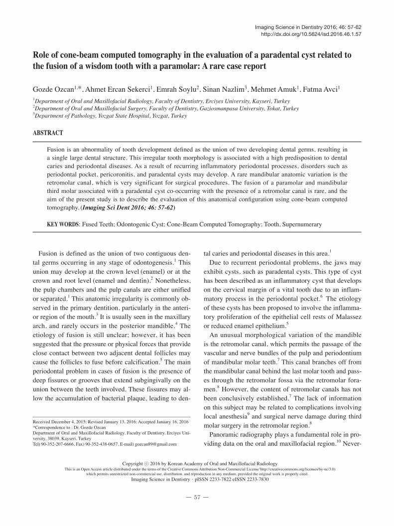
An intraoral examination revealed irregular morphol-ogy of the mandibular permanent third molar. This mor-phology suggested the presence of the union of an extra cusp on the lingual aspect of a wisdom tooth. This union led to the development of a groove formation that caused periodontal problems. The gingiva around the right man-dibular third molar appeared to be reddish in color, with loss of stippling and an inflamed appearance (Fig. 1A).
A panoramic radiograph indicated the fusion of a super-
numerary tooth with the permanent third molar and the presence of a dilated follicular space associated with this abnormal formation. This irregular enlargement exhibited a sharply delineated area of radiolucency around the fused supernumerary tooth (Fig. 1B). However, the panoramic radiograph was not sufficient to visualize this dental for-mation and its surrounding tissues because it only provid-ed information in the mesiodistal plane. With the consent of the patient, a CBCT image was taken to observe the abovementioned teeth, the periradicular lesion, and the relationship thereof with the peripheral structures.
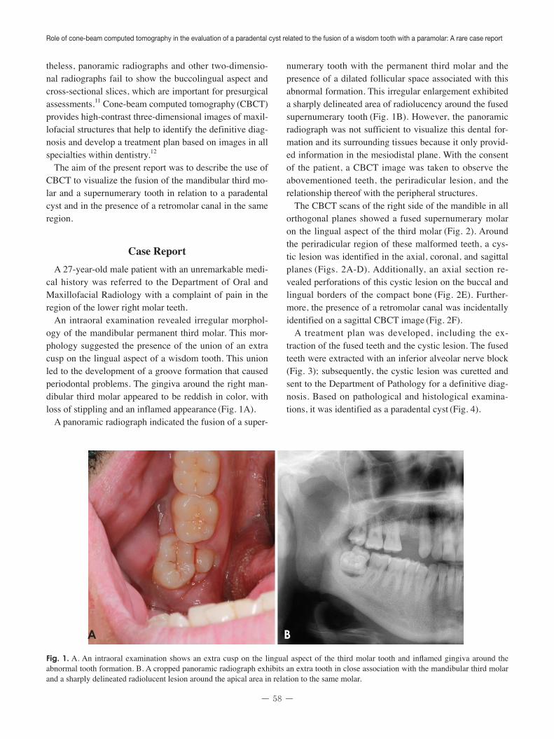
The CBCT scans of the right side of the mandible in all orthogonal planes showed a fused supernumerary molar on the lingual aspect of the third molar (Fig. 2). Around the periradicular region of these malformed teeth, a cys-tic lesion was identified in the axial, coronal, and sagittal planes (Figs. 2A-D). Additionally, an axial section re-vealed perforations of this cystic lesion on the buccal and lingual borders of the compact bone (Fig. 2E). Further-more, the presence of a retromolar canal was incidentally identified on a sagittal CBCT image (Fig. 2F).
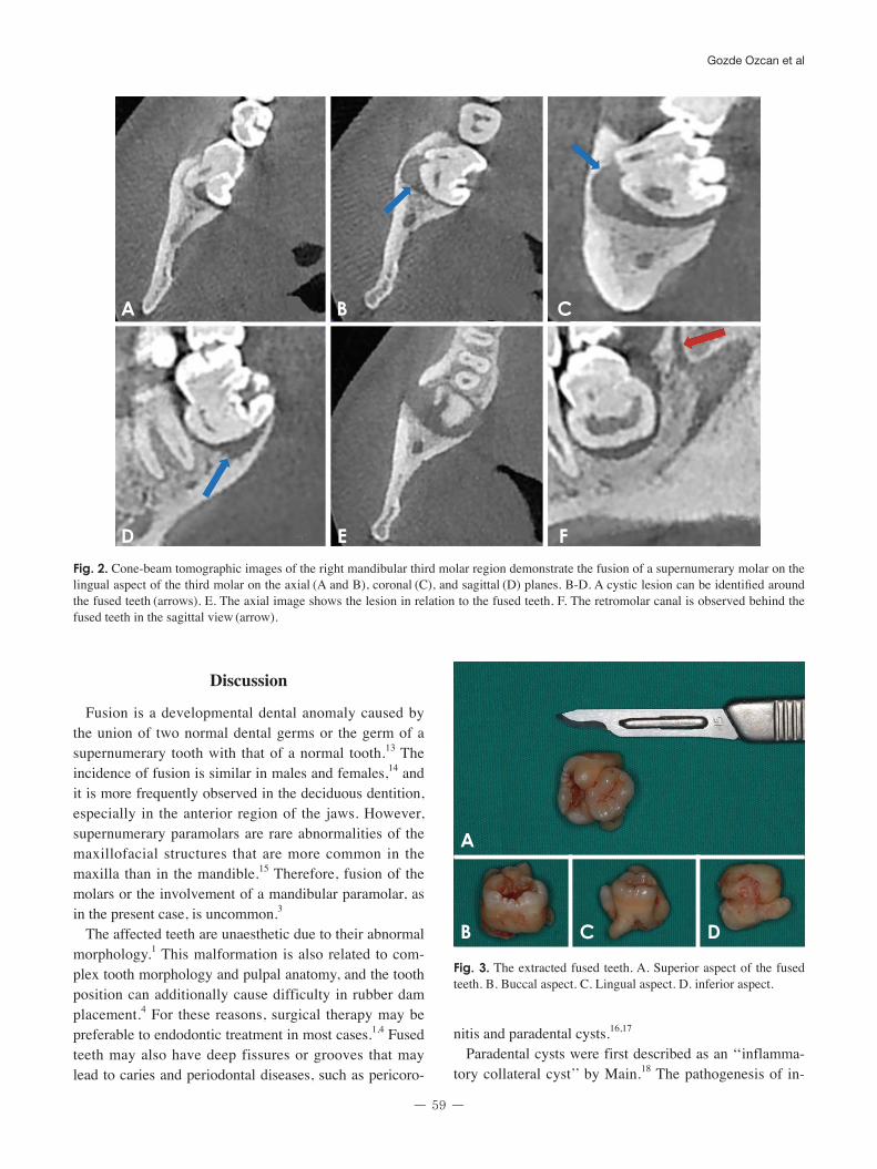
A treatment plan was developed, including the ex-traction of the fused teeth and the cystic lesion. The fused teeth were extracted with an inferior alveolar nerve block
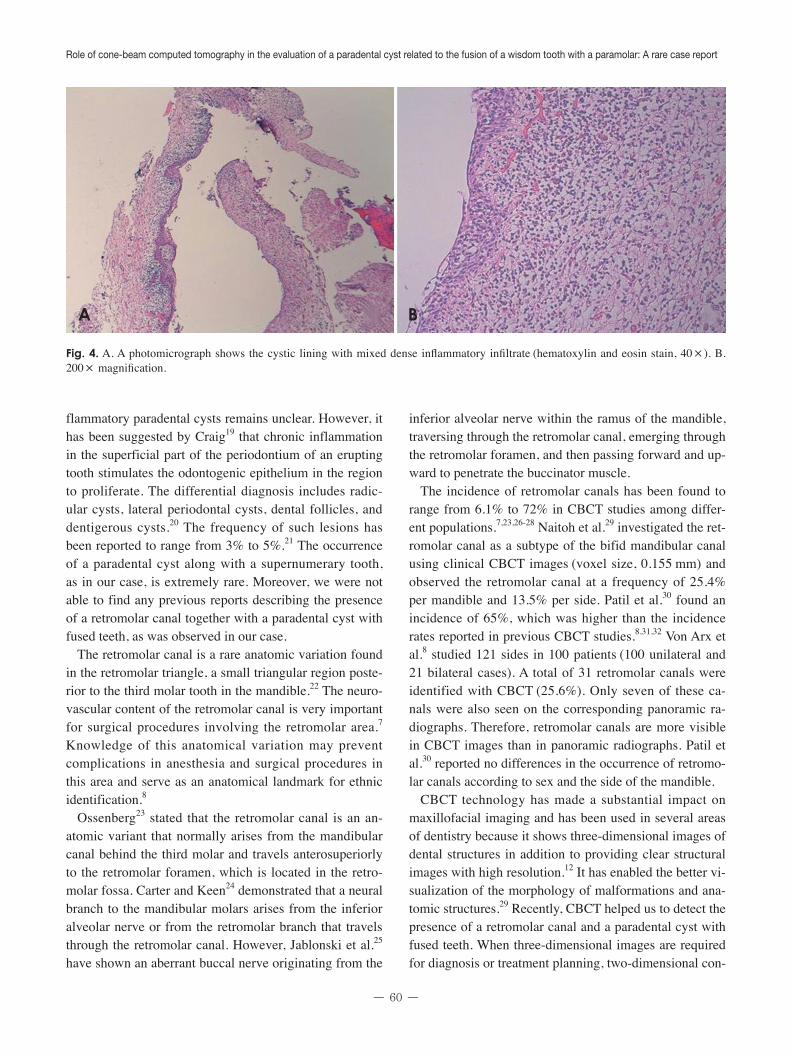
(Fig. 3); subsequently, the cystic lesion was curetted and sent to the Department of Pathology for a definitive diag-nosis. Based on pathological and histological examina-tions, it was identified as a paradental cyst (Fig. 4).
Fig. 1. A. An intraoral examination shows an extra cusp on the lingual aspect of the third molar tooth and inflamed gingiva around the abnormal tooth formation. B. A cropped panoramic radiograph exhibits an extra tooth in close association with the mandibular third molar and a sharply delineated radiolucent lesion around the apical area in relation to the same molar.
A B
- 59 -
Gozde Ozcan et al
discussion
Fusion is a developmental dental anomaly caused by the union of two normal dental germs or the germ of a supernumerary tooth with that of a normal tooth.13 The incidence of fusion is similar in males and females,14 and it is more frequently observed in the deciduous dentition, especially in the anterior region of the jaws. However, supernumerary paramolars are rare abnormalities of the maxillofacial structures that are more common in the maxilla than in the mandible.15 Therefore, fusion of the molars or the involvement of a mandibular paramolar, as in the present case, is uncommon.3
The affected teeth are unaesthetic due to their abnormal morphology.1 This malformation is also related to com-plex tooth morphology and pulpal anatomy, and the tooth position can additionally cause difficulty in rubber dam placement.4 For these reasons, surgical therapy may be preferable to endodontic treatment in most cases.1,4 Fused teeth may also have deep fissures or grooves that may lead to caries and periodontal diseases, such as pericoro-
nitis and paradental cysts.16,17
Paradental cysts were first described as an ‘‘inflamma-tory collateral cyst’’ by Main.18 The pathogenesis of in-
Fig. 2. Cone-beam tomographic images of the right mandibular third molar region demonstrate the fusion of a supernumerary molar on the lingual aspect of the third molar on the axial (A and B), coronal (C), and sagittal (D) planes. B-D. A cystic lesion can be identified around the fused teeth (arrows). E. The axial image shows the lesion in relation to the fused teeth. F. The retromolar canal is observed behind the fused teeth in the sagittal view (arrow).
A B C
D E F
Fig. 3. The extracted fused teeth. A. Superior aspect of the fused teeth. B. Buccal aspect. C. Lingual aspect. D. inferior aspect.
A
B C D
Role of cone-beam computed tomography in the evaluation of a paradental cyst related to the fusion of a wisdom tooth with a paramolar: A rare case report
- 60 -
flammatory paradental cysts remains unclear. However, it has been suggested by Craig19 that chronic inflammation in the superficial part of the periodontium of an erupting tooth stimulates the odontogenic epithelium in the region to proliferate. The differential diagnosis includes radic-ular cysts, lateral periodontal cysts, dental follicles, and dentigerous cysts.20 The frequency of such lesions has been reported to range from 3% to 5%.21 The occurrence of a paradental cyst along with a supernumerary tooth, as in our case, is extremely rare. Moreover, we were not able to find any previous reports describing the presence of a retromolar canal together with a paradental cyst with fused teeth, as was observed in our case.
The retromolar canal is a rare anatomic variation found in the retromolar triangle, a small triangular region poste-rior to the third molar tooth in the mandible.22 The neuro-vascular content of the retromolar canal is very important for surgical procedures involving the retromolar area.7 Knowledge of this anatomical variation may prevent complications in anesthesia and surgical procedures in this area and serve as an anatomical landmark for ethnic identification.8
Ossenberg23 stated that the retromolar canal is an an-atomic variant that normally arises from the mandibular canal behind the third molar and travels anterosuperiorly to the retromolar foramen, which is located in the retro-molar fossa. Carter and Keen24 demonstrated that a neural branch to the mandibular molars arises from the inferior alveolar nerve or from the retromolar branch that travels through the retromolar canal. However, Jablonski et al.25
have shown an aberrant buccal nerve originating from the
inferior alveolar nerve within the ramus of the mandible, traversing through the retromolar canal, emerging through the retromolar foramen, and then passing forward and up-ward to penetrate the buccinator muscle.
The incidence of retromolar canals has been found to range from 6.1% to 72% in CBCT studies among differ-ent populations.7,23,26-28 Naitoh et al.29 investigated the ret-romolar canal as a subtype of the bifid mandibular canal using clinical CBCT images (voxel size, 0.155 mm) and observed the retromolar canal at a frequency of 25.4% per mandible and 13.5% per side. Patil et al.30 found an incidence of 65%, which was higher than the incidence rates reported in previous CBCT studies.8,31,32 Von Arx et al.8 studied 121 sides in 100 patients (100 unilateral and 21 bilateral cases). A total of 31 retromolar canals were identified with CBCT (25.6%). Only seven of these ca-nals were also seen on the corresponding panoramic ra-diographs. Therefore, retromolar canals are more visible in CBCT images than in panoramic radiographs. Patil et al.30 reported no differences in the occurrence of retromo-lar canals according to sex and the side of the mandible.
CBCT technology has made a substantial impact on maxillofacial imaging and has been used in several areas of dentistry because it shows three-dimensional images of dental structures in addition to providing clear structural images with high resolution.12 It has enabled the better vi-sualization of the morphology of malformations and ana-tomic structures.29 Recently, CBCT helped us to detect the presence of a retromolar canal and a paradental cyst with fused teeth. When three-dimensional images are required for diagnosis or treatment planning, two-dimensional con-
Fig. 4. A. A photomicrograph shows the cystic lining with mixed dense inflammatory infiltrate (hematoxylin and eosin stain, 40 × ). B. 200 × magnification.
A B
- 61 -
Gozde Ozcan et al
ventional radiographs may be unsatisfactory. Moreover, in comparison with conventional computed tomography, CBCT has the advantage of lower radiation exposure.33
Additionally, CBCT is more useful in the accurate iden-tification of anatomical variations.34 Additionally, CBCT machines are convenient for routine investigations in the dental office.
In conclusion, fusion of the teeth is rarely seen in the mandibular molars, and to the best of our knowledge, this case report is the first in the literature to describe fusion between a mandibular third molar and a paramolar associ-ated with a paradental cyst and the presence of a retromo-lar canal. An accurate assessment of these morphological and pathological formations was carried out using CBCT.
References
1. Nunes E, de Moraes IG, de Novaes PM, de Sousa SM. Bilat-eral fusion of mandibular second molars with supernumerary teeth: case report. Braz Dent J 2002; 13: 137-41.
2. Neves AA, Neves ML, Farinhas JA. Bilateral connation of permanent mandibular incisors: a case report. Int J Paediatr Dent 2002; 12: 61-5.
4. Muthukumar RS, Arunkumar S, Sadasiva K. Bilateral fusion of mandibular second premolar and supernumerary tooth: a rare case report. J Oral Maxillofac Pathol 2012; 16: 128-30.
5. Prakash AR, Reddy PS, Rajanikanth M. Paradental cyst asso-ciated with supernumerary tooth fused with third molar: a rare case report. J Oral Maxillofac Pathol 2012; 16: 131-3.
6. Mufeed A, Chatra L, Shenai P. Diagnostic features of the paradental cyst and report of a case. Dentomaxillofac Radiol 2009; 38: 125-6.
7. Bilecenoglu B, Tuncer N. Clinical and anatomical study of retromolar foramen and canal. J Oral Maxillofac Surg 2006; 64: 1493-7.
8. von Arx T, Hänni A, Sendi P, Buser D, Bornstein MM. Radio-graphic study of the mandibular retromolar canal: an anatomic structure with clinical importance. J Endod 2011; 37: 1630-5.
9. Rossi AC, Freire AR, Prado GB, Prado FB, Botacin PR, Caria PH. Incidence of retromolar foramen in human mandibles: ethnic and clinical aspects. Int J Morphol 2012; 30: 1074-8.
10. Pires CA, Bissada NF, Becker JJ, Kanawati A, Landers MA. Mandibular incisive canal: cone beam computed tomography. Clin Implant Dent Relat Res 2012; 14: 67-73.
11. Watson RM, Davis DM, Forman GH, Coward T. Consider-ations in design and fabrication of maxillary implant-support-ed prostheses. Int J Prosthodont 1991; 4: 232-9.
12. Neves FS, Souza TC, Almeida SM, Haiter-Neto F, Freitas DQ, Bóscolo FN. Correlation of panoramic radiography and cone beam CT findings in the assessment of the relationship between impacted mandibular third molars and the mandibu-lar canal. Dentomaxillofac Radiol 2012; 41: 553-7.
13. Chen HS, Huang YL. Fusion of third and fourth mandibular molars? Oral Surg Oral Med Oral Pathol 1992; 73: 767.
14. Järvinen S, Lehtinen L. Supernumerary and congenitally missing primary teeth in Finnish children. An epidemiologic study. Acta Odontol Scand 1980; 39: 83-6.
15. Parolia A, Kundabala M, Dahal M, Mohan M, Thomas MS. Management of supernumerary teeth. J Conserv Dent 2011; 14: 221-4.
16. Ghoddusi J, Zarei M, Jafarzadeh H. Endodontic treatment of a supernumerary tooth fused to a mandibular second molar: a case report. J Oral Sci 2006; 48: 39-41.
17. Indra R, Srinivasan MR, Farzana H, Karthikeyan K. End-odontic management of a fused maxillary lateral incisor with a supernumerary tooth: a case report. J Endod 2006; 32: 1217-9.
18. Main DM. Epithelial jaw cysts: a clinicopathological reap-praisal. Br J Oral Surg 1970; 8: 114-25.
19. Craig GT. The paradental cyst. A specific inflammatory odon-togenic cyst. Br Dent J 1976; 141: 9-14.
20. de Sousa SO, Corrêa L, Deboni MC, de Araújo VC. Clinico-pathologic features of 54 cases of paradental cyst. Quintes-sence Int 2001; 32: 737-41.
21. Ackermann G, Cohen MA, Altini M. The paradental cyst: a clinicopathologic study of 50 cases. Oral Surg Oral Med Oral Pathol 1987; 64: 308-12.
22. Gadbail AR, Mankar Gadbail MP, Hande A, Chaudhary MS, Gondivkar SM, Korde S, et al. Tumor angiogenesis: role in locally aggressive biological behavior of ameloblastoma and keratocystic odontogenic tumor. Head Neck 2013; 35: 329-34.
23. Ossenberg NS. Retromolar foramen of the human mandible. Am J Phys Anthropol 1987; 73: 119-28.
24. Carter RB, Keen EN. The intramandibular course of the infe-rior alveolar nerve. J Anat 1971; 108: 433-40.
25. Jablonski NG, Cheng CM, Cheng LC, Cheung HM. Unusual origins of the buccal and mylohyoid nerves. Oral Surg Oral Med Oral Pathol 1985; 60: 487-8.
26. Ikeda K, Ho KC, Nowicki BH, Haughton VM. Multiplanar MR and anatomic study of the mandibular canal. AJNR Am J Neuroradiol 1996; 17: 579-84.
27. Pyle MA, Jasinevicius TR, Lalumandier JA, Kohrs KJ, Saw-yer DR. Prevalence and implications of accessory retromolar foramina in clinical dentistry. Gen Dent 1999; 47: 500-5.
28. Sawyer DR, Kiely ML. Retromolar foramen: a mandibular variant important to dentistry. Ann Dent 1991; 50: 16-8.
29. Naitoh M, Hiraiwa Y, Aimiya H, Ariji E. Observation of bifid mandibular canal using cone-beam computerized tomography. Int J Oral Maxillofac Implants 2009; 24: 155-9.
30. Patil S, Matsuda Y, Nakajima K, Araki K, Okano T. Retromo-lar canals as observed on cone-beam computed tomography: their incidence, course, and characteristics. Oral Surg Oral Med Oral Pathol Oral Radiol 2013; 115: 692-9.
31. Kawai T, Asaumi R, Sato I, Kumazawa Y, Yosue T. Observa-tion of the retromolar foramen and canal of the mandible: a CBCT and macroscopic study. Oral Radiol 2012; 28: 10-4.
32. Lizio G, Pelliccioni GA, Ghigi G, Fanelli A, Marchetti C. Radiographic assessment of the mandibular retromolar canal using cone-beam computed tomography. Acta Odontol Scand 2013; 71: 650-5.
Role of cone-beam computed tomography in the evaluation of a paradental cyst related to the fusion of a wisdom tooth with a paramolar: A rare case report
- 62 -
33. Naitoh M, Nakahara K, Suenaga Y, Gotoh K, Kondo S, Ariji E. Comparison between cone-beam and multislice computed tomography depicting mandibular neurovascular canal struc-tures. Oral Surg Oral Med Oral Pathol Oral Radiol Endod 2010; 109: e25-31.
34. Kuribayashi A, Watanabe H, Imaizumi A, Tantanapornkul W, Katakami K, Kurabayashi T. Bifid mandibular canals: cone beam computed tomography evaluation. Dentomaxillofac Ra-diol 2010; 39: 235-9.

![Case Report Orthokeratinized Odontogenic Cyst: A Report of … · 2019. 7. 31. · such as dentigerous cyst or paradental cyst [ , ]. Odon-togenic tumours such as ameloblastoma and](https://static.documents.pub/doc/80x56/614074aa1664f1518558c43e/case-report-orthokeratinized-odontogenic-cyst-a-report-of-2019-7-31-such-as.jpg)


