Role of corin in trophoblast invasion and uterinespiral artery remodelling in pregnancyYujie Cui1*{, Wei Wang1*{, Ningzheng Dong2,3*, Jinglei Lou1*, Dinesh Kumar Srinivasan1{, Weiwei Cheng4, Xiaoyi Huang4, Meng Liu2,Chaodong Fang2, Jianhao Peng1, Shenghan Chen1, Shannon Wu1, Zhenzhen Liu2, Liang Dong2, Yiqing Zhou2 & Qingyu Wu1,2
In pregnancy, trophoblast invasion and uterine spiral artery remodel-ling are important for lowering maternal vascular resistance andincreasing uteroplacental blood flow. Impaired spiral artery remodel-ling has been implicated in pre-eclampsia, a major complication ofpregnancy, for a long time but the underlying mechanisms remainunclear1,2. Corin (also known as atrial natriuretic peptide-convertingenzyme) is a cardiac protease that activates atrial natriuretic peptide(ANP), a cardiac hormone that is important in regulating blood pres-sure3. Unexpectedly, corin expression was detected in the pregnantuterus4. Here we identify a new function of corin and ANP in pro-moting trophoblast invasion and spiral artery remodelling. We showthat pregnant corin- or ANP-deficient mice developed high bloodpressure and proteinuria, characteristics of pre-eclampsia. In thesemice, trophoblast invasion and uterine spiral artery remodelling weremarkedly impaired. Consistent with this, the ANP potently stimu-lated human trophoblasts in invading Matrigels. In patients with pre-eclampsia, uterine Corin messenger RNA and protein levels weresignificantly lower than that in normal pregnancies. Moreover, wehave identified Corin gene mutations in pre-eclamptic patients, whichdecreased corin activity in processing pro-ANP. These results indicatethat corin and ANP are essential for physiological changes at thematernal–fetal interface, suggesting that defects in corin and ANPfunction may contribute to pre-eclampsia.
Pregnancy poses a serious challenge for maintaining normal bloodpressure. Pregnancy-induced hypertension, a major cause of maternaland fetal deaths, occurs in approximately 10% of pregnancies5,6.During pregnancy, the uterus undergoes profound morphologicalchanges, including trophoblast invasion and spiral artery remodelling.In pre-eclampsia, impaired spiral artery remodelling is common, butthe underlying mechanisms are unclear1,2,7–9. Studies indicate thatvascular growth factor receptors, angiotensin and oestradiol areinvolved in the disease10–14.
Corin is a cardiac protease that activates ANP, which is a cardiachormone that regulates blood pressure and sodium homeostasis15. Inmice, lack of CORIN prevents ANP generation and causes hyper-tension16. In humans, CORIN variants are associated with hyper-tension17. Interestingly, Corin expression was detected in the pregnantmouse4 (Fig. 1A) and human uterus (Supplementary Fig. 1). As a trans-membrane protein, CORIN is expected to act at the expression sites,suggesting a possible function in the pregnant uterus.
To understand the role of CORIN in pregnancy, we created a mousemodel in which a Corin transgene was expressed under a cardiacpromoter (Fig. 1B). The transgenic and Corin knockout mice werecrossed to generate mice expressing Corin only in the heart (‘knock-out/transgenic mice’; Fig. 1C, D). In knockout/transgenic mice, trans-genic Corin expression restored pro-ANP processing in the heart(Supplementary Fig. 2) and normalized blood pressure (Fig. 1E),
indicating that cardiac CORIN was sufficient to maintain normalblood pressure in non-pregnant mice.
In pregnant Corin knockout mice, blood pressure increased atapproximately 17 days post coitus and rose further before returningto the non-pregnant blood pressure level after delivery (Fig. 1F), whichresembled late gestational hypertension in pre-eclamptic women. InCorin knockout/transgenic mice, which were normotensive, bloodpressure increased similarly during pregnancy (Fig. 1G), indicatingthat cardiac Corin expression did not prevent pregnancy-inducedhypertension. The data also show that in these mice, hypertension inpregnancy was not due to pre-existing high blood pressure. As well asin the uterus, Corin mRNA was detected in the umbilical cord andplacenta (Supplementary Fig. 3). To distinguish the role of maternalCorin from that of placental or other fetal organs, Corin knockoutfemales were mated with either wild-type or knockout males. Theresulting fetuses carried one or no copy of the functional Corin gene.Normally, enzymes that are encoded by one gene copy are able tofunction. As shown in Fig. 1H, pregnant Corin knockout females thatwere mated with either wild-type or knockout males had similarlyincreased blood pressure, indicating that lack of maternal, but not fetal,Corin caused hypertension in pregnancy.
Proteinuria is a hallmark of pre-eclampsia. Wild-type, Corin knock-out and knockout/transgenic mice had similar urinary protein levelsbefore pregnancy and at mid gestation. However, the levels increasedin Corin knockout and knockout/transgenic mice at late gestation(Fig. 1I), consistent with reported proteinuria in mouse models ofpre-eclampsia18. Ischaemic glomeruli, indicated by fewer red bloodcells, were found in pregnant Corin knockout and knockout/transgenicmice (Fig. 1J, a–f) but not in non-pregnant mice (Supplementary Fig. 4).Periodic acid–Schiff staining revealed increased extracellular matrixesand collapsed glomerular capillaries in pregnant Corin knockout andknockout/transgenic mice (Fig. 1J, g–i). Electron microscopy showednarrow glomerular capillary lumens and thick basement membranes(Fig. 1K), suggesting endotheliosis and increased extracellular mat-rices. Additional pathological features such as necrotic cells and cal-cium deposits in the placental labyrinth also existed in these mice(Supplementary Fig. 5), indicating insufficient uteroplacental per-fusion. Consistent with this, Corin knockout and knockout/transgenicmice had smaller litters (7.1 6 2.3 (n 5 28) and 6.8 6 2.7 (n 5 28) pupsper litter, respectively, versus wild-type mice, which had 9.1 6 1.2(n 5 21) pups per litter; P , 0.001 in both cases).
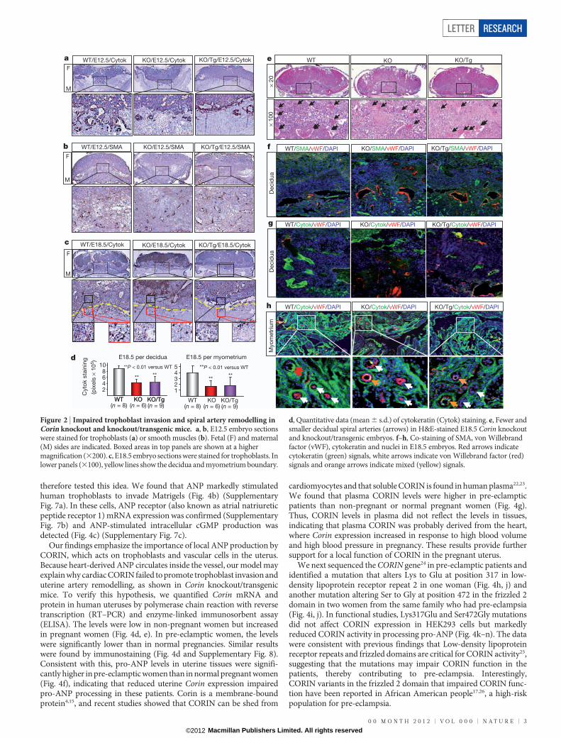
We examined embryos at embryonic day 12.5 (E12.5), an early timepoint before blood pressure increase in Corin knockout and knockout/transgenic mice, and E18.5 (two days before delivery). Wild-type E12.5embryos showed obvious trophoblast invasion, shown by cytokeratinstaining (Fig. 2a), and large vessels mostly in the deep decidua, shownby smooth-muscle a-actin (SMA) staining (Fig. 2b), indicating that
*These authors contributed equally to this work.
1Molecular Cardiology, Nephrology and Hypertension, Lerner Research Institute, Cleveland Clinic, 9500 Euclid Avenue, Cleveland, Ohio 44195, USA. 2Cyrus Tang Hematology Center, Jiangsu Institute ofHematology, the First AffiliatedHospital, Soochow University, 199 RenAi Road, Suzhou215123,China. 3Key Lab of Thrombosis and Hemostasis, Jiangsu Institute of Hematology, the First AffiliatedHospital,Soochow University, 188 Shi Zhi Street, Suzhou 215006, China. 4The International Peace Maternity and Child Health Hospital, Shanghai Jiaotong University School of Medicine, 910 Hengshan Road,Shanghai 200030, China. {Present address: School of Laboratory Science, Tianjin Medical University, Tianjin 300203, China (Y.C.); Department of Cardiology, Peking Union Medical College, Beijing100730, China (W.W.); Lee Kong Chian School of Medicine, Singapore 637553 (D.K.S.).
0 0 M O N T H 2 0 1 2 | V O L 0 0 0 | N A T U R E | 1
smooth muscles in the superficial decidua were replaced by invadingtrophoblasts. In contrast, trophoblast invasion in Corin knockout andknockout/transgenic embryos was markedly reduced (Fig. 2a) andsmaller arteries were found in both superficial and deep decidua(Fig. 2b). In E18.5 wild-type embryos, more abundant trophoblastswere found in the decidua and myometrium compared with those inCorin knockout and knockout/transgenic mice (Fig. 2c, d). By haema-toxylin and eosin staining, larger and more abundant decidual spiralarteries were observed in wild-type than in Corin knockout or knock-out/transgenic mice (Fig. 2e). Figure 2f–h shows strong cytokeratin(trophoblasts) staining but weak von Willebrand factor (endothelial)and SMA (smooth muscle) staining in wild-type decidual and myo-metrial arteries. These data indicate that trophoblast invasion andspiral artery remodelling were impaired in Corin knockout and knock-out/transgenic mice, and that this defect occurred before blood pres-sure increased in these mice.
CORIN activates ANP in the heart15 but it was unknown whether theCORIN function in pregnancy was also mediated by ANP. Pro-ANP isexpressed in the non-pregnant and pregnant uterus (SupplementaryFig. 6). If CORIN acts on pro-ANP to promote trophoblast invasionand spiral artery remodelling, thereby preventing hypertension in preg-nancy, ANP (also known as Nppa) and Corin knockout mice shouldhave similar phenotypes. ANP knockout mice are hypertensive (Fig. 3a)
but their blood pressure was not monitored during pregnancy19. Wefound similarly increased blood pressure in pregnant ANP knockoutmice (Fig. 3b). The mice also had late gestational proteinuria (Fig. 3c)and smaller litters (4.4 6 1.7 (n 5 25) versus wild-type, 9.1 6 1.2(n 5 21) pups per litter, P , 0.001). By immunostaining, impairedtrophoblast invasion and smaller spiral arteries were observed inE12.5 embryos (Fig. 3d, e). In E18.5 embryos, ANP knockout micehad far fewer trophoblasts (Fig. 3f, g) and smaller arteries (Fig. 3h) inthe decidua and myometrium than those in wild-type mice. Consistentwith this, weak cytokeratin-staining but strong von Willebrand factor-staining were found in arteries in ANP knockout mice (Fig. 3i). Thus,ANP and Corin knockout mice had very similar phenotypes, indicatingthat the role of CORIN in pregnancy is probably mediated by ANP.
In the heart, CORIN produces ANP, which then regulates bloodpressure by promoting natriuresis and vasodilation3. Here we foundthat lack of CORIN and ANP impaired trophoblast invasion and spiralartery remodelling, which was not rescued by cardiac Corin expressionin Corin knockout/transgenic mice. ANP is known to relax vascularsmooth muscles. Recently, ANP and its downstream cyclic GMP-dependent protein kinase were shown to be important in angiogenicprocesses by promoting endothelial regeneration20,21. Thus, ANP mayfunction locally to remodel uterine arteries. Our results also indicatethat ANP may directly promote trophoblast invasion (Fig. 4a), and we
pAB MHC Corin DNA
CORIN
GAPDH
KO
heart KO/Tg
WT
heart
Heart
Liv
er
Lung
Kid
ney
Sp
leen
C
**
40
60
80
100
120
Systolic
BP
(m
m H
g)
**P < 0.01(n = 8 per group)
WT KO KO/TG WT KO KO/TG
**
** **
E
G
80
100
120
140
Delivery
Systo
lic (m
m H
g)
† † †
† †
KO/Tg n = 6
H
80
100
120
140 KO × KO n = 3
KO × WT
Delivery
Systo
lic (m
m H
g)
n = 3
50
100
150
200
Non-pregnant D16–18
Urinary
pro
tein
(m
g d
l–1)
WT
KOKO/Tg **
**
I
*
22 2380
100
120
140
NP 1 5 10 15 16 17 18 19 20 21
22 23
22 23
NP 1 5 10 15 16 17 18 19 20 21
NP 1 5 10 15 16Days
Days
Days
17 18 19 20 21
WT n = 6
KO n = 7
†† †
†
††
Delivery
Systo
lic (m
m H
g)
* * **
* * ** *
*
*
FWT uterus
NP
E7.5
E10.5
E14.5
E18.5
WT
heart
KO
ute
rus
KO
heart
Gapdh
Corin
A
D
CORIN
GAPDH
WT
NP
Uterus
WT
heart
KO
heart
WT
E18.5
KO
NP
KO
E18.5
KO
/Tg
NP
KO
/Tg
E18.5
Masso
nH
&E
PA
S
WT KO
a b c
d e f
g h i
J
WT
KO
KO
/Tg
× 2,650 × 7,100K
L
Diastolic D8–10
KO/Tg
*
†
Figure 1 | Hypertension, proteinuria and renal pathology in pregnantCorin knockout and knockout/transgenic mice. A, Corin mRNA expressionin mouse uteruses. B, Corin transgenic (Tg) construct. C, D, Western blotanalysis of CORIN protein in wild-type (WT), Corin knockout (KO) andknockout/transgenic mice. E, Blood pressure (BP, mean 6 s.d.) in non-pregnant females. F, G, Blood pressure increased in Corin knockout (F) andknockout/transgenic (G) mice in pregnancy. Data are mean 6 s.d. *P , 0.05versus WT of the same time point. {P , 0.05 versus non-pregnant level of thesame genotype. H, Similar blood-pressure changes in Corin knockout femalesmated with knockout or WT males. I, Late gestational proteinuria in Corin
knockout and knockout/transgenic mice. Data are mean 6 s.d. **P , 0.01, n 5
7 or 8 per group. J, a–i, Renal ischaemia in pregnant Corin knockout andknockout/transgenic mice. E18.5 sections are stained with haematoxylin andeosin (H&E), Masson trichrome or periodic acid–Schiff (PAS). Scale bar, 20mm. Red blood cells (yellow arrows) and open capillaries (red arrows) in WTglomeruli are shown. K, Narrow glomerular capillary lumen (L) and thickbasement membranes (red arrows) in Corin knockout and knockout/transgenic mice at E18.5 shown by electron microscopy. GAPDH,glyceraldehyde 3-phosphate dehydrogenase; NP, non pregnant; pA, poly A.
RESEARCH LETTER
2 | N A T U R E | V O L 0 0 0 | 0 0 M O N T H 2 0 1 2
therefore tested this idea. We found that ANP markedly stimulatedhuman trophoblasts to invade Matrigels (Fig. 4b) (SupplementaryFig. 7a). In these cells, ANP receptor (also known as atrial natriureticpeptide receptor 1) mRNA expression was confirmed (SupplementaryFig. 7b) and ANP-stimulated intracellular cGMP production wasdetected (Fig. 4c) (Supplementary Fig. 7c).
Our findings emphasize the importance of local ANP production byCORIN, which acts on trophoblasts and vascular cells in the uterus.Because heart-derived ANP circulates inside the vessel, our model mayexplain why cardiac CORIN failed to promote trophoblast invasion anduterine artery remodelling, as shown in Corin knockout/transgenicmice. To verify this hypothesis, we quantified Corin mRNA andprotein in human uteruses by polymerase chain reaction with reversetranscription (RT–PCR) and enzyme-linked immunosorbent assay(ELISA). The levels were low in non-pregnant women but increasedin pregnant women (Fig. 4d, e). In pre-eclamptic women, the levelswere significantly lower than in normal pregnancies. Similar resultswere found by immunostaining (Fig. 4d and Supplementary Fig. 8).Consistent with this, pro-ANP levels in uterine tissues were signifi-cantly higher in pre-eclamptic women than in normal pregnant women(Fig. 4f), indicating that reduced uterine Corin expression impairedpro-ANP processing in these patients. Corin is a membrane-boundprotein4,15, and recent studies showed that CORIN can be shed from
cardiomyocytes and that soluble CORIN is found in human plasma22,23.We found that plasma CORIN levels were higher in pre-eclampticpatients than non-pregnant or normal pregnant women (Fig. 4g).Thus, CORIN levels in plasma did not reflect the levels in tissues,indicating that plasma CORIN was probably derived from the heart,where Corin expression increased in response to high blood volumeand high blood pressure in pregnancy. These results provide furthersupport for a local function of CORIN in the pregnant uterus.
We next sequenced the CORIN gene24 in pre-eclamptic patients andidentified a mutation that alters Lys to Glu at position 317 in low-density lipoprotein receptor repeat 2 in one woman (Fig. 4h, j) andanother mutation altering Ser to Gly at position 472 in the frizzled 2domain in two women from the same family who had pre-eclampsia(Fig. 4i, j). In functional studies, Lys317Glu and Ser472Gly mutationsdid not affect CORIN expression in HEK293 cells but markedlyreduced CORIN activity in processing pro-ANP (Fig. 4k–n). The datawere consistent with previous findings that Low-density lipoproteinreceptor repeats and frizzled domains are critical for CORIN activity25,suggesting that the mutations may impair CORIN function in thepatients, thereby contributing to pre-eclampsia. Interestingly,CORIN variants in the frizzled 2 domain that impaired CORIN func-tion have been reported in African American people17,26, a high-riskpopulation for pre-eclampsia.
2468
10
WT(n = 8)
KO(n = 6)
KO/Tg(n = 9)
WT(n = 8)
KO(n = 6)
KO/Tg(n = 9)
Cyto
k s
tain
ing
(pix
els
× 1
05)
** **
**P < 0.01 versus WT
d
12345 **P < 0.01 versus WT
****
E18.5 per decidua E18.5 per myometrium
g WT/Cytok/vWF/DAPI KO/Cytok/vWF/DAPI KO/Tg/Cytok/vWF/DAPI
Figure 2 | Impaired trophoblast invasion and spiral artery remodelling inCorin knockout and knockout/transgenic mice. a, b, E12.5 embryo sectionswere stained for trophoblasts (a) or smooth muscles (b). Fetal (F) and maternal(M) sides are indicated. Boxed areas in top panels are shown at a highermagnification (3200). c, E18.5 embryo sections were stained for trophoblasts. Inlower panels (3100), yellow lines show the decidua and myometrium boundary.
d, Quantitative data (mean 6 s.d.) of cytokeratin (Cytok) staining. e, Fewer andsmaller decidual spiral arteries (arrows) in H&E-stained E18.5 Corin knockoutand knockout/transgenic embryos. f–h, Co-staining of SMA, von Willebrandfactor (vWF), cytokeratin and nuclei in E18.5 embryos. Red arrows indicatecytokeratin (green) signals, white arrows indicate von Willebrand factor (red)signals and orange arrows indicate mixed (yellow) signals.
LETTER RESEARCH
0 0 M O N T H 2 0 1 2 | V O L 0 0 0 | N A T U R E | 3
Figure 3 | Hypertension, proteinuria and uteroplacental pathology inpregnant ANP knockout mice. a, Blood pressure (mean 6 s.d.) in non-pregnant females, **P , 0.01. b, Elevated blood pressure (mean 6 s.d.) inpregnant ANP knockout mice. {P , 0.05 versus non-pregnant level.c, Gestational proteinuria in ANP knockout mice. Data are mean 6 s.d.d, e, Impaired trophoblast invasion and smooth muscle remodelling in E12.5embryos stained for cytokeratin (d) or SMA (e). Boxed areas in top panels are
shown at a higher magnification (3200). f, Impaired trophoblast invasion inE18.5 embryos stained for cytokeratin. g, Quantitative data (mean 6 s.d.) ofcytokeratin staining in E18.5 ANP knockout embryos. h, Impaired decidual andmyometrial artery remodelling (arrows) in H&E-stained E18.5 ANP knockoutembryos. i, Co-staining of cytokeratin, von Willebrand factor and nuclei inE18.5 ANP knockout embryos. Red arrows indicate cytokeratin (green) signalsand white arrows indicate von Willebrand factor (red) signals.
Patient a
Normal
/G
h
Patient b A/G
Patient c A/G
Normali
Pro
-AN
Pco
nvers
ion
(%
)
20406080
100
WT Lys317Glu
**P < 0.01
m
pro-ANP
WT
Arg
80
1A
la
Ser9
85
Ala
Vecto
r
Lys3
17
Glu
ANP
k
Corin
pro-ANP
ANP
Corin
WT
Vecto
r
Ser4
72
Gly
l
20406080
100
WT Ser472Gly
P < 0.01
n
Pro
-AN
Pco
nvers
ion
(%
)
**
j
ProteaseTM Fz1 Fz2LDLR1–5 LDLR1 SR
Lys317Glu Ser472Gly
Non-pregnant(n = 18)
Normalpregnant(n = 21)
Pre-eclampsia(n = 14)
5
10
15
20
25
Rela
tive C
orin
mR
NA
levels
P < 0.001 P = 0.026
P = 0.02d
P < 0.001
0.5
1
1.5
2
Pla
sm
a C
orin
(n
g m
l–1)
Non-pregnant(n = 77)
Normalpregnant(n = 108)
P < 0.001
P = 0.001
g
Pre-eclampsia(n = 56)
Pla
centa
a
Myo
metr
ium
Decid
ua
Pregnant
pro-ANP
ANP
Sp
iral art
ery
lu
men
Blood flow
Trophoblasts
Corin
Non-pregnant
Sm
oo
th m
uscle
cells
En
do
thelia
l cells
Myo
metr
ium
End
om
etr
ium
Remodelling
Intervillous space
Non-pregnant(n = 18)
Normalpregnant(n = 19)
Pre-eclampsia(n = 15)
2
4
6
8
10
12
14
Co
rin
pro
tein
(pg
10
μg
–1 t
ota
l p
rote
in) P < 0.001 P = 0.012
P = 0.22e
Non-pregnant(n =11)
Normalpregnant(n =19)
Pre-eclampsia(n =15)
1
2
3
pro
-AN
P
(pm
ol m
g–1 t
ota
l p
rote
in)
P < 0.01
P < 0.01
P < 0.01f
Matr
igel in
vasio
n (%
)b
100
200
300
400BeWo
****
ANP (μM)
100
200
300
400
0 .01 0.1 10 .01 0.1 1
Primary
**
ANP (nM)
Intr
acellu
lar
cG
MP
(fm
ol μg
–1 p
rote
in)
c
10
20
30
40
50 BeWo
****
10
20
30
40
50
0 1 10 250 1 10 25
Primary
**
*
Figure 4 | ANP-stimulated humantrophoblast invasion, and impaireduterine Corin expression and Corinmutations in pre-eclampticpatients. a, A model showing thatCORIN-produced ANP in thepregnant uterus promotestrophoblast invasion (red arrows)and vascular-wall remodelling (blackarrows). b, c, ANP-stimulatedhuman BeWo trophoblasts andprimary trophoblasts in Matrigelinvasion (b) and intracellular cGMPproduction (c). Data are mean 6 s.d.*P , 0.05; **P , 0.01 versus control.d–f, Corin mRNA (d) and protein(e), and pro-ANP levels (f) in humanuterus samples. Horizontal linesindicate mean values. g, Plasma-soluble CORIN levels (mean 6 s.d.)in pre-eclamptic patients and normalcontrols. h–j, CORIN gene mutationscausing Lys317Glu (h) andSer472Gly (i) changes in CORIN(j). k, l, Expression of Lys317Glu andSer472Gly mutants in HEK293 cells(top panels). Vector, WT CORINand inactive CORIN Arg801Ala andSer985Ala mutants were controls.Lys317Glu and Ser472Gly mutationsreduced pro-ANP processing activity(bottom panels). m, n, Quantitativedata (mean 6 s.d.) from threeexperiments or more. Fz, frizzled;LDLR, LDL receptor; SR, scavengerreceptor; TM, transmembrane.
RESEARCH LETTER
4 | N A T U R E | V O L 0 0 0 | 0 0 M O N T H 2 0 1 2
Previously, high levels of plasma pro-ANP or ANP were detected inpre-eclamptic patients27,28. As shown by our plasma soluble CORINdata, plasma protein levels may not reflect those in tissues. Takentogether, our data show a novel local function of CORIN and ANPin promoting trophoblast invasion and spiral artery remodelling toprevent hypertension in pregnancy. The data suggest that impairedCorin expression or function in the pregnant uterus may be an import-ant mechanism underlying pre-eclampsia. Studies to better under-stand impaired uterine Corin expression in pre-eclamptic patientsmay help to develop new strategies to enhance the CORIN–ANPpathway and prevent or treat this life-threatening disease.
METHODS SUMMARYCorin and ANP knockout mice have been described previously16,19. Transgenicmice with cardiac Corin expression were generated using a heart-specific pro-moter. Blood pressure was measured by radiotelemetry16. Tissue sections fromnon-pregnant and pregnant mice were stained with haematoxylin and eosin,Masson’s trichrome, periodic acid–Schiff or von Kossa, or immunostained withantibodies against cytokeratin, SMA, von Willebrand factor or CORIN. Renalsections were also examined by electron microscopy. Trans-well invasion assaywas carried out with human primary villous trophoblasts (ScienCell) and tropho-blastic JEG3, BeWo, JAR cell lines (ATCC) in Matrigel Invasion Chambers (BDBiosciences). ANP-stimulated cGMP production in trophoblasts was assayed in96-well plates. Intracellular cGMP levels were determined using an enzymeimmunoassay kit (Enzo Life Sciences). Corin levels in human blood and uterustissue samples were measured using ELISA22. Pro-ANP levels in human uterustissues were also measured using ELISA (Alpco Diagnostics). Corin gene exons24
from pre-eclamptic patients were PCR-amplified and sequenced directly. Coringene mutations that were identified were studied by expressing mutant CORINproteins in HEK293 cells and testing their activities in pro-ANP processing assays,as described previously26.
Full Methods and any associated references are available in the online version ofthe paper at www.nature.com/nature.
Received 21 May 2010; accepted 25 January 2012.
Published online 21 March 2012.
1. Pijnenborg, R., Vercruysse, L. & Hanssens, M. The uterine spiral arteries in humanpregnancy: facts and controversies. Placenta 27, 939–958 (2006).
2. Red-Horse, K. et al. Trophoblast differentiation during embryo implantation andformation of the maternal-fetal interface. J. Clin. Invest. 114, 744–754 (2004).
3. Wu, Q., Xu-Cai, Y. O., Chen, S. & Wang, W. Corin: new insights into the natriureticpeptide system. Kidney Int. 75, 142–146 (2009).
4. Yan, W., Sheng, N., Seto, M., Morser, J. & Wu, Q. Corin, a mosaic transmembraneserine protease encoded by a novel cDNA from human heart. J. Biol. Chem. 274,14926–14935 (1999).
5. Lain, K. Y. & Roberts, J. M. Contemporary concepts of the pathogenesis andmanagement of preeclampsia. J. Am. Med. Assoc. 287, 3183–3186 (2002).
6. Sibai,B.,Dekker,G.&Kupferminc,M.Pre-eclampsia.Lancet365,785–799 (2005).7. Brosens, I. A., Robertson, W. B. & Dixon, H. G. The role of the spiral arteries in the
pathogenesis of preeclampsia. Obstet. Gynecol. Annu. 1, 177–191 (1972).8. Kaufmann, P., Black, S. & Huppertz, B. Endovascular trophoblast invasion:
implications for the pathogenesis of intrauterine growth retardation andpreeclampsia. Biol. Reprod. 69, 1–7 (2003).
9. Norwitz, E. R., Schust, D. J. & Fisher, S. J. Implantation and the survival of earlypregnancy. N. Engl. J. Med. 345, 1400–1408 (2001).
10. Kanasaki, K. et al. Deficiency in catechol-O-methyltransferase and2-methoxyoestradiol is associated with pre-eclampsia. Nature 453, 1117–1121(2008).
11. Levine, R. J. et al. Circulating angiogenic factors and the risk of preeclampsia. N.Engl. J. Med. 350, 672–683 (2004).
12. Redman, C. W. & Sargent, I. L. Latest advances in understanding preeclampsia.Science 308, 1592–1594 (2005).
13. Venkatesha, S. et al. Soluble endoglin contributes to the pathogenesis ofpreeclampsia. Nature Med. 12, 642–649 (2006).
14. Zhou, C. C. et al. Angiotensin receptor agonistic autoantibodies induce pre-eclampsia in pregnant mice. Nature Med. 14, 855–862 (2008).
16. Chan, J. C. et al. Hypertension in mice lacking the proatrial natriuretic peptideconvertase corin. Proc. Natl Acad. Sci. USA 102, 785–790 (2005).
17. Dries, D. L. et al. Corin gene minor allele defined by 2 missense mutations iscommon in blacks and associated with high blood pressure and hypertension.Circulation 112, 2403–2410 (2005).
18. Davisson, R. L. et al. Discovery of a spontaneous genetic mouse model ofpreeclampsia. Hypertension 39, 337–342 (2002).
19. John, S. W. et al. Genetic decreases in atrial natriuretic peptide and salt-sensitivehypertension. Science 267, 679–681 (1995).
20. Kuhn, M. et al. The natriuretic peptide/guanylyl cyclase—a system functions as astress-responsive regulator of angiogenesis in mice. J. Clin. Invest. 119,2019–2030 (2009).
21. Tokudome, T. et al. Impaired recovery of blood flow after hind-limb ischemia inmice lackingguanylyl cyclase-A, a receptor for atrial andbrain natriureticpeptides.Arterioscler. Thromb. Vasc. Biol. 29, 1516–1521 (2009).
22. Dong, N. et al. Plasma soluble corin in patients with heart failure. Circ. Heart Fail. 3,207–211 (2010).
23. Jiang, J. et al. Ectodomain shedding and autocleavage of the cardiac membraneprotease corin. J. Biol. Chem. 286, 10066–10072 (2011).
24. Pan, J. et al. Genomic structures of the human and murine corin genes andfunctional GATA elements in their promoters. J. Biol. Chem. 277, 38390–38398(2002).
25. Knappe, S., Wu, F., Madlansacay, M. R. & Wu, Q. Identification of domain structuresin the propeptide of corin essential for the processing of proatrial natriureticpeptide. J. Biol. Chem. 279, 34464–34471 (2004).
26. Wang, W. et al. Corin variant associated with hypertension and cardiachypertrophy exhibits impaired zymogen activation and natriuretic peptideprocessing activity. Circ. Res. 103, 502–508 (2008).
27. Irons, D. W., Baylis, P. H., Butler, T. J. & Davison, J. M. Atrial natriuretic peptide inpreeclampsia: metabolic clearance, sodium excretion and renal hemodynamics.Am. J. Physiol. 273, F483–F487 (1997).
28. Tihtonen, K. M., Koobi, T., Vuolteenaho, O., Huhtala, H. S. & Uotila, J. T. Natriureticpeptides and hemodynamics in preeclampsia. Am. J. Obstet. Gynecol. 196,328.e1–328.e7 (2007).
Supplementary Information is linked to the online version of the paper atwww.nature.com/nature.
Acknowledgements We thank J. Robbins for the a-myosin heavy chain promoterconstruct and L. Zhang for help with statistical analysis. This work was partly supportedby grants from the Ralph Wilson Medical Foundation, the Bakken Heart-Brain Instituteand the National Institutes of Health (HL089298, HD064634), and by grants from theNational Natural Science Foundation of China (31070716, 81170247 and31161130356) and the Priority Academic Program Development of Jiangsu HigherEducation Institutions.
Author Contributions Y.C., W.W., N.D., J.L., D.K.S., M.L., C.F., J.P., S.C., S.W., Z.L. and L.D.designed and performed experiments. N.D., W.C. and X.H. collected patient samplesand analysed clinical data. Q.W. conceived the study and designed experiments. Y.Z.and Q.W. wrote the manuscript. All authors analysed and interpreted data, and criticallyread the manuscript.
Author Information Reprints and permissions information is available atwww.nature.com/reprints. The authors declare no competing financial interests.Readers are welcome to comment on the online version of this article atwww.nature.com/nature. Correspondence and requests for materials should beaddressed to Q.W. ([email protected]).
LETTER RESEARCH
0 0 M O N T H 2 0 1 2 | V O L 0 0 0 | N A T U R E | 5
METHODSKnockout and transgenic mice. Corin knockout mice were described previously16.ANP knockout mice (B6.129P2-Nppa tm1Unc/J)19 were from the Jackson Laboratory.To make transgenic mice expressing Corin in the heart, the full-length mouse CorincDNA was inserted into a construct driven by the mouse a-myosin heavy chain(a-MHC) promoter. Pro-nuclear microinjection and breeding of transgenic micewere carried out at the Case Western Reserve University Transgenic Core. Corinknockout and transgenic mice were crossed to generate knockout/transgenic mice.Littermates were used as controls. The animal study was conducted in accordancewith the National Institutes of Health guidelines and approved by the InstitutionalAnimal Care and Use Committee at the Cleveland Clinic.Blood-pressure monitoring. Radiotelemetry was used for real-time blood-pressure monitoring in conscious and unrestrained mice16. Female mice (8–12weeks old) were chronically instrumented in the left carotid artery with a PA-C10device (Data Sciences International) and rested for at least 7 days to recover fromthe surgery. The mice were mated and checked for vaginal plugs to establishgestation timing. The day on which a plug was observed was defined as E0.5.The mating mice were homozygous except for those in the fetal testing experi-ment. Telemetry receivers (model RPC-1) were placed under individual cages fordata acquisition using the Dataquest A.R.T. 4.0 Gold System (Data SciencesInternational). Data presented were from continuous recording of at least 6 hper day (10:00 to 16:00).Urinary protein measurement. Urine samples were collected from non-pregnantmice and pregnant mice at mid (8–10 days post coitus) and late (16–18 days postcoitus) gestational stages. Urinary protein levels were measured using a colorimetricassay based on a modified Bradford method (Bio-Rad).RT–PCR, western blot analysis and ELISA. Total RNAs were isolated fromcultured cells or mouse and human tissues using TRIzol reagent (Invitrogen) oran RNeasy kit (Qiagen), and were used to synthesize the first strand cDNAs. RT–PCR was carried out using oligonucleotide primers that were specific for themouse or human CORIN, mouse ANP, or human ANP receptor genes.Quantitative RT–PCR for human CORIN mRNA expression in uterus tissueswas carried out using the PRISM 7500 System (Applied Biosystems). The b-actingene was used as an internal control. Quantitative RT–PCR for mouse ANPmRNA in uteruses was carried out using the iCycler system (Bio-Rad). For westernblot analysis of CORIN protein, membrane fractions from tissue homogenateswere isolated by ultracentrifugation, as described previously29. Proteins wereanalysed by SDS–polyacylamide agarose gel electrophoresis (SDS–PAGE) andwestern blot using a polyclonal antibody (Berlex Biosciences). Western blotanalysis of pro-ANP in heart samples was carried out using a polyclonal antibody(Santa Cruz). Processing pro-ANP by CORIN in transfected cells was analysed bywestern blot analysis, as described previously30. Pro-ANP in human uterus tissueswas measured by an amino-terminal (NT) pro-ANP ELISA kit from AlpcoDiagnostics. Human CORIN in uterus tissues or plasma was measured byELISA, as described previously31.Histology and immunohistochemistry. Tissues were fixed with 4% para-formaldehyde and embedded in paraffin. Sections were stained with H&E,Masson’s trichrome, PAS or von Kossa. For immunohistochemical or immuno-fluorescent analysis, antibodies against SMA (Sigma-Aldrich), von Willebrandfactor (Sigma-Aldrich) and cytokeratin (Dako) were used to label smooth musclecells, endothelial cells and trophoblasts, respectively. For human CORIN, anantibody from Berlex Biosciences was used. Secondary antibodies were conjugatedwith horseradish peroxidase or Alexa Fluor 488 (green) or Alexa Fluor 594 (red)(Invitrogen). Tissue sections were mounted with or without DAPI-containing(blue) mounting medium (Dako). For ANP expression in mouse uterus tissues,a polyclonal antibody from Millipore was used. Control sections were treatedsimilarly but without the primary antibodies. Photographs were taken with a lightor fluorescent microscope equipped with a digital camera (Olympus). Data arefrom experiments using five or more mice per study group.
For immunohistochemical analysis of trophoblast invasion in mouse embryos,tissue samples from at least five mice per group, and at least two implant sites permouse were used. Serial sections (.50 per embryo) of 5 mm in thickness wereprepared. The position of the maternal artery was used as a guide to orient sectionpositions. At least 4–6 sections from the centre of the placenta of each embryo wereused for immunohistochemical analysis. Slides that were stained for cytokeratinwere examined by two individuals. The sections that showed the deepest tropho-blast invasion are presented. These sections were also analysed by ImagePro soft-ware to quantify cytokeratin staining. For each section that was analysed, the entirearea of the decidua and myometrium was scanned by the software.Electron microscopy. Kidneys from pregnant mice at E18.5 were fixed in 3%glutaraldehyde, treated with 1% osmium tetroxide and embedded in an Araldite–Epon mixture. Semi-thin sections (0.6 mm) were prepared and examined with atransmission electron microscope (JEOL JEM-1210) at the Lerner Image Core of the
Cleveland Clinic. Data are from experiments using at least three mice per studygroup.Trans-well invasion assay. Human trophoblastic JEG3, BeWo and JAR cells fromthe American Type Culture Collection were cultured in Minimum EssentialMedium (JEG3), RPMI1640 (JAR) and F-12K (BeWo) medium, respectively, with10% FBS at 37 uC. Primary human villous trophoblasts from ScienCell ResearchLabs (Carlsbad) were cultured in the Trophoblast Medium (ScienCell) with 10%FBS. Transwell invasion assays were carried out using the BioCoat Growth FactorReduced Matrigel Invasion Chambers (pore size of 8 mm) and control inserts (noMatrigel coating) (BD Biosciences) in 24-well plates. Culture medium containinghuman ANP (Calbiochem) was added to the bottom wells, and cell suspension(5 3 104) was added to the top wells and incubated at 37 uC for 24 h. Non-invadingcells were removed from the upper surface of the Matrigel layer by gentlescrubbing. The cells on the lower surface of the membrane were stained usingDiff-Quick staining solutions. The membranes were excised and mounted ontoglass slides. Invasion indices were determined by counting the number of stainedcells on the membrane under a light microscope. The assay was carried out induplicate in at least three independent experiments.cGMP assay. ANP-stimulated intracellular cGMP production assay was per-formed with JEG3, JAR and BeWo cells and primary human trophoblasts usinga method described previously32. The cells were grown in 96-well plates. Confluentcells were washed once with serum-free medium. Human ANP was added toserum-free medium and incubated with cells at 37 uC for 30 min. In these experi-ments, ANP was more potent in stimulating intracellular cGMP production whenserum-free medium was used (data not shown). The cells were lysed with 0.1 MHCl. Intracellular cGMP levels in ANP-stimulated cells were determined using anEIA kit (Enzo Life Sciences). Each experimental condition was assayed in duplicatein at least three independent experiments.Human blood and tissue samples. The study was approved by local ethicscommittees and participants gave informed consent. Women of normal preg-nancy or with pre-eclampsia, and age-matched non-pregnant normal controlswho underwent routine medical check-ups were recruited. All participants wereethnic Han Chinese. Hypertension was defined as diastolic pressure .90 mm Hgand/or systolic pressure .140 mm Hg on at least two occasions. Pre-eclampsiawas defined as hypertension that appeared after 20 weeks of gestation withproteinuria (.300 mg urinary protein per 24 h). Patients with chronic hyper-tension, chronic kidney disease, diabetes and heart disease were excluded. Uterustissues were obtained during caesarean sections in pregnant women or operationsfor uterine leiomyoma in non-pregnant women. Clinical characteristics of womenwho provided blood and those who provided uterus tissue samples are summarizedin Supplementary Tables 1 and 2, respectively.CORIN gene sequences in patients. Blood samples from 56 patients with pre-eclampsia were collected into tubes containing EDTA as an anticoagulant.Genomic DNA was extracted from white blood cells using the QIAamp DNAMini kit (Qiagen) and used in PCR to amplify exon sequences of the CORINgene24. PCR products were used for direct DNA sequencing. Mutations that wereidentified were verified by independent PCR and DNA sequencing. AdditionalPCR and DNA sequencing were carried out with DNA samples from more than100 normal controls to verify that mutations that were identified in patients didnot exist in the normal population.Expression and functional analysis of Corin mutants. Plasmids expressinghuman wild-type Corin and two inactive mutants Arg801Ala and Ser985Ala, in whichthe activation cleavage site and catalytic site residues were mutated, respectively, weredescribed previously33. Plasmids expressing Corin mutants Lys317Glu or Ser472Glywere constructed by PCR-based mutagenesis. Recombinant CORIN proteins thatwere expressed by these plasmids contained a carboxy-terminal V5 tag to be detectedby an anti-V5 antibody (Invitrogen)30. Plasmids were transfected into HEK293 cellsusing Lipofectamine 2000 (Invitrogen). Cells were lysed and proteins were analysedby western blot using an anti-V5 antibody. To analyse the function of CORIN,recombinant human pro-ANP in conditioned medium was added to HEK293 cellsexpressing Corin wild-type or mutants and incubated at 37 uC for 2 h. Pro-ANP andANP in the medium were immunoprecipitated and analysed by western blot. Proteinbands on X-ray films were scanned by densitometry. The percentage of pro-ANP toANP conversion was calculated as described previously30.Statistical analysis. Results are presented as mean 6 s.d. Differences between twogroups were analysed with the Student’s t-test. Data involving more than twogroups were analysed by analysis of variance followed by the Tukey multiplecomparison test. Comparisons for Corin mRNA and protein and pro-ANP levelsin human uterus samples were carried out using the Mann–Whitney–Wilcoxontest. A P value of less than 0.05 was considered statistically significant.
29. Chen, S. et al. Protease corin expression and activity in failing hearts. Am. J. Physiol.Heart Circ. Physiol. 299, H1687–H1692 (2010).
30. Liao, X., Wang, W., Chen, S. & Wu, Q. Role of glycosylation in corin zymogenactivation. J. Biol. Chem. 282, 27728–27735 (2007).
31. Dong, N. et al. Effects of anticoagulants on human plasma soluble corin levelsmeasured by ELISA. Clin. Chim. Acta 411, 1998–2003 (2010).
32. Wu, F., Yan, W., Pan, J., Morser, J. & Wu, Q. Processing of pro-atrial natriureticpeptide by corin in cardiac myocytes. J. Biol. Chem. 277, 16900–16905 (2002).
33. Qi, X., Jiang, J., Zhu, M. & Wu, Q. Human corin isoforms with different cytoplasmictails that alter cell surface targeting. J. Biol. Chem. 286, 20963–20969 (2011).