Role of epithelial Na+ channels in endothelial functionDongqing Guo1, Shenghui Liang1, Su Wang1, Chengchun Tang2, Bin Yao3, Wenhui Wan3, Hailing Zhang4,Hui Jiang5, Asif Ahmed6, Zhiren Zhang7,* and Yuchun Gu1,*
ABSTRACTAn increasing numberofmechano-sensitive ion channels in endothelialcells have been identified in response to blood flow and hydrostaticpressure. However, how these channels respond to flow under differentphysiological andpathological conditions remainsunknown.Our resultsshow that epithelial Na+ channels (ENaCs) colocalize withhemeoxygenase-1 (HO-1) and hemeoxygenase-2 (HO-2) within thecaveolae on the apical membrane of endothelial cells and are sensitiveto stretch pressure and shear stress. ENaCs exhibited low levels ofactivity until their physiological environment was changed; in this case,theupregulationofHO-1,which in turn facilitated hemedegradation andhence increased the carbon monoxide (CO) generation. CO potentlyincreased the bioactivity of ENaCs, releasing the channel frominhibition. Endothelial cells responded to shear stress by increasingthe Na+ influx rate. Elevation of intracellular Na+ concentrationhampered the transportation of L-arginine, resulting in impaired nitricoxide (NO) generation. Our data suggest that ENaCs that areendogenous to human endothelial cells are mechano-sensitive.Persistent activation of ENaCs could inevitably lead to endotheliumdysfunction and even vascular diseases such as atherosclerosis.
KEY WORDS: Mechanical stress, ENaC, Heme, NO, Endotheliumdysfunction
INTRODUCTIONThe endothelium is a thin layer of cells that line the interior surface ofentire blood vessels, forming a physical barrier between circulatingblood elements and underlying tissues. Endothelial cells are involvedin many aspects of vascular biology, including the response to shearforce, modulation of blood vessel tone and blood flow (Buchananet al., 2014). There are many diverse responses of endothelial cells tohemodynamically related mechanical stress, ranging from ionchannel activation to gene regulatory events (Davies et al., 1992).Endothelial dysfunction is associated with most forms of
cardiovascular disease, such as hypertension (Antonello et al.,2007), coronary artery diseases (Goel et al., 2007), peripheral arterydiseases (Rhodes et al., 2015), diabetes (Prattichizzo et al., 2015)and chronic renal failure (Panizo et al., 2015). In hypertensivesubjects with hyperaldosteronism, endothelium-dependent flow-mediated vasodilatation is impaired (Nishizaka et al., 2004).
Increased pulmonary blood flow in immature animals producesendothelial cell dysfunction with loss of endothelium-dependentvasodilatation before the onset of pulmonary vascular remodeling(Vitvitsky et al., 1998).
Ion channels expressed in endothelial cells are considered tomediate ‘short-term’ responses (in a range of seconds and minutes) toshear stress and subsequently affect cytoskeleton rearrangement andthe synthesis and/or release of pro- and anticoagulants, growth factorsand vasomotor regulators (Nilius and Droogmans, 2001). EpithelialNa+ channels (ENaCs) are expressed in a variety of endothelial celltypes (Wang et al., 2009). These channels play a central role incontrolling Na+ transport across epithelia and are thus of immenseimportance in all aspects of fluid clearance, as well as havingnumerous other functions. Administration of amiloride and benzamil,both antagonists of ENaCs, results in blockade of myogenicconstriction of blood vessels (Jernigan and Drummond, 2005),suggesting a potential role of ENaCs in mediating vascular tone.Changes in plasma [Na+] are known to affect endothelial cellfunction, suggesting that ENaCs expressed in endothelial cells mightcontribute indirectly to the regulation of myogenic activity(Oberleithner et al., 2007). Additionally, aldosterone, a stimulatorof ENaC activity and of the translocation of ENaCs in endothelial cellmembranes (Oberleithner et al., 2006), has been shown to causeswelling of human umbilical vein endothelial cells (HUVECs)(Oberleithner et al., 2003), endothelium stiffening (Jeggle et al.,2013) and a decrease in NO production, resulting in endotheliumdysfunction. Thus, a role for endothelial ENaCs in the control ofvascular tone appears to be likely, but the nature of this role remainsunclear. Additionally, inward rectified K+ (Kir) channels have longbeen considered to be the principal shear-force-sensing ion channelsin endothelial cells because Kir channels are predominantly expressedin endothelial cells (Coleman et al., 2004) and the increase in Kir-mediated K+ conductance is one of the most immediate cellularresponses to shear force (Olesen et al., 1988). The primary cellularevents following the activation of Kir eventually lead to the generationand release of NO, causing the underlying smooth muscle cells torelax, leading to vasodilatation (Nilius et al., 1997). In our previouswork, we showed that CO inhibits Kir channels (Liang et al., 2014).Thus, it seems that CO as the product of hemeoxygenases is the signalto switch from Kir- to ENaC-mediated control. However, whetherENaC can specifically contribute to flow sensing upon stimulationwith inflammatory factors is unknown and is addressed in this study.
RESULTSENaCs are activated by fluid flowWe used whole-cell patch-clamping to record the Na+ currents(Fig. 1A). A flow rate of 5 ml/min (equivalent to 8.42 dyne)significantly augmented inward currents (Fig. 1B). This effect wascompletely reversed by benzamil (10 μM), an antagonist of ENaCs(Fig. 1B). This indicated that the shear-induced inward current wasdue to activation of ENaCs. The potentiation of inward current(normalized to control current) by fluid flow was directly dependentReceived 18 January 2015; Accepted 21 November 2015
1Institute of Molecular Medicine, Peking University, Beijing, China,100871. 2Department of Cardiology, The School of Medicine, South EastUniversity, Nanjing, China, 210009. 3Department of Cardiology, Nanjing GeneralHospital, Nanjing, China, 210000. 4Department of Pharmacology, Hebei MedicalUniversity, Shijiazhuang, China, 050017. 5Beijing Key Laboratory of ReproductiveEndocrinology and Assisted Reproduction, Department of Urology, PekingUniversity Third Hospital, Beijing, China, 100191. 6Aston Medical School, AstonUniversity, Birmingham, B4 7ET, UK. 7Department of Pharmacology, 2nd affiliatedhospital of Harbin Medical University, Harbin, China, 150000.
upon the amplitude of the flow-induced shear force, saturating atapproximately 12 dyne (Fig. 1C).
ENaCs are activated by hydrostatic pressure and stretchPositive pressure rather than suction was applied to the pipette, thusmimicking the effects of hydrostatic pressure on the apical surfaceof the membrane. Application of 10 mmHg increased activity ofENaCs almost 2.5-fold (Fig. 2A; P<0.05). Channel activity reacheda maximum within 1 s of pressure application and decayed overa period of several seconds after release of pressure. Repeatedstimulation elicited a similar response on every application
(Fig. 2A). Intriguingly, the flow-induced increase of channel-opening probability was also present in a cell-attached recordingconfiguration where target channels had no direct contact with flow(Fig. 2B), suggesting that ENaCs can be, at least, activated by cellmembrane tension and curvature.
Regulation of ENaCs by heme and COUsing the inside-out configuration with a pipette voltage of+80 mV, we obtained traces that showed that differentconcentrations of heme could inhibit the activity of ENaCs(Fig. 3Aa), and the inhibitory effects were dose dependent
Fig. 1. Flow activated ENaCs. (A) In aconventional whole-cell recording setup,ENaC currents were elicited by using avoltage-ramp protocol. (B) In aconventional whole-cell recording setup,flow at a speed of 5 ml/min, which isequivalent to 8.42 dyne, significantlyenhanced the inward currents mediatedby ENaCs, which was reversed andblocked by benzamil (10 μM). Each pointrepresents the mean±s.e.m., *P<0.05(Student’s t-test), n=6–8. (C) Flow atspeeds of 1, 3 and 5 ml/min, which areequivalent to 0.49, 3.67 and 8.42 dyne,respectively, substantially enhanced theinward currents mediated by ENaCs in adose-dependent manner, which wasreversed and blocked by stopping theflow. In the bottom graph, the x-axisrepresents shear stress acting on the cellmembrane, and the y-axis represents theincreased ratio of whole-cell currentdensity carried by ENaCs. Each pointrepresents the mean±s.e.m., n=4–5.
291
RESEARCH ARTICLE Journal of Cell Science (2016) 129, 290-297 doi:10.1242/jcs.168831
Journal
ofCe
llScience
(Fig. 3B). However, addition of NADPH to heme-inhibited cellsresulted in ENaC activity (Fig. 3Ab). This effect was reversedwhenHO-1 expression was knocked down (HO-1−) (Fig. 3Ac,Ad,C).CO, which is produced when cells are treated with heme plusNADPH, also activated ENaCs (data not shown).
ENaCs colocalize with caveolin-1, HO-1 and HO-2Previous reports have demonstrated that ENaCs are presentwithin lipid rafts (Sagi-Eisenberg et al., 1985). Accordingly,HUVECs were lysed, and the components of the membrane wereseparated by their density using a sucrose gradient. Twelvefractions were obtained; each fraction was precipitated and thenimmunoblotted with antibodies against the α-subunit of ENaC(also known as SCNN1A), caveolin-1, HO-1 and HO-2. Low-density membranes, which correspond to lipid rafts, were foundnear the top of the gradient in fractions 4 and 5, whereas non-raftmarkers were found at higher sucrose densities in fraction 8–12.Caveolin-1 was found in fractions 4 and 5. As well as ENaCα-subunit, HO-1 and HO-2 were detected in these fractions(Fig. 4A), suggesting that ENaC, HO-1 and HO-2 colocalizewith caveolin-1.
TPA-induced increase in hemeoxygenase expressionenhances ENaC activity and shear sensitivityPhorbol 12-myristate 13-acetate (TPA) is a potent tumor promoterand is also regarded as a common inflammatory factor (DeRiemeret al., 1985). TPA at a concentration of 100 ng/ml was added into thegrowth medium 24 h before experiments. Western blot analysisshowed that HO-1 expression was significantly increased after 24 h
of stimulation with TPA, whereas the expression of caveolin-1,HO-2 and the α subunit of ENaC remained unchanged (Fig. 4B).
We then performed perforated whole-cell patch clamp analysis,a non-invasive method that monitors whole-cell currents whilekeeping the intracellular signaling molecules active. The results ofperforated whole-cell patch clamping demonstrated a significantincrease in Na+ conductance in response to a flow rate of 3 ml/min(equivalent to 3.67 dyne) in TPA-treated HUVECs, which wasnegligible in the results acquired from the non-TPA-treatedHUVEC cells under the same conditions (Fig. 4C).Additionally, stimulation with TNF-α (25 ng/ml) exerted similareffects to those observed with TPA (data not shown). Taking allthese data together, we conclude that the inflammatory factorscould enhance ENaC activity and sensitivity to shear stress.
Increasing ENaC activity impedes cationic amino acidtransporter activity and NO productionIncreased ENaC activity leads to elevated intracellular Na+
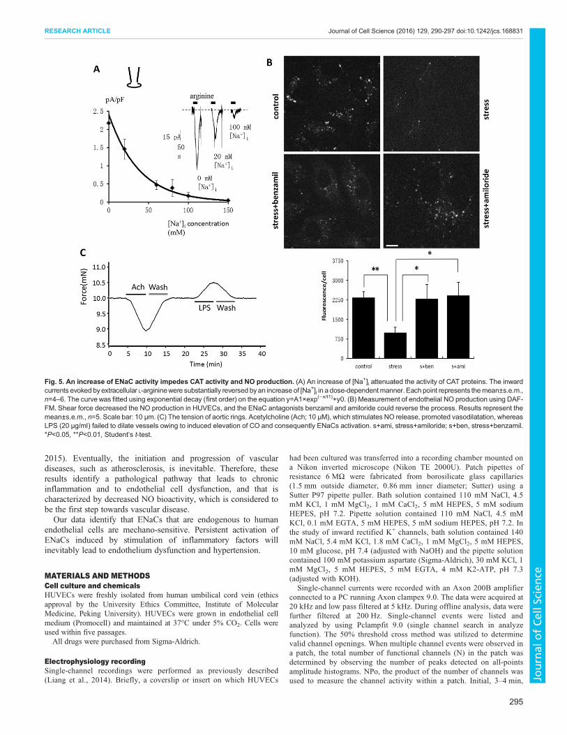
concentration ([Na+]i) in endothelial cells. We used whole-cellpatch clamping to examine the corresponding currents that areinduced by cationic amino acid transporter (CAT) proteins underdifferent concentration of [Na+]i. The inward currents evoked byextracellular L-arginine were significantly reversed, in a dose-dependent manner, by increasing [Na+]i (Fig. 5A). The cellularavailability of L-arginine is decided by the transport capacity of CATproteins. Increased [Na+]i, as we have demonstrated, reduces CATactivity and, in turn, impairs L-arginine entry. Therefore, ENaC-mediated shear-force sensing is likely to exert a reverse effect onendothelial cell function as it impairs endothelial-dependent NO
Fig. 2. Stretch-activated ENaCs. (A) In an inside-out recording, positive pressure (10 mmHg) applied through the patch pipette on the outside membrane ofHUVECs activated ENaC currents in a repeatable manner. The pipette voltage was held at 80 mV. Stretch significantly increased ENaC activity by 1.5-fold.Each point represents the mean±s.e.m., n=8 (right side). (B) Indirect flow-mediated activation of ENaCs. In a cell-attached recording, flow significantly increasedthe open probability of ENaCs, suggesting the presence of an indirect mechanism. Application of pressure is shown by the black bars above the uppercurrent trace. Selected parts of currents (marked by bars below the upper trace) are shown with an expanded time scale in the lower panel, correspondinglynumbered. Each point represents the mean±s.e.m., n=5. *P<0.05, **P<0.01, Student’s t-test.
292
RESEARCH ARTICLE Journal of Cell Science (2016) 129, 290-297 doi:10.1242/jcs.168831
Journal
ofCe
llScience
generation. We subjected HUVECs to shear force and found thatNO production decreased owing to the resultant activation ofENaCs, whereas the ENaC antagonists benzamil and amiloridecould reverse the process (Fig. 5B). Next, we isolated aortas fromWistar rats and explored the tension of aortic rings. The data showsthat lipopolysaccharide (LPS; 20 μg/ml) failed to dilate vesselsowing to induced elevation of CO and consequently ENaCactivation, whereas acetylcholine (10 μM), which stimulates NOrelease, promoted vasodilatation (Fig. 5C).
DISCUSSIONWe have identified the expression and the mechano-sensitivity ofENaCs in endothelial cells, determined the upregulation of HO-1expression by inflammatory factors and validated the effects ofthe HO-1-mediated heme degradation pathway on ENaCs. Bypiecing all these findings together, we deduce that ENaCs areexpressed on endothelial cells, and this expression provides aNa+ entry pathway and confers a mechanism by which cells canrespond to mechanical stress. However, ENaC expression issuppressed until the physiological environment changes; in thiscase, the appearance of inflammation. Pro-inflammatory factorsgreatly upregulate the highly inducible protein HO-1, which inturn facilitates heme degradation and hence increases COgeneration. CO potently increases the bioactivity of ENaCs,releasing the channels from inhibition, by contrast, CO exerts a
strong inhibitory effect on Kir channels (Liang et al., 2014). Thisaspect of CO impact on endothelial cells can be therefore thoughtof as CO changing the responsibility of mechano-sensing fromKir channels to ENaCs. Eventually, endothelial cells start torespond to shear stress by increasing the Na+ influx rate.Elevated [Na+]i impairs L-arginine entry, resulting in impairedNO generation, whereas benzamil and amiloride, bothantagonists of ENaCs, could rescue NO production. Thus, it ispredicted that long-term ENaC hyper activation induced bychronic inflammation leads to decreased NO generation inendothelial cells and vasoconstriction.
Our results are consistent with many previous clinical reports(Nishizaka et al., 2004). For example, decreased L-arginineconversion to NO has been shown in hypertensive individualsafter salt loading (Ni and Vaziri, 2001). Flow-mediatedvasodilatation was significantly reduced in affected individualscompared to that in healthy control subjects (Pemp et al., 2009).Hemeoxygenases are the rate-limiting enzymes in hemecatabolism, which leads to the generation of CO (Maines,1997). Hemeoxygenases and their metabolic product, CO, havebeen implicated in the regulation of basal tone and blood pressure(Johnson et al., 1996). CO is a promising molecule withtherapeutic potential in a number of vascular disorders owing toits cytoprotective and homeostatic properties (Dulak et al., 2008).CO inhibits sprouting angiogenesis and phosphorylation of
Fig. 3. Heme alone inhibits ENaCs, whereas heme plus NADPH activates ENaCs in HUVECs. (A) Traces were obtained using the inside-out configurationwith a pipette voltage of +80 mV. (Aa) Different concentrations of heme inhibited ENaC activity. (Ab) Heme plus NADPH (1 μM) activated ENaCs. (Ac) Hemeplus NADPH (1 μM) with knockdown of HO-1 (HO-1−) inhibited ENaCs. (Ad) Western blot analysis of HO-1 knockdown. Con, control. (B) Concentration-dependent effects of heme or heme plus NADPH on ENaC activity. Data were fitted with a sigmoidal dose-effect curve. The Hill coefficient is 0.99, based onthe equation Y=minimum+(maximum−minimum)/[1+(X/X0)p]. The blue linewith black square symbols represents the effect of heme alone, and the black linewithred circle symbols represents the effect of heme plus NADPH. Each point represents the mean±s.e.m., n=6–8. (C) Statistical analysis of data shown in panel Ac.Heme (20 nM) significantly inhibited ENaC activity, whereas in presence of heme (20 nM) plus NADPH, ENaCs from HUVECs in which HO-1 (HO–) had beenknocked down were inhibited. Each point represents the mean±s.e.m., n=10. *P<0.05, Student’s t-test.
293
RESEARCH ARTICLE Journal of Cell Science (2016) 129, 290-297 doi:10.1242/jcs.168831
Journal
ofCe
llScience
vascular endothelial growth factor receptor 2 (VEGF2) (Ahmadet al., 2015). CO-releasing molecule (CORM-2) promotesendothelial repair and inhibits neointima formation after carotidartery balloon injury (Hu et al., 2015).Many studies have shown the relationship between NO and
HO-1. NO and CO are both gaseous messengers and mediate intra-and intercellular communication with high specificity. It has beensuggested that the binding of CO and NO to heme-containingproteins regulates the signaling of CO and NO (Ding et al., 1999).NO can inhibit HO-2 catalytic activity, which inhibits COproduction (Ding et al., 1999). In isolated hearts (Maulik et al.,1996) and porcine aortic endothelial cells, NO increases COproduction. NO could have a direct inhibitory effect on HO-2 that ismasked in the intact system by cGMP-induced stimulation.Alternatively, extravascular sources of CO in vivo might
contribute to pial arteriolar dilation through the generation ofglutamate. In isolated microvessels, NO synthase (NOS) inhibitiontotally abolishes glutamate-induced CO production (Leffler et al.,2005). Additionally, CO is much less effective than NO atstimulating guanylyl cyclases (Kim et al., 2005). Normal dose-dependent dilation in response to CO occurs when cGMP levels areheld constant. COmight exert its effect by stimulating the activationof BKCa; NO also can also affect BKCa channels, but throughprotein kinase G (Leffler et al., 1999).
Once endothelium-dependent NO production is hampered,vessels start to lose their ability to mitigate increased bloodpressure through dilatation, which can further endothelial celldysfunction or even cause cell damage. Moreover, decreased NObioactivity leaves vessels unprotected and vulnerable to plateletaggregation and the expression of adhesion molecules (Mury et al.,
Fig. 4. TPA facilitates the flow response mediated by ENaCs in HUVECs. (A) Caveolae compartmentalization of ENaCs, HO-1 and HO-2 in HUVECs.Total cell lysate was loaded onto the discontinuous sucrose gradients for 18 h (39,000 rpm, SW41 rotor). Each sample was separated into 12 fractions, and eachwas subjected towestern blot analysis. Caveolin-1 was concentrated in fractions 4–5 (arrows), where ENaCs, HO-1 and HO-2were also present. (B) Incubation ofHUVECs with TPA (100 ng/ml) in the medium significantly augmented HO-1 expression but not that of HO-2 and ENaCs over 24 h. NC, non-TPA-treated cells.(C) Treatment with TPA significantly enhanced ENaC-mediated whole-cell currents and flow response. Flow was applied at a speed of 3 ml/min, which isequivalent to 3.67 dyne. Each point represents the mean±s.e.m., n=10. *P<0.05, **P<0.01, Student’s t-test.
294
RESEARCH ARTICLE Journal of Cell Science (2016) 129, 290-297 doi:10.1242/jcs.168831
Journal
ofCe
llScience
2015). Eventually, the initiation and progression of vasculardiseases, such as atherosclerosis, is inevitable. Therefore, theseresults identify a pathological pathway that leads to chronicinflammation and to endothelial cell dysfunction, and that ischaracterized by decreased NO bioactivity, which is considered tobe the first step towards vascular disease.Our data identify that ENaCs that are endogenous to human
endothelial cells are mechano-sensitive. Persistent activation ofENaCs induced by stimulation of inflammatory factors willinevitably lead to endothelium dysfunction and hypertension.
MATERIALS AND METHODSCell culture and chemicalsHUVECs were freshly isolated from human umbilical cord vein (ethicsapproval by the University Ethics Committee, Institute of MolecularMedicine, Peking University). HUVECs were grown in endothelial cellmedium (Promocell) and maintained at 37°C under 5% CO2. Cells wereused within five passages.
All drugs were purchased from Sigma-Aldrich.
Electrophysiology recordingSingle-channel recordings were performed as previously described(Liang et al., 2014). Briefly, a coverslip or insert on which HUVECs
had been cultured was transferred into a recording chamber mounted ona Nikon inverted microscope (Nikon TE 2000U). Patch pipettes ofresistance 6 MΩ were fabricated from borosilicate glass capillaries(1.5 mm outside diameter, 0.86 mm inner diameter; Sutter) using aSutter P97 pipette puller. Bath solution contained 110 mM NaCl, 4.5mM KCl, 1 mM MgCl2, 1 mM CaCl2, 5 mM HEPES, 5 mM sodiumHEPES, pH 7.2. Pipette solution contained 110 mM NaCl, 4.5 mMKCl, 0.1 mM EGTA, 5 mM HEPES, 5 mM sodium HEPES, pH 7.2. Inthe study of inward rectified K+ channels, bath solution contained 140mM NaCl, 5.4 mM KCl, 1.8 mM CaCl2, 1 mM MgCl2, 5 mM HEPES,10 mM glucose, pH 7.4 (adjusted with NaOH) and the pipette solutioncontained 100 mM potassium aspartate (Sigma-Aldrich), 30 mM KCl, 1mM MgCl2, 5 mM HEPES, 5 mM EGTA, 4 mM K2-ATP, pH 7.3(adjusted with KOH).
Single-channel currents were recorded with an Axon 200B amplifierconnected to a PC running Axon clampex 9.0. The data were acquired at20 kHz and low pass filtered at 5 kHz. During offline analysis, data werefurther filtered at 200 Hz. Single-channel events were listed andanalyzed by using Pclampfit 9.0 (single channel search in analyzefunction). The 50% threshold cross method was utilized to determinevalid channel openings. When multiple channel events were observed ina patch, the total number of functional channels (N) in the patch wasdetermined by observing the number of peaks detected on all-pointsamplitude histograms. NPo, the product of the number of channels wasused to measure the channel activity within a patch. Initial, 3–4 min,
Fig. 5. An increase of ENaC activity impedes CAT activity and NO production. (A) An increase of [Na+]i attenuated the activity of CAT proteins. The inwardcurrents evoked byextracellular L-argininewere substantially reversed byan increase of [Na+]i in a dose-dependentmanner. Each point represents themean±s.e.m.,n=4–6. The curve was fitted using exponential decay (first order) on the equation y=A1×exp(−x/t1)+y0. (B) Measurement of endothelial NO production using DAF-FM. Shear force decreased the NO production in HUVECs, and the ENaC antagonists benzamil and amiloride could reverse the process. Results represent themean±s.e.m., n=5. Scale bar: 10 μm. (C) The tension of aortic rings. Acetylcholine (Ach; 10 μM), which stimulates NO release, promoted vasodilatation, whereasLPS (20 μg/ml) failed to dilate vessels owing to induced elevation of CO and consequently ENaCs activation. s+ami, stress+amiloride; s+ben, stress+benzamil.*P<0.05, **P<0.01, Student’s t-test.
295
RESEARCH ARTICLE Journal of Cell Science (2016) 129, 290-297 doi:10.1242/jcs.168831
Journal
ofCe
llScience
single-channel recordings were normally used as the control. Theactivity of ENaCs during application of chemicals was normalized toactivity during the control period to assess the effects of chemicals onENaC activity. In some cases, ENaC activity during the application ofchemicals was also compared to that of ENaCs when chemicals werewashed off. These data were used to confirm the effects of chemicals.Data are presented as means±s.e.m. Means were compared usingStudent’s paired t-test. Statistical significance was set as <0.05.
Whole-cell recording was performed using pipettes of 6 MΩ resistance asdetailed above. Bath saline was as described above but with the addition of10 mM D-glucose. The pipette solution contained 40 mM KCl, 100K-gluconate, 1 mM MgCl2, 1 mM CaCl2, 0.1 mM EGTA, 4 mM Na2ATP, 10mM glucose, 10 mM HEPES and 2 mM GTP, and the pH was adjusted to7.2 with KOH. For perforated patch whole-cell recording, the pipettesolution was supplemented with 10–20 μg/ml amphotericin B. On the day ofexperiment, 0.1 g amphotericin B (Sigma-Aldrich) was dissolved in 0.1–0.5 ml DMSO to make a stock solution. 0.5 μl of this stock solution wasadded into 1 ml of pipette solution to make up the final pipette solution,giving a final amphotericin B concentration of 10–20 μg/ml. DMSO(vehicle for amphotericin B) at the same dilution was tested in controlexperiments, but no effect was observed.
Flow setupFlow setup was performed as described previously by David E. Claphamwith modifications (Oancea et al., 2006). A pipette with a wide opening inthe range of 100–200 μm, which was connected to a syringe pump (HarvardApparatus, Harvard), was placed within 160 µm of the cell of interest. Whenthe whole-cell configuration was obtained, and the cell membrane potentialwas clamped at −100 mV, the pump was switched to generate a relativelaminar flow over the cell of interest. The flow-induced shear force appliedto the cell of interest was calculated according to the equation:
T =rV2
2� 0:664
ffiffiffiffiffiffi
Rxp
where ρ is the density of water (1025 kg/m3), V is the fluid velocity[calculated using V=Q/A, where A=πd2/4 (d is the flow application pipettediameter) and Q is the flow rate generated by the syringe pump measured inm3/s]. Rx=V×X/µ, where X is the distance between pipette and cell; µ is thekinematic viscosity of the water (1.139×10⁻⁶ m2/s).
In our setup, d was approximately 120 µm and X approximately160 µm.Accordingly, 1 ml/min of flow generated 0.49 dyne stress; 3 ml/min of flowwas 3.67 dyne; 5 ml/min was 8.42 dyne; 8 ml/min was 17 dyne.
All data were obtained consistently. Whole-cell recording is a perfect wayto monitor and valuate the channel response to shear force. Therefore, it isused in the study of shear force response of ENaCs. However, cell-attachedrecording (single channel recording) is a good way, with less bias, tomeasure the response to hydrostatic pressure and was used to study themechanical response of ENaCs. In these studies, 10 mmHg of hydrostaticpressure was applied.
Isolation of caveolae compartments, and western blottingHUVECs were grown to confluence in 75-ml culture flasks and used forfractionation. The homogenate solution, in MBS buffer [25 mM 2-(N-morpholino)-ethanesulfonic acid (MES), pH 6.5, 0.15 M NaCl] containing1% Triton X-100, was adjusted to 40% sucrose by the addition of 2 ml of80% sucrose (in MBS) and was placed at the bottom of an ultracentrifugetube. A 5–30% discontinuous sucrose gradient was formed above (4 ml of5% sucrose, 4 ml of 30% sucrose, both in MBS lacking detergent) andcentrifuged at 39,000 rpm for 18 h in an SW41 rotor (BeckmanInstruments). A light-scattering band at the 5–30% sucrose interface wascollected or fractionated into 12 sub-fractions.
Each fractionated protein sample was loaded and separated on a 10%Bis-Tris Gel. The membranes were then blocked with 5% non-fat dry milk,probed with appropriate primary antibodies, followed by incubation withhorseradish peroxidase (HRP)-conjugated secondary antibodies at a dilutionof 1:3000. Antibodies against the following proteins were used: ENaC
Cell transfectionThe HUVECs were seeded, and one day after seeding, were transfected withsmall interfering (si)RNA against HO-1 (GenePharma, Shanghai, China).In short, siRNA was mixed with PromoFectin-HUVECs (Promocell) inOpti-MEM reduced serummedium (Invitrogen) and incubated for 20 min atroom temperature before being added to the apical side of the monolayer andfurther incubated for 5 h at 37°C. The transfection medium was thenreplaced with endothelial cell medium without antibiotics. Knockdowneffects were examined by western blot analysis after 24 h.
The sequences targeted to silence HO-1 mRNA were 5′-GGGAAUUU-AUGCCAUGUAATT-3′ (sense) and 5′-UUACAUGGCAUAAAUUCC-CTT-3′ (antisense).
Measurement of endothelial NO productionHUVECs were stimulated with shear force for 8 h and treated with benzamiland amiloride. Then endothelial cells were incubated with 5 μM DAF-FM(Biyuntian, China) diacetate in Phenol-Red-free Dulbecco’s modifiedEagle’s medium for 30 min at 37°C. The cells werewashed gently with PBSthree times; DAF fluorescence was observed by using ×60 oil objectivelenses and analyzed with laser scanning confocal microscopy (Nikon).
Tension studies of aortic ringsAorta tension studies were performed as previously described, withmodifications (Faury et al., 1995). Male Wistar rats (250 g) wereanesthetized with sodium pentobarbital (55 mg/kg, intraperitoneally) andkilled by cervical dislocation. Rat aortas were quickly removed to a bathcontaining cold physiological salt solution (PSS) for dissection (154.7 mMNaCl, 5.4 mM KCl, 11.0 mM D-glucose, 2.5 mM CaCl2, 6.0 mM Tris,pH 7.4). The distal aorta was dissected carefully to be free of surroundingtissue, cut as rings and mounted in a temperature-controlled myographsystem (DanisMyo Technology A/S model: 610 M). The bath solution(PSS) was gassed with 100% O2 at 37°C. Each ring was initially stretchedto give an optimal pressure of 200 m/s2, and the preparation was allowed tostabilize for 60 min. 10 μM of acetylcholine and 20 μg/ml LPS were used toperfuse. Tension data was relayed from the pressure transducers to a signalamplifier (600 series eight-channel amplifier, Gould Electronics). Data wereacquired and analyzed with Pclamp software.
Statistical analysisStudent’s t-test was used for the statistical analysis of all the independentexperiments, with significance accepted at P<0.05. In the patch-clampingassays, data from one coverslip were averaged and are presented in thefigures. The same experiment was repeated on different coverslips (n). Dataare presented as mean±s.e.m., and statistical differences were assessed usingStudent’s paired t-test. P<0.05 was considered significant.
Competing interestsThe authors declare no competing or financial interests.
Author contributionsD.G., S.W. and Y.G. designed the experiments. D.G., S.L. and S.W. performed theexperiments and analyzed the data. C.T., B.Y. and W.W. helped with the cell cultureassays. H.Z. and H.J. helped with the tension studies of aortic rings. D.G. and Y.G.wrote the manuscript, and A.A., H.J. and Z.Z. revised the manuscript.
FundingThis work was supported by the research grants held by Y.G. from the 973 Project[grant numbers 2013CB531206 and 2012CB517803]; and the National NaturalScience Foundation of China [grant numbers 81170236, 31127001 and 31221002].
ReferencesAhmad, S., Hewett, P. W., Fujisawa, T., Sissaoui, S., Cai, M., Gueron, G., Al-Ani,
B., Cudmore, M., Ahmed, S. F., Wong, M. K. K. et al. (2015). Carbon monoxideinhibits sprouting angiogenesis and vascular endothelial growth factor receptor-2phosphorylation. Thromb. Haemost. 113, 329-337.
296
RESEARCH ARTICLE Journal of Cell Science (2016) 129, 290-297 doi:10.1242/jcs.168831
Antonello, M., Montemurro, D., Bolognesi, M., Di Pascoli, M., Piva, A., Grego,F., Sticchi, D., Giuliani, L., Garbisa, S. and Rossi, G. P. (2007). Prevention ofhypertension, cardiovascular damage and endothelial dysfunction with green teaextracts. Am. J. Hypertens. 20, 1321-1328.
Buchanan, C. F., Verbridge, S. S., Vlachos, P. P. and Rylander, M. N. (2014).Flow shear stress regulates endothelial barrier function and expression ofangiogenic factors in a 3D microfluidic tumor vascular model. Cell. Adh. Migr. 8,517-524.
Coleman, H. A., Tare, M. and Parkington, H. C. (2004). Endothelial potassiumchannels, endothelium-dependent hyperpolarization and the regulation ofvascular tone in health and disease. Clin. Exp. Pharmacol. Physiol. 31, 641-649.
Davies, P. F., Robotewskyj, A., Griem, M. L., Dull, R. O. and Polacek, D. C.(1992). Hemodynamic forces and vascular cell communication in arteries. Arch.Pathol. Lab. Med. 116, 1301-1306.
DeRiemer, S. A., Strong, J. A., Albert, K. A., Greengard, P. and Kaczmarek, L. K.(1985). Enhancement of calcium current in Aplysia neurones by phorbol ester andprotein kinase C. Nature 313, 313-316.
Ding, Y., McCoubrey, W. K., Jr. and Maines, M. D. (1999). Interaction of hemeoxygenase-2 with nitric oxide donors. Is the oxygenase an intracellular ‘sink’ forNO? Eur. J. Biochem. 264, 854-861.
Dulak, J., Loboda, A. and Jozkowicz, A. (2008). Effect of heme oxygenase-1 onvascular function and disease. Curr. Opin. Lipidol. 19, 505-512.
Faury, G., Ristori, M. T., Verdetti, J., Jacob, M. P. and Robert, L. (1995). Effect ofelastin peptides on vascular tone. J. Vasc. Res. 32, 112-119.
Goel, R., Majeed, F., Vogel, R., Corretti, M. C., Weir, M., Mangano, C., White, C.,Plotnick, G. D. andMiller, M. (2007). Exercise-induced hypertension, endothelialdysfunction, and coronary artery disease in a marathon runner.Am. J. Cardiol. 99,743-744.
Hu, Q. S., Chen, Y. X., Huang, Q. S., Deng, B. Q., Xie, S. L., Wang, J. F. and Nie,R.Q. (2015).Carbonmonoxide releasingmoleculeaccelerates reendothelializationafter carotid artery balloon injury in rat. Biomed. Environ. Sci. 28, 253-262.
Jeggle, P., Callies, C., Tarjus, A., Fassot, C., Fels, J., Oberleithner, H., Jaisser,F. and Kusche-Vihrog, K. (2013). Epithelial sodium channel stiffens the vascularendothelium in vitro and in Liddle mice. Hypertension 61, 1053-1059.
Jernigan, N. L. andDrummond, H. A. (2005). Vascular ENaC proteins are requiredfor renal myogenic constriction. Am. J. Physiol. Renal Physiol. 289, F891-F901.
Johnson, R. A., Lavesa, M., DeSeyn, K., Scholer, M. J. and Nasjletti, A. (1996).Heme oxygenase substrates acutely lower blood pressure in hypertensive rats.Am. J. Physiol. 271, H1132-H1138.
Kim, H. P., Wang, X., Nakao, A., Kim, S. I., Murase, N., Choi, M. E., Ryter, S. W.and Choi, A. M. K. (2005). Caveolin-1 expression by means of p38beta mitogen-activated protein kinase mediates the antiproliferative effect of carbon monoxide.Proc. Natl. Acad. Sci. USA 102, 11319-11324.
Leffler, C. W., Nasjletti, A., Yu, C., Johnson, R. A., Fedinec, A. L. and Walker, N.(1999). Carbon monoxide and cerebral microvascular tone in newborn pigs.Am. J. Physiol. 276, H1641-H1646.
Leffler, C. W., Balabanova, L., Fedinec, A. L. and Parfenova, H. (2005). Nitricoxide increases carbon monoxide production by piglet cerebral microvessels.Am. J. Physiol. Heart Circ. Physiol. 289, H1442-H1447.
Liang, S., Wang, Q., Zhang, W., Zhang, H., Tan, S., Ahmed, A. and Gu, Y. (2014).Carbon monoxide inhibits inward rectifier potassium channels in cardiomyocytes.Nat. Commun. 5, 4676.
Maines, M. D. (1997). The heme oxygenase system: a regulator of secondmessenger gases. Annu. Rev. Pharmacol. Toxicol. 37, 517-554.
Maulik, N., Engelman, D. T., Watanabe, M., Engelman, R. M. and Das, D. K.(1996). Nitric oxide–a retrograde messenger for carbon monoxide signaling inischemic heart. Mol. Cell. Biochem. 157, 75-86.
Mury, W. V., Brunini, T. M., Abrantes, D. C., Mendes, I. K., Campos, M. B.,Mendes-Ribeiro, A. C. and Matsuura, C. (2015). Hyperaggregability andimpaired nitric oxide production in platelets from postmenopausal women.Maturitas. 80, 75-81.
Ni, Z. and Vaziri, N. D. (2001). Effect of salt loading on nitric oxide synthaseexpression in normotensive rats. Am. J. Hypertens. 14, 155-163.
Nilius, B. and Droogmans, G. (2001). Ion channels and their functional role invascular endothelium. Physiol. Rev. 81, 1415-1459.
Nilius, B., Viana, F. and Droogmans, G. (1997). Ion channels in vascularendothelium. Annu. Rev. Physiol. 59, 145-170.
Nishizaka, M. K., Zaman, M. A., Green, S. A., Renfroe, K. Y. and Calhoun, D. A.(2004). Impaired endothelium-dependent flow-mediated vasodilation inhypertensive subjects with hyperaldosteronism. Circulation 109, 2857-2861.
Oancea, E., Wolfe, J. T. and Clapham, D. E. (2006). Functional TRPM7 channelsaccumulate at the plasma membrane in response to fluid flow. Circ. Res. 98,245-253.
Oberleithner, H., Schneider, S. W., Albermann, L., Hillebrand, U., Ludwig, T.,Riethmuller, C., Shahin, V., Schafer, C. and Schillers, H. (2003). Endothelialcell swelling by aldosterone. J. Membr. Biol. 196, 163-172.
Oberleithner, H., Riethmuller, C., Ludwig, T., Shahin, V., Stock, C., Schwab, A.,Hausberg, M., Kusche, K. and Schillers, H. (2006). Differential action of steroidhormones on human endothelium. J. Cell Sci. 119, 1926-1932.
Oberleithner, H., Riethmuller, C., Schillers, H., MacGregor, G. A., de Wardener,H. E. and Hausberg, M. (2007). Plasma sodium stiffens vascular endotheliumand reduces nitric oxide release. Proc. Natl. Acad. Sci. USA 104, 16281-16286.
Olesen, S.-P., Claphamt, D. and Davies, P. (1988). Haemodynamic shear stressactivates a K+ current in vascular endothelial cells. Nature 331, 168-170.
Panizo, N., Rubio-Navarro, A., Amaro-Villalobos, J. M., Egido, J. andMoreno J.A. (2015). Molecular Mechanisms and Novel Therapeutic Approaches toRhabdomyolysis-Induced Acute Kidney Injury. Kidney Blood Press. Res. 40,520-532.
Pemp, B., Weigert, G., Karl, K., Petzl, U., Wolzt, M., Schmetterer, L. andGarhofer, G. (2009). Correlation of flicker-induced and flow-mediatedvasodilatation in patients with endothelial dysfunction and healthy volunteers.Diabetes Care 32, 1536-1541.
Prattichizzo, F., Giuliani, A., Ceka, A., Rippo, M. R., Bonfigli, A. R., Testa, R.,Procopio, A. D. and Olivieri, F. (2015). Epigenetic mechanisms of endothelialdysfunction in type 2 diabetes. Clin. Epigenet. 7, 56.
Rhodes, C. J., Im, H., Cao, A., Hennigs, J. K.,Wang, L., Sa, S., Chen, P.-I., Nickel,N. P., Miyagawa, K., Hopper, R. K. et al. (2015). RNA sequencing analysisdetection of a novel pathway of endothelial dysfunction in pulmonary arterialhypertension. Am. J. Respir. Crit. Care Med. 192, 356-366.
Sagi-Eisenberg, R., Lieman, H. and Pecht, I. (1985). Protein kinase C regulation ofthe receptor-coupled calcium signal in histamine-secreting rat basophilicleukaemia cells. Nature 313, 59-60.
Vitvitsky, E. V., Griffin, J. P., Collins, M. H., Spray, T. L. andGaynor, J. W. (1998).Increased pulmonary blood flow produces endothelial cell dysfunction in neonatalswine. Ann. Thorac. Surg. 66, 1372-1377.
Wang, S., Meng, F., Mohan, S., Champaneri, B. and Gu, Y. (2009). FunctionalENaC channels expressed in endothelial cells: a new candidate for mediatingshear force. Microcirculation 16, 276-287.
297
RESEARCH ARTICLE Journal of Cell Science (2016) 129, 290-297 doi:10.1242/jcs.168831


![1 Tim M. Heping Xu · Age-related multinucleate cells have been observed in various tissues and cells, such as vascular endothelial cells [4] and retinal pigment epithelial (RPE)](https://static.documents.pub/doc/80x56/5ed88e2c6714ca7f476824db/1-tim-m-heping-xu-age-related-multinucleate-cells-have-been-observed-in-various.jpg)


