Cezmi A. Akdis,* Thorsten Blesken,* Mübeccel Akdis,* Brunello Wüthrich,
‡
and Kurt Blaser*
*
Swiss Institute of Allergy and Asthma Research, CH-7270 Davos, Switzerland; and
‡
Allergy Unit, Department of Dermatology, University of Zurich, CH-8091 Zurich, Switzerland
Abstract
The induction of allergen-specific anergy in peripheral Tcells represents a key step in specific immunotherapy (SIT).Here we demonstrate that the anergic state results from in-creased IL-10 production. In bee venom (BV)-SIT the spe-cific proliferative and cytokine responses against the main
allergen, the phospholipase A
2
(PLA), and T cell epitope-containing PLA peptides were significantly suppressed after7 d of treatment. Simultaneously, the production of IL-10increased during BV-SIT. After 28 d of BV-SIT the anergicstate was established. Intracytoplasmic cytokine staining ofPBMC combined with surface marker detection revealed
that IL-10 was produced initially by activated CD4
1
CD25
1
,allergen-specific T cells, and followed by B cells and mono-cytes. Neutralization of IL-10 in PBMC fully reconstitutedthe specific proliferative and cytokine responses. A similarstate of IL-10–associated T cell anergy, as induced in BV-SIT, was found in hyperimmune individuals who recentlyhad received multiple bee stings. The addition of IL-10 tosoluble CD40 ligand IL-4–stimulated PBMC or purifiedB cells inhibited the PLA-specific and total IgE and en-hanced the IgG4 formation. Accordingly, increased IL-10production by SIT causes specific anergy in peripheral T cells,and regulates specific IgE and IgG4 production toward nor-
mal IgG4-related immunity. (
J. Clin. Invest.
1998. 102:98–
106.) Key words: immunotherapy
•
interleukin 10
•
anergy
•
T cell epitopes
•
isotype regulation
•
bee venom allergy
Introduction
It is well established that allergy is associated with high serumlevels of allergen-specific IgE and eosinophilia that are in-
duced by a spectrum of Th2 type cytokines. In particular, gen-eration of IL-4 and IL-13 is associated with IgE productionand IL-5 with increased eosinophil numbers (1–3). In contrast,normal immunity to allergen is characterized by the predomi-nant IgG antibody formation, especially of the IgG4 class (4).IgG4, like IgE, requires IL-4 for isotype switch. The produc-tion of IgG4 antibodies by memory B cells depends on thepresence of IFN-
g
, whereas IgE remains IL-4 dependent and is
suppressed by IFN-
g
(5–7). IL-10 is a major regulatory cyto-kine of inflammatory responses. It was identified originally asan inhibitor of IFN-
g
and IL-2 synthesis in Th1 cells. However,IL-10 was found to be a general inhibitor of proliferative andcytokine responses in T cells and is produced by mononuclearphagocytes (8, 9), natural killer cells (10), and by both Th1 andTh2 type lymphocytes (11). It can suppress cytokine synthesisin T cells by inhibiting accessory CD28/B7.1 receptor interac-tion (12).
The response to the soluble phospholipase A
2
(PLA)
1
ofbee venom (BV) represents a well suited human model tostudy regulatory mechanisms of cytokine- and antigen-specificIgE and IgG antibodies. PLA represents the major antigenand allergen in individuals sensitized to bee sting and can in-duce both allergy and normal immunity (13, 14). Normally, atan initial response to bee sting, low-affinity IgG1 anti-PLA an-tibodies are elicited (4). Repeated exposure to BV generateshigh-affinity IgG4 anti-PLA antibodies (4, 15, 16). In contrast,individuals allergic to bee sting develop excessive IgE antibod-ies mainly to PLA (13, 14).
Although specific immunotherapy (SIT) is applied fre-quently in allergy to hymenoptera venom, the mechanisms bywhich clinical improvement is achieved in allergic patients re-main to be elucidated. Stimulation of IgG4 antibodies and adecline of the IgE/IgG4 anti-PLA antibody ratio (15–20) wereclaimed to be responsible for successful SIT. Also, the genera-tion of IgE downregulating CD8
1
T cells and reduced numbersof mediator-secreting mast cells and eosinophils (21–25) werefound in SIT. Moreover, successful SIT of allergic rhinitis wasshown to be associated with decreased numbers of Th2 and in-creased IFN-
g
–producing cells (26). A shift from a Th2 cyto-kine pattern toward increased IFN-
g
production in SIT of al-lergy to BV, Lol p, and Der p (26–29) indicated an importantinvolvement of cytokine-producing CD4
1
T cells. However,the induction of an anergic state in peripheral T cells, which ischaracterized by suppressed proliferative and cytokine re-sponses against the major allergen(s), appeared to be a pivotalstep in SIT (30).
In this study we demonstrate that the induction and main-tenance of epitope-specific T cell anergy is associated with in-creased IL-10 production. At the beginning of a so-called ul-tra-rush SIT and initiation of anergy, IL-10 was mostlyproduced by PLA-specific T cells. The anergic state was main-tained by IL-10 production in B cells and monocytes. In-creased IL-10 production also accounts for a change in PLA-specific IgE and IgG4 ratio in favor of IgG4. Interestingly,such a state of naturally induced tolerance could be observedin hyperimmune subjects who had been frequently stung bybees, such as beekeepers.
Address correspondence to Cezmi A. Akdis, M.D., Swiss Institute ofAllergy and Asthma Research (SIAF), Obere Strasse 22, CH-7270Davos, Switzerland. Phone: 41-81-413-70-83; FAX: 41-81-413-16-07;E-mail: [email protected]
Received for publication 12 November 1997 and accepted in re-vised form 4 May 1998.
1.
Abbreviations used in this paper:
APC, antigen-presenting cells;BV, bee venom; PLA, bee venom phospholipase A
2
; sCD40L, solubleCD40 ligand; SIT, specific immunotherapy; TT, tetanus toxoid.
IL-10–induced T Cell Anergy in Allergen-specific Immunotherapy
99
Methods
Study population.
12 BV allergic individuals (mean age: 29 yr) with ahistory of severe systemic allergic reactions of grade III–IV (31, 32)after a bee sting were studied. All patients demonstrated positive in-tracutaneous reactions to honey BV (Pharmalgen Venom, ALK,Horsholm, Denmark) at a concentration of
,
10
2
4
g/liter and BV-specific serum IgE antibodies
.
2 kU/liter as estimated by CAP im-munoassay (Pharmacia Diagnostics AG, Uppsala, Sweden). Bloodsamples were taken before BV-SIT and 1, 7, and 28 d after startingBV-SIT. Two healthy nonallergic BV hyperimmune subjects (bee-keepers, age 37 and 57 yr), who had been stung
.
20 times by beesduring the last 2 mo, were included in the study.
Reagents.
PLA was from Boehringer-Mannheim (Mannheim,Germany). T cell epitope–containing peptides PLA
45-62
(PI), PLA
81-92
(PII), and PLA
113-124
(PIII) were synthesized by Drs. C.H. Schneiderand H.P. Rolli (Institute for Clinical Immunology and Allergology,Inselspital, University of Bern, Switzerland) by solid phase synthesisand subsequent HPLC purification (33). The three peptides did notbind anti-PLA antibodies of any isotype and did not show any skintest reactivity. Tetanus toxoid (TT) was from the Swiss Serum andVaccine Institute (Bern, Switzerland). Recombinant human IL-2 andIL-4 were from Novartis (Basel, Switzerland). IL-10 and rabbitanti–IL-10 were from PeproTech Inc. (Rocky Hill, NJ) and mouseanti–IL-10 JES3-9D7 was from PharMingen (San Diego, CA). Solu-ble CD40 ligand (sCD40L) was obtained from the transfected cellline 8-40-1, generated by Dr. P. Lane (34) (sCD40L-CD8-
a
fusionprotein; Institute for Immunology, Basel, Switzerland), and culturedfor 3 d in CG medium (Vitromex, Vilshofen, Germany). The sCD40Lactivity was standardized according to highest IgE inducing capacityin a 12-d PBMC culture costimulated with 25 ng/ml of IL-4 (35). Su-pernatants from the corresponding untransfected cell line J558L(kindly provided by Dr. M. Reth, University of Freiburg, Freiburg,Germany) were used as controls.
BV-SIT.
Under intensive care conditions 0.1, 1, 10, and 20
m
g ofBV (ALK) were administered subcutaneously in the upper arms at30-min intervals and then 30 and 50
m
g at 60-min intervals, reaching acumulative dose of 111.1
m
g. On day 7, two booster injections of 50
m
gwere administered, followed by 100-
m
g boosters given at 4-wk inter-vals (32).
T cell cultures.
PBMC were isolated by Ficoll (Biochrom, Berlin,Germany) density gradient centrifugation of peripheral venousblood. Cells were washed three times and resuspended in RPMI 1640medium supplemented with 1 mM sodium pyruvate, 1% MEM non-essential amino acids and vitamins, 2 mM
L
-glutamine, 100 U/ml pen-icillin, 100
m
g/ml streptomycin, 50
m
M 2-ME (all from Life Technolo-gies, Basel, Switzerland), and 10% heat-inactivated FCS (Sera-Lab,Sussex, United Kingdom). As described previously (5, 30), 10
6
cells/ml were stimulated in a 24-well plate with 1
m
M of PLA, 3
m
M ofPLA peptides I, II, and III, or 0.01 U/ml TT. Supernatants were har-vested on day 5 for cytokine detection and parallel cultures were ex-panded in medium supplemented with a mixture of IL-2 (25 U/ml)and IL-4 (25 ng/ml). After 12 d, cells were washed three times withPBS and 10
6
cells were restimulated with the same antigen at thesame concentrations as before, in the presence of 10
6
autologous,3,000 rad irradiated PBMC in 24-well tissue culture plates, in dupli-cates. Previously it was demonstrated that stimulation of PBMC withantigen, expansion of the responding antigen-specific T cells by IL-2and IL-4, and boosting with the same antigen allowed the detection ofantigen-induced IL-4, and conferred the same cytokine pattern as ob-served in the primary response for IL-5, IL-10, IL-13, and IFN-
g
(30).According to previous kinetic studies, supernatants were harvested16 h later for determination of IL-4 and 48 h later for IL-5, IL-10, IL-13,and IFN-
g
(5, 30). To determine T cell proliferative response, 2
3
10
5
PBMC were stimulated with 1
m
M PLA, a 3
m
M mixture of the threePLA T cell epitope peptides, and 0.01 U/ml TT in 200
m
l medium in96-well flat-bottomed tissue culture plates in triplicates for 6 d andpulsed for 20 h with 1
m
Ci of [
3
H]thymidine (DuPont/New England
Nuclear, Boston, MA). Incorporation of the labeled nucleotide wasdetermined after 20 h in a LKB beta plate reader (Wallac-Phormacia,Turku, Finland). IL-10 was neutralized in cultures with 20
m
g/ml rab-bit anti–IL-10 or anti–IL-10 mAb JES3-9D7. Rabbit IgG (SigmaChemical Co., St. Louis, MO) or mouse IgG1 (Coulter Corp., Miami,FL) served as a control antibody.
Quantification of cytokines.
The solid-phase sandwich ELISAsfor IFN-
g
, IL-4, IL-5, IL-10, and IL-13 have been described previ-ously (5, 6, 30). In brief, 96-well microtiter plates (Maxisorb; Nunc,Roskilde, Denmark) were coated with mAb 43-11 to human IFN-
g
and developed with biotinylated mAb 45-15. The sensitivity of theIFN-
g
ELISA was
,
10 pg/ml (mAbs and IFN-
g
standard were giftsfrom Dr. S.S. Alkan, Novartis).
IL-4 was measured by using mAb 3H4 for coating and bioti-nylated mAb 8F12 for detection (mAbs and IL-4 standard were pro-vided by Dr. C.H. Heusser, Novartis).
IL-5 was determined by using mAb TRFK5 for coating and bioti-nylated mAb JES15A10 for detection (mAbs and IL-5 standard werefrom PharMingen). The detection limit of the IL-5 ELISA was 50 pg/ml.
IL-10 was determined by using a combination of mAb JES3-9D7and biotinylated JES3-12G8 (mAb and rIL-10 standard were fromPharMingen). The sensitivity of the IL-10 ELISA was
#
50 pg/ml.The IFN-
g
, IL-4, IL-5, and IL-10 ELISAs were developed by peroxi-dase-labeled ExtrAvidine (Sigma Chemical Co.), and
o
-phenylendi-amine HCl in citrate buffer, pH 5.5, was used as a substrate. Opticaldensity was measured at 490 nm in an ELISA reader (Molecular De-vices, Menlo Park, CA) after stopping the reaction with 0.5 N H
2
SO
4
.For the detection of IL-13 the mAb JES10-2F9 (kindly provided
by DNAX Research Institute, Palo Alto, CA) was used for coating.Recombinant IL-13 from PeproTech Inc. was used as a standard.Rabbit anti–IL-13 (PeproTech Inc.) and alkaline phosphatase–labeledgoat anti–rabbit antibodies (Zymed Laboratories Inc., South SanFrancisco, CA) were used for detection. The detection limit was 300pg/ml of IL-13. In this case, the chromogenic substrate was 4-nitro-phenyl-phosphate-disodium-hexahydrate (E. Merck, Darmstadt,Germany) in diethanolamine buffer, pH 9.8. Optical density wasmeasured at 405 nm.
Detection of intracytoplasmic IL-10.
Immediately after purifica-tion of PBMC, intracellular IL-10 was stained by fixing and perme-abilizing the cells with a paraformaldehyde and saponin solution(PermeaFix™, Ortho Diagnostic Systems Inc., Raritan, NJ) (36). Af-ter washing with PBS containing 5% FCS, 1.5% BSA (Sigma Chemi-cal Co.), and 0.0055% EDTA (Fluka Chemie AG, Buchs, Switzer-land), cells were incubated with 0.5
m
g/ml PE-labeled anti–IL-10, orPE-labeled rat IgG1 control antibodies (all from PharMingen) for 40min at room temperature. IL-10–containing monocytes were deter-mined with anti–CD14-FITC, B cells with anti–CD19-FITC, and acti-vated CD4
1
T cells with anti–CD4-ECD and anti–CD25-FITC for 30min at room temperature. The multicolor fluorescence analysis wasperformed on an Epics Profile (Coulter Corp.) with Argon laser (488nm). Emitted fluorescence was determined by means of 525-nm, 575-nm, and 635-nm band filters. The intracytoplasmic IL-10 content ofspecifically stimulated PBMC was analyzed at day 10 of culture aftera 5-h stimulation with a mixture of 4
m
g/ml anti-CD3 (Ortho Diag-nostic Systems Inc.) and anti-CD28 mAbs (CLB, Amsterdam, TheNetherlands), in the presence of 2
m
M monensin (Sigma ChemicalCo.).
Generation of IgE and IgG4 anti-PLA in vitro.
As described pre-viously (5, 30), PBMC (2.5
3
10
6
/well per 5 ml) were cultured in 6-welltissue culture plates in the presence of 0.1 ng/ml of PLA, 25 ng/ml IL-4,and 25% sCD40L-containing 8-40-1 cell supernatant in the above me-dium, which was further supplemented with 40
m
g/ml human trans-ferrin and 4
m
g/ml bovine insulin (both from Sigma Chemical Co.).B cells were purified from PBMC using the magnet activated cell sep-aration system (MACS
®
; Miltenyi Biotech, Marburg, Germany) andanti-CD19 mAb after depletion of monocytes by anti-CD14 as de-scribed (5, 7, 37). The purity of cells was
.
95% as determined byFACS
®
analysis with FITC-labeled anti-CD19 (Coulter Corp.). B cells
100
Akdis et al.
were stimulated with IL-4 and sCD40L (50,000/well in 200
m
l) in96-well tissue culture plates. IL-10 was added in different concentra-tions to the PBMC and B cell cultures. Supernatants were harvestedat day 12 for determination of PLA-specific and total IgE and IgG4antibodies.
Quantification of specific antibodies and total Ig isotypes.
The IgEand IgG4 anti-PLA antibody contents in serum and culture superna-tants were measured in duplicates by ELISA (5–7, 30). PLA-specificantibodies were detected in ELISA plates (Maxisorb; Nunc) coatedwith 0.5
m
g/well PLA and incubated with culture supernatants at dif-ferent dilutions. Biotinylated anti-IgE mAb 6-7 (Novartis) and perox-idase-labeled ExtrAvidine (Sigma Chemical Co.) were used to de-velop IgE anti-PLA. Anti-IgG4 mAb RJ4 (Oxoid Ltd., Basingstoke,United Kingdom) and peroxidase-labeled anti–mouse Ig antibodies(Tago, Burlingame, CA) were used in IgG4 anti-PLA ELISA. Serafrom BV allergic patients, calibrated by BV RAST (Pharmacia Diag-nostics AG), were used as IgE anti-PLA standards. Human PLA-spe-cific IgG4 mAb BVA2 (38) was used as an IgG4 anti-PLA standard.The sensitivity of these assays was
,
0.1 ng/ml of IgE and IgG4 anti-PLA. Total IgE and IgG4 were assayed by sandwich ELISA as de-scribed (5–7, 30).
Statistical analysis.
Student’s
t
test for paired samples was usedfor statistical analysis to compare results at different time points ofimmunotherapy.
Results
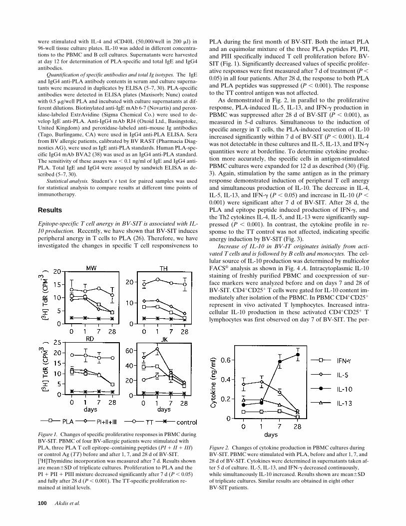
Epitope-specific T cell anergy in BV-SIT is associated with IL-10 production.
Recently, we have shown that BV-SIT inducesperipheral anergy in T cells to PLA (26). Therefore, we haveinvestigated the changes in specific T cell responsiveness to
PLA during the first month of BV-SIT. Both the intact PLAand an equimolar mixture of the three PLA peptides PI, PII,and PIII specifically induced T cell proliferation before BV-SIT (Fig. 1). Significantly decreased values of specific prolifer-ative responses were first measured after 7 d of treatment (
P
,
0.05) in all four patients. After 28 d, the response to both PLAand PLA peptides was suppressed (
P
,
0.001). The responseto the TT control antigen was not affected.
As demonstrated in Fig. 2, in parallel to the proliferativeresponse, PLA-induced IL-5, IL-13, and IFN-
g
production inPBMC was suppressed after 28 d of BV-SIT (
P
,
0.001), asmeasured in 5-d cultures. Simultaneous to the induction ofspecific anergy in T cells, the PLA-induced secretion of IL-10increased significantly within 7 d of BV-SIT (
P
,
0.001). IL-4was not detectable in these cultures and IL-5, IL-13, and IFN-
g
quantities were at borderline. To determine cytokine produc-tion more accurately, the specific cells in antigen-stimulatedPBMC cultures were expanded for 12 d as described (30) (Fig.3). Again, stimulation by the same antigen as in the primaryresponse demonstrated induction of peripheral T cell anergyand simultaneous production of IL-10. The decrease in IL-4,IL-5, IL-13, and IFN-
g
(
P
,
0.05) and increase in IL-10 (
P
,
0.001) were significant after 7 d of BV-SIT. After 28 d, thePLA and epitope peptide induced production of IFN-
g
, andthe Th2 cytokines IL-4, IL-5, and IL-13 were significantly sup-pressed (
P
,
0.001). In contrast, the cytokine profile in re-sponse to the TT control was not affected, indicating specificanergy induction by BV-SIT (Fig. 3).
Increase of IL-10 in BV-IT originates initially from acti-vated T cells and is followed by B cells and monocytes.
The cel-lular source of IL-10 production was determined by multicolorFACS
®
analysis as shown in Fig. 4
A
. Intracytoplasmic IL-10staining of freshly purified PBMC and coexpression of sur-face markers were analyzed before and on days 7 and 28 ofBV-SIT. CD4
1
CD25
1
T cells were gated for IL-10 content im-mediately after isolation of the PBMC. In PBMC CD4
1
CD25
1
represent in vivo activated T lymphocytes. Increased intra-cellular IL-10 production in these activated CD4
1
CD25
1
Tlymphocytes was first observed on day 7 of BV-SIT. The per-
Figure 1. Changes of specific proliferative responses in PBMC during BV-SIT. PBMC of four BV-allergic patients were stimulated with PLA, three PLA T cell epitope–containing peptides (PI 1 II 1 III) or control Ag (TT) before and after 1, 7, and 28 d of BV-SIT. [3H]Thymidine incorporation was measured after 7 d. Results shown are mean6SD of triplicate cultures. Proliferation to PLA and the PI 1 PII 1 PIII mixture decreased significantly after 7 d (P , 0.05) and fully after 28 d (P , 0.001). The TT-specific proliferation re-mained at initial levels.
Figure 2. Changes of cytokine production in PBMC cultures during BV-SIT. PBMC were stimulated with PLA, before and after 1, 7, and 28 d of BV-SIT. Cytokines were determined in supernatants taken af-ter 5 d of culture. IL-5, IL-13, and IFN-g decreased continuously, while simultaneously IL-10 increased. Results shown are mean6SD of triplicate cultures. Similar results are obtained in eight otherBV-SIT patients.
IL-10–induced T Cell Anergy in Allergen-specific Immunotherapy
101
centage of IL-10–containing cells increased from 7.2% beforeBV-SIT, to 28.9% at day 7, and 44.2% at day 28 (
P , 0.001)(Fig. 4 A). Because only a fraction of CD41CD251 T cells isPLA specific, PBMC were stimulated with PLA for 10 d andintracytoplasmic IL-10 was stained after additional stimulationwith anti-CD3 and anti-CD28 mAbs. IL-10–positive PLA-spe-cific T cells increased from 8.1% before BV-IT to 43.8% onday 7 and 49.2% on day 28 of BV-SIT (P , 0.001).
Beside PLA-specific and activated T cells, we have ana-lyzed IL-10 content in monocytes and B cells during BV-SIT.CD141 monocytes and CD191 B cells were gated and intracy-
toplasmic IL-10 was determined immediately after isolation ofPBMC. As shown in Fig. 4 B from the CD141 stained mono-cytes only 1.2% contained intracellular IL-10 before and 3.3%on day 7 of BV-SIT. After 28 d of BV-SIT, 41.2% of themonocyte population was IL-10 positive and showed a fullshift in the FACS® histogram (P , 0.001). The mean fluores-cence intensity of IL-10–positive monocytes increased from0.713 to 1.520. From the CD191 B cells 17.9% showed intra-cellular IL-10 before and 19.1% on day 7 of BV-SIT. Similarto monocytes, intracytoplasmic IL-10–containing B cells in-creased to 41.3% after 28 d of BV-SIT (P , 0.001). The mean
Figure 3. Changes of IL-4, IL-5, IL-10, IL-13, and IFN-g secretion during BV-SIT in response to PLA, PI 1 PII 1 PIII mixture, and control Ag (TT). PBMC were stimulated before and after 1, 7, and 28 d of BV-SIT. Cells were expanded inIL-2/IL-4–supplemented cultures, washed, and re-stimulated with the same antigens in the presence of autologous irradiated PBMC on day 12. Super-natants were harvested after 16 h for the determi-nation of IL-4 and 48 h for IL-5, IL-10, IL-13, and IFN-g. PLA and PI 1 PII 1 PIII–specific IL-4, IL-5, IL-13, and IFN-g production decreased, whereas IL-10 production simultaneously in-creased. TT induced cytokines did not show any change.
102 Akdis et al.
fluorescent intensity of IL-10–containing B cells increasedfrom 0.491 to 1.330.
The possibility that these changes in intracytoplasmic IL-10content in activated T cells, monocytes, and B cells, could oc-cur spontaneously was ruled out by determination of intracy-toplasmic IL-10 in three BV allergic individuals who receivedno treatment for 28 d. There was no significant change in intra-cytoplasmic IL-10 content observed in the respective cells ofthese individuals. The intracytoplasmic IL-10 was detectable in5.5564.07% of the CD41CD251 T cells on day 0 and5.1563.28% after 28 d without any treatment. 3.1262.83% ofthe monocytes contained intracytoplasmic IL-10 at day 0 and3.9562.52% after 28 d. 13.4764.78% of the B cells containedintracytoplasmic IL-10 at day 0 and 14.8764.91% after 28 d.
Anergized specific T cell response in BV-SIT can be rees-tablished by neutralization of endogenous IL-10 in cultures.Because the specific T cell anergy in BV-SIT was associatedwith increased IL-10 production, we investigated whether spe-cific responses of in vivo anergized T cells can be restored byIL-10 neutralization in vitro. For this purpose PBMC were cul-tured with PLA or PLA peptides in the presence or absence ofneutralizing anti–IL-10 mAb. As shown in Fig. 5 A, withoutanti–IL-10 addition, the PLA and PLA peptide-specific prolif-erative responses started to decrease after 7 d of BV-SIT and
reached their lowest values after 28 d. With IL-10 neutraliza-tion, the abrogated specific T cell proliferation (Fig. 5 A) andproduction of both Th1 (IFN-g) and Th2 (IL-4, IL-5, IL-13)cytokines (Fig. 5 B) were fully reestablished, indicating thatIL-10 is actively involved in the generation of anergy in PLA-specific T cells. Due to neutralization of IL-10, the prolifera-tive responses to PLA and PLA peptides, but also to TT, wereslightly higher than without mAb treatment, but remained atthis level throughout.
IL-10 inhibits the production of allergen-specific and totalIgE and enhances IgG4 in vitro. Although knowledge of themolecular and cellular basis of human IgE antibody regulationhas increased during recent years, the entire mechanism con-trolling allergen-specific IgE and IgG4 production needs fur-ther elucidation. For this purpose, a PBMC-based cell culturesystem has been established (5). This procedure, which was al-ready successfully applied for studies of drug-induced IgE reg-ulation (39), allows the in vitro generation of human allergen-specific IgE antibodies. PBMC from BV allergic subjects werespecifically stimulated with allergen and polyclonally withsCD40L, in the presence of IL-4. As demonstrated in Fig. 6 A,addition of IL-10 to the culture exerted clear counterregula-tory effects on both the PLA-specific and total IgE and IgG4synthesis by inhibiting IgE and enhancing IgG4 antibodies in a
Figure 4. Intracytoplasmic IL-10 content of T cells, monocytes, and B cells during BV-SIT. Immediately after isolation, PBMC were stained for surface markers CD4, CD25, CD14 (monocytes), and CD19 (B cells), then fixed, permeabilized, and counterstained for intracytoplasmic IL-10. (A) Intracytoplasmic IL-10 content in CD41CD251 T cells was significantly increased after 7 d and continued to increase during 28 d of BV-SIT. To determine changes in IL-10 content of PLA-specific T cells, PLA-stimulated PBMC were cultured for 10 d and T cells were restimulated with an anti-CD3/anti-CD28 mixture in the presence of monensin for 5 h. The yellow area represents the isotype control antibody. The IL-10 content of T cells was significantly increased already after 7 d of BV-SIT. (B) Intracytoplasmic IL-10 content of monocytes and B cells was detected from PBMC. CD141 monocytes and CD191 B cells were gated and intracytoplasmic IL-10 was determined. IL-10 content in monocytes and B cells only increased after 28 d of BV-SIT. Similar intracytoplasmic IL-10 pattern was obtained in two other BV-SIT patients.
IL-10–induced T Cell Anergy in Allergen-specific Immunotherapy 103
dose-dependent manner. To analyze whether this was a directeffect on B cells, purified B cells from PBMC were stimulatedin the presence or absence of IL-10, using the same culture sys-tem as described above. As shown in Fig. 6 B, IL-10 inhibitedthe total IgE synthesis by 84% and simultaneously enhancedIgG4 up to 4.5 times. The IgE/IgG4 ratio changed on averagefrom z 0.5 to 0.01, by a factor of 50 in favor of IgG4. Accord-ingly, IL-10 acts not only on T cells but also directly on Ig iso-type synthesis by B cells.
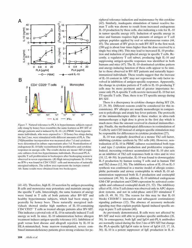
IL-10 induced natural T cell tolerance in hyperimmune in-dividuals after exposure to bee stings. It is generally observedthat hyperimmune individuals (beekeepers) after multiple beestings develop a state of low responsiveness to BV allergen. Asshown in Fig. 7 A, PBMC from such persons, who were previ-ously stung more often (. 20 times), showed abolished PLA-specific proliferative and IL-5 and IFN-g cytokine responses.This state of specific anergy corresponds to that observed inpatients after BV-SIT, and similar high levels of IL-10 werefound after PLA-specific stimulation. IL-4 and IL-13 were notdetectable. Specific stimulation in the presence of neutralizinganti–IL-10 resulted in enhanced PLA-specific proliferationand IL-5 and IFN-g production. Also, in these beekeepers whoare hyperimmune to PLA, the intracytoplasmic IL-10 contentof activated T cells and monocytes was analyzed directly fromPBMC. Again, similar to patients after 28 d of BV-SIT, acti-vated T cells and monocytes expressed high intracytoplasmicIL-10 contents and no IFN-g (Fig. 7 B).
Discussion
Recently, we demonstrated that BV-SIT induces a state ofepitope-specific anergy in T cells against PLA, the major aller-gen of BV (30). The anergy, which could also be generated inT cell clones after peptide-ligand activation in the absence ofaccessory signals (40), was characterized by suppressed prolif-erative and cytokine responses. Circulating anergic T cellsfrom peripheral blood could be reactivated to produce Th1 cy-tokines by culturing the cells with IL-2 or IL-15 (30). The pre-sented data indicate that the specific anergic state in T cellswas induced by IL-10, initially produced by the specific periph-eral T cells themselves after high-dose allergen administrationduring BV-SIT. The IL-10 production in epitope-specific Tcells was already significantly enhanced after 7 d of SIT, whilesimultaneously the specific proliferative and cytokine re-sponses were decreasing. This was confirmed by demonstrat-ing increased intracytoplasmic IL-10 content in the activatedCD251CD41 T cell subset. In contrast, the synthesis of IL-10in B cells and monocytes did not change until BV-SIT was pro-longed for 1 mo. Probably while the anergic state in activatedT cells progressed during BV-SIT, IL-10 production in thesecells influenced the antigen-presenting cells (APC) and shiftedto B cells and monocytes.
It appears from the multicolor FACS® analyses that in BV-SIT IL-10 production is initially triggered in Ag-specific Tcells, followed by the APC population and activated BV non-specific bystander T cells. IL-10 plays an important role inboth induction and maintenance of specific T cell tolerance
Figure 5. T cell proliferation and cytokine response was not sup-pressed in IL-10–neutralized PBMC cultures of a BV-SIT patient. PBMC were stimulated with PLA, the PLA peptides PI 1 PII 1 PIII, or with TT control antigen, with and without neutralizing anti–IL-10 antibodies before and after 1, 7, and 28 d of BV-SIT. [3H]Thymidine incorporation was measured after 7 d. Cells were expanded in IL-2– and IL-4–supplemented cultures for 7 d, then washed, and restimu-lated with the same antigen in the presence of autologous irradiated PBMC at day 12. Abolished PLA and PLA peptide-specific (A) pro-liferative and (B) cytokine responses were reconstituted by neutral-ization of endogenously produced IL-10. Results shown are mean6SD of triplicate cultures. Same results were obtained from two other BV-SIT patients.
Figure 6. IL-10 counterregulates total and PLA-specific IgE and IgG4 synthesis. (A) PBMC were stimulated with PLA, in the pres-ence of IL-4 and sCD40L and different amounts of IL-10. PLA-spe-cific and total IgE and IgG4 were determined in supernatants from 12-d cultures. (B) Isotype synthesis in purified B cells stimulated with IL-4 and sCD40L with and without IL-10. In both PBMC and B cell cultures IL-10 exerted counterregulatory effects by inhibiting IgE and enhancing IgG4 formation. The results shown are mean6SD of triplicate cultures, representative of two BV allergic and two healthy hyperimmune individuals with the same results.
104 Akdis et al.
(41–43). Therefore, high IL-10 secretion by antigen-presentingB cells and monocytes may potentiate and maintain anergy inthe specific T cells. Interestingly, the same features of anergywere found in T cells of both patients after BV-SIT andhealthy hyperimmune subjects, which had been stung re-peatedly by honey bees. These naturally anergized indi-viduals showed similar high numbers of IL-10–containingCD41CD251 T cells as treated allergic patients after BV-SIT.This indicates a pivotal role of IL-10 in naturally induced T cellanergy as well. In mice, IL-10 administration before allergentreatment induces antigen-specific tolerance (41). Inhibition ofgraft versus host disease and allograft rejection by IL-10 inHLA-mismatched, bone marrow–transplanted, severe com-bined immunodeficiency patients gives strong evidence for pe-
ripheral tolerance induction and maintenance by this cytokine(42). Similarly, inadequate stimulation of tumor reactive hu-man T cells was shown to result from increased endogenousIL-10 production by these cells, also indicating a role for IL-10in tumor specific anergy (43). Induction of specific anergy inmice and humans requires high amounts of antigen or T cellepitope peptides applied by oral or subcutaneous routes (44,45). The amount of BV given in rush BV-SIT in the first week(200 mg) is about four times higher than the dose received by asingle bee sting (46). This may lead to increased IL-10 produc-tion and induction of peripheral anergy in specific T cells. Re-cently, a regulatory T cell subset, producing high IL-10 andsuppressing antigen-specific responses was identified in bothhumans and mice (47). The IL-10–dominated cytokine patternand anergy-inducing function of these cells appear to be simi-lar to those observed in BV-SIT patients and naturally hyper-immunized individuals. These results suggest that the increaseof IL-10 content in APC may not represent the only factor in-volved in inhibition of antigen-specific responses. Apparently,the change in cytokine pattern of T cells to IL-10–producing Tcells may be more pertinent and of greater importance be-cause only PLA-specific T cells secrete increased IL-10 but notTT-specific T cells. Thus, there is no TT-specific anergy duringBV-SIT.
There is a discrepancy in cytokine changes during SIT (26,27, 29, 30). Different reasons could be considered for this in-consistency. BV allergics are usually monoallergic in compari-son to polyallergic and atopic hay fever patients. The protocolsof the immunotherapies differ in these studies: in ultra-rushimmunotherapy a high dose is given in the first day which ismuch more than the amount received in classical immunother-apy. Finally, the methodological differences in restimulation ofT cells by anti-CD3 instead of antigen-specific stimulation maybe responsible for differences in cytokine production (27).
IL-10 was originally described as a mouse Th2 cell factor,inhibiting cytokine synthesis by Th1 cells (48). However, neu-tralization of IL-10 in PBMC cultures reconstituted both type1 and type 2 cytokine production and proliferative response.Indeed, increasing evidence accumulated that IL-10 also actsas an inhibitor of Th2 cell responses both in vitro and in vivo(10, 12, 48–50). In particular, IL-10 was found to downregulateIL-5 production by human resting T cells and in human Th0and Th2 clones (12, 50). The inhibitory action of IL-10 on IL-5synthesis was confirmed in a murine model of allergic eosino-philic peritonitis and airway eosinophilia in which IL-10 ad-ministration suppressed both IL-5 production and eosinophilrecruitment (49, 50). In addition, IL-10 inhibited endogenousGM-CSF production and CD40 expression by activated eosin-ophils and enhanced eosinophil death (51, 52). The inhibitoryeffect of IL-10 in T cell clones was observed only in APC-depen-dent systems, and not in solid-phase anti-CD3 stimulated Tcells (9, 49, 50, 53). The reason for this may be that IL-10blocks CD28/B7.1 interaction and subsequent costimulatorysignaling pathways (12). The absence of accessory moleculesignaling may thus explain peptide ligand–induced specific an-ergy in pure T cell systems (40).
As previously demonstrated, B cells were not affected byBV-SIT and were still able to produce specific antibodies (26,30). In consequence, both IgE and IgG4 anti-PLA antibodiesare normally raised during the first 2 mo of BV-SIT but changethe PLA-specific IgE/IgG4 ratio in favor of IgG4 (15, 17, 18,30). IL-10 is a potent suppressor of IgE production in IL-4–
Figure 7. Natural tolerance to PLA in hyperimmune subjects repeat-edly stung by honey bees resembles the same features as BV-SIT of allergic patients and is induced by IL-10. (A) PBMC from hyperim-mune individuals, who were exposed to . 20 honey bee stings during the last 2 mo, were stimulated with different amounts of PLA. The [3H]thymidine incorporation was measured after 7 d and cytokines were determined in culture supernatants after 5 d. Neutralization of endogenous IL-10 fully reconstituted the proliferative and cytokine responses in anergic cells. The results shown are mean6SD of tripli-cate cultures from two hyperimmune individuals. Decreased PLA-specific T cell proliferation in beekeepers after beekeeping season is observed in seven experiments. (B) High intracytoplasmic IL-10 but no IFN-g was found in CD41CD251 cells and monocytes of naturally anergized subjects. The yellow area represents the isotype control Ab. Same results were obtained from two beekeepers.
IL-10–induced T Cell Anergy in Allergen-specific Immunotherapy 105
stimulated PBMC (54) and, as we have shown, of specific IgEin IL-4/sCD40L–stimulated cultures, while simultaneously in-creasing IgG4 formation. In this way IL-10 appears to partici-pate in the counterregulatory mechanisms of antigen-specificIgE and IgG4 synthesis by memory B cells.
Although definite decreases in IgE antibody synthesis andIgE-mediated skin sensitivity require 6–12 mo of treatment(55, 56), most patients are protected when restung by a beeduring the first months of BV-SIT. The protective effect of theSIT may primarily originate from the suppression of cytokineand mediator release from mast cells and basophils. IL-10 wasshown to reduce TNF-a, GM-CSF, and IL-6 generation frommouse bone marrow and rat peritoneal mast cells in responseto specific IgE cross-linking (57, 58). Recent studies indicatethat mast cells may represent important targets of IL-10 duringBV-SIT, and it was shown that histamine and sulfidoleuko-triens release from PLA-stimulated blood basophils is alreadygreatly reduced at the very early phase of BV-SIT (59). Themechanism of this decrease in basophil releasability may be as-sociated with anergy induction in T cells, since effector cells inallergy require priming by T cell cytokines for full activity andmediator release (60, 61).
The results of this study provide additional insight into thephysiological mechanisms of SIT. Administration of high aller-gen doses, as applied in immunotherapy, enhances endoge-nous production of IL-10, first in specific T cells and later inAPC. This results in specific T cell anergy and selective coun-terregulation of antigen-specific IgE and IgG4 by B cells in fa-vor of IgG4. In consequence, increased IL-10 synthesis in aller-gic inflammation may also inhibit effector cells of allergy.Anergized T cells can be reactivated by cytokines produced inthe microenvironment that can govern the secondary induc-tion of distinct Th1 or Th2 cytokine patterns associated witheither normal immunity against an allergen or with further de-velopment of allergy and inflammation (30). In this state of im-mune response it was shown that high antigen doses also gen-erate Th0 cytokine patterns in newly activated T cells, which isrequired for eliciting a protective IgG4 response (62).
Acknowledgments
We are grateful to Dr. Jan De Vries (DNAX Research Institute,Dept. of Human Immunology) for anti–IL-13 mAb and Dr. Chris-toph H. Heusser and Dr. Sefik S. Alkan for anti–IL-4 and anti–IFN-gmAbs.
This work was sponsored by the Swiss National Foundation(grant no. 31.39.177.93).
References
1. Del Prete, G., E. Maggi, P. Paola, I. Ghretien, A. Tiri, D. Macchia, J.Banchereau, J.E. De Vries, and S. Romagnani. 1988. IL-4 is an essential factorfor the IgE synthesis induced in vitro by human T cell clones and their superna-tants. J. Immunol. 140:4193–4198.
2. Punnonen, J., G. Aversa, B.G. Cocks, A.N.J. McKenzie, S. Menon, G.Zurawski, R. De Waal Malefyt, and J.E. De Vries. 1993. Interleukin-13 inducesinterleukin-4 independent IgG4 and IgE synthesis and CD23 expression by hu-man B cells. Proc. Natl. Acad. Sci. USA. 90:3730–3734.
3. Walker, C., J.C. Virchow, Jr., P.L.B. Bruijnzeel, and K. Blaser. 1991. Tcell subsets and their soluble products regulate eosinophilia in allergic and non-allergic asthma. J. Immunol. 146:1829–1835.
4. Aalberse, R.C., R. van der Gaag, and J. van Leuwen. 1983. Serologicaspects of IgG4 antibodies. I. Prolonged immunization results in an IgG4-restricted response. J. Immunol. 130:722–726.
5. Akdis, C.A., T. Blesken, M. Akdis, S.S. Alkan, B. Wüthrich, C.H.
Heusser, and K. Blaser. 1997. Induction and differential regulation of beevenom phospholipase A2-specific human IgE and IgG4 antibodies in vitro re-quires allergen-specific and nonspecific activation of T and B cells. J. AllergyClin. Immunol. 99:345–353.
6. Carballido, J.M., N. Carballido-Perrig, A. Oberli-Schrämmli, C.H.Heusser, and K. Blaser. 1994. Regulation of IgE and IgG4 responses by aller-gen-specific T-cell clones to bee venom phospholipase A2 in vitro. J. AllergyClin. Immunol. 93:758–767.
7. Akdis, M., C.A. Akdis, L. Weigl, R. Disch, and K. Blaser. 1997. Skinhoming, CLA1 memory T cells are activated in atopic dermatitis and regulateIgE by IL-13 dominated cytokine pattern. IgG4 counter-regulation by CLA2
memory T cells. J. Immunol. 159:4611–4619.8. Fiorentino, D.F., A. Zlotnik, T.R. Mosmann, M. Howard, and A.
O’Garra. 1991. IL-10 inhibits cytokine production by activated macrophages. J.Immunol. 147:3815–3822.
9. De Waal Malefyt, R., J. Abrams, B. Bennett, C.G. Figdor, and J.E. DeVries. 1991. Interleukin 10 (IL-10) inhibits cytokine synthesis by human mono-cytes: an autoregulatory role of IL-10 produced by monocytes. J. Exp. Med.174:1209–1220.
10. Hsu, D.-H., K.W. Moore, and H. Spits. 1992. Differential effects of in-terleukin-4 and -10 on interleukin-2 induced interferon-g synthesis and lym-phokine activated killer activity. Int. Immunol. 4:563–569.
11. Del Prete, G., M. DeCarli, F. Almerigogna, M.G. Giudizi, R. Biagiotti,and S. Romagnani. 1993. Human IL-10 is produced by both type 1 helper (Th1)and type 2 helper (Th2) T cell clones and inhibits their antigen-specific prolifer-ation and cytokine production. J. Immunol. 150:353–360.
12. Schandene, L., C. Alonso-Vega, F. Willems, C. Gerard, A. Delvaux, T.Velu, R. Devos, M. de Boer, and M. Goldman. 1994. B7/CD28-dependent IL-5production by human resting T cells is inhibited by IL-10. J. Immunol. 152:4368–4374.
13. Kagey-Sabotka, A., R.M. Franklin, N.F. Adkinson, Jr., M. Valentine, H.Bauer, and L.M. Lichtenstein. 1976. Allergy to insect sting. II. PhospholipaseA: the major allergen in honey bee venom. J. Allergy Clin. Immunol. 57:29–40.
14. Müller, U.R., T. Dudler, T. Schneider, R. Crameri, H. Fischer, D.Skrbic, R. Maibach, K. Blaser, and M. Suter. 1995. Type 1 skin reactivity to na-tive and recombinant phospholipase A2 from honey bee venom is similar. J. Al-lergy Clin. Immunol. 96:395–402.
15. Carballido, J.M., N. Carballido-Perrig, M.K. Kägi, R.H. Meloen, B.Wüthrich, C.H. Heusser, and K. Blaser. 1993. T cell epitope specificity in hu-man allergic and non-allergic subjects to bee venom phospholipase A2. J. Im-munol. 150:3582–3591.
17. Randolph, C.C., and R.E. Reismann. 1986. Evaluation of decline in se-rum venom specific IgE as a criterion for stopping venom immunotherapy. J.Allergy Clin. Immunol. 77:823–827.
18. Wyss, M., T. Scheitlin, B.M. Stadler, and B. Wüthrich. 1993. Immuno-therapy with aluminum hydroxide adsorbed insect venom extracts (AlutardSQ): immunologic and clinical results of a prospective study over 3 years. Al-lergy. 48:81–86.
19. Reid, M.J., R.B. Moss, Y.P. Hsu, J.M. Kwasnicki, T.M. Commerford,and B.L. Nelson. 1986. Seasonal asthma in northern California: allergic causesand efficacy of immunotherapy. J. Allergy Clin. Immunol. 78:590–600.
20. Hussain, R., R.W. Poindexter, and E.A. Ottesen. 1992. Control of aller-gic reactivity in human filariasis. Predominant localization of blocking antibod-ies to the IgG4 subclass. J. Immunol. 148:2731–2739.
21. Rocklin, R.E., A. Sheffer, D.K. Greineder, and K.L. Melmon. 1980.Generation of antigen-specific suppressor cells during allergy desensitization.N. Engl. J. Med. 302:1213–1219.
22. Otsuka, H., A. Mezawa, M. Ohnishi, K. Okubo, H. Seki, and M. Okuda.1991. Changes in nasal metachromatic cells during allergen immunotherapy.Clin. Exp. Allergy. 21:115–120.
23. Crecitos, P.S., N. Franklin Adkinson, Jr., A. Kagey-Sobotka, D. Proud,H.L. Meier, R.M. Naclerio, L.M. Lichtenstein, and P.S. Norman. 1983. Nasalchallenge with ragweed in hay fever patients. Effect of immunotherapy. J. Clin.Invest. 76:2247–2253.
24. Furin, M.J., P.S. Norman, P.S. Creticos, D. Proud, A. Kagey-Sobotka,L.M. Lichtenstein, and R.M. Naclerio. 1991. Immunotherapy decreases anti-gen-induced eosinophil migration into the nasal cavity. J. Allergy Clin. Immu-nol. 88:27–32.
25. Rak, S., O. Rowhagen, and P. Venge. 1988. The effect of immunother-apy on bronchial hyper-responsiveness and eosinophil cationic protein in pollenallergic patients. J. Allergy Clin. Immunol. 82:470–480.
26. Secrist, H., C.J. Chelen, Y. Wen, J.D. Marshall, and D.T. Umetsu. 1993.Allergen immunotherapy decreases interleukin 4 production in CD4 T cellsfrom allergic individuals. J. Exp. Med. 178:2123–2130.
27. Jutel, M., W.J. Pichler, D. Skrbic, A. Urwyler, C. Dahinden, and U.R.Müller. 1995. Bee venom immunotherapy results in decrease of IL-4 and IL-5and increase of IFN-g secretion in specific allergen stimulated T cell cultures. J.Immunol. 154:4187–4194.
28. Secrist, H., R.H. DeKruyff, and D.T. Umetsu. 1995. Interleukin 4 pro-duction by CD41 T cells from allergic individuals is modulated by antigen con-
106 Akdis et al.
centration and antigen-presenting cell type. J. Exp. Med. 181:1081–1089.29. Bellinghousen, I., G. Metz, A.H. Enk, S. Christmann, J. Knop, and J.
Saloga. 1997. Insect venom immunotherapy induces interleukin-10 productionand a Th2-to-Th1 shift, and changes surface marker expression in venom-aller-gic subjects. Eur. J. Immunol. 27:1131–1139.
30. Akdis, A.C., M. Akdis, T. Blesken, D. Wymann, S.S. Alkan, and K. Bla-ser. 1996. Epitope specific T cell tolerance to phospholipase A2 in bee venomimmunotherapy and recovery by IL-2 and IL-15 in vitro. J. Clin. Invest. 98:1676–1683.
31. Mueller, H.L. 1966. Diagnosis and treatment of insect sensitivity. J.Asthma Res. 3:331–333.
32. Birnbaum, J., D. Charpin, and D. Vervloet. 1993. Rapid hymenopteravenom immuno-therapy: comparative safety of three protocols. Clin. Exp. Al-lergy. 23:226–230.
33. Von Grüningen, R., and C.H. Schneider. 1989. Antigenic structure ofthe hexacosa-peptide mellitin: evidence for three determinants, one with a heli-cal conformation. Immunology. 66:339–342.
34. Lane, P., T. Brocker, S. Hubele, E. Padovan, A. Lanzavecchia, and F.F.McConnell. 1993. Soluble CD40 ligand can replace the normal T cell derivedCD40 ligand signal to B cells in T cell-dependent activation. J. Exp. Med. 177:1209–1213.
35. Gascan, H., J.-F. Gauchat, G. Aversa, P. Van Vlasselaer, and J.E. DeVries. 1991. Anti-CD40 monoclonal antibodies or CD41 T cell clones and IL-4induce IgG4 and IgE switching in purified B cells via different signaling path-ways. J. Immunol. 147:8–13.
36. Prussin, C., and D. Metcalfe. 1995. Detection of intracytoplasmic cyto-kine using flow cytometry and directly conjugated anti-cytokine antibodies. J.Immunol. Methods. 188:117–125.
37. Miltenyi, S., W. Muller, W. Weichel, and A. Radbruch. 1990. A highgradient magnetic cell separation with MACS. Cytometry. 11:231–238.
38. Schneider, T., A.B. Lang, J.M. Carballido, L.F. Santamaria Babi, T.Dudler, M.K. Kägi, K. Blaser, and M. Suter. 1994. Human monoclonal andpolyclonal antibodies recognize predominantly discontinuous epitopes on beevenom phospholipase A2. J. Allergy Clin. Immunol. 94:61–69.
39. Akdis, C.A., T. Blesken, M. Akdis, S.S. Alkan, C.H. Heusser, and K.Blaser. 1997. Glucocorticoids inhibit human antigen-specific and enhance totalIgE and IgG4 production due to differential effects on T and B cells in vitro.Eur. J. Immunol. 27:2351–2357.
40. Faith, A., C.A. Akdis, M. Akdis, H.-U. Simon, and K. Blaser. 1997. De-fective TCR stimulation in anergized type 2 T helper cells correlates with abro-gated p56lck and Zap 70 tyrosine kinase activities. J. Immunol. 159:53–60.
41. Enk, A.H., J. Saloga, D. Becker, M. Mohamadzadeh, and J. Knop. 1994.Induction of hapten-specific tolerance by interleukin 10 in vivo. J. Exp. Med.179:1397–1402.
42. Bacetta, R., M. Bigler, J.-L. Touraine, R. Parkman, P.-A. Tovo, J.Abrams, R. de Waal Malefyt, J.E. De Vries, and M.G. Roncarolo. 1994. Highlevels of interleukin 10 production in vivo are associated with tolerance inSCID patients transplanted with HLA mismatched hematopoietic stem cells. J.Exp. Med. 179:493–502.
43. Becker, J.C., C. Czerny, and E.-B. Bröcker. 1994. Maintenance of clonalanergy by endogenously produced IL-10. Int. Immunol. 6:1605–1612.
44. Müller, U., C.A. Akdis, M. Fricker, M. Akdis, T. Blesken, F. Bettens,and K. Blaser. 1998. Successful immunotherapy with T-cell epitope peptides ofbee venom phospholipase A2 induces specific T-cell anergy in patients allergicto bee venom. J. Allergy Clin. Immunol. In press.
45. Hoyne, G.F., R.E. O’Hehir, D.C. Wraith, W.R. Thomas, and J.R. Lamb.1993. Inhibition of T cell and antibody responses to house dust mite allergen by
inhalation of the dominant T cell epitope in naive and sensitized mice. J. Exp.Med. 178:1783–1788.
46. Lichtenstein, L.M., M.D. Valentine, and A. Kagey-Sobotka. 1974. Acase for venom treatment in anaphylactic sensitivity to hymenoptera sting. N.Engl. J. Med. 290:1223–1227.
47. Groux, H., A. O’Garra, M. Bigler, M. Rouleau, S. Antonenko, J.E. DeVries, and M.G. Roncarolo. 1997. A CD41 T-cell subset inhibits antigen-spe-cific T cell responses and prevents colitis. Nature. 389:737–742.
48. Fiorentino, D.F., M.W. Bond, and T.R. Mosmann. 1989. Two types ofmouse T helper cell IV. Th2 clones secrete a factor that inhibits cytokine pro-duction by Th1 clones. J. Exp. Med. 170:2081–2095.
49. Zuany-Amorim, C., S. Hailé, D. Leduc, C. Dumarey, M. Huerre, B.B.Vergaftig, and M. Pretolani. 1995. Interleukin 10 inhibits antigen-induced cellu-lar recruitment into the airways of sensitized mice. J. Clin. Invest. 95:2644–2651.
50. Zuany-Amorim, C., C. Creminon, M.C. Nevers, M.-A. Nahori, B.B.Vergaftig, and M. Pretolani. 1996. Modulation by IL-10 of antigen-induced IL-5generation, and CD41 T lymphocyte and eosinophil infiltration into the mouseperitoneal cavity. J. Immunol. 157:377–384.
51. Takanaski, S., R. Nonaka, Z. Xing, P. O’Byrne, J. Dolovich, and M. Jor-dana. 1994. Interleukin 10 inhibits lipopolysaccharide-induced survival and cy-tokine production by human peripheral blood eosinophils. J. Exp. Med. 180:711–715.
52. Ohkawara, Y., K.G. Lim, M. Glibetic, K. Nakano, J. Dolovich, K. Croitoru,P.F. Weller, and M. Jordana. 1996. CD40 expression by human peripheralblood eosinophils. J. Clin Invest. 97:1761–1766.
53. Ding, L., and E.M. Shevach. 1992. IL-10 inhibits mitogen-induced T cellproliferation by selectively inhibiting macrophage co-stimulatory function. J.Immunol. 148:3133–3139.
54. Punnonen, J.R., R. de Waal Malefyt, P. van Vlasselaer, J.-F. Gauchat,and J.E. De Vries. 1993. IL-10 and viral IL-10 prevent IL-4-induced IgE synthe-sis by inhibiting the accessory cell function of monocytes. J. Immunol. 151:1280–1289.
55. Müller, U., and H. Mosbech. 1993. Position paper: immunotherapy withhymenoptera venoms. Allergy. 48(Suppl. 14):36–46.
56. Hunt, K., M. Valentine, A. Sobotka, A. Benton, F. Amoido, and L. Lich-tenstein. 1978. A controlled trial of immunotherapy in insect hypersensitivity.N. Engl. J. Med. 299:157–161.
57. Arock, M., C. Zuany-Amorim, M. Singer, M. Benhamou, and M. Preto-lani. 1996. Interleukin-10 inhibits cytokine generation from mast cells. Eur. J.Immunol. 26:166–170.
58. Marshall, J.S., I. Leal-Berumen, L. Nielsen, M. Glibetic, and M. Jor-dana. 1996. Interleukin (IL)-10 inhibits long-term IL-6 production but not pre-formed mediator release from rat peritoneal mast cells. J. Clin. Invest. 97:1122–1128.
59. Jutel, M., U. Müller, M. Fricker, S. Rihs, W. Pichler, and C. Dahinden.1996. Influence of bee venom immunotherapy on degranulation and leuko-triene generation in human blood basophils. Clin. Exp. Allergy. 26:1112–1118.
60. Schleimer, R.P., C.P. Derse, B. Friedmann, S. Gillis, M. Plaut, M.L. Lich-tenstein, and D.W. Mac Glashan, Jr. 1989. Regulation of human basophil medi-ator release by cytokines. I. Interaction with anti-inflammatory steroids. J. Im-munol. 143:1310–1317.
61. Brunner, T., C.H. Heusser, and C.A. Dahinden. 1993. Human periph-eral blood basophils primed by interleukin 3 (IL-3) produce IL-4 in response toimmunoglobulin E receptor stimulation. J. Exp. Med. 177:605–611.
62. Carballido, J.M., A. Faith, N. Carballido-Perrig, and K. Blaser. 1997.The intensity of T cell receptor engagement determines the cytokine pattern ofhuman allergen-specific Th cells. Eur. J. Immunol. 27:515–521.