Role of long non-coding RNAs in glucosemetabolism in cancerChunmei Fan1,2,3, Yanyan Tang2, Jinpeng Wang2, Fang Xiong1, Can Guo2, Yumin Wang1,2, Shanshan Zhang1,Zhaojian Gong2, Fang Wei2, Liting Yang2, Yi He2, Ming Zhou1,2,3, Xiaoling Li1,2,3, Guiyuan Li1,2,3, Wei Xiong1,2,3*
and Zhaoyang Zeng1,2,3*
Abstract
Long-noncoding RNAs (lncRNAs) are a group of transcripts that are longer than 200 nucleotides and do not code forproteins. However, this class of RNAs plays pivotal regulatory roles. The mechanism of their action is highly complex.Mounting evidence shows that lncRNAs can regulate cancer onset and progression in a variety of ways. They can notonly regulate cancer cell proliferation, differentiation, invasion and metastasis, but can also regulate glucose metabolism incancer cells through different ways, such as by directly regulating the glycolytic enzymes and glucose transporters(GLUTs), or indirectly modulating the signaling pathways. In this review, we summarized the role of lncRNAs in regulatingglucose metabolism in cancer, which will help understand better the pathogenesis of malignant tumors. Theunderstanding of the role of lncRNAs in glucose metabolism may help provide new therapeutic targets and noveldiagnostic and prognosis markers for human cancer.
BackgroundMetabolism is one of the basic attributes of life. In the1920s, Warburg found that tumor cells exhibit a specialmetabolic phenotype. One of the features of this phenotypeis that despite adequate availability of oxygen, cancer cellsstill tend to generate energy from glycolysis, rather thandepending on oxidative phosphorylation, which producesmore ATP per molecule of glucose. This phenomenonis known as the “Aerobic glycolysis” or “Warburg effect”[1, 2]. It often results in increased glucose uptake andaccumulation of ATP and lactic in the cancer cells.T1Initially, Warburg speculated that the mitochondrial
function in tumor cells might be impaired, making itobligatory for the tumor cells to depend on aerobicglycolysis [3]. But later work found that mitochondrialfunction is not damaged in most tumor cell types [4].Further studies have shown that proliferating cellsrequire not only ATP, but also nucleic acids, fatty acids,proteins, and membrane phospholipids. Glycolysis canprovide substrates and intermediates required for the
synthesis of the aforementioned biological macromolecules.Glycolysis generates small molecule precursors or interme-diates that contribute to cell proliferation, such as acetyl-CoA, intermediates of non-essential amino acids, andribose for nucleotide synthesis to meet the needs of rapidDNA replication [3, 5]. Glycolysis produces lower quantitiesof reactive oxygen species (ROS). ROS can induce apoptosisor senescence in tumor cells under oxygen stress [6]. Sincemitochondrial oxidative phosphorylation produces higherlevels of ROS, it is advantageous for the tumor cells todepend on glycolysis for their energy needs. Although gly-colysis produces less ATP than oxidative phosphorylation,glycolytic intermediates provide the carbon sources that arerequired for rapid cell proliferation [7]. The lactate gener-ated by glycolysis lowers the pH of the extracellular matrix(ECM) [8]. Acidic microenvironment promotes tumor inva-sion and metastasis and confers resistance to radiation ther-apy [9, 10]. Thus, the Warburg effect is an optimized waythat tumor cells harness cellular stress to thrive. It alsosuggests that cancer is a metabolic disease. The most directway of altering glucose metabolism is by affecting the meta-bolic enzymes or kinases. However, some signaling path-ways also play important roles in glucose metabolism.Regulation of enzymes, kinases and signaling pathways may
* Correspondence: [email protected]; [email protected] Key Laboratory of Carcinogenesis of the Chinese Ministry of Health,Xiangya Hospital, Central South University, Changsha, Hunan, ChinaFull list of author information is available at the end of the article
directly or indirectly affect glucose metabolism in cancer.Changes at mRNA and protein levels have been shown tobe involved in reprogramming the glucose metabolism intumor cells [11, 12].A very large part of the more than 3 billion base pair
long human genome is transcribed, but less than 2% ofthe genome encodes proteins. Most of the transcripts arenot translated into proteins. These are referred to as non-coding RNAs (ncRNAs), which are longer than 200 nucle-otides (NT), are called long non-coding RNAs (lncRNAs)[13–20]. LncRNAs are involved in a variety of importantregulatory processes, at the transcriptional and post-transcriptional levels [21–27], and in epigeneticmodifications [28–31] that play complex and preciseregulatory roles in development and gene expression.LncRNAs can also regulate glucose metabolism in tumorcells [32–35]. The regulatory mechanism of lncRNAs isextremely complicated and merits systematic and in-depthresearch. A large number of studies have shown thatlncRNAs can affect genes involved in glucose metabolism[36]. Therefore, we focused on the ways and mechanismsby which lncRNAs regulate glucose metabolism in cancer,which may help advance the understanding the complex
regulatory network of cancer metabolism and provide abetter theoretical basis for clinical diagnosis and treatment.LncRNAs and their targets in the regulation of glucose me-tabolism in cancer are summarized in Table 1.
LncRNAs regulate enzymes, regulatory molecules,and oncogenes involved in glucose metabolism incancerLncRNAs regulate glucose uptake via altering theexpression of glucose transportersGlucose transporters (GLUTs) are membrane proteinsthat transport glucose from the capillaries into cells andplay an important role in cellular glucose metabolism. Sofar, 13 members of the GLUT family have been identified,out of which GLUT1, GLUT3, and GLUT4 are closely in-volved in glucose metabolism in cancer. Under normalphysiological conditions, GLUTs transport glucose rapidly.GLUTs are often upregulated in malignant tumor cells,expediting the glucose transport further.LncRNA NBR2 regulates AMPK activity and is in-
duced by glucose starvation. However, Liu et al. showedthat knocking out NBR2 does not affect phenformin-induced AMPK activity, but attenuates the expression
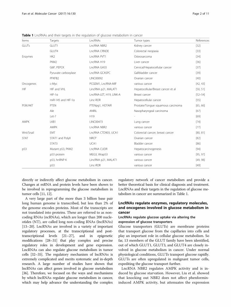
Table 1 LncRNAs and their targets in the regulation of glucose metabolism in cancer
Items Targets LncRNAs Tumor types References
GLUTs GLUT1 LncRNA NBR2 Kidney cancer [32]
GLUT4 LncRNA CRNDE Colorectal neopasia [33]
Enzymes HK2 LncRNA PVT1 Osteosarcoma [34]
PKM2 LncRNA H19 Liver cancer [36]
G6P, PEPCK LncRNA GAS5 Cervical/Hepatocellular cancer [37]
Pyruvate carboxylase LncRNA GCASPC Gallbladder cancer [39]
PFKFB2 LINC00092 Ovarian cancer [40]
Oncogenes c-Myc PCGEM1, LncRNA-MIF various cancer [42, 43]
HIF HIF and VHL LincRNA p21, MALAT1 Hepatocellular/Breast cancer et al [50, 51]
HIF-1α LncRNA-LET, H19, LINK-A Breast cancer [52–54]
miR-145 and HIF-1α Linc-ROR Hepatocellular cancer [55]
p53, hnRNP-K LincRNA p21, MALAT1 various cancer [49, 98]
p53 Linc-ROR various cancer [49]
Fan et al. Molecular Cancer (2017) 16:130 Page 2 of 11
of GLUT1, thereby reducing glucose uptake [37].LncRNA Colorectal neoplasia differentially expressed(LncRNA-CRNDE) regulates gene expression by epi-genetic modification. The intron region of this gene hasa highly conserved sequence (gVC-In4). Ellis demon-strated that knocking out gVC-In4 in HT29 cellsreduced the amount of lactic acid produced in cancercells. They further showed that the reduction in lacticacid production was due to the decrease in the effi-ciency of aerobic glycolysis or conversion of pyruvate toacetyl-CoA. They also found that the expression ofGLUT4 was reduced, indicating that CRNDE modulatesthe level of GLUT4 positively [38] (Fig. 1).
LncRNAs influence glycolysis by regulating enzymes orkinasesHK2 was a direct target of miR-497, long non-codingRNA PVT1 acts as molecular sponge to repress miR-497,as a result, PVT1 promotes glycolysis and cell prolifera-tion in osteosarcoma and form a PVT1/miR-497 axis inthe Warburg effect through regulation of HK2 expression[39] (Fig. 1).Pyruvate kinase (PK) is the last rate-limiting enzyme in
glycolysis. Allosteric as well as covalent modifications canaffect PK activity. Four isoenzymes of PK have been iden-tified so far: M, K, L, and R types. The aberrant expressionof pyruvate kinase M2 (PKM2) is most common in tumorcells [40]. PKM2 determines the proportion of carbons
derived from glucose that are used for glycolytic energyproduction [41]. In the breast cancer cell line MCF-7, thecytoplasmic promyelocytic leukemia tumor suppressorprotein (PML-TSP) interacts directly with PKM2. Overex-pression of a mutated form of PML-TSP, which was gener-ated by mutagenesis of the nuclear localization signals ofPML-TSP, suppressed PKM2 activity and accumulation oflactate [42]. Li et al. illustrated that miR675 inhibits theexpression of heterochromatin protein 1α (HP1α), leadingto changes in histones. miR675 also upregulates lncRNAH19 via EGR1 activation. H19 can induce and activatePKM2, which is essential for Waburg effect and tumori-genesis in liver cancer [43] (Fig. 1).LncRNA GAS5 binds to the DNA binding domain of
the adrenocorticotropic hormone receptor, thereby pre-venting its binding to the regulatory region of the gene.GAS5 inhibits the expression of 6-phosphoglucanase(G6Pase) and phosphoenolpyruvate carboxykinase(PEPCK) [44], enzymes that play key roles in glucosemetabolism, thereby inhibiting gluconeogenesis andglycogenolysis [45]. Thus, the role of GAS5 in glucosemetabolism is undoubtedly of great significance (Fig. 1).Pyruvate carboxylase (PC), an enzyme that convert pyru-
vate to oxaloacetate, has been proved to play an importantrole in cancer cell metabolism and proliferation. In gallblad-der cancer, GCASPC binds to pyruvate carboxylase,reduces its level and activity by promoting the instability ofPC, thereby inhibiting cell proliferation [46] (Fig. 1).
Glucose
G6P
F6P
F-1,6-BP
FBP
3-PG
PEP
Pyruvate
Lactate LDH
F-2,6-BP
LINC00092
GAS5
H19
CRNDEANRIL
Enzyme
LncRNA
Stimulation
GLUT
Oxaloaceticacid
Inhibition
PVT1
GCASPC
PKM2PC
PFK2
PFKFB
HK2
Fig. 1 LncRNAs regulate the molecules involved in glucose metabolism in cancer. LncRNAs regulate glucose uptake and glycolytic flux bymodulating GLUTs and glycolic enzymes
Fan et al. Molecular Cancer (2017) 16:130 Page 3 of 11
LINC00092 is upregulated in ovarian cancer. It in-hibits one of the glycolytic enzymes, fructose-2,6-bispho-sphatase (PFKFB2), thereby altering glycolysis, which inturn promotes metastasis and sustains the local supportivefunction of cancer-associated fibroblasts (CAFs) [47–50](Fig. 1). Although many enzymes involved in glucosemetabolism have been described, there are few reportsthat discuss how lncRNAs affect the levels of metabolismby influencing these enzymes. It is also necessary to inves-tigate whether lncRNAs are associated with other en-zymes involved in glucose metabolism.
LncRNAs affect glycolysis by regulating oncogenesAccumulating evidence shows that MYC oncogene dys-regulation is a common event in tumorigenesis. MYConcogene encodes the transcription factor, c-Myc, whichpromotes cell growth and proliferation. Jung-whan Kimdemonstrated that hypoxia-inducible factor 1 (HIF-1)cooperates with dysregulated c-Myc to promote glycoly-sis by inducing hexokinase 2, which catalyzes the firststep of glycolysis, and pyruvate dehydrogenase kinase 1,which inactivates pyruvate dehydrogenase and dimin-ishes mitochondrial respiration [51]. The prostate cancermarker, lncRNA PCGEM1, can influence a variety ofmetabolic pathways such as glucose metabolism, PPP,
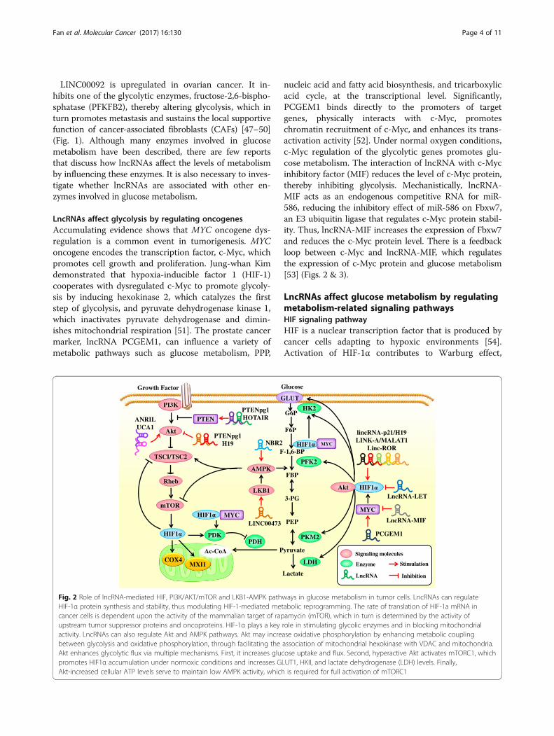
nucleic acid and fatty acid biosynthesis, and tricarboxylicacid cycle, at the transcriptional level. Significantly,PCGEM1 binds directly to the promoters of targetgenes, physically interacts with c-Myc, promoteschromatin recruitment of c-Myc, and enhances its trans-activation activity [52]. Under normal oxygen conditions,c-Myc regulation of the glycolytic genes promotes glu-cose metabolism. The interaction of lncRNA with c-Mycinhibitory factor (MIF) reduces the level of c-Myc protein,thereby inhibiting glycolysis. Mechanistically, lncRNA-MIF acts as an endogenous competitive RNA for miR-586, reducing the inhibitory effect of miR-586 on Fbxw7,an E3 ubiquitin ligase that regulates c-Myc protein stabil-ity. Thus, lncRNA-MIF increases the expression of Fbxw7and reduces the c-Myc protein level. There is a feedbackloop between c-Myc and lncRNA-MIF, which regulatesthe expression of c-Myc protein and glucose metabolism[53] (Figs. 2 & 3).
LncRNAs affect glucose metabolism by regulatingmetabolism-related signaling pathwaysHIF signaling pathwayHIF is a nuclear transcription factor that is produced bycancer cells adapting to hypoxic environments [54].Activation of HIF-1α contributes to Warburg effect,
Glucose
F-1,6-BP
FBP
3-PG
PEP
Pyruvate
Lactate
HK2PI3K
PFK2
PKM2
LDH
Akt
TSCI/TSC2
mTOR
AMPK
LKB1
PTEN
Growth Factor
Rheb
COX4MXI1
Ac-CoA
PDKPDH
MYC
PTENpg1HOTAIRANRIL
UCA1PTENpg1
H19 NBR2
LINC00473
LncRNA-LET
PCGEM1
LncRNA-MIF
lincRNA-p21/H19LINK-A/MALAT1
Linc-ROR
Enzyme
LncRNA
Stimulation
Inhibition
GLUT
Akt
Signaling molecules
MYC
MYC
F6P
G6P
Fig. 2 Role of lncRNA-mediated HIF, PI3K/AKT/mTOR and LKB1-AMPK pathways in glucose metabolism in tumor cells. LncRNAs can regulateHIF-1α protein synthesis and stability, thus modulating HIF-1-mediated metabolic reprogramming. The rate of translation of HIF-1a mRNA incancer cells is dependent upon the activity of the mammalian target of rapamycin (mTOR), which in turn is determined by the activity ofupstream tumor suppressor proteins and oncoproteins. HIF-1α plays a key role in stimulating glycolic enzymes and in blocking mitochondrialactivity. LncRNAs can also regulate Akt and AMPK pathways. Akt may increase oxidative phosphorylation by enhancing metabolic couplingbetween glycolysis and oxidative phosphorylation, through facilitating the association of mitochondrial hexokinase with VDAC and mitochondria.Akt enhances glycolytic flux via multiple mechanisms. First, it increases glucose uptake and flux. Second, hyperactive Akt activates mTORC1, whichpromotes HIF1α accumulation under normoxic conditions and increases GLUT1, HKII, and lactate dehydrogenase (LDH) levels. Finally,Akt-increased cellular ATP levels serve to maintain low AMPK activity, which is required for full activation of mTORC1
Fan et al. Molecular Cancer (2017) 16:130 Page 4 of 11
partly through the upregulation of GLUTs, thereby in-creasing glucose uptake [55] or by increasing the expres-sion of glycolytic enzymes [56, 57] or by inhibitingoxidative phosphorylation [58]. These studies indicatethat the Warburg effect is not caused just by hypoxia,but rather through a more specific regulation of tran-scription, in which HIF-1 increases the expression ofmost glycolytic enzymes.Hypoxia is thought to be related to Warburg ef-
fect, although the underlying mechanism is not yetclear. LincRNA-p21 was originally thought to be ap53-induced lncRNA that regulated P53-triggeredapoptosis in murine models [59]. However, it is notassociated with apoptosis in human tissues.LincRNA-p21 is a hypoxia-responsive lincRNA thatcompetes with HIF-1α to bind to the von Hippel-Lindau tumor suppressor protein (pVHL) and pre-vents the formation of HIF-1α-pVHL, thus inhibitingthe ubiquitinated degradation of HIF-1α. pVHL is acomponent of ubiquitin ligase complex that binds toHIF-1α and routes it to the proteasome degradationpathway. Thus, lincRNA-p21 plays an important rolein hypoxia-induced glycolysis. Under hypoxic condi-tions, HIF-1α-induced lincRNA-p21 stabilizes HIF-1α, forming a positive feedback loop. But this loop isnot always activated because hypoxic stimulationmay slow down [60]. In human hepatic epithelialcells (L-02), arsenite increases the expression of
glycolysis-related genes, including HK2, Eno-1, andGlut-4. In L-02 cells exposed to arsenite, the lncRNA,metastasis-associated lung adenocarcinoma transcript 1(MALAT1), and HIF-α, are overexpressed. Moreover,MALAT1 enhances arsenite-induced glycolysis by pro-moting the disassociation of HIF-1α from VHL, prevent-ing VHL-mediated ubiquitination of HIF-1α, whichcauses the accumulation of HIF-1α [61]. However,the overexpression of lncRNA-LET results in a de-crease in the expression of HIF-1α [62]. Hypoxiaalso induces LncRNA H19, which is involved inhypoxia-induced signal transduction processes incancer cells, thereby altering glucose metabolism[63]. Lin reported that an lncRNA in cytoplasm, longintergenic non-coding RNA for kinase activation(LINK-A), is involved in the metabolic reprogram-ming in triple-negative breast cancer [64]. LINK-Afacilitates the recruitment of BRK to the EGFR-GPNMB complex and activates BRK kinase. TheBRK-dependent phosphorylation of HIF1α at tyrosine565 interferes with hydroxylation of proline 564,thereby stabilizing HIF1α. LINK-A promotes themetabolic reprogramming and tumor progression intriple negative breast cancer by activating HIF1α. Takaha-shi et al. reported that linc-ROR is associated with hypoxiaresponse and can act as a molecular sponge of miR-145 toregulate HIF-1α and its target genes such as VEGF, TGF-β,and PDK1 [65] (Fig. 2).
Citrate Ac-coA
Glucose
F-1,6-BP
FBP
3-PG
PEP
Pyruvate
Lactate
HK2
F-2,6-BP
Pyruvate
Krebscycle
PFK2
PKM2
LDH
ACL
PFKFB
MYC
MYC
ACCLipid synthesis
Citrate
6-P-Gluconolactone
PentosePhosphate
Shunt
Glutamine
Ribulose-5P
MYCSTAT1
STAT3
Wnt/snail
P53TIGAR
P53
Nuleotide synthesis
6-P-gluconate
PGM
NRCP
UCA1
MALAT1lncRNA-p21
MEG3Wrap53
CUDR
CTD903
UCA1
Enzyme
LncRNA
Stimulation
Inhibition
GL1UT
Ac-coA
P53
Signaling molecules
F6P
G6P
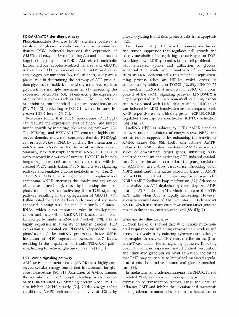
Fig. 3 Role of lncRNA–mediated Wnt/Snail, STAT and p53 pathways in glucose metabolism in tumor cells. LncRNA can modulate the expressionof Wnt/Snail, STAT and p53 expression and exert regulatory effect on glucose metabolism. p53 plays a key role in the process of glycolysis andoxidative phosphorylation, through interacting with various molecules or enzymes, such as TIGAR, GLUTs and PGM, thus affecting several keybiological processes including glucose uptake and pyruvate conversion. LncRNAs can affect expression of glycolic enzymes through STATpathways and modulate mitochondrial activity via Wnt/Snail
Fan et al. Molecular Cancer (2017) 16:130 Page 5 of 11
PI3K/AKT/mTOR signaling pathwayPhosphoinositide 3-kinase (PI3K) signaling pathway isinvolved in glucose metabolism even in insulin-freetissues. PI3K indirectly increases the expression ofGLUTs and enzymes by modulating Akt and mammaliantarget of rapamycin (mTOR). Akt-related metabolicfactors include apoptosis-related kinases and GLUTs.Activation of Akt can increase cellular ATP productionand oxygen consumption [66, 67]. In short, Akt plays apivotal role in determining the pathway of ATP produc-tion; glycolysis or oxidative phosphorylation. Akt regulatesglycolysis via multiple mechanisms: (1) increasing theexpression of GLUTs [68]; (2) enhancing the expressionof glycolytic enzymes such as HK2, PKM2 [67, 69, 70]or inhibiting mitochondrial oxidative phosphorylation[71, 72]; (3) activating mTORC1, which in turn in-creases HIF-1 levels [73, 74].Polisenno found that PTEN pseudogene (PTENpg1)
can regulate the expression level of PTEN, and inhibittumor growth by inhibiting Akt signaling pathway [75].The PTENpg1 and PTEN 3′ UTR contain a highly con-served domain, and a non-conserved domain. PTENpg1can protect PTEN mRNA by blocking the interaction ofmiRNA and PTEN in the form of miRNA decoy.Similarly, hox transcript antisense RNA (HOTAIR) isoverexpressed in a variety of tumors. HOTAIR in humantongue squamous cell carcinoma is associated with in-creased PTEN methylation. PTEN inhibits Akt signalingpathway and regulates glucose metabolism [76] (Fig. 2).LncRNA ANRIL is upregulated in nasopharyngeal
carcinoma. ANRIL increases the uptake and utilizationof glucose in aerobic glycolysis by increasing the phos-phorylation of Akt and activating the mTOR signalingpathway, resulting in the upregulation of GLUT1 [77].Kallen noted that H19 harbors both canonical and non-canonical binding sites for the let-7 family of micro-RNAs, which plays important roles in development,cancer, and metabolism. LncRNA H19, acts as a molecu-lar sponge to inhibit miRNA Let-7 activity [78]. H19 ishighly expressed in a variety of human cancers. H19expression is inhibited via PI3K/AKT-dependent phos-phorylation of the miRNA processing factor KSRP.Inhibition of H19 expression increases let-7 levels,resulting in the impairment of insulin/PI3K/AKT path-way, leading to reduced glucose uptake [79] (Fig. 2).
LKB1-AMPK signaling pathwayAMP activated protein kinase (AMPK) is a highly con-served cellular energy sensor that is necessary for glu-cose homeostasis [80, 81]. Activation of AMPK triggersthe activation of TSC2 complex, leading to inactivationof mTOR-activated GTP-binding protein Rheb. mTORalso inhibits AMPK directly [56]. Under energy deficitconditions, AMPK enhances the activity of TSC2 by
phosphorylating it and thus protects cells from apoptosis[82].Liver kinase B1 (LKB1) is a threonine/serine kinase
and tumor suppressor that regulates cell growth andenergy metabolism by regulating the activity of m-TOR.Knocking down LKB1 promotes tumor cell proliferation,with increased uptake and utilization of glucose,enhanced ATP levels, and biosynthesis of macromole-cules. In LKB1-deficient cells, this metabolic reprogram-ming process relies on HIF-1α, which exerts itsantagonism by inhibiting m-TORCI [12, 83]. LINC00473is a nuclear lncRNA that interacts with NONO, a com-ponent of the cAMP signaling pathway. LINC00473 ishighly expressed in human non-small cell lung cancerand is associated with LKB1 dysregulation. LINC00473was induced by LKB1 inactivation and subsequent cyclicAMP-responsive element-binding protein (CREB)/CREB-regulated transcription coactivator (CRTC) activation[84] (Fig. 2).LncRNA NBR2 is induced by LKB1-AMPK signaling
pathway under conditions of energy stress. NBR2 canact as tumor suppressor by enhancing the activity ofAMPK kinase [85, 86]. LKB1 can activate AMPK,followed by AMPK phosphorylation. AMPK activates aseries of downstream target genes, inhibiting ATP-depleted anabolism and activating ATP-induced catabol-ism. Glucose starvation can induce the phosphorylationof AMPK or acetyl-CoA carboxylase. Knocking downNBR2 significantly attenuates phosphorylation of AMPKand mTORC1 inactivation, suggesting the presence of aNBR2-AMPK feedback loop mechanism [87]. Adenosinekinase alleviates ATP depletion by converting two ADPsinto one ATP and one AMP, which maintains the ATP/ADP ratio when ATP is rapidly decreasing. However,excessive accumulation of AMP activates LKB1-dependentAMPK, which in turn activates downstream target genes toreplenish the energy currency of the cell [88] (Fig. 2).
Wnt/snail signaling pathwaySu Yeon Lee et al. showed that Wnt inhibits mitochon-drial respiration via inhibiting cytochrome c oxidase andpromotes glycolysis by inducing pyruvate carboxylase, akey anaplerotic enzyme. This process relies on the β-ca-tenin/T-cell factor 4/Snail signaling pathway. Knockingdown E-cadherin repressed mitochondrial respirationand stimulated glycolysis via Snail activation, indicatingthat EMT may contribute to Wnt/Snail-mediated regula-tion of mitochondrial respiration and glucose metabol-ism [89].In metastatic lung adenocarcinoma, lncRNA-CTD903
inhibited Wnt/β-catenin and subsequently inhibited theexpression of transcription factors, Twist and Snail, toinfluence EMT and inhibit the invasion and metastasisof lung adenocarcinoma cells [90]. In the breast cancer
Fan et al. Molecular Cancer (2017) 16:130 Page 6 of 11
cell line MDA-MB-231, lncRNA UCA1 contributes tothe stimulation of EMT through Wnt/β-catenin signal-ing pathway, thus promoting the invasion and metastasisof breast cancer cells [91]. We speculate that lncRNAcould indirectly alter glucose metabolism in cancer byaffecting EMT via Wnt/Snail pathway (Fig. 3).
STAT signaling pathwayLncRNA NRCP is upregulated in ovarian cancer andpromotes tumor cell growth and proliferation by stimu-lating glycolysis. Rupaimoole demonstrated that NRCPpromotes STAT1 binding to RNA polymerase II. Whenthe expression of NRCP was silenced by the introductionof siRNA–NRCP into the tumor microenvironment,binding of RNA polymerase II to STAT1 decreased, indi-cating that NRCP acts as an intermediate in the bindingof STAT1-RNA polymerase II. Further studies haveshown that NRCP binds to STAT1 and RNA polymeraseII, leading to an increase in the expression of down-stream target genes such as glucose-6-phosphate isomer-ase, which in turn affects glycolysis in tumor cells [92].LncRNA UCA1 plays an important role in bladder
cancer via the activation of PI3K/AKT/mTOR pathway.Li et al. discovered that UCA1 can stimulate glycolysisby upregulating HK2. Earlier research has shown thatSTAT3 is a direct transcriptional activator of HK2. It isalso a downstream effector of mTOR [93–95]. Theauthors further validated the association of UCA1with the mTOR-STAT3 signaling pathway. The re-sults showed that both rapamycin and STAT3 siRNAcould decrease glucose consumption and lactateproduction, indicating that UCA1 can induce the ex-pression of HK2 via mTOR-STAT3 pathway, thusregulating glycolysis [96] (Fig. 3).
p53 signaling pathwayThe absence of p53 in the cell can lead to mitochondrialrespiratory damage and increased glycolysis [97, 98]. p53not only inhibits the expression of GLUT1 and GLUT4[99], but, it also acts as a transcription factor that regulatesmultiple metabolism-related enzymes [100]. Activation ofp53 increases the ubiquitination of phosphoglyceratemutase (PGM), preventing the conversion of fructose-1,6-bisphosphate to pyruvate [101]. In gastric cancer, p53inhibits glycolysis by activating TP53-induced glycolysisand apoptosis regulator (TIGAR) [102]. TIGAR is a p53-induced gene that encodes a protein, which degradesfructose 2,6-bisphosphate, which in turn prevents theactivation of 6-phosphofructokinase 1 (PFK1), therebyinhibiting glycolysis. Therefore, glucose gets shunted intopentose phosphate pathway, which produces moreNADPH. NADPH can produce a simplified form of gluta-thione, which is the main substance that protects the cellfrom ROS damage [103]. In conclusion, the multifaceted
role of p53 in glucose metabolism in cancer is manifestedin the inhibition of glycolysis and facilitation of TCA cycleand oxidative phosphorylation.Wu et al. showed that a double mutant of p53
(N340Q/L344R) could facilitate the progression of HCCby upregulating PKM2. The p53 mutant forms a com-plex with LncRNA CUDR. The complex binds to thepromoter regions of PKM2, enhancing the phosphoryl-ation of PKM2 and its polymer formation [104]. ManylncRNAs can regulate the expression of p53 directly orindirectly. Maternally expressed gene 3 (MEG3) is usu-ally absent in a variety of human tumor cell lines. MEG3overexpression leads to an increase in p53 protein andactivation of p53 downstream target genes [105]. MEG3promotes p53-regulating transactivation in meningiomacell lines [106]. Wrap53, a natural antisense transcript ofp53, regulates the mRNA level of endogenous p53 andinduces its expression by targeting the 5′ UTR [107].LincRNA p21 is a downstream transcript of p53. It caninhibit the transcription of p53 and induce apoptosis bybinding to hnRNP-K [59]. MALAT1 is highly expressedin lung cancer, pancreatic cancer, non-small cell lungcancer, and is closely associated with cancer metastasisin patients with non-small cell lung cancer. Tripathi etal. found that knocking out MALAT1 in normal humanfibroblasts stimulated DNA damage repair and resultedin the activation of p53 and its downstream target genes.The cell cycle defects observed in MALAT1-depleted cellswere sensitive to p53 levels, indicating that MALAT1 maybe an important inhibitor of p53 [108]. ROR is a speciallncRNA in p53 signaling pathway. It can inhibit p53 andin turn be regulated by p53 [59]. These results suggest thatlncRNAs may play a crucial role in p53-mediated regula-tion of glucose metabolism (Fig. 3).
Therapeutic potential of lncRNAs in targetedtreatment of cancerTargeted therapy has attracted significant attention in re-cent times. Detailed understanding of lncRNA-mediatedregulation of glucose metabolism in tumor cells may facili-tate the development lncRNA inhibitors, which blocktumor progression. Anti-miRNAs have been developedfor treating hepatocellular carcinoma and are now inclinical trials [109]. Understanding the role of lncRNA inregulating glucose metabolism in cancer is important toexplore the possibility of using lncRNA for targetedtherapy.In a recent study of lung adenocarcinoma, reversing
the Warburg effect by inhibiting the EDFR signalpathway inhibited tumor development [65]. Pusapatiet al. identified the mTORC1-dependent reprogram-ming of metabolism that allowed cancer cells escapedependence on glycolysis. Using a combination oftargeted glycolysis and mTOR inhibitors to prevent
Fan et al. Molecular Cancer (2017) 16:130 Page 7 of 11
metabolic reprogramming induced cancer cell apoptosis[110]. In the MCF-7 breast cancer cells, combinationtreatment using acarindine (AICAR) and Methotrexate(aminoglucuric acid) reversed the Warburg effect. Monodrug therapy may induce drug resistance, but combinationtherapy can induce the expression of AMPK and FOX1,resulting in increased mitochondrial oxidative phosphoryl-ation and decreased glycolysis. These metabolic changessuggest an anti-Warburg effect that blocked the G1/S andthe G2/M transition, slowing down cell cycle [111]. Theseresults highlight the potential of targeting glucosemetabolism for cancer treatment.Fluorodeoxyglucose positron emission tomography
(FDG-PET) has been employed to measure glucosemetabolism, for detecting cancer and predicting theprognosis [112]. Current methods, including positronemission tomography (PET), autoradiography andmagnetic resonance imaging (MRI), can measure therate of primary metabolism of glucose. The limitationsof these methods include their inability to distinguishmarkers and intermediate products. Germline mutationsin succinate dehydrogenase and fumarate hydratase ofTCA cycle have been reported in kidney and ganglioncell tumors [113]. One of the effects of these mutationsis the activation of HIF-1α-regulated glucose metabol-ism. HIF plays a pivotal role in tumor metabolism; butHIF also regulates a variety of target genes, such as thoseinvolved in cell proliferation, angiogenesis and glucosemetabolism. Therefore, therapies targeting HIF maycause unpredictable pathophysiological changes. Hence,it seems more reasonable to develop specific inhibitorstargeting lncRNA.In contrast to gene therapy, oligonucleotide therapy is
more similar to small molecule therapy. Oligonucleotidescan be synthesized artificially, do not integrate into the hostgenome and are highly specific. Therefore, they have min-imal non-specific and generalized effects. Oligonucleotide-based therapies include using siRAN, anti-miRs, miRNAmimics, antisense oligonucleotides, targeting the upregula-tion of mRNA by lncRNA, and oligonucleotide-induceddifferential splicing [114]. Locked nucleic acid gapmers caneffectively interfere with lncRNA. Gapmers have been re-ported to be effective in targeting primate PSCK9, but failedin phase 1 clinical trials [115]. Survivin and HIF-1α gap-mers have been used for one year without safety issues[116, 117]. LncRNA-based targeted therapies still have along way to go. Future studies need to address theseexciting hypotheses.
ProspectReprogramming glucose metabolism is a recently identi-fied hallmark of cancer cells. Mounting evidence showsthat numerous factors are involved in this process. Wehave highlighted the special roles of lncRNAs in this
review. As discussed above, the interaction of lncRNAswith crucial transcription factors or metabolic enzymesinvolved in the processes of glycolysis can effectivelymodulate glucose metabolism and promote tumorprogression. In addition to these critical molecules, othermetabolic pathways are also pivotal for glucose metabolismin cancer, especially the PI3K/AKT/mTOR pathway andthe AMPK pathway. LncRNA, as a regulator of metabol-ism, may provide novel attractive targets for cancer therapy.Therefore, detailed understanding of the role of lncRNA inregulating glucose metabolism and the mechanism bywhich it accomplishes this regulation will help to developnovel means to control aberrant metabolic phenotypeand find more effective therapeutic strategies to sup-press the “Warburg effect”, ultimately paving the wayfor better treatment of cancer.
FundingThis work was supported in part by grants from the National Natural ScienceFoundation of China (81372907, 81301757, 81472531, 81402009, 81572787,81672993 and 81672683) and the Natural Science Foundation of HunanProvince (13JJ3039 and 2015JJ1022).
Availability of data and materialsNot applicable.
Authors’ contributionsCF, YT, JW, FX, CG, YW, SZ, ZG, FW, LY, YH collected the related paper anddrafted the manuscript. MZ, XL, GL, WX and ZZ participated in the design ofthe review and draft the manuscript. All authors read and approved the finalmanuscript.
Ethics approval and consent to participateNot applicable.
Consent for publicationNot applicable.
Competing interestsThe authors declare that they have no competing interests.
Fan et al. Molecular Cancer (2017) 16:130 Page 8 of 11
Author details1The Key Laboratory of Carcinogenesis of the Chinese Ministry of Health,Xiangya Hospital, Central South University, Changsha, Hunan, China. 2The KeyLaboratory of Carcinogenesis and Cancer Invasion of the Chinese Ministry ofEducation, Cancer Research Institute, Central South University, Changsha,Hunan, China. 3Hunan Key Laboratory of Nonresolving Inflammation andCancer, Disease Genome Research Center, The Third Xiangya Hospital,Central South University, Changsha, Hunan, China.
Received: 14 March 2017 Accepted: 11 July 2017
References1. Warburg O. On the origin of cancer cells. Science. 1956;123:309–14.2. Warburg O. The chemical constitution of respiration ferment. Science. 1928;
effect: the metabolic requirements of cell proliferation. Science. 2009;324:1029–33.4. Fantin VR, St-Pierre J, Leder P. Attenuation of LDH-A expression uncovers a
link between glycolysis, mitochondrial physiology, and tumor maintenance.Cancer Cell. 2006;9:425–34.
5. DeBerardinis RJ, Mancuso A, Daikhin E, Nissim I, Yudkoff M, Wehrli S, et al.Beyond aerobic glycolysis: transformed cells can engage in glutaminemetabolism that exceeds the requirement for protein and nucleotidesynthesis. Proc Natl Acad Sci U S A. 2007;104:19345–50.
6. Wang Y, Wu Y, Wang Y, Fu A, Gong L, Li W, et al. Bacillus amyloliquefaciensSC06 alleviates the oxidative stress of IPEC-1 via modulating Nrf2/Keap1signaling pathway and decreasing ROS production. Appl MicrobiolBiotechnol. 2016;
7. Brooks GA. Cell-cell and intracellular lactate shuttles. J Physiol. 2009;587:5591–600.8. Held-Warmkessel J, Dell DD. Lactic acidosis in patients with cancer. Clin J
Oncol Nurs. 2014;18:592–4.9. Peppicelli S, Bianchini F, Calorini L. Extracellular acidity, a “reappreciated”
trait of tumor environment driving malignancy: perspectives in diagnosisand therapy. Cancer Metastasis Rev. 2014;33:823–32.
10. Shiraishi T, Verdone JE, Huang J, Kahlert UD, Hernandez JR, Torga G, et al.Glycolysis is the primary bioenergetic pathway for cell motility andcytoskeletal remodeling in human prostate and breast cancer cells.Oncotarget. 2015;6:130–43.
11. Zhao M, Fan J, Liu Y, Yu Y, Xu J, Wen Q, et al. Oncogenic role of the TP53-induced glycolysis and apoptosis regulator in nasopharyngeal carcinomathrough NF-kappaB pathway modulation. Int J Oncol. 2016;48:756–64.
12. Faubert B, Vincent EE, Griss T, Samborska B, Izreig S, Svensson RU, et al. Lossof the tumor suppressor LKB1 promotes metabolic reprogramming ofcancer cells via HIF-1alpha. Proc Natl Acad Sci U S A. 2014;111:2554–9.
13. Rinn JL, Chang HY. Genome regulation by long noncoding RNAs. Annu RevBiochem. 2012;81:145–66.
14. Gong Z, Zhang S, Zhang W, Huang H, Li Q, Deng H, et al. Long non-codingRNAs in cancer. Sci China Life Sci. 2012;55:1120–4.
15. Wang Y, Xue D, Li Y, Pan X, Zhang X, Kuang B, et al. The long noncodingRNA MALAT-1 is a novel biomarker in various cancers: a meta-analysisbased on the GEO database and literature. J Cancer. 2016;7:991–1001.
16. Lian Y, Li XY, Tang YY, Yang LT, Li XL, Xiong W, et al. Long non-codingRNAs function as competing endogenous RNAs to regulate cancerprogression. Prog Biochem Biophys. 2016;43:219–25.
17. Zeng Z, Fan S, Zhang X, Li S, Zhou M, Xiong W, et al. Epstein-Barr virus-encoded small RNA 1 (EBER-1) could predict good prognosis innasopharyngeal carcinoma. Clin Transl Oncol. 2016;18:206–11.
18. Zeng ZY, Bo H, Gong ZJ, Lian Y, Li XY, Li XL, et al. AFAP1-AS1, a longnoncoding RNA upregulated in lung cancer and promotes invasion andmetastasis. Tumor Biol. 2016;37:729–37.
19. Xu K, Xiong W, Zhou M, Wang HR, Yang J, Li XY, et al. Integrating ChIP-sequencing and digital gene expression profiling to identify BRD7downstream genes and construct their regulating network. Mol CellBiochem. 2016;411:57–71.
20. Wang YM, Mo YZ, Gong ZJ, Yang X, Yang M, Zhang SS, et al. Circular RNAsin human cancer. Mol Cancer. 2017;16
21. Gong Z, Zhang S, Zeng Z, Wu H, Yang Q, Xiong F, et al. LOC401317, a p53-regulated long non-coding RNA, inhibits cell proliferation and inducesapoptosis in the nasopharyngeal carcinoma cell line HNE2. PLoS One.2014;9:e110674.
22. Bo H, Gong Z, Zhang W, Li X, Zeng Y, Liao Q, et al. Upregulated long non-coding RNA AFAP1-AS1 expression is associated with progression and poorprognosis of nasopharyngeal carcinoma. Oncotarget. 2015;6:20404–18.
23. Gong Z, Yang Q, Zeng Z, Zhang W, Li X, Zu X, et al. An integrativetranscriptomic analysis reveals p53 regulated miRNA, mRNA, and lncRNAnetworks in nasopharyngeal carcinoma. Tumour Biol. 2016;37:3683–95.
24. He B, Li W, Wu Y, Wei F, Gong Z, Bo H, et al. Epstein-Barr virus-encodedmiR-BART6-3p inhibits cancer cell metastasis and invasion by targeting longnon-coding RNA LOC553103. Cell Death Dis. 2016;7:e2353.
25. Yang L, Tang Y, He Y, Wang Y, Lian Y, Xiong F, et al. High expression ofLINC01420 indicates an unfavorable prognosis and modulates cell migrationand invasion in nasopharyngeal carcinoma. J Cancer. 2017;8:97–103.
26. Yu J, Liu Y, Gong Z, Zhang S, Guo C, Li X, et al. Overexpression long non-codingRNA LINC00673 is associated with poor prognosis and promotes invasion andmetastasis in tongue squamous cell carcinoma. Oncotarget. 2017;8:16621–32.
27. Yu J, Liu Y, Guo C, Zhang S, Gong Z, Tang Y, et al. Upregulated long non-coding RNA LINC00152 expression is associated with progression and poorprognosis of tongue squamous cell carcinoma. J Cancer. 2017;8:523–30.
28. Costa FF. Non-coding RNAs, epigenetics and complexity. Gene. 2008;410:9–17.29. Amaral PP, Dinger ME, Mercer TR, Mattick JS. The eukaryotic genome as an
RNA machine. Science. 2008;319:1787–9.30. Bernstein E, Allis CD. RNA meets chromatin. Genes Dev. 2005;19:1635–55.31. Tang Y, Wang J, Lian Y, Fan C, Zhang P, Wu Y, et al. Linking long non-
coding RNAs and SWI/SNF complexes to chromatin remodeling in cancer.Mol Cancer. 2017;16:42.
32. Gupta RA, Shah N, Wang KC, Kim J, Horlings HM, Wong DJ, et al. Long non-coding RNA HOTAIR reprograms chromatin state to promote cancermetastasis. Nature. 2010;464:1071–6.
33. Marques AC, Ponting CP. Catalogues of mammalian long noncoding RNAs:modest conservation and incompleteness. Genome Biol. 2009;10:R124.
34. Yang F, Xue X, Bi J, Zheng L, Zhi K, Gu Y, et al. Long noncoding RNACCAT1, which could be activated by c-Myc, promotes the progression ofgastric carcinoma. J Cancer Res Clin Oncol. 2013;139:437–45.
35. Flockhart RJ, Webster DE, Qu K, Mascarenhas N, Kovalski J, Kretz M, et al.BRAFV600E remodels the melanocyte transcriptome and induces BANCR toregulate melanoma cell migration. Genome Res. 2012;22:1006–14.
36. Xiao ZD, Zhuang L, Gan B. Long non-coding RNAs in cancer metabolism.BioEssays. 2016;38:991–6.
37. Liu X. Gan B: lncRNA NBR2 modulates cancer cell sensitivity to phenforminthrough GLUT1. Cell Cycle. 2016;15:3471–81.
38. Ellis BC, Graham LD, Molloy PL. CRNDE, a long non-coding RNA responsiveto insulin/IGF signaling, regulates genes involved in central metabolism.Biochim Biophys Acta. 1843;2014:372–86.
39. Song J, Wu X, Liu F, Li M, Sun Y, Wang Y, et al. Long non-coding RNA PVT1promotes glycolysis and tumor progression by regulating miR-497/HK2 axisin osteosarcoma. Biochem Biophys Res Commun. 2017;
40. Li Q, Chen P, Zeng ZY, Liang F, Song YL, Xiong F, et al. Yeast two-hybridscreening identified WDR77 as a novel interacting partner of TSC22D2.Tumor Biol. 2016;37:12503–12.
41. Liang F, Li Q, Li XY, Li Z, Gong ZJ, Deng H, et al. TSC22D2 interacts with PKM2and inhibits cell growth in colorectal cancer. Int J Oncol. 2016;49:1046–56.
42. Shimada N, Shinagawa T, Ishii S. Modulation of M2-type pyruvate kinaseactivity by the cytoplasmic PML tumor suppressor protein. Genes Cells.2008;13:245–54.
43. Li H, Li J, Jia S, Wu M, An J, Zheng Q, et al. miR675 upregulates longnoncoding RNA H19 through activating EGR1 in human liver cancer.Oncotarget. 2015;6:31958–84.
44. Kino T, Hurt DE, Ichijo T, Nader N, Chrousos GP. Noncoding RNA gas5 is agrowth arrest- and starvation-associated repressor of the glucocorticoidreceptor. Sci Signal. 2010;3:ra8.
45. Barthel A, Schmoll D. Novel concepts in insulin regulation of hepaticgluconeogenesis. Am J Phys Endocrinol Metab. 2003;285:E685–92.
46. Ma M-z, Zhang Y, Weng MZ, Wang SH, Hu Y, Hou ZY, et al. Longnoncoding RNA GCASPC, a target of miR-17-3p, negatively regulatesPyruvate Carboxylase–dependent cell proliferation in gallbladder cancer.Cancer Res. 2016;76:5361–71.
47. Zhao L, Ji G, Le X, Wang C, Xu L, Feng M, et al. Long noncoding RNALINC00092 acts in cancer-associated fibroblasts to drive Glycolysis andprogression of ovarian cancer. Cancer Res. 2017;77:1369–82.
48. Wang MN, Zhao JZ, Zhang LS, Wei F, Lian Y, Wu YF, et al. Role of tumormicroenvironment in tumorigenesis. J Cancer. 2017;8:761–73.
Fan et al. Molecular Cancer (2017) 16:130 Page 9 of 11
49. Song YL, Li XL, Zeng ZY, Li Q, Gong ZJ, Liao QJ, et al. Epstein-Barr virusencoded miR-BART11 promotes inflammation-induced carcinogenesis bytargeting FOXP1. Oncotarget. 2016;7:36783–99.
50. Xiao K, Yu ZY, Li XY, Li XL, Tang K, Tu CF, et al. Genome-wide analysis ofEpstein-Barr virus (EBV) integration and strain in C666-1 and Raji cells. JCancer. 2016;7:214–24.
51. Kim JW, Gao P, Liu YC, Semenza GL, Dang CV. Hypoxia-inducible factor 1and dysregulated c-Myc cooperatively induce vascular endothelial growthfactor and metabolic switches hexokinase 2 and pyruvate dehydrogenasekinase 1. Mol Cell Biol. 2007;27:7381–93.
52. Hung CL, Wang LY, Yu YL, Chen HW, Srivastava S, Petrovics G, et al. A longnoncoding RNA connects c-Myc to tumor metabolism. Proc Natl Acad Sci US A. 2014;111:18697–702.
53. Zhang P, Cao L, Fan P, Mei Y, Wu M. LncRNA-MIF, a c-Myc-activated longnon-coding RNA, suppresses glycolysis by promoting Fbxw7-mediated c-Myc degradation. EMBO Rep. 2016;17:1204–20.
54. Kaelin WG Jr, Ratcliffe PJ. Oxygen sensing by metazoans: the central role ofthe HIF hydroxylase pathway. Mol Cell. 2008;30:393–402.
55. Starska K, Forma E, Jozwiak P, Brys M, Lewy-Trenda I, Brzezinska-Blaszczyk E,et al. Gene and protein expression of glucose transporter 1 and glucosetransporter 3 in human laryngeal cancer-the relationship with regulatoryhypoxia-inducible factor-1alpha expression, tumor invasiveness, and patientprognosis. Tumour Biol. 2015;36:2309–21.
56. Iyer NV, Kotch LE, Agani F, Leung SW, Laughner E, Wenger RH, et al. Cellularand developmental control of O2 homeostasis by hypoxia-inducible factor1alpha. Genes Dev. 1998;12:149–62.
57. Hussien R, Brooks GA. Mitochondrial and plasma membrane lactatetransporter and lactate dehydrogenase isoform expression in breast cancercell lines. Physiol Genomics. 2011;43:255–64.
58. Kim JW, Tchernyshyov I, Semenza GL, Dang CV. HIF-1-mediated expressionof pyruvate dehydrogenase kinase: a metabolic switch required for cellularadaptation to hypoxia. Cell Metab. 2006;3:177–85.
59. Huarte M, Guttman M, Feldser D, Garber M, Koziol MJ, Kenzelmann-Broz D,et al. A large intergenic noncoding RNA induced by p53 mediates globalgene repression in the p53 response. Cell. 2010;142:409–19.
60. Yang F, Zhang H, Mei Y, Wu M. Reciprocal regulation of HIF-1alpha andlincRNA-p21 modulates the Warburg effect. Mol Cell. 2014;53:88–100.
61. Luo F, Liu X, Ling M, Lu L, Shi L, Lu X, et al. The lncRNA MALAT1, actingthrough HIF-1alpha stabilization, enhances arsenite-induced glycolysis inhuman hepatic L-02 cells. Biochim Biophys Acta. 1862;2016:1685–95.
62. Yang F, Huo XS, Yuan SX, Zhang L, Zhou WP, Wang F, et al. Repression ofthe long noncoding RNA-LET by histone deacetylase 3 contributes tohypoxia-mediated metastasis. Mol Cell. 2013;49:1083–96.
63. Matouk IJ, DeGroot N, Mezan S, Ayesh S, Abu-lail R, Hochberg A, et al. The H19non-coding RNA is essential for human tumor growth. PLoS One. 2007;2:e845.
64. Lin A, Li C, Xing Z, Hu Q, Liang K, Han L, et al. The LINK-A lncRNA activatesnormoxic HIF1alpha signalling in triple-negative breast cancer. Nat Cell Biol.2016;18:213–24.
65. Takahashi K, Yan IK, Haga H, Patel T. Modulation of hypoxia-signalingpathways by extracellular linc-RoR. J Cell Sci. 2014;127:1585–94.
66. Robey RB, Hay N. Is Akt the “Warburg kinase”?-Akt-energy metabolisminteractions and oncogenesis. Semin Cancer Biol. 2009;19:25–31.
67. Gottlob K, Majewski N, Kennedy S, Kandel E, Robey RB, Hay N. Inhibition ofearly apoptotic events by Akt/PKB is dependent on the first committed stepof glycolysis and mitochondrial hexokinase. Genes Dev. 2001;15:1406–18.
68. van Dam EM, Govers R, James DE. Akt activation is required at a late stageof insulin-induced GLUT4 translocation to the plasma membrane. MolEndocrinol. 2005;19:1067–77.
69. Salani B, Ravera S, Amaro A, Salis A, Passalacqua M, Millo E, et al. IGF1 regulatesPKM2 function through Akt phosphorylation. Cell Cycle. 2015;14:1559–67.
70. Whiteman EL, Cho H, Birnbaum MJ. Role of Akt/protein kinase B inmetabolism. Trends Endocrinol Metab. 2002;13:444–51.
71. Miyamoto S, Murphy AN, Brown JH. Akt mediates mitochondrial protectionin cardiomyocytes through phosphorylation of mitochondrial hexokinase-II.Cell Death Differ. 2007;15:521–9.
72. Robey RB, Hay N. Mitochondrial hexokinases, novel mediators of theantiapoptotic effects of growth factors and Akt. Oncogene. 2006;25:4683–96.
73. Majumder PK, Febbo PG, Bikoff R, Berger R, Xue Q, McMahon LM, et al.mTOR inhibition reverses Akt-dependent prostate intraepithelial neoplasiathrough regulation of apoptotic and HIF-1-dependent pathways. Nat Med.2004;10:594–601.
74. Skeen JE, Bhaskar PT, Chen CC, Chen WS, Peng XD, Nogueira V, et al. Aktdeficiency impairs normal cell proliferation and suppresses oncogenesis in a p53-independent and mTORC1-dependent manner. Cancer Cell. 2006;10:269–80.
75. Poliseno L, Salmena L, Zhang J, Carver B, Haveman WJ, Pandolfi PP. Acoding-independent function of gene and pseudogene mRNAs regulatestumour biology. Nature. 2010;465:1033–8.
76. Li D, Feng J, Wu T, Wang Y, Sun Y, Ren J, et al. Long intergenic noncodingRNA HOTAIR is overexpressed and regulates PTEN methylation in laryngealsquamous cell carcinoma. Am J Pathol. 2013;182:64–70.
77. Zou ZW, Ma C, Medoro L, Chen L, Wang B, Gupta R, et al. LncRNA ANRIL isup-regulated in nasopharyngeal carcinoma and promotes the cancerprogression via increasing proliferation, reprograming cell glucosemetabolism and inducing side-population stem-like cancer cells.Oncotarget. 2016;7:61741–54.
78. Kallen AN, Zhou XB, Xu J, Qiao C, Ma J, Yan L, et al. The imprinted H19lncRNA antagonizes let-7 microRNAs. Mol Cell. 2013;52:101–12.
79. Gao Y, Wu F, Zhou J, Yan L, Jurczak MJ, Lee HY, et al. The H19/let-7 double-negative feedback loop contributes to glucose metabolism in muscle cells.Nucleic Acids Res. 2014;42:13799–811.
80. Hardie DG. AMP-activated protein kinase: a cellular energy sensor with a keyrole in metabolic disorders and in cancer. Biochem Soc Trans. 2011;39:1–13.
81. Kahn BB, Alquier T, Carling D, Hardie DG. AMP-activated protein kinase:ancient energy gauge provides clues to modern understanding ofmetabolism. Cell Metab. 2005;1:15–25.
82. Inoki K, Zhu T, Guan KL. TSC2 mediates cellular energy response to controlcell growth and survival. Cell. 2003;115:577–90.
83. Shackelford DB, Vasquez DS, Corbeil J, Wu S, Leblanc M, Wu CL, et al. mTORand HIF-1alpha-mediated tumor metabolism in an LKB1 mouse model ofPeutz-Jeghers syndrome. Proc Natl Acad Sci U S A. 2009;106:11137–42.
84. Chen Z, Li JL, Lin S, Cao C, Gimbrone NT, Yang R, et al. cAMP/CREB-regulated LINC00473 marks LKB1-inactivated lung cancer and mediatestumor growth. J Clin Invest. 2016;126:2267–79.
85. Xiao ZD, Liu X, Zhuang L, Gan B. NBR2: a former junk gene emerges as akey player in tumor suppression. Mol Cell Oncol. 2016;3:e1187322.
86. Liu X, Xiao ZD, Gan B. An lncRNA switch for AMPK activation. Cell Cycle.2016;15:1948–9.
87. Liu X, Xiao ZD, Han L, Zhang J, Lee SW, Wang W, et al. LncRNA NBR2engages a metabolic checkpoint by regulating AMPK under energy stress.Nat Cell Biol. 2016;18:431–42.
89. Lee SY, Jeon HM, Ju MK, Kim CH, Yoon G, Han SI, et al. Wnt/snail signalingregulates cytochrome C oxidase and glucose metabolism. Cancer Res.2012;72:3607–17.
90. Yuan Z, Yu X, Ni B, Chen D, Yang Z, Huang J, et al. Overexpression of longnon-coding RNA-CTD903 inhibits colorectal cancer invasion and migrationby repressing Wnt/beta-catenin signaling and predicts favorable prognosis.Int J Oncol. 2016;48:2675–85.
91. Xiao C, Wu CH, Hu HZ. LncRNA UCA1 promotes epithelial-mesenchymaltransition (EMT) of breast cancer cells via enhancing Wnt/beta-cateninsignaling pathway. European review for medical and pharmacologicalsciences. 2016;20:2819–24.
92. Rupaimoole R, Lee J, Haemmerle M, Ling H, Previs RA, Pradeep S, et al.Long noncoding RNA Ceruloplasmin promotes cancer growth by alteringGlycolysis. Cell Rep. 2015;13:2395–402.
93. Seto AG, Kingston RE, Lau NC. The coming of age for Piwi proteins. MolCell. 2007;26:603–9.
94. Gunawardane LS, Saito K, Nishida KM, Miyoshi K, Kawamura Y, Nagami T, etal. A slicer-mediated mechanism for repeat-associated siRNA 5′ endformation in drosophila. Science. 2007;315:1587–90.
95. Esteller M. Non-coding RNAs in human disease. Nat Rev Genet. 2011;12:861–74.96. Li Z, Li X, Wu S, Xue M, Chen W. Long non-coding RNA UCA1 promotes
glycolysis by upregulating hexokinase 2 through the mTOR-STAT3/microRNA143 pathway. Cancer Sci. 2014;105:951–5.
97. Bensaad K, Vousden KH. p53: new roles in metabolism. Trends Cell Biol.2007;17:286–91.
98. Matoba S, Kang JG, Patino WD, Wragg A, Boehm M, Gavrilova O, et al. p53regulates mitochondrial respiration. Science. 2006;312:1650–3.
99. Rajeshkumar NV, Dutta P, Yabuuchi S, de Wilde RF, Martinez GV, Le A, et al.Therapeutic targeting of the Warburg effect in pancreatic cancer relies onan absence of p53 function. Cancer Res. 2015;75:3355–64.
Fan et al. Molecular Cancer (2017) 16:130 Page 10 of 11
100. Aquilano K, Baldelli S, Pagliei B, Cannata SM, Rotilio G, Ciriolo MR. p53orchestrates the PGC-1alpha-mediated antioxidant response upon mildredox and metabolic imbalance. Antioxid Redox Signal. 2013;18:386–99.
101. Chen H, Untiveros GM, McKee LA, Perez J, Li J, Antin PB, et al. Micro-RNA-195 and -451 regulate the LKB1/AMPK signaling axis by targeting MO25.PLoS One. 2012;7:e41574.
102. Kim SH, Choi SI, Won KY, Lim SJ. Distinctive interrelation of p53 with SCO2,COX, and TIGAR in human gastric cancer. Pathol Res Pract. 2016;212:904–10.
103. Bensaad K, Tsuruta A, Selak MA, Vidal MN, Nakano K, Bartrons R, et al. TIGAR,a p53-inducible regulator of glycolysis and apoptosis. Cell. 2006;126:107–20.
104. Wu M, An J, Zheng Q, Xin X, Lin Z, Li X, et al. Double mutant P53 (N340Q/L344R) promotes hepatocarcinogenesis through upregulation of Pim1mediated by PKM2 and LncRNA CUDR. Oncotarget. 2016;7:66525–39.
105. Zhang X, Rice K, Wang Y, Chen W, Zhong Y, Nakayama Y, et al. Maternallyexpressed gene 3 (MEG3) noncoding ribonucleic acid: isoform structure,expression, and functions. Endocrinology. 2010;151:939–47.
106. Zhang X, Gejman R, Mahta A, Zhong Y, Rice KA, Zhou Y, et al. Maternallyexpressed gene 3, an imprinted noncoding RNA gene, is associated withmeningioma pathogenesis and progression. Cancer Res. 2010;70:2350–8.
107. Mahmoudi S, Henriksson S, Corcoran M, Mendez-Vidal C, Wiman KG,Farnebo M. Wrap53, a natural p53 antisense transcript required for p53induction upon DNA damage. Mol Cell. 2009;33:462–71.
108. Tripathi V, Shen Z, Chakraborty A, Giri S, Freier SM, Wu X, et al. Longnoncoding RNA MALAT1 controls cell cycle progression by regulatingthe expression of oncogenic transcription factor B-MYB. PLoS Genet.2013;9:e1003368.
109. Janssen HL, Reesink HW, Lawitz EJ, Zeuzem S, Rodriguez-Torres M, Patel K,et al. Treatment of HCV infection by targeting microRNA. N Engl J Med.2013;368:1685–94.
110. Pusapati RV, Daemen A, Wilson C, Sandoval W, Gao M, Haley B, et al.mTORC1-dependent metabolic reprogramming underlies escape fromGlycolysis addiction in cancer cells. Cancer Cell. 2016;29:548–62.
111. Fodor T, Szanto M, Abdul-Rahman O, Nagy L, Der A, Kiss B, et al. Combinedtreatment of MCF-7 cells with AICAR and Methotrexate, arrests cell cycleand reverses Warburg metabolism through AMP-activated protein Kinase(AMPK) and FOXO1. PLoS One. 2016;11:e0150232.
112. Ben-Haim S, Ell P. 18F-FDG PET and PET/CT in the evaluation of cancertreatment response. Journal of nuclear medicine: official publication, Societyof Nuclear Medicine. 2009;50:88–99.
113. Baysal BE, Ferrell RE, Willett-Brozick JE, Lawrence EC, Myssiorek D, Bosch A,et al. Mutations in SDHD, a mitochondrial complex II gene, in hereditaryparaganglioma. Science. 2000;287:848–51.
114. Goodchild J. Therapeutic oligonucleotides. Methods Mol Biol. 2011;764:1–15.115. Krieg AM. Targeting LDL cholesterol with LNA. Molecular therapy Nucleic
acids. 2012;1:e6.116. Jeong W, Rapisarda A, Park SR, Kinders RJ, Chen A, Melillo G, et al. Pilot trial
of EZN-2968, an antisense oligonucleotide inhibitor of hypoxia-induciblefactor-1 alpha (HIF-1alpha), in patients with refractory solid tumors. CancerChemother Pharmacol. 2014;73:343–8.
117. Wiechno P, Somer BG, Mellado B, Chlosta PL, Cervera Grau JM, Castellano D,et al. A randomised phase 2 study combining LY2181308 sodium (survivinantisense oligonucleotide) with first-line docetaxel/prednisone in patientswith castration-resistant prostate cancer. Eur Urol. 2014;65:516–20.
• We accept pre-submission inquiries
• Our selector tool helps you to find the most relevant journal
• We provide round the clock customer support
• Convenient online submission
• Thorough peer review
• Inclusion in PubMed and all major indexing services
• Maximum visibility for your research
Submit your manuscript atwww.biomedcentral.com/submit
Submit your next manuscript to BioMed Central and we will help you at every step:
Fan et al. Molecular Cancer (2017) 16:130 Page 11 of 11