+ All Categories
Home > Documents > Role of MRI in Evaluation of Chronic Shoulder Pain · Shoulder pain due to cervical spondylosis....

Role of MRI in Evaluation of Chronic Shoulder Pain · Shoulder pain due to cervical spondylosis....

Date post: 08-Jul-2020
Category:
Upload: others
View: 1 times
Download: 0 times
Share this document with a friend
9
International Journal of Science and Healthcare Research Vol.5; Issue: 1; Jan.-March 2020 Website: ijshr.com Original Research Article ISSN: 2455-7587 International Journal of Science and Healthcare Research (www.ijshr.com) 150 Vol.5; Issue: 1; January-March 2020 Role of MRI in Evaluation of Chronic Shoulder Pain Shilpa Chudasama 1 , Daxa Khunt 2 1 Associate Professor, 2 Resident; Department of Radiodiagnosis, Shri M.P. Shah Government Medical College and Shri Gurugobind Singh Government Hospital, Jamnagar. Corresponding Author: Daxa Khunt ABSTRACT Objectives: - The objective of this study was to evaluate the causes of chronic shoulder pain using Magnetic resonance imaging with in depth evaluation of rotator cuff pathologies and to assess the usefulness and accuracy of MR imaging in rotator cuff pathologies, their characteristics and also evaluation of associated bony injuries or bony pathologies of shoulder joint, gleno-humeral joint instability and its correlation with surrounding rotator cuff pathology. Materials and methods:- 150 patients presenting with chronic shoulder pain and other complaints like restriction of movement or recurrent dislocation of shoulder joint were assessed with 1.5 Tesla Magnetic Resonance Imaging system using Axial T1WI, T2WI, PD fat sat; Coronal PD fat sat and STIR; Sagittal PD fat sat; Axial, coronal and sagittal-Contrast T1 Fat sat sequences. Results: - The age distribution in our study was in range of 15 years to 70 years with maximum population within 45-54 year range. The majority of cases of chronic shoulder pain included rotator cuff injuries followed by biceps pathologies, acromio-clavicular arthritis, gleno-humeral instability, gleno- humeral arthritis and lastly miscellaneous conditions like tumors or cysts. The rotator cuff tears were more common in non-traumatic causes and supraspinatus was the most commonly involved tendon followed by subscapularis, infraspinatus and teres minor in decreasing order of frequency. Type I and type II acromion morphology was most commonly observed. Anterior instability was most common type of gleno- humeral instability. Anterior instability was strongly associated with Hill-Sachs and Bankart lesion. Conclusions: - MRI is highly accurate and non- invasive modality for evaluating rotator cuff disorders like rotator cuff tendinopathy, partial tears, and complete tears because of multi- planar imaging and comprehensive display of soft tissue anatomy, unlike CT which has a limited role in the setting of soft tissue pathologies. Key Words: Shoulder, Rotator cuff, Acromion, MRI INTRODUCTION Shoulder pain is the third most common musculoskeletal complaint in the general population, and accounts for 5% of all musculoskeletal consults. The rotator cuff is the most commonly affected structure in the shoulder. The imaging modalities used for evaluating rotaor cuff pathologies include conventional radiography, USG, CT, MR, arthrography. Conventional radiography and CT has very little role. USG is very much operator dependent. MRI has significant advantages over computed tomography, conventional arthroscopy and radiography because of its excellent soft tissue contrast, high resolution, reduced artifacts, shorter imaging time and improved accuracy. MRI also has additional advantages of providing good multiplaner delineation even without contrast and absence of radiation hazards and detailed information can be obtained regarding cuff defects, adjacent structures, muscle
Transcript

International Journal of Science and Healthcare Research

Vol.5; Issue: 1; Jan.-March 2020

Website: ijshr.com

Original Research Article ISSN: 2455-7587

International Journal of Science and Healthcare Research (www.ijshr.com) 150

Vol.5; Issue: 1; January-March 2020

Role of MRI in Evaluation of Chronic Shoulder Pain

Shilpa Chudasama1, Daxa Khunt

2

1Associate Professor,

2Resident;

Department of Radiodiagnosis, Shri M.P. Shah Government Medical College and Shri Gurugobind Singh

Government Hospital, Jamnagar.

Corresponding Author: Daxa Khunt

ABSTRACT

Objectives: - The objective of this study was to

evaluate the causes of chronic shoulder pain

using Magnetic resonance imaging with in depth

evaluation of rotator cuff pathologies and to

assess the usefulness and accuracy of MR

imaging in rotator cuff pathologies, their

characteristics and also evaluation of associated

bony injuries or bony pathologies of shoulder

joint, gleno-humeral joint instability and its

correlation with surrounding rotator cuff

pathology.

Materials and methods:- 150 patients

presenting with chronic shoulder pain and other

complaints like restriction of movement or

recurrent dislocation of shoulder joint were

assessed with 1.5 Tesla Magnetic Resonance

Imaging system using Axial T1WI, T2WI, PD

fat sat; Coronal PD fat sat and STIR; Sagittal

PD fat sat; Axial, coronal and sagittal-Contrast

T1 Fat sat sequences.

Results: - The age distribution in our study was

in range of 15 years to 70 years with maximum

population within 45-54 year range.

The majority of cases of chronic shoulder

pain included rotator cuff injuries followed

by biceps pathologies, acromio-clavicular

arthritis, gleno-humeral instability, gleno-

humeral arthritis and lastly miscellaneous

conditions like tumors or cysts.

The rotator cuff tears were more common in

non-traumatic causes and supraspinatus was

the most commonly involved tendon

followed by subscapularis, infraspinatus and

teres minor in decreasing order of

frequency.

Type I and type II acromion morphology

was most commonly observed. Anterior

instability was most common type of gleno-

humeral instability. Anterior instability was

strongly associated with Hill-Sachs and

Bankart lesion.

Conclusions: - MRI is highly accurate and non-

invasive modality for evaluating rotator cuff

disorders like rotator cuff tendinopathy, partial

tears, and complete tears because of multi-

planar imaging and comprehensive display of

soft tissue anatomy, unlike CT which has a

limited role in the setting of soft tissue

pathologies.

Key Words: Shoulder, Rotator cuff, Acromion,

MRI

INTRODUCTION

Shoulder pain is the third most common

musculoskeletal complaint in the general

population, and accounts for 5% of all

musculoskeletal consults.

The rotator cuff is the most commonly

affected structure in the shoulder.

The imaging modalities used for

evaluating rotaor cuff pathologies

include conventional radiography, USG,

CT, MR, arthrography. Conventional

radiography and CT has very little role.

USG is very much operator dependent.

MRI has significant advantages over

computed tomography, conventional

arthroscopy and radiography because of

its excellent soft tissue contrast, high

resolution, reduced artifacts, shorter

imaging time and improved accuracy.

MRI also has additional advantages of

providing good multiplaner delineation

even without contrast and absence of

radiation hazards and detailed

information can be obtained regarding

cuff defects, adjacent structures, muscle

Shilpa Chudasama et.al. Role of MRI in evaluation of chronic shoulder pain

International Journal of Science and Healthcare Research (www.ijshr.com) 151

Vol.5; Issue: 1; January-March 2020

atrophy, size of muscle cross-sectional

area and fatty degeneration which have

implications for the physiologic and

mechanical status of the rotator cuff.

MATERIALS AND METHODS

This prospective study was

conducted at the Department of

Radiodiagnosis, M. P. Shah govt. medical

college and Shri Guru Gobind Singh

Government Hospital, Jamnagar, Gujarat

during June 2018 to January 2020. After

taking informed consent, total 150 patients

presenting to orthopaedic out-patient

department with complaints of shoulder

pain for at least for 3-6 months’ duration

associated with swelling, stiffness,

dislocation, with or without a history of

trauma to shoulder were included and

assessed with 1.5 Tesla Magnetic

Resonance Imaging system. All patients

were of age ranging from 15 years to 70

years.

Inclusion criteria:

Chronic causes of shoulder pain.

Compound injuries of shoulder.

Cases of all age groups irrespective of

sex.

Exclusion criteria:

Shoulder pain of less than 3 months’

duration.

Shoulder pain due to cervical

spondylosis.

Post treatment patient

Cardiac pacemaker.

Claustrophobia.

Patients who are unwilling for imaging.

Protocol:

After enrolment of the case, detailed history

with clinical examination was done.

Specific shoulder tests were done wherever

needed and MRI shoulder were done by 1.5

Tesla Magnetic Resonance Imaging system

(Magnetom Essenza, Siemens health care,

Germany).

Following different criteria was used for the

pathologies.

Normal tendon: normal in signal

intensity and morphology

Tendinopathy: increased signal intensity

on proton density with fading of signal

on T2-weighted image without any

tendon defect.

Partial tear: partial intra-substance tear

or extending to any one of the surface

with increased signal intensity on T2-

weighted image.

Complete tear: tendon defect extending

to both surfaces with increased signal

intensity on T2-weighted image.

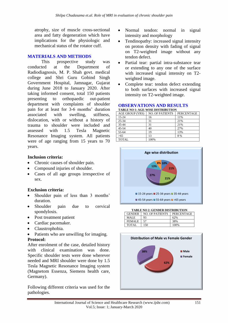
OBSERVATIONS AND RESULTS TABLE NO 1: AGE-WISE DISTRIBUTION

AGE GROUP (YRS) NO. OF PATIENTS PERCENTAGE

15-24 16 11%

25-34 31 21%

35-44 31 21%

45-54 40 27%

55-64 19 13%

>65 13 8 %

TOTAL 100%

TABLE NO 2: GENDER DISTRIBUTION

GENDER NO. OF PATIENTS PERCENTAGE

MALE 93 62%

FEMALE 57 38%

TOTAL 150 100%

10%

21%

21% 27%

13% 8%

Age wise distribution

15-24 years 25-34 years 35-44 years

45-54 years 55-64 years >65 years

62%

38%

Distribution of Male vs Female Gender

Male

Female

Shilpa Chudasama et.al. Role of MRI in evaluation of chronic shoulder pain

International Journal of Science and Healthcare Research (www.ijshr.com) 152

Vol.5; Issue: 1; January-March 2020

TABLE NO 3: AFFECTED SHOULDER SIDE

SIDE NO. OF PATIENTS PERCENTAGE

RIGHT 94 62.66%

LEFT 56 37.33%

TOTAL 150 100%

TABLE NO 4: DISTRIBUTION OF ETIOLOGIES OF CHRONIC SHOULDER PAIN

ETIOLOGIES OF CHRONIC SHOULDER PAIN FREQUENCY OF ETIOLOGIES (MORE

THAN ONE ETIOLOGY

CAN BE SEEN IN ONE PATIENT)

PERCENTAGE

OF

ETIOLOGY

AMONG THE STUDY GROUP

ROTATOR CUFF PATHOLOGIES 119 79%

ACROMIO-CLAVICULAR JOINT ARTHRITIS 75 50%

BICEPS PATHOLOGIES 48 30%

SHOULDER INSTABILITY 31 21%

GLENO-HUMERAL JOINT ARTHRITIS

(INFECTIVE/INFLAMMATORY/DEGENERATIVE) 8 5%

MISCELLANEOUS (PATHOLOGICAL FRACTURES DUE TO TUMORS,

METASTASES ETC)

13 9%

TABLE NO 5: ETIOLOGY FOR ROTATOR CUFF PATHOLOGIES

ETIOLOGY NUMBER OF CASES PERCENTAGE

NON-TRAUMATIC 78 65%

HISTORY OF TRAUMA/ DISLOCATION/INSTABILITY 42 35%

TOTAL 120 100%

65%

35%

Distribution of Right vs Left Side

Right Left

0 20

40

60

80

100

ROTATOR CUFF PATHOLOGIES

ACROMIO-CLAVICULAR JOINT ARTHRITIS

BICEPS PATHOLOGIES

SHOULDER INSTABILITY (LABRAL TEARS)

GLENO-HUMERAL JOINT ARTHRITIS (INFECTIVE/INFLAMMATORY/DEGENERATIVE)

MISCELLANEOUS (PATHOLOGICAL FRACTURES DUE TO TUMORS, METASTASES ETC)

79

50

30

21

5

9

DISTRIBUTION OF ETIOLOGIES OF CHRONIC SHOULDER PAIN

Shilpa Chudasama et.al. Role of MRI in evaluation of chronic shoulder pain

International Journal of Science and Healthcare Research (www.ijshr.com) 153

Vol.5; Issue: 1; January-March 2020

TABLE NO 6: DISTRIBUTION OF ROTATOR CUFF PATHOLOGIES [N=126]

ROTATOR CUFF

TENDON

NUMBER OF CASES (MORE THAN ONE MUSCLE TENDON

INVOLVEMENT CAN BE SEEN IN ONE PATIENT)

PERCENTAGE OF

INVOLVMENT OF

CUFF TENDON

SUPRASPINATUS 114 90%

INFRASPINATUS 18 14%

SUBSCAPULARIS 77 61%

TERES MINOR 3 2%

TABLE NO 7: INVOLVEMENT OF ROTATOR CUFF MUSCLES [N=126]

ROTATOR CUFF TENDON

INVOLVEMENT

NUMBER OF CASES

(MORE THAN ONE MUSCLE TENDON INVOLVEMENT CAN BE SEEN IN ONE PATIENT)

PERCENTAGE

PARTIAL TEAR OF SUPRASPINATUS 72 58%

PARTIAL TEAR OF SUBSCAPULARIS 64 51%

PARITAL TEAR OF INFRASPINATUS 11 9%

COMPLETE TEAR OF SUPRASPINATUS 9 7%

COMPLETE TEAR OF SUBSCAPULARIS 1 0.8%

COMPLETE TEAR OF INFRASPINATUS 1 0.8%

65%

35%

ETIOLOGY FOR ROTATOR CUFF PATHOLOGIES

NON-TRAUMATIC

HISTORY OF TRAUMA/ DISLOCATION/INSTABILITY

90

14

61

2 0 10 20 30 40 50 60 70 80 90

100

DISTRIBUTION OF ROTATOR CUFF PATHOLOGIES

No. of patients

0

10

20

30

40

50

60

70

80

PARTIAL TEAR OF

SUPRASPINATUS

PARTIAL TEAR OF

SUBSCAPULARIS

PARITAL TEAR OF

INFRASPINATUS

COMPLETE TEAR OF

SUPRASPINATUS

COMPLETE TEAR OF

SUBSCAPULARIS

COMPLETE TEAR OF

INFRASPINATUS

72

64

11 9

INVOLVEMENT OF ROTATOR CUFF MUSCLES

Shilpa Chudasama et.al. Role of MRI in evaluation of chronic shoulder pain

International Journal of Science and Healthcare Research (www.ijshr.com) 154

Vol.5; Issue: 1; January-March 2020

TABLE NO 8: TYPES OF ACROMION

TYPE FREQUENCY PERCENTAGE

TYPE 1 54 36%

TYPE 2 90 60%

TYPE 3 6 4%

TYPE 4 0 0%

TOTAL 100 100%

Figure 1 Above PD-FAT SAT MR images show articular surface partial tear (red arrows) of supraspinatus tendon.

Figure 2 Above PD-FAT SAT MR images show partial tear at myo-tendinous junction (green arrows) of subscapularis muscle.

36%

60%

4%

TYPES OF ACROMION

Type1 Type2

Type3 Type4

Shilpa Chudasama et.al. Role of MRI in evaluation of chronic shoulder pain

International Journal of Science and Healthcare Research (www.ijshr.com) 155

Vol.5; Issue: 1; January-March 2020

Figure 3 Above PD FAT SAT MR images show complete tear of supraspinatus at myo-tendinous junction (red arrow) and partial

tear of subscapularis (blue arrow) and infraspinatus (black arrow). Subchondral cystic changes are noted involving glenoid (green

arrow).

Figure 4 Above PD FAT SAT MR images show Hill-Sach’s lesion (blue arrow) and bony Bankart lesion (red arrow) as described

above.

Figure 5 Above PD FAT SAT MR images show detachment of the antero’-inferior labrum (3-6 o'clock) with medially stripped but

intact periosteum – Perthe’s injury (red arrows)

Shilpa Chudasama et.al. Role of MRI in evaluation of chronic shoulder pain

International Journal of Science and Healthcare Research (www.ijshr.com) 156

Vol.5; Issue: 1; January-March 2020

Figure 6

PD-FAT SAT MR IMAGES

Above images show significant gleno-humeral joint space narrowing (red arrows) and

cartilage loss associated with synovial hypertrophy (green arrow). Large punched out

erosions (yellow arrow) are noted involving humeral head predominantly at rotator cuff

attachment sites. Synovial hypertrophy with mild joint effusion and rice bodies are noted

(orange arrow).

Figure 7 PD-FAT SAT IMAGES

T1 FAT SAT POST CONTRAST

Shilpa Chudasama et.al. Role of MRI in evaluation of chronic shoulder pain

International Journal of Science and Healthcare Research (www.ijshr.com) 157

Vol.5; Issue: 1; January-March 2020

Above MR images show large lesion with

soft tissue component involving metaphysis

extending to involve epiphyses of head,

neck and proximal shaft of left humerus

associated with sunburst periosteal reaction.

DISCUSSION

The age distribution in our study was in

range of 15 years to 70 years with

maximum population within 45-54 year

range.

The majority of the cases in our study

were male.

Most commonly involved side was right

shoulder.

The majority of cases of chronic

shoulder pain included rotator cuff

injuries followed by biceps pathologies,

acromio-clavicular arthritis, gleno-

humeral instability, gleno-humeral

arthritis and lastly miscellaneous

conditions like tumors or cysts.

The findings of rotator cuff injuries are

as followed:

The rotator cuff tears are more

common in non-traumatic causes.

Trauma mostly only aggravates the

prior inflammatory and degenerative

tendon alterations.

Supraspinatus was the most

commonly involved tendon followed

by subscapularis, infraspinatus and

Teres minor in decreasing order of

frequency.

Among these, partial tears of the

rotator cuff were the most common

tendon abnormality where partial

tears of supraspinatus tendon were

most common.

Among partial tears, articular

surface tears were most common.

Acromio-clavicular joint showed

abnormality showed strong association

with rotator cuff injuries.

Decreased acromio-humeral distance

has been associated with rotator cuff

injuries especially supraspinatus tendon

injuries. As the distance reduces,

chances of rotator cuff injury increase.

Type I and type II acromion morphology

was most commonly observed. Rotator

cuff injuries were more commonly seen

in type II and type III acromion with

type III acromion showing strong

association with rotator cuff injuries.

Biceps tendon showed positive

association with rotator cuff injuries.

Anterior instability was most common

type of gleno-humeral instability.

Anterior instability was strongly

associated with Hill-Sach’s and

Bankart’s lesion.

Rotator cuff injuries were seen in

the 73% cases of the shoulder

instability.

CONCLUSIONS

According to our study MRI is more

sensitive than clinical tests to evaluate

chronic shoulder pain because of rotator

cuff disorders like rotator cuff

tendinopathy, partial tears, and complete

tears.

MRI being non -invasive does not

involve morbidity associated with other

tests like arthroscopy.

MRI is unique in its ability to evaluate

the labral pathologies, bone marrow,

cartilage and deeper intra-articular

pathologies.

Its advantages are: no ionizing radiation,

multi-planar imaging and

comprehensive display of soft tissue

anatomy, demonstration of causes for

impingement and also characterisation

and staging of bone tumours.

MR imaging used for soft tissue as well

as bony changes involving shoulder

region. MR is even better than CT in

detecting smaller erosions and acromio-

clavicular arthritis. Owing to the better

soft tissue resolution of MRI, it was

primarily used for detecting the rotator

cuff, labral and acromio-clavicular joint

pathologies.

Source of Support: None

Conflict of Interest: None

Shilpa Chudasama et.al. Role of MRI in evaluation of chronic shoulder pain

International Journal of Science and Healthcare Research (www.ijshr.com) 158

Vol.5; Issue: 1; January-March 2020

REFERENCES

1. Neer CS 2nd. Anterior acromioplasty for the

chronic impingement syndrome in the shoulder:

a preliminary report. J Bone Joint Surg Am

1972;54(l):41-50.

2. Umer M, Qadir I, Azam M. Subacromial

impingement syndrome. Orthop Rev (Pavia).

2012 May 9;4(2):el8.

3. Morrison DS, Biglani LU. The clinical

significance of variations in acromial

morphology. Orthop Trans. 1987;11:234.

4. Ogata S, Uhthoff HK. Acromial enthesopathy

and rotator cuff tear: a radiologic and histologic

postmortem investigation of the coracoacromial

arch. Clin Orthop Relat Res. 1990

May;(254):39-48.

5. Meister K, Thesing J, Montgomery WJ,

Indelicato PA, Walczak S, Fontenot W. MR

arthrography of partial thickness tears of the

undersurface of the rotator cuff: an arthroscopic

correlation. Skeletal Radiol. 2004 Mar;33(3):

136-41.

6. Morag Y, Jamadar DA, Miller B, Dong Q,

Jacobson JA.The subscapularis: anatomy, injury,

and imaging. Skeletal Radiol. 2011 Mar;

40(3):255-69.

7. Bencardino JT, Garcia Al, Palmer WE:

Magnetic resonance imaging of the shoulder:

Rotator cuff. Top Magn Reson Imaging 2003

Feb; 14(1):51-67.

8. Kjellin I, Ho CP, Cervilla V, Haghighi P, Kerr

R, Vangness CT et al. Alterations in

supraspinatus tendon at MR imaging :

correlation with histopathologic findings in

cadavers. Radiology. 1991 Dec;181(3):837-41.

9. Kieft GJ, Bloem JL, Rozing PM, Obermann

WR. Rotator cuff impingement syndrome: MR

imaging. Radiology. 1988 Jan;166(l Pt 1):211-4.

10. Bureau NJ, Dussault RG, Keats TE. Imaging of

the bursae of the shoulder joint. Skeletal Radiol.

1996 Aug;25(6):513-7.

11. Kuhn JE. A new classification system for

shoulder instability. Br J Sports Med. 2010

Apr;44(5):341-6.

12. WamserG, Bohndorf K, Vollert K, Bucklein W,

Schalm J. Power Doppler sonography with and

without echo-enhancing contrast agent and

contrast-enhanced MRI for the evaluation of

rheumatoid arthritis of the shoulder joint:

differentiation between synovitis and joint

effusion.

13. Maeseneer M D, Roy F V, Lenchik L,

Shahabpour M, Jacobson J, Ryu K N, et al. CT

and MR arthrography of the normal and

pathologic anterosuperior labrum and labral-

bicipital complex. Radiographics 2000 Oct;

20(1):S67-S81.

Dr. Shilpa Chudasama – Associate professor in Radiology, G.G. Hospital, M.P. Shah

Medical College, Jamnagar-368001

Dr. Daxa Khunt – 2nd year Resident Doctor in

Radiology, G.G. Hospital, M.P. Shah

Medical College, Jamnagar-368001

How to cite this article: Chudasama

S, Khunt D.

Role of MRI in evaluation of chronic shoulder

pain. International Journal of Science &

Healthcare Research. 2020; 5(1): 150-158.

******


Recommended