ROLE OF ORGANISMS OF THE PLEUROPNEUMONIA GROUP IN HUMAN GENITAL INFECTIONS*t BY C. S. NICOL and D. G. ff. EDWARD St. Bartholomew's Hospital and the London Hospital, London, and the Wellcome Research Laboratories, Beckenham, Kent The isolation of organisms of the pleuropneu- monia group from the human genital tract has aroused considerable interest in view of the attention now being paid to the aetiology of non-specific (non-gonococcal) urethritis. The first isolation of a pleuropneumonia-like organism from man was made by Dienes and Edsall (1937) from a Bartholin's abscess. Dienes and his associates (Dienes, 1940; 'Dienes and Smith, 1942; Dienes and others, 1948) later showed that organisms of the pleuropneumonia group were frequent inhabitants of both male and female genital tracts, and suggested that these organisms might cause infection. Beveridge, Campbell, and Lind (1946) isolated pleuropneu- monia-like organisms from the urethra of 20 per cent. of men with non-specific urethritis; urethral cultures from healthy men were negative. Harkness and Henderson-Begg (1948) obtained sinilar results, so that it appeared possible that pleuropneumonia- like organisms might cause non-specific urethritis. More recently, however, pleuropneumonia-like organisms were found in urethral cultures from 10-16 per cent. of men without evidence of infection (Harkness, 1950; Mel6n and Linnross, 1952). Positive isolations from the female were even more frequent; in one series of 300 cervical cultures, 26 per cent. were positive (Randall, Stein, and Ayres, 1950). The organisms were found more frequently in women with genital infection than in those with no evidence of infection; cultures from a small number of virgins were negative (Mel6n and Odeblad, 1952). The significance of organisms of the pleuro- pneumonia group in genital infection was discussed by Edward (1952), who did not consider that the evidence was sufficient for conclusions to be drawn. Although the organisms had sometimes been * Received for publication July 10, 1953. t Paper read before the M.S.S.V.D. April 24, 1953. isolated in pure culture from suppurative lesions, the frequency with which they were found in the vagina and their isolation from the urethra of healthy men suggested that they might be part of the normal flora. It was, however, possible that the organisms isolated from patients with infection belonged to a different species of the pleuropneumonia group from those isolated from healthy persons. Pleuro- pneumonia-like organisms belonging to at least three different species have been isolated from the vagina of bitches (Edward and Fitzgerald, 1951a). Few attempts have been made to find out whether the pleuropneumonia-like organisms isolated from the human genital tract belong to more than one species. In the investigations to be reported cultures from both infected and healthy persons were exaniined and the strains of pleuropneumonia-like organisms isolated were compared by biological and serqlogical methods to find out whether any particu- lar species or type of organism was associated with disease. Material and Methods The investigation was carried out on 468 patients; 336 of them, including ninety police candidates examined as controls, were seen at the Special Treatment Centre at St. Battholomew's Hospital, and 132 at the White- chapel Clinic of the London Hospital. All patients had smears and cultures to exclude gonococcal infection and an examination for Trichomonas vaginalis was made by the moist-slide method. Routine serological tests (W.R., Kahn, and G.C.F.T.) were also made. Altogether 1,155 cultures were examined for organisms of the pleuropneumonia group. Specimens were collected using sterile cotton wool swabs, which were afterwards placed in 2-ml. amounts of a horse serum broth medium in screw-cap bottles. Early in the investigation the male urethra was irrigated with the broth medium, but this practice was discontinued after a patient developed an allergic reaction at the, third time of testing. The swab method appeared to give as many positive cultures as 141 copyright. on August 25, 2019 by guest. Protected by http://sti.bmj.com/ Br J Vener Dis: first published as 10.1136/sti.29.3.141 on 1 September 1953. Downloaded from
Transcript
ROLE OF ORGANISMS OF THE PLEUROPNEUMONIAGROUP IN HUMAN GENITAL INFECTIONS*t
BY
C. S. NICOL and D. G. ff. EDWARDSt. Bartholomew's Hospital and the London Hospital, London,and the Wellcome Research Laboratories, Beckenham, Kent
The isolation of organisms of the pleuropneu-monia group from the human genital tract hasaroused considerable interest in view of the attentionnow being paid to the aetiology of non-specific(non-gonococcal) urethritis. The first isolation of apleuropneumonia-like organism from man was madeby Dienes and Edsall (1937) from a Bartholin'sabscess. Dienes and his associates (Dienes, 1940;'Dienes and Smith, 1942; Dienes and others, 1948)later showed that organisms of the pleuropneumoniagroup were frequent inhabitants of both male andfemale genital tracts, and suggested that theseorganisms might cause infection. Beveridge,Campbell, and Lind (1946) isolated pleuropneu-monia-like organisms from the urethra of 20 percent. of men with non-specific urethritis; urethralcultures from healthy men were negative. Harknessand Henderson-Begg (1948) obtained sinilar results,so that it appeared possible that pleuropneumonia-like organisms might cause non-specific urethritis.More recently, however, pleuropneumonia-likeorganisms were found in urethral cultures from10-16 per cent. of men without evidence of infection(Harkness, 1950; Mel6n and Linnross, 1952).Positive isolations from the female were even morefrequent; in one series of 300 cervical cultures, 26per cent. were positive (Randall, Stein, and Ayres,1950). The organisms were found more frequentlyin women with genital infection than in those withno evidence of infection; cultures from a smallnumber of virgins were negative (Mel6n andOdeblad, 1952).The significance of organisms of the pleuro-
pneumonia group in genital infection was discussedby Edward (1952), who did not consider that theevidence was sufficient for conclusions to be drawn.Although the organisms had sometimes been
* Received for publication July 10, 1953.t Paper read before the M.S.S.V.D. April 24, 1953.
isolated in pure culture from suppurative lesions,the frequency with which they were found in thevagina and their isolation from the urethra of healthymen suggested that they might be part of the normalflora. It was, however, possible that the organismsisolated from patients with infection belonged to adifferent species of the pleuropneumonia groupfrom those isolated from healthy persons. Pleuro-pneumonia-like organisms belonging to at leastthree different species have been isolated from thevagina of bitches (Edward and Fitzgerald, 1951a).Few attempts have been made to find out whetherthe pleuropneumonia-like organisms isolated fromthe human genital tract belong to more than onespecies. In the investigations to be reported culturesfrom both infected and healthy persons wereexaniined and the strains of pleuropneumonia-likeorganisms isolated were compared by biological andserqlogical methods to find out whether any particu-lar species or type of organism was associated withdisease.
Material and MethodsThe investigation was carried out on 468 patients;
336 of them, including ninety police candidates examinedas controls, were seen at the Special Treatment Centreat St. Battholomew's Hospital, and 132 at the White-chapel Clinic of the London Hospital. All patients hadsmears and cultures to exclude gonococcal infection andan examination for Trichomonas vaginalis was made bythe moist-slide method. Routine serological tests (W.R.,Kahn, and G.C.F.T.) were also made.
Altogether 1,155 cultures were examined for organismsof the pleuropneumonia group. Specimens were collectedusing sterile cotton wool swabs, which were afterwardsplaced in 2-ml. amounts of a horse serum broth mediumin screw-cap bottles. Early in the investigation the maleurethra was irrigated with the broth medium, but thispractice was discontinued after a patient developed anallergic reaction at the, third time of testing. The swabmethod appeared to give as many positive cultures as
141
copyright. on A
ugust 25, 2019 by guest. Protected by
http://sti.bmj.com
/B
r J Vener D
is: first published as 10.1136/sti.29.3.141 on 1 Septem
the instillation method. In the female, specimens weretaken from the cervix without first removing any vaginaldischarge; thus the swab collected both cervical andvaginal secretions.The broth samples were usually cultured within a few
hours, but could be stored satisfactorily at -20° C.;pleuropneumonia-like organisms were isolated fromsamples even after storage for several months at thistemperature. The broth samples were inoculated directly,without preliminary incubation, on to special media,made from ox heart infusion broth containing 1 per cent.added peptone with the further addition of 10 per cent.yeast extract and 20 per cent. horse serum, thepH beingadjusted to 8-0. Both a solid medium and a semi-solidmedium were employed. Thallium acetate and penicillinwere added as bacteriostatics to provide a selectivemedium (Edward, 1947); only on a few occasions werecultures overgrown by bacteria. Penicillin was onlyapplied to half of the surface of a plate. When coloniesof pleuropneumonia-like organisms were present, theyappeared on both halves of the plate, unless the penicillin-free half was overgrown with bacteria. There was there-fore no suggestion that bacteria were induced by thepenicillin to form L-phase variants, which were mistakenfor organisms of the pleuropneumonia group.The plates were examined with a dissecting microscope
using slightly oblique transmitted light; this is con-sidered to be the easiest and most reliable method foridentifying an organism of the pleuropneumonia group(Edward, 1953). From every positive culture a sub-culture was made from a single colony; each of thestrains thus isolated was examined to determine itsbiological and serological properties. The type of growthin a semi-solid medium was noted. Cultures on horseserum agar were examined for the formation of a filmand spots. The ability to grow on rabbit serum agar,the ability to ferment glucose, and the ability to producehaemolysis of horse blood agar were also tested (Edward,1950b, 1953). Reasons are given elsewhere for believingthat organisms of the pleuropneumonia group can bedistinguished with certainty from bacteria in the L-phaseby an adequate examination of their cultural appearancesand properties (Edward, 1953).The serological reactions were tested with antisera
prepared in rabbits against several of the strains. Theagglutination tests were carried out by the techniquepreviously described (Edward, 1950a), using livingsuspensions obtained by washing off the organisms fromcultures on agar plates with saline. The growth of apleuropneumonia-like organism is inhibited by incor-porating in the culture medium an antiserum preparedagainst that strain (Edward and Fitzgerald, 1953). Anantiserum may be inhibitory even at a dilution of 1 : 1,000,the presence of complement not being required. Since allstrains which were agglutinated by a particular antiserumwere also inhibited by that antiserum, a convenient testfor antigenicity was provided. A suitably dilutedsuspension of an organism was inoculated on a horseserum agar plate containing antiserum and growth wascompared with the growth on a control horse serum agarplate containing normal rabbit serum.
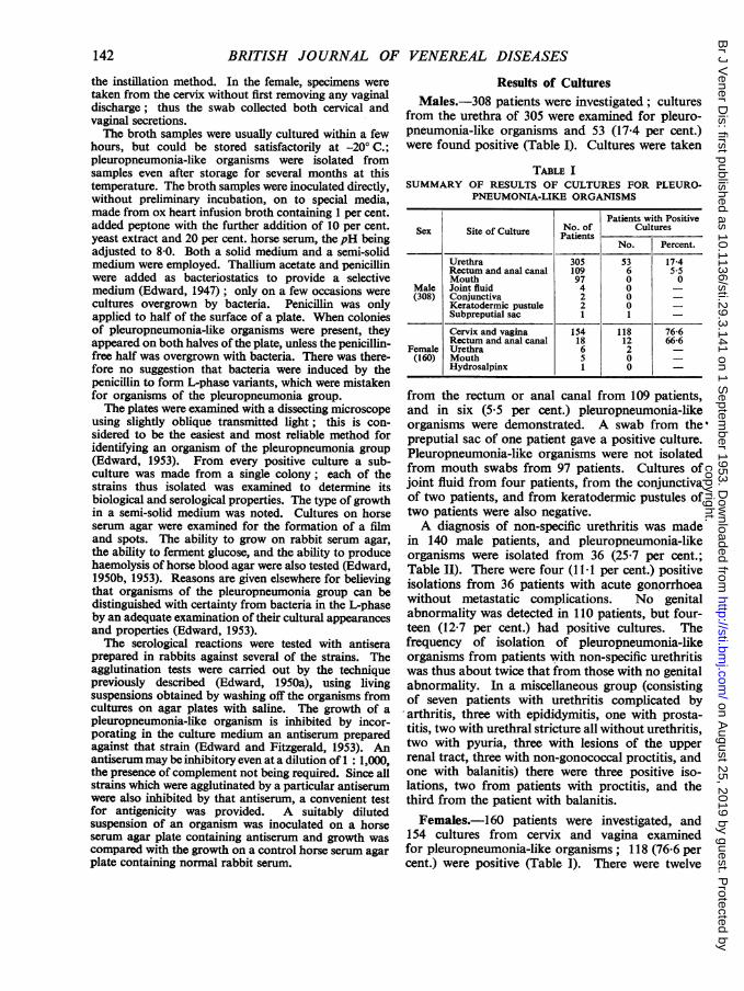
Results of CulturesMales.-308 patients were investigated; cultures
from the urethra of 305 were examined for pleuro-pneumonia-like organisms and 53 (17-4 per cent.)were found positive (Table I). Cultures were taken
TABLE ISUMMARY OF RESULTS OF CULTURES FOR PLEURO-
PNEUMONIA-LIKE ORGANISMS
Patients with PositiveSex Site of Culture Patients Cultures
Cervix and vagina 154 118 76-6Rectum and anal canal 18 12 66-6
Female Urethra 6 2 -
(160) Mouth 5 0 -Hydrosalpinx 1 0
from the rectum or anal canal from 109 patients,and in six (5.5 per cent.) pleuropneumonia-likeorganisms were demonstrated. A swab from the'preputial sac of one patient gave a positive culture.Pleuropneumonia-like organisms were not isolatedfrom mouth swabs from 97 patients. Cultures ofjoint fluid from four patients, from the conjunctivaof two patients, and from keratodermic pustules oftwo patients were also negative.A diagnosis of non-specific urethritis was made
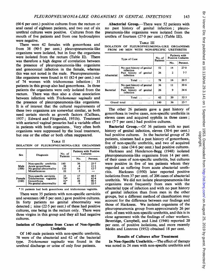
in 140 male patients, and pleuropneumonia-likeorganisms were isolated from 36 (25-7 per cent.;Table II). There were four (1I1 per cent.) positiveisolations from 36 patients with acute gonorrhoeawithout metastatic complications. No genitalabnormality was detected in 110 patients, but four-teen (12.7 per cent.) had positive cultures. Thefrequency of isolation of pleuropneumonia-likeorganisms from patients with non-specific urethritiswas thus about twice that from those with no genitalabnormality. In a miscellaneous group (consistingof seven patients with urethritis complicated byarthritis, three with epididymitis, one with prosta-titis, two with urethral stricture all without urethritis,two with pyuria, three with lesions of the upperrenal tract, three with non-gonococcal proctitis, andone with balanitis) there were three positive iso-lations, two from patients with proctitis, and thethird from the patient with balanitis.Females.-160 patients were investigated, and
154 cultures from cervix and vagina examinedfor pleuropneumonia-like organisms; 118 (76-6 percent.) were positive (Table 1). There were twelve
142
copyright. on A
ugust 25, 2019 by guest. Protected by
http://sti.bmj.com
/B
r J Vener D
is: first published as 10.1136/sti.29.3.141 on 1 Septem
PLEUROPNEUMONIA-LIKE ORGANISMS IN GENITAL INFECTIONS
(66-6 per cent.) positive cultures from the rectum or
anal canal of eighteen patients, and two out of sixurethral cultures were positive. Cultures from themouth of five patients and from one hydrosalpinxwere negative.There were 42 females with gonorrhoea and
from 38 (905 per cent.) pleuropneumonia-likeorganisms were isolated, but in four the organismswere isolated from the rectum (Table II). Therewas therefore a high degree of correlation betweenthe presence of pleuropneumonia-like organismsand gonococcal infection in the female, whereasthis was not noted in the male. Pleuropneumonia-like organisms were found in 61 (82-4 per cent.) outof 74 women with trichomonas infection; 31patients in this group also had gonorrhoea. In threepatients the organisms were only isolated from therectum. There was thus also a close associationbetween infection with Trichomonas vaginalis andthe presence of pleuropneumonia-like organisms.It is of interest that the cultural requirements ofthese two organisms are somewhat similar, as bothneed certain sterols as growth factors (Cailleau,1937; Edward and Fitzgerald, 1951b). Treatmentwith acetarsol vaginal pessaries had a variable effectin patients with both organisms. Very often bothorganisms were suppressed by the local treatment,but one or the other or both often reappeared.
TABLE IIISOLATION OF PLEUROPNEUMONIA-LIKE ORGANISMS
Patients with PositiveSex Diagnosis No. of Cultures
* 31 patients had both gonorrhoea and trichomonas vaginitis.
There were 35 patients with non-specific cervicitisand seventeen (48 5 per cent.) gave positive cultures.In forty patients no genital abnormality wasdetected; nine (22 5 per cent.) of these had positivecultures, one being in the rectum only. There werethree virgins in this group and they all had negativecultures.
Isolation of Organisms from Cases of Non-SpecificUrethritis
Of 140 male patients with non-specific urethritis,78 were of the abacterial and 62 of the bacterialtype. Trichomonas vaginalis was found in theurethral discharge or urine of only four patients.
Abacterial Group.-There were 52 patients withno past history of genital infection; pleuro-pneumonia-like organisms were isolated from theurethra of fourteen (27-0 per cent.) (Table III).
TABLE IIIISOLATION OF PLEUROPNEUMONIA-LIKE ORGANISMSFROM 140 MEN WITH NON-SPECIFIC URETHRITIS
Patients withType of Case No. of Positive CulturesTypeofCase ~Patients
No. Percent.
No past history of genital 52 14 27-0infection
Past history of genital 26 2 7-7Abacterial infection
Total .. 78 16 20-5
No past history of genital 36 11 30-6infection
Bacterial Past history of genital 26 9 34-6infection
Total 62 20 32-3
Grand total .140 36 25-7
The other 26 patients gave a past history ofgonorrhoea in twelve cases, non-specific urethritis ineleven cases and acquired syphilis in three cases;two (7-7 per cent.) had positive cultures.
Bacterial Group.-Of 36 patients with no pasthistory of genital infection, eleven (30-6 per cent.)had positive cultures. In the bacterial group of 26patients, nineteen had a past history of gonorrhoea,five of non-specific urethritis, and two of acquiredsyphilis; nine (34-6 per cent.) had positive cultures.
Harkness and Henderson-Begg (1948) isolatedpleuropneumonia-like organisms from 14 per cent.of their cases of non-specific urethritis, but cultureswere positive in five of ten patients whom theyregarded as suffering from acute abacterial ureth-ritis. Harkness (1950) later reported positiveisolations from 37 per cent. of 206 cases of abacterialurethritis. We did not isolate pleuropneumonia-likeorganisms more frequently from men with theabacterial type of infection and with no past historyof genital infection than from men in the othergroups, but a different method of classification mayaccount for the difference between our findings andthose of Harkness. We isolated organisms of thepleuropneumonia group from approximately 26 percent. ofmen with non-specific urethritis, and this is inclose agreement with the findings of other workers.Beveridge, Campbell, and Lind (1946) reported 20per cent. of positive isolations, and more recentlyMelen and Linnross (1952) obtained 18 per cent.
Results of Cultures after TreatmentIn Non-Specific Urethritis.-The effect of therapy
was noted in 24 men with non-specific urethritis and
143
copyright. on A
ugust 25, 2019 by guest. Protected by
http://sti.bmj.com
/B
r J Vener D
is: first published as 10.1136/sti.29.3.141 on 1 Septem
with positive cultures for pleuropneumonia-likeorganisms. Streptomycin calcium chloride (1g. intra-muscularly) was administered daily for five days.There were fourteen patients whose immediate clinicalresponse to treatment was satisfactory, but culturesfor pleuropneumonia-like organisms remainedpositive. In five patients the clinical response totreatment was satisfactory and the cultures becamenegative. One patient who relapsed after treatmentstill had a positive culture, but in four others whorelapsed pleuropneumonia-like organisms were notfound in the urethra after treatment.
Treatment with aureomycin (aureomycin hydro-chloride, 500 mg. -8-hrly for 5 days) was given notless than 2 weeks later to twelve patients who hadpositive cultures in spite of clinical recovery afterstreptomycin therapy. All patients passed tests forcure 3 months after treatment, but in two of themcultures for pleuropneumonia-like organisms re-
mained persistently positive.
In Consorts.-Pleuropneumonia-like organismswere isolated from the consorts of six men with posi-tive cultures and non-specific urethritis. The womenwere given the same antibiotic therapy as the malepatients and its effect on the persistence of theorganisms was noted. After streptomycin in fourpairs of consorts, cultures from the male becamenegative while those in the female remained positive;one of the males had a clinical relapse (Table IV).In one pair the female became negative and themale remained positive. Both partners in the sixthpair remained positive and were subsequentlytreated with aureomycin. Cultures in the woman
then became negative, but the man remainedpositive, although he had no clinical relapse.Antibiotic therapy was thus not equally effective inboth partners in eliminating the organisms. In vitrotests for sensitivity to the drugs were not carried outto determine whether the strains isolated from'thetwo partners differed in sensitivity.
In Cases of Clinical Relapse.-The courseof disease and response to treatment were ob-served in men'with non-specific urethritis and nohistory of genital infection. They were treated withstreptomycin and final tests for cure were completednot less than 3 months later. Excluding patients whodefaulted before the final tests were complete, therewere 44 patients with the abacterial type of infection;twelve of them originally had positive cultures forpleuropneumonia-like organisms and two had aclinical relapse. Among the 32 patients with negativecultures, there were nine who relapsed. In the groupwith the bacterial type of infection not one of eightpatients with positive cultures relapsed, but there werethree who relapsed among nineteen patients withnegativecultures. Thus clinical relapseoccurred rathermore frequently among menwho originally had nega-tive cultures than among those with positive cultures.The presence of organisms of the pleuropneumoniagroup in the genital tract did not appear to affectthe course of the disease or to increase the risk ofcomplications or relapse. It was also noted thatamong 19 patients treated for gonorrhoea withpenicillin, three had non-gonococcal relapses,although pleuropneumonia-like organisms were notdemonstrated in the urethra at any time.
TABLE IVRESPONSE TO TREATMENT IN PAIRS OF CONSORTS
Pairs Culture Treatment Culture Treatment Culture Final Test of Cure
IMale ..Positive S Negative - - SatisfactoryFemale .. .. JS Positive A Negative Satisfactory
fMale .. .. S Negative - - Satisfactory2 ~~~~~~~Positive _ _ _ _ _Female .. .. J S Positive A Negative Satisfactory
{ Male .. PositiveS Negative, relapse A Negative Satisfactory
Female .. . . S Positive Defaulted -
Male .. Positive S Positive A Negative SatisfactoryFemale .. .. S Negative - 1 _ Satisfactory
{
Male.. Positive
S Positive A Positive SatisfactoryFemale ... J S Positive A Negative Satisfactory
6{ Male .... S Negative _ SatisfactoryPositive Positive_________________________ Negative___-_______________Female .. .. f S Positive A Negative Satisfactory
Positive = Cultures for pleuropneumonia-like organisms positive. S = Streptomycin.Negative = Cultures for pleuropneumonia-like organisms negative. A Aureomycin.
144
copyright. on A
ugust 25, 2019 by guest. Protected by
http://sti.bmj.com
/B
r J Vener D
is: first published as 10.1136/sti.29.3.141 on 1 Septem
No genital abnormality Positive 4 3 INegative 4 2 2
Total Positive 19 16 4Negative 49 18 31
Cultures were examined from twenty contacts ofnineteen men who had positive cultures; sixteen(80 per cent.) of the women were also positive.Each of 49 men with negative cultures brought inone contact and eighteen (36-7 per cent.) of thewomen had positive cultures. Thus positive cultureswere more ftequent in the contacts of men who werethemselves positive than in the contacts of men withnegative cultures, but there were many instanceswhere the cultural findings in the two partnersdiffered. Pleuropneumonia-like organisms were notisolated from several female contacts of men withpositive cultures, and there were even more menwho had negative cultures although the femalecontact was positive.
In Patients with Arthritis.-Three males andone female suffering from arthritis associatedwith gonococcal infection and four males witharthritis and non-specific urethritis were observed.Pleuropneumonia-like organisms were demonstratedin the genital tract of the female, but could not befound in the urethra of the five men examined.The joint fluid from four patients was sterile onculture. Pleuropneumonia-like organisms were notisolated from lesions of keratodermia blennorrhagicain two patients or from the conjunctiva of twopatients.From the Anal Canal of Females.-Pleuro-
pneumonia-like organisms were isolated from therectum and anal canal of twelve our of eighteenwomen examined. All except two of the women hadclinical proctitis. Cultures were examined from bothcervix and anal canal from eight patients, and thecervical cultures were positive in addition to the
anal cultures in all except one. Nine patients hadgonorrhoea and six of these also had trichomonasinfection. One of three patients without evidence ofgonorrhoea had trichomonas infection. Observationof a larger group of women, including virgins andwomen with no evidence of genital or rectal infection,would be necessary to determine the frequency withwhich pleuropneumonia-like organisms inhabit theanal canal in the female. It is not certain whetherthe organisms reach the anal canal from the vaginaor whether the reverse commonly happens. From afew patients swabs were taken both from the analcanal and from the rectum using a proctoscope.The anal swab gave at least as--many positives as therectal swab, and could therefore be used in theexamination of a larger group of women.
In a Control Group of Healthy Males.-Organismsof the pleuropneumonia group were isolated fromfour out of twenty men who on attendance at theclinic showed no evidence of genital infection. Inorder to extend the findings in healthy males,cultures were made from urethra and anal canalof ninety police candidates who attended forroutine serological tests and were found to haveno genital abnormality. There were 31 marriedmen and 31 single men who admitted having hadsexual intercourse; a further 28 men denied havinghad sexual intercourse at any time. Pleuro-pneumonia-like organisms were isolated from theurethra of seven (11-3 per cent.) men with sexualexperience; two also had positive anal cultures.There were three (10-7 per cent.) positive urethralisolations from men who denied intercourse,although it is of course impossible to be sure of thetruth of their denial. There were therefore 11 1 percent. positive isolations from the urethra in thisgroup of apparently healthy men and two (2-3 percent.) positive isolations from 88 anal cultures.The cultures were repeated and confirmed to be
positive in seven candidates. In three the organismaswere still present in the urethra 3 months later, inanother 6 weeks later, and in three others 4 weekslater. Two men gave a past history of non-specificurethritis and one was found at a later examinationto have developed signs of a recurrence of thisinfection. The female contacts of four candidateswere examined, but from only one of these werepleuropneumonia-like organisms isolated, althoughanother woman gave a past history of vaginitistreated with pessaries.
Serological Examination of StrainsAntisera were prepared against five- strains;
three had been isolated in the course of this investi-
145
copyright. on A
ugust 25, 2019 by guest. Protected by
http://sti.bmj.com
/B
r J Vener D
is: first published as 10.1136/sti.29.3.141 on 1 Septem
gation, one strain in France, and one in America.In cross-agglutination tests one strain, " CampoL ", isolated in America, was agglutinated by itsown antiserum but not by any of the other antisera,and the antiserum against "Campo L" onlyagglutinated the homologous organism (Table VI).
TABLE VIRESULTS OF CROSS-AGGLUTINATION TESTS
Antisera
Antigen Strain Strain Strain Strain StrainH.23 H.26 H.50 D.419 Campo
Complement-fixation tests and inhibition ofgrowth tests confirmed that " Campo L" differedantigenically from the other four strains. StrainH 23was not agglutinated by the heterologous antisera,but the H 23 antiserum agglutinated strain H 26 tofull titre. Strain H 26 itself was agglutinated byantisera against strains H 50 and D 419. The fourstrains, H 23, H 26, H 50, and D 419, thus sharecommon antigens, although differences exist be-tween them in their antigenic constitution. Thesefour strains will be regarded as belonging to onespecies of the pleuropneumonia group and will beprovisionally called Human Type 1 strains.The serological reactions of 91 strains isolated in
the course of this investigation were tested against
TABLE VIISEROLOGICAL IDENTIFICATION OF STRAINS ISOLATED
StrainsIdentified as
Human Type 1Source of Strains Total - Other
Strains By By StrainsAgglu- Inhibi-tiain tiontiasn Test
Females (cervix, vagina, or 39 19A 20) 0urethra)
Males with non-specific 31 18 13 0urethritis (urethra)
these antisera. Strains from both male and femalegenital tracts, and from both healthy persons andfrom persons with genital infection, including maleswith non-specific urethritis, were included in thisseries; there were also eight strains isolated fromthe anal canal (Table VII). Altogether 54 strainswere tested for agglutination, and were agglutinatedto a significant titre by at least one of the Type 1antisera; the identification of many of these strainsas Type 1 was confirmed by the inhibition of growthtest. An additional 36 strains, tested only forinhibition of growth, were inhibited by the anti-serum against strain H 50 (or by that against strainD 419). Thus, all but one of the 91 strains wereserologically related to each other and were identi-fied as Type 1. The one exception, strain H 106,will be described later; it had been isolated from apolice candidate, who had acquired non-specificurethritis 3 years previously in Malaya, althoughthere was no evidence of disease at the time ofexamination.The strains, identified serologically as Type 1,
differed from each other in the titres to which theywere agglutinated by the four antisera; somestrains gave the highest titre with one antiserum andsome with another. These differences were notrelated to the source of the strains. There was noconstant difference between strains isolated frompatients with infection and strains from apparentlyhealthy persons; strains isolated from the analcanal did not differ from the genital strains.A number of additional strains which had been
isolated by other workers were available for study.Five strains, isolated by Stokes (1953) in London,were identified as Type 1 serologically; one ofthese strains had been isolated from the blood of awoman with puerperal pyrexia (Table VIII). Twostrains, isolated by Durel (1953) in Paris, were alsofound to be Type 1. Another five strains which hadbeen isolated in the U.S.A. were received fromMorton (1953). Two of these were identified asType 1. A third strain, " Campo L ", was sero-
TABLE VIIIIDENTIFICATION OF ALL STRAINS EXAMINED
IdentificationSources of Total No.
Strains Examined Human OtherType 1 Types
Isolated by Nicol and 91 90 1 (strainEdward (London) H.106)
Received from E. J. Stokes 5 5 0(London) (including onestrain from blood)
Received from P. Durel 2 2 0(Paris)
Received from H. E. 5 2 3 (Type 2)Morton (U.S.A.)
Received from M. Ruiter 1 0 1 (Type 3)(Holland)
146
copyright. on A
ugust 25, 2019 by guest. Protected by
http://sti.bmj.com
/B
r J Vener D
is: first published as 10.1136/sti.29.3.141 on 1 Septem
PLEUROPNEUMONIA-LIKE ORGANISMS IN GENITAL INFECTIONS
logically different from the Type 1 strains, and theremaining two strains were serologically similar toit. These last three strains are regarded as belongingto a species of the pleuropneumonia group differentfrom the Type 1 strains and will provisionally becalled Human Type 2 strains. Strain " Campo L"was originally isolated by Dienes (1953), whoshowed that it was serologically similar to each offive other genital strains isolated by him in 1939-40.The five strains, examined by Norman, Saslaw, andKuhn (1950) and shown to be serologically identicalwith each other, included strain " Campo L ".Thus the strains which had previously been studiedserologically were Type 2 strains, all of which hadbeen isolated in America.
Ruiter and Wentholt (1952) isolated pleuro-pneumonia-like organisms, which appeared todiffer from the type usually isolated from the genitaltract, from four patients with ulcerative balanitis;they called them " G " strains. One of these strains,examined by us, was serologically different from allthe other strains and will be called a Human Type 3strain.
Biological Properties of Species of the Pleuro-pneumonia Group of Organisms isolated from the
Human Genital TractThe various species of the pleuropneumonia
group of organisms not only differ from each otherserologically, but often have distinctive cultural andbiochemical properties which assist their identifi-cation (Edward, 1950b, 1953) Strains of each of thespecies isolated from man were therefore examinedto determine their properties. The Type 1 strainswhen grown on horse serum agar did not form thefilm and spots which are noticeable in cultures ofsome species of pleuropneumonia-like organisms(Edward, 1950a). The strains grew as well on mediaenriched with rabbit serum as they did on horseserum agar (Table IX). They grew throughout asemi-solid medium, usually producing a markedly
granular growth. They produced little or nohaemolysis of horse blood agar and did not fermentglucose.The Type 2 strains had similar properties, but
differed from the Type 1 strains in their pathogeni-city for mice. Cultures of each of the three Type 2strains in semi-solid media (i.e. cultures containinga small proportion of agar) produced local abscessesafter subcutaneous inoculation, even though thestrains had been through a large number of sub-cultures over several years. Similar cultures of fiveType 1 strains did not affect mice after subcutaneousinoculation.When the Type 3 strain (" G " strain of Ruiter
and Wentholt, 1952, 1953) was grown on horseserum agar, it produced a film and spots similar tothose produced by certain other organisms of thepleuropneumonia group. In a semi-solid mediumthe strain grew best near the bottom where theoxygen tension was lowest; the growth was smooth.The strain fermented glucose and a number of othercarbohydrates. Ruiter and Wentholt (1952) pro-duced local abscesses in mice by injecting thisstrain into the foot pads, but the strain had lost thisproperty when re-examined in this laboratory.Growth of the strain was improved by adding 20[±g/ml. thymic nucleic acid to the medium.
Strain H 106, the only strain which was notType 1 to be isolated during the investigation,resembled -the Type 3 strain in fermenting the samecarbohydrates and in forming a film and spots. Itwas not, however, agglutinated by an antiserumprepared against the Type 3 strain, and its growthwas not inhibited by that antiserum. Since only asingle strain was studied, it was not possible todetermine whether strain H 106 belonged to aseparate species or whether it was a Type 3 strain.
Comparison of the Genital Strains with Strainsisolated from the Mouth.-Since organisms of thepleuropneumonia group have been found in the
TABLE IXPROPERTIES OF SPECIES OF PLEUROPNEUMONIA-LIKE ORGANISMS ISOLATED FROM MAN
Species Formation Growth on Growth in Granular or Haemolysis Fermenta- Pathogenicity(orSrain) of Film Rabbit Semi-solid Smoh of Horse tion of Patogenicet(or Strain) and Spots Serum Agar Medium Smooth Blood Agar GlucoseMice
Human Type 1* - Good Throughout Usually granular (or trace) None
Human Type 2 - Good Throughout Usually granular - - Abscesses after s/c inoc.
Human Type 3t$ + Good At bottom only Smooth + Abscesses after s/c inoc.
Strain H.106t + Good At bottom only Indefinite _ + Not tested
Human Type 4tf + or- Very good At bottom only Smooth Not tested
* Some strains grew better anaerobically.t Grew better anaerobically.t Growth improved by adding thymic nucleic acid to the medium.
147
copyright. on A
ugust 25, 2019 by guest. Protected by
http://sti.bmj.com
/B
r J Vener D
is: first published as 10.1136/sti.29.3.141 on 1 Septem
mouth (Smith and Morton, 1951 ; Dienes andMadoff, 1953), it was necesasry to compare strainsfrom the mouth with genital strains. Saliva,collected from healthy persons immediately afterrising in the morning, was cultivated aerobically andanaerobically. All the aerobic cultures were nega-tive, but colonies of pleuropneumonia-like organismsappeared on plates incubated anaerobically, pro-vided that the medium contained 20 Fg/ml. addedthymic nucleic acid. Growth was better if rabbitserum was used for enrichment in place of horseserum. As already noted, pleuropneumonia-likeorganisms were not isolated from mouth swabs, eventhough the cultures were incubated anaerobically onmedia containing thymic nucleic acid. The Americanworkers isolated their strains under aerobic condi-tions on media to which nucleic acid had not beenadded (Smith and Morton, 1951; Dienes andMadoff, 1953). It is not clear why we obtaineddifferent results. No serological or other comparisonhas yet been made between our strains and theAmerican strains.
Five strains isolated from saliva were examined.Three of them formed a film and spots on horseserum agar; the other two did not. All the strainsgrew best near the bottom of semi-solid mediaproducing a smooth growth. They did not fermentglucose. An antiserum, prepared in a rabbitagainst one strain, inhibited growth of the homo-logous strain and of each of the other strains.Agglutination tests were unsatisfactory because ofthe poor agglutinability of some of the strains. Thestrains from saliva were antigenically different fromthe genital strains, there being no cross-reactionswith genital types in agglutination and inhibition ofgrowth tests. These strains will therefore be pro-visionally called Human Type 4.
Effect of Anaerobiosis and of an Atmosphere of 10per cent. CO2 upon Growth.-It has been reportedthat some strains of pleuropneumonia-like organismscould be isolated from the genital tract and genitalorgans only if anaerobic methods of cultivationwere employed (Dienes and others, 1948; Ruiterand Wentholt, 1952). On a number of occasions inthis investigation, colonies of pleuropneumonia-likeorganisms appeared on plates incubated anaero-bically, whereas corresponding plates incubatedaerobically were negative. Unfortunately duplicateaerobic and anaerobic cultivation was not carriedout throughout the investigation, but in a series of35 urethral cultures from cases of non-specificurethritis, the use of both methods of cultivationdid not give a higher proportion of positive isolationsthan in the whole series. There was therefore no
evidence that, if anaerobic cultivation had been usedthroughout, there would have been a significantlygreater number of positive isolations.The need of certain Type 1 strains for anaerobiosis
appears to depend upon conditions in the medium,particularly upon pH. When a urethral samplecontaining large numbers of Type 1 pleuropneu-monia-like organisms was inoculated on media atpH 8, there was growth on a plate incubated anaero-bically and on a plate incubated in an atmosphere of10 per cent. CO2, whereas there was no growth on aplate incubated aerobically (Table X). The samesample was inoculated on to media of similar con-stitution adjusted to pH values of 7, 6*5, and 6, andthe cultures were incubated aerobically. There wasgrowth on the media at pH 6 5 and 6, but not onthe medium atpH 7. Thus this strain was capable ofgrowing aerobically in the particular culture mediumprovided the conditions were acid, whereas in amore alkaline medium anaerobiosis was necessary.
The Type 3 strain grew well on the medium atpH 8 incubated anaerobically, there being nogrowth on the same medium incubated aerobically.Growth was only moderately good on the samemedium incubated in an atmosphere of 10 per cent.CO2. There was also moderately good growth onmedia adjusted topH 6 5 and 6 and incubated aero-bically. Reducing the pH of the medium did notpermit satisfactory growth under aerobic conditionsof a Type 4 strain which had been isolated fromsaliva by anaerobic cultivation.
It is concluded that cultures from the humangenital tract should be incubated both aerobicallyand anaerobically to ensure growth of all strains ofpleuropneumonia-like organisms. Cultivation in anatmosphere of 10 per cent. CO2 could be used as analternative to anaerobiosis, although Morton,Smith, and Leberman (1951) noted that an atmo-sphere of CO2 suppressed growth of some strains ontheir media.
DiscussionOrganisms of the pleuropneumonia group were
isolated from 26 per cent. of cases of non-specific
148
copyright. on A
ugust 25, 2019 by guest. Protected by
http://sti.bmj.com
/B
r J Vener D
is: first published as 10.1136/sti.29.3.141 on 1 Septem
PLEUROPNEUMONIA-LIKE ORGANISMS IN GENITAL INFECTIONS
urethritis. This percentage of positive isolations wasat least as high as the percentages reported by otherworkers. Although the pleuropneumonia-likeorganisms which were isolated grew well andeasily on the media employed, it is possible thatsometimes cultures failed to reveal pleuropneumonia-like organisms when they were present in theurethra. It is however unlikely that the culturalmethods failed in 75 per cent. of patients. In three-quarters of all cases of non-specific urethritis,therefore, organisms of the pleuropneumonia groupwere not found and could not be incriminated asthe cause of the infection. Clinically no differencewas noted between the cases with positive culturesand those with negative cultures. The course of thedisease and the response to treatment did notappear to be affected by the cultural findings. Itwould thus appear that a cause for non-specificurethritis must be sought other than pleuropneu-monia-like organisms of the types already isolated.
Pleuropneumonia-like organisms, which had thesame serological and biological properties as strainsisolated from men with non-specific urethritis, wereisolated from 11 per cent. of a group of policecandidates, who had no evidence of genital infection.These organisms were regarded as belonging to onespecies of the pleuropneumonia group and werecalled Human Type 1 strains. All except one of 91strains isolated in the course of the investigationwere identified as Type 1. The presence in theurethra of organisms of Type 1 did not appear toincrease the liability of patients with non-specificurethritis to complications. Relapses occurred inpatients with negative cultures. Ruiter and Wentholt(1953) investigated the pathogenicity of a strain ofpleuropneumonia-like organism isolated from apatient with non-specific urethritis by inoculating aculture into the urethra of two male volunteers.The inoculation was without effect. If the patho-genicity of Type 1 pleuropneumonia-like organismsis considered in the light of these separate findings,it would seem probable that the organisms do notplay a significant role in non-specific urethritis,although the possibility cannot be excluded thatthey may produce suppurative lesions in the genitaltract under certain circumstances.Thus the evidence suggests that organisms of
Type 1 are commensals. Both in males and femalesthe organisms are found more frequently wherethere is evidence of inflammation. Possibly per-sistence in the genital tract is favoured by inflam-matory conditions, but the organisms persisted inthe apparently healthy urethra of three males for atleast 3 months. The organisms probably reach themale urethra during sexual intercourse, at least in
the majority of instances, but a non-venerealmethod of spread may possibly occur in the female.Type 1 organisms were sometimes found in theanal canal. In most instances cultures from bothanal canal and genital tract were positive so that itis impossible to establish where the organisms residedfirst, but spread from the anus to the vagina wouldseem a possibility.
Several species of the pleuropneumonia group oforganisms have a tendency to localize in joints, andso particular attention has been paid to pleuro-pneumonia-like organisms in considering the aetio-logy of arthritis, when it occurs as a complication ofurethritis. Pleuropneumonia-like organisms havebeen isolated directly from joint fluids on a fewoccasions (Dienes and others, 1948; Kuzell andMankle, 1950), but in many other attempts thecultures have been negative. We failed to isolatethese organisms from the urethral tract, joint fluid,and other lesions of several cases of gonococcal andnon-specific urethritis. It is difficult to assess thesignificance of the few positive isolations madefrom joint fluid, because pleuropneumonia-likeorganisms are known to be capable of lying latentinside the body, for instance in the brains of healthymice (Sabin, 1941). All the successful isolationsfrom joint fluid were made in the U.S.A. It is to benoted that strains of a different species of thepleuropneumonia group have been isolated inAmerica from the genital tract. The Type 2 strainswere pathogenic for mice, but pathogenicity formice does not necessarily indicate pathogenicity forman. Further work will be required to determinethe significance of the Type 2 strains.
SummaryOrganisms of the pleuropneumonia group were
isolated from the urethra of 26 per cent. of 140 menwith non-specific urethritis. Relapses occurredduring treatment in men with negative cultures.Similar organisms were found in the urethra of 11per cent. of ninety men with no genital abnormality,there being three positive isolations from men whodenied sexual intercourse. Cultures from the cervixand vagina of 154 women showed that 77 per cent.were positive, the proportion positive being evengreater in women with gonorrhoea or trichomonasinfection. Pleuropneumonia-like organisms werealso isolated from the anal canal of males andfemales, both in association with proctitis and inthe absence of signs of infection.
All except one of 91 strains were shown by theirserological and biological properties to belong toone species of the pleuropneumonia group of
149
copyright. on A
ugust 25, 2019 by guest. Protected by
http://sti.bmj.com
/B
r J Vener D
is: first published as 10.1136/sti.29.3.141 on 1 Septem
organisms; they were called Human Type 1 strains.Strains from healthy persons did not differ fromstrains isolated from patients with infection;genital strains were similar to anal strains. A fewstrains isolated in America from the genital tract,one strain isolated in Holland from an ulceratedglans penis, and strains isolated from saliva,belonged to three other different species of thepleuropneumonia group.
Type 1 pleuropneumonia-like organisms cannotbe incriminated in the aetiology of 75 per cent. ofcases of non-specific urethritis, and another causefor this infection must therefore be sought. Thepleuropneumonia-like organisms appear to becommensals and no evidence was obtained that theyplayed a significant role in genital infection, althoughthe possibility that they may produce suppurativelesions under certain circumstances cannot beexcluded.
Our thanks are due to Mr. A. J. King, SeniorPhysician, and to other members of the medical andnursing staff of the Whitechapel Clinic of the LondonHospital, and to the medical and nursing st-aff of theSpecial Treatment Centre of St. Bartholomew's Hospital,for their help in collecting the clinical material, and toDr. D. R. Crabb for allowing us to take tests fromPolice candidates. We also wish to thank Prof. M.Ruiter, Prof. H. E. Morton, Dr. L. Dienes, and Dr. E. J.Stokes for supplying us with cultures.
REFERENCESBeveridge, W. I. B., Campbell, A. D., and Lind, P. E. (1946). Med.
J. Aust., 1, 179.Cailieau, R. (1937). Ann. Inst. Pasteur, 59, 293.Dienes, L. (1940). Proc. Soc. exp. Biol., N.Y., 44, 468.-(1953). Personal communication.-, and Edsall, G. (1937). Proc. Soc. exp. Biol., N. Y., 36, 740.-, and Smith, W. E. (1942). Ibid., 50, 99.
, and Madoff, S. (1953). Ibid., 82, 36.-, Ropes, M. W., Smith, W. E., Madoff, S., and Bauer, W. (1948).
New Engl. J. Med., 238, 509, 563.Durel, P. (1953). Personal communication.Edward, D. G. ff. (1947). J. gen. Microbiol., 1, 238.
(1950a). Ibid., 4, 4.-(1950b). Ibid., 4, 311.-(1952). British Journal of Venereal Diseases, 28, 89.-(1953). J. gen. Microbiol., 8, (In the press).
, and Fitzgerald, W. A. (1951a). Ibid., 5, 566.-, -(1951b). Ibid., 5, 576.
, -(1953). J. Path. Bact., 65, (In the press).Harkness, A. H. (1950). " Non-gonococcal Urethritis ". Livingstone,
Edinburgh.-, and Henderson-Begg, A. (1948). British Journal of Venereal
Diseases, 24, 50.Kuzell, W. C., and Mankle, E. A. (1950). Proc. Soc. exp. Biol.,
N. Y., 74, 677.Melen, B., and Linnross, B. (1952). Acta derm. venereol., Stockh.,
32, 77.-, and Odeblad, E. (1952). Ibid., 32, 74.Morton, H. E. (1953). Personal communication.-, Smith, P. F., and Leberman, P. R. (1951). Amer. J. Syph.,
35, 14.Norman, M. C., Saslaw, S., and Kuhn, L. R. (1950). Proc. Soc.
exp. Biol., N.Y., 75, 718.Randall, J. H., Stein, R. J., and Ayres, J. C. (1950). Amer. J.
Obstet. Gynec., 59, 404.Ruiter, M., and Wentholt, H. M. M. (1952). J. invest. Dermat.,