Biochemistry 1988, 27, 7671-7677 767 1 Eds.) pp 279-324, Academic, New York. Galante, Y. M., Lee, Y., & Hatefi, Y. (1980) J. Biol. Chem. Kobashi, K. (1968) Biochim. Biophys. Acta 158, 239-245. Laemmli, U. K. (1970) Nature (London) 227, 680-685. Lee, C.-P., Simard-Dequesne, N., Ernster, L., & Hoberman, H. D. (1965) Biochim. Biophys. Acta 105, 397-409. Low, H., & Vallin, I. (1963) Biochim. Biophys. Acta 69, Mitchell, P., & Moyle, J. (1965) Nature (London) 208, Moyle, J., & Mitchell, P. (1973) Biochem. J. 132, 571-585. O'Neal, S. G., & Fisher, R. R. (1977) J. Biol. Chem. 252, Persson, B., & Rydstrom, J. (1987) Biochem. Biophys. Res. Robillard, G. T., & Konings, W. N. (1982) Eur. J. Biochem. 255, 9641-9646. 361-374. 1205-1 206. 4552-4556. Commun. 142, 573-578. 125, 597-604. Rydstrom, J. (1981) in Mitochondria and Microsomes (Lee, C. P., Schatz, G., & Dallner, G., Eds.) pp 317-335, Ad- dison-Wesley, Reading, MA. Sanadi, D. R., Pharo, R. L., & Sordahl, L. A. (1967) Methods Enzymol. 10, 297-302. Sedmak, J. J., & Grossberg, S. E. (1977) Anal. Biochem. 79, Skulachev, V. P. (1974) Ann. N.Y. Acad. Sci. 227, 188-202. Wu, L. N. Y., & Fisher, R. R. (1983) J. Biol. Chem. 258, Wu, L. N. Y., Pennington, R. M., Everett, T. D., & Fisher, Wu, L. N. Y., Lubin, I. M., & Fisher, R. R. (1985) J. Biol. Wu, L. N. Y., Alberta, J. A,, & Fisher, R. R. (1986) Methods Yamaguchi, M., Hatefi, Y., Trach, K., & Hoch, J. A. (1988) 544-552. 7 847-78 5 1 R. R. (1981) J. Biol. Chem. 257, 4052-4055. Chem. 260, 6361-6366. Enzymol. 126, 353-360. J. Biol. Chem. 263, 2761-2767. Role of Peptide Conformation in the Rate and Mechanism of Deamidation of Asparaginyl Residues? Richard Lural and Verne Schirch* Received March 28, 1988; Revised Manuscript Received June 15, 1988 Department of Biochemistry, Virginia Commonwealth University, Richmond, Virginia 23298 ABSTRACT: The tetrapeptides Val-Asn-Gly-Ala and N-acetyl-Val-Asn-Gly-Ala undergo deamidation of the asparaginyl residue at pH 7.0 at similar rates. However, they form different products. The N-acetyl peptide gave a 3: 1 ratio of N-acetyl-Val-isoAsp-Gly-Ala and N-acetyl-Val-Asp-Gly-Ala, respectively. The non- acetylated peptide gave no detectable amounts of these products but rather gave a cyclic peptide formed from the nucleophilic displacement of the asparaginyl side chain amide by the amino terminus of valine. This compound was slowly inverted at carbon 2 of the asparaginyl residue. At pH values above 7.5, the nonacetylated peptide also underwent deamidation to form Val-isoAsp-Gly-Ala and Val-Asp-Gly-Ala in the 3:l ratio. Proton N M R spectra of the acetylated and nonacetylated tetrapeptides show that below pH 7.5 they have very different preferred conformations, and it is these different conformations which result in the different mechanisms of deamidation. Above pH 9.0, both peptides have similar conformations and deamidate by the same mechanism to give equivalent products. Neither mechanism of deamidation was subject to general base catalysis by the buffer. These results suggest that deamidation rates of the as- paraginyl-glycyl sequence in proteins will vary according to the conformation of the peptide backbone of each respective protein. The results also show that asparaginyl residues which are penultimate to the amino terminus can react to form an N-terminal-blocked seven-membered ring. Asparagine and glutamine residues in proteins have been observed to undergo nonenzymatic deamidation in neutral to mildly alkaline solutions. The rate of these reactions has been shown to be dependent on the amino acid residues near the asparagine or glutamine residue (Robinson & Rudd, 1974). The time frames of these deamidation reactions are similar to the time frames of protein turnover in the cell (Midelfort & Mehler, 1972). Robinson has proposed that these deam- idation reactions serve as biological clocks in determining the function and catabolism of proteins. Recently, the deamidation of an asparagine residue has been implicated as a signal for 'This work was supported by Grant DMB-8704031 from the National Science Foundation and Grant AGO7369 from the National Institutes of Health. *Present address: Department of Chemistry, Milligan College, Mil- ligan College, TN 37682. the degradation of triosephosphate isomerase and in adreno- corticotropic hormone, being a substrate for protein carboxyl methyltransferase(Yuksel & Gracy, 1986; Murray & Clarke, 1984; Aswad, 1984). The deamidation of asparagine residues, which occur in the sequence Asn-Gly, has been shown to be particularly suscep- tible to deamidation and to undergo a rearrangement to an isoaspartyl peptide bond (Sandheimer & Holley, 1954; Bat- tersby & Robinson, 1955; Haley et al., 1966; Geiger & Clarke, 1987; Meinwald et al., 1987). Mechanism studies on model peptides have shown that an intermediate in this reaction is a succinimide compound, which hydrolyzes in approximately a 3:l ratio to the is0 and normal peptide bond, respectively. Most of the information on the mechanism and rate of deamidation of Asn-Gly sequences has been obtained from studies on short model peptides. The determinationof the rate and mechanism of this reaction in proteins is complicated by 0006-2960/88/0427-7671$01.50/0 0 1988 American Chemical Society
Transcript
Biochemistry 1988, 27, 7671-7677 767 1
Eds.) pp 279-324, Academic, New York. Galante, Y. M., Lee, Y., & Hatefi, Y. (1980) J . Biol. Chem.
Kobashi, K. (1968) Biochim. Biophys. Acta 158, 239-245. Laemmli, U. K. (1970) Nature (London) 227, 680-685. Lee, C.-P., Simard-Dequesne, N., Ernster, L., & Hoberman,
H. D. (1965) Biochim. Biophys. Acta 105, 397-409. Low, H., & Vallin, I. (1963) Biochim. Biophys. Acta 69,
Mitchell, P., & Moyle, J. (1965) Nature (London) 208,
Moyle, J., & Mitchell, P. (1973) Biochem. J . 132, 571-585. O'Neal, S. G., & Fisher, R. R. (1977) J . Biol. Chem. 252,
Persson, B., & Rydstrom, J. (1987) Biochem. Biophys. Res.
Robillard, G. T., & Konings, W. N. (1982) Eur. J . Biochem.
255, 9641-9646.
361-374.
1205-1 206.
4552-4556.
Commun. 142, 573-578.
125, 597-604.
Rydstrom, J. (1981) in Mitochondria and Microsomes (Lee, C. P., Schatz, G., & Dallner, G., Eds.) pp 317-335, Ad- dison-Wesley, Reading, MA.
Sanadi, D. R., Pharo, R. L., & Sordahl, L. A. (1967) Methods Enzymol. 10, 297-302.
Sedmak, J. J., & Grossberg, S. E. (1977) Anal. Biochem. 79,
Skulachev, V. P. (1974) Ann. N.Y. Acad. Sci. 227, 188-202. Wu, L. N. Y., & Fisher, R. R. (1983) J . Biol. Chem. 258,
Wu, L. N. Y., Pennington, R. M., Everett, T. D., & Fisher,
Wu, L. N. Y., Lubin, I. M., & Fisher, R. R. (1985) J . Biol.
Wu, L. N. Y., Alberta, J. A,, & Fisher, R. R. (1986) Methods
Yamaguchi, M., Hatefi, Y., Trach, K., & Hoch, J. A. (1988)
544-5 52.
7 847-78 5 1
R. R. (1981) J . Biol. Chem. 257, 4052-4055.
Chem. 260, 6361-6366.
Enzymol. 126, 353-360.
J . Biol. Chem. 263, 2761-2767.
Role of Peptide Conformation in the Rate and Mechanism of Deamidation of Asparaginyl Residues?
Richard Lural and Verne Schirch*
Received March 28, 1988; Revised Manuscript Received June 15, 1988 Department of Biochemistry, Virginia Commonwealth University, Richmond, Virginia 23298
ABSTRACT: The tetrapeptides Val-Asn-Gly-Ala and N-acetyl-Val-Asn-Gly-Ala undergo deamidation of the asparaginyl residue at pH 7.0 at similar rates. However, they form different products. The N-acetyl peptide gave a 3: 1 ratio of N-acetyl-Val-isoAsp-Gly-Ala and N-acetyl-Val-Asp-Gly-Ala, respectively. The non- acetylated peptide gave no detectable amounts of these products but rather gave a cyclic peptide formed from the nucleophilic displacement of the asparaginyl side chain amide by the amino terminus of valine. This compound was slowly inverted at carbon 2 of the asparaginyl residue. At pH values above 7.5, the nonacetylated peptide also underwent deamidation to form Val-isoAsp-Gly-Ala and Val-Asp-Gly-Ala in the 3:l ratio. Proton N M R spectra of the acetylated and nonacetylated tetrapeptides show that below pH 7.5 they have very different preferred conformations, and it is these different conformations which result in the different mechanisms of deamidation. Above pH 9.0, both peptides have similar conformations and deamidate by the same mechanism to give equivalent products. Neither mechanism of deamidation was subject to general base catalysis by the buffer. These results suggest that deamidation rates of the as- paraginyl-glycyl sequence in proteins will vary according to the conformation of the peptide backbone of each respective protein. The results also show that asparaginyl residues which are penultimate to the amino terminus can react to form an N-terminal-blocked seven-membered ring.
Aspa rag ine and glutamine residues in proteins have been observed to undergo nonenzymatic deamidation in neutral to mildly alkaline solutions. The rate of these reactions has been shown to be dependent on the amino acid residues near the asparagine or glutamine residue (Robinson & Rudd, 1974). The time frames of these deamidation reactions are similar to the time frames of protein turnover in the cell (Midelfort & Mehler, 1972). Robinson has proposed that these deam- idation reactions serve as biological clocks in determining the function and catabolism of proteins. Recently, the deamidation of an asparagine residue has been implicated as a signal for
'This work was supported by Grant DMB-8704031 from the National Science Foundation and Grant AGO7369 from the National Institutes of Health.
*Present address: Department of Chemistry, Milligan College, Mil- ligan College, T N 37682.
the degradation of triosephosphate isomerase and in adreno- corticotropic hormone, being a substrate for protein carboxyl methyltransferase (Yuksel & Gracy, 1986; Murray & Clarke, 1984; Aswad, 1984).
The deamidation of asparagine residues, which occur in the sequence Asn-Gly, has been shown to be particularly suscep- tible to deamidation and to undergo a rearrangement to an isoaspartyl peptide bond (Sandheimer & Holley, 1954; Bat- tersby & Robinson, 1955; Haley et al., 1966; Geiger & Clarke, 1987; Meinwald et al., 1987). Mechanism studies on model peptides have shown that an intermediate in this reaction is a succinimide compound, which hydrolyzes in approximately a 3:l ratio to the is0 and normal peptide bond, respectively. Most of the information on the mechanism and rate of deamidation of Asn-Gly sequences has been obtained from studies on short model peptides. The determination of the rate and mechanism of this reaction in proteins is complicated by
0006-2960/88/0427-7671$01.50/0 0 1988 American Chemical Society
7672 B I O C H E M I S T R Y L U R A A N D S C H I R C H
not having a simple system to detect the deamidation and rearrangement products. An unanswered question is whether all Asn-Gly bonds in proteins undergo the deamidation re- actions at similar rates. Previous studies have not determined to what extent these deamidation reactions are dependent on general base catalysis by solvent buffer and the conformation of the peptide.
Cytosolic serine hydroxymethyltransferase from rabbit liver contains two Asn-Gly sequences, one of which occurs at res- idues 5-6 (Martini et al., 1987). During the determination of the sequence of this protein, we obtained evidence that there had been some deamidation and rearrangement of the Asn- Gly bond at position 5 . We have also found that during the heat step in our purification procedure a number of subforms of the enzyme are formed which are more negatively charged and, thus, could be the result of deamidation occurring at either Asn or Gln residues (Schirch et al., 1984). We have also found that chymotrypsin rapidly cleaves the enzyme at position 14, leaving a fully active enzyme. The 14-residue peptide from the amino terminus can be rapidly isolated by high-perform- ance liquid chromatography and subdigested with thermolysin and trypsin to a heptapeptide containing the Asn-Gly sequence (Schirch et al., 1986).
The ability to rapidly isolate the amino-terminal peptide from serine hydroxymethyltransferase provides a system for determining the rate and mechanism of deamidation of an Asn-Gly sequence in a protein. Our initial studies were to determine the mechanism and rate of deamidation of the Asn-Gly bond in this protein by looking at the reactivity of a tetrapeptide corresponding to positions 3-6. The sequence of this tetrapeptide is Val-Asn-Gly-Ala (VNGA).' We wanted to determine not only the rate and mechanism of this reaction but also whether it was susceptible to general base catalysis. Our studies showed that a novel reaction occurred which involved the amino-terminal group of valine. To cir- cumvent this problem, we also studied the properties of Ac- VNGA. NMR analyses of these two peptides as a function of pH suggest that preferred conformations of the peptides play an important role in determining the rate of deamidation and the nature of the reaction.
EXPERIMENTAL PROCEDURES Materials. D20, 99.8% and 99.96%, was obtained from
Aldrich Chemical Co. t-Boc-Amino acids and t-Boc-amino acid resins were from Advanced Chemtech, Inc. All organic solvents and inorganic compounds were of ACS reagent grade or better.
Peptide Synthesis. Most peptides were synthesized on a Du Pont 1000 peptide synthesizer. We also used a manual method for peptide synthesis (Stewart & Young, 1969). V-isoDGA was prepared by substituting the 1-blocked aspartate for the 4-blocked amino acid in the normal protocol for peptide syn- thesis. The presence of the isopeptide bond was verified by showing the peptide would not sequence beyond the N-terminal valine. The structure and purity of each peptide were verified by HPLC, amino acid analysis, and amino acid sequencing as described below. Acetylated VNGA, V-isoDGA, and VDGA were prepared by incubating 20 mg of the respective peptide in 10 mM potassium phosphate, pH 5.5, with 100 pmol of acetic anhydride at 23 OC for 30 min with stirring. The
Abbreviations: HPLC, high-performance liquid chromatography; NMR, nuclear magnetic resonance; VNGA, the peptide Val-Asn-Gly- Ala; Ac-VNGA, N-acetylated Val-Asn-Gly-Ala; VDGA, the peptide Val-Asp-Gly-Ala; V-isoDGA, the peptide Val-Asp-Gly-Ala where the amide bond between Asp and Gly is through the 4-carboxyl group of Asp.
acetylated peptides were purified by HPLC. The structures were verified by natural-abundance 13C NMR spectroscopy, by retention times on HPLC, and by failure to show any amino-terminal residue during amino acid sequence analysis.
Deamidation Reaction Conditions. Initially, reactions were performed in 200-pL tubes which had been sealed under vacuum. However, reactions performed in unsealed tubes without the removal of air were found to give the same results. Most of the results in this study were obtained from reactions performed in 10-mm NMR tubes. Concentrations of peptide varied from 1 to 10 mM. Unless otherwise stated, the buffer was 20 mM potassium phosphate, pH 7.0, at 60 OC. At various times, an NMR spectrum was taken, and an aliquot was removed for analyses on HPLC. Studies on the effect of pH were performed the same way except the initial pH was adjusted with either KOH or HCl. The pH was not found to change during the reaction or the recording of NMR spectra.
Kinetic Analysis. All reactions of VNGA and Ac-VNGA were first order. The value of kOM was determined either from the disappearance of the parent peptide or from the appearance of a product as determined from the areas under the absor- bance curves from HPLC separations. For several reactions, kobsd was also determined from either the appearance or the disappearance of a NMR resonance band unique to either the reactant or the product.
Separation of Deamidation Products. The reactants and products of peptide deamidation were separated by HPLC on a 4.6 mm X 25 cm 5-pm Rainin Microsorb C-18 column. The reactants and products were eluted with a 0-20% linear gra- dient in 35 min of 0.1% (v/v) trifluoroacetic acid, in water (solvent A) and in acetonitrile (solvent B). The flow rate was 0.7 mL/min. Peptides were detected by an Isco variable- wavelength monitor at 215 nm.
Amino Acid Analysis. The amino acid composition of peptides was determined on an amino acid analyzer after hydrolysis in 6 N HCl for 24 h as previously described (Martini et al., 1987). The sequence of amino acids in the peptides was determined from the N-terminus on an Applied Biosystems model 470 gas-phase sequencer and from the C-terminus by digestion with carboxypeptidase Y followed by amino acid analysis (Martini et al., 1987).
N M R Analysis. All NMR spectra were recorded on an IBM/Bruker Model AF270 instrument. Proton spectra were recorded by using a proton-dedicated probe while carbon spectra were recorded by using a carbon-tuned broad-band probe. I3C NMR spectra were obtained in 20 mM potassium phosphate buffer (80% H20/20% D20) while 'H spectra were obtained in either deuteriomethanol, D20/potassium phosphate buffer, or H20/potassium phosphate buffer. In all cases, for 'H spectra, the water or residual solvent peak was preirradiated for solvent suppression. All 13C NMR experiments were re- corded as proton-decoupled spectra utilizing the probe's broad-band decoupler. Peak assignments were based on comparisons to literature values, known standards, and one- dimensional spin decoupling for hydrogen spectra (Breitmaier & Voelter, 1987).
Fast Atom Mass Spectrometry. Mass spectra were re- corded on a Finnigan MAT 112s instrument in the fast atom bombardment mode using xenon with a glycerol matrix. The mass range was 70-1000 amu.
RESULTS HPLC Analysis of Reactions of VNGA and Ac-VNGA.
Previous investigators have shown that the -NG- sequence in a peptide or protein can undergo deamidation to form both
P E P T I D E C O N F O R M A T I O N A N D A S P A R A G I N Y L D E A M I D A T I O N
Scheme I
COO
I VDGA 1
0-2
t
N-3 v-3
V O L . 2 7 , N O . 2 0 , 1 9 8 8 7673
I I
I
I I
C-NE2
0
E O
II It
( succininide intermediate ) ( V N G A )
0 c-0 0
A
A f -
16 24 m i n u t e s
I I B 500 Houri
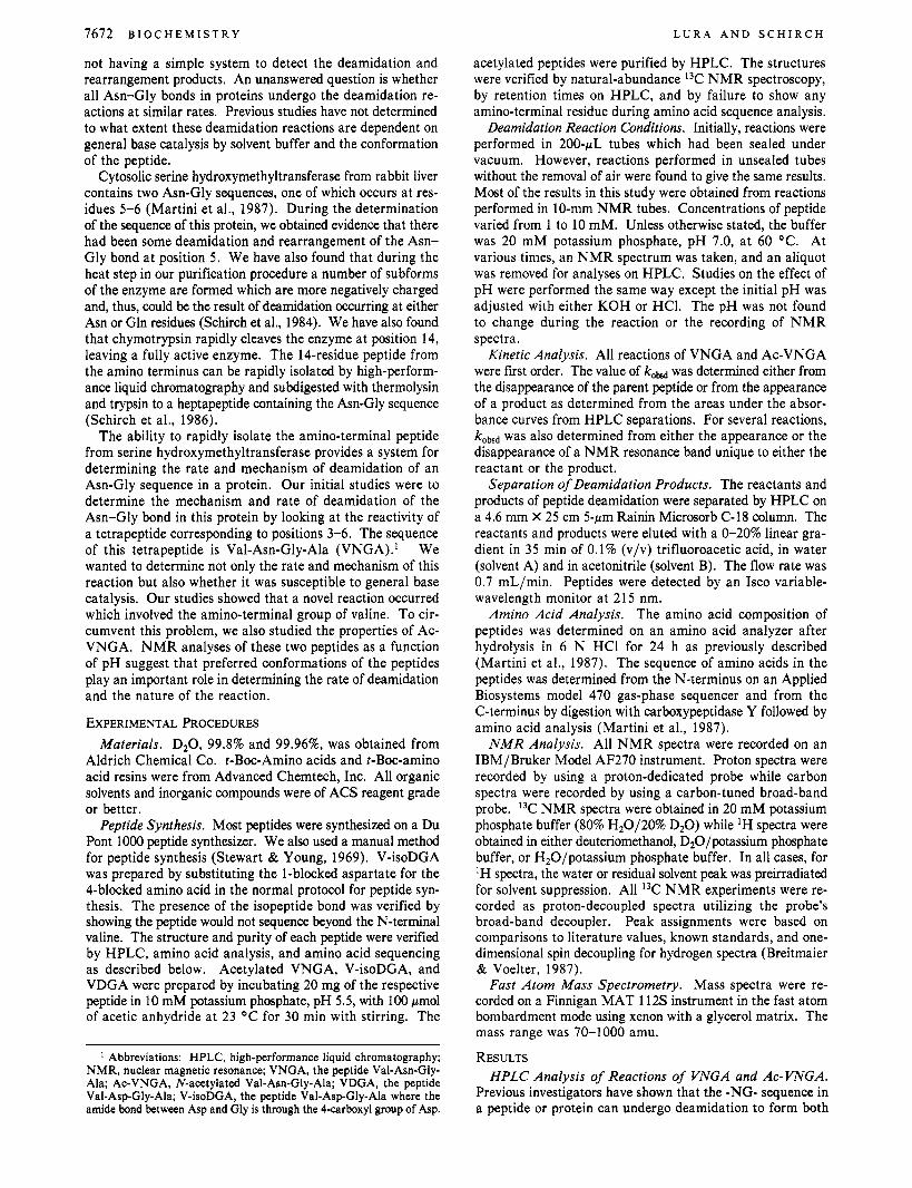
FIGURE 1 : (A) Elution profile of VNGA from a C-18 HPLC column after 0,8, and 500 h of incubation. (B) 13C NMR spectra of VNGA solutions after 0, 8, and 500 h of incubation. VNGA (10 mM) solutions were in 20 mM potassium phosphate, pH 7.0, at 60 OC.
the -DG- and -isoDG- sequences (Haley et al., 1966; Geiger & Clark, 1987; Meinwald et al., 1986). Incubation of VNGA and Ac-VNGA at pH 7.3 resulted in the appearance of two products as detected by elution from an HPLC C-18 column (Figures 1A and 2A). We conclude that the data reported in our study support the interpretation that Ac-VNGA is reacting to form Ac-VDGA and Ac-V-isoDGA while VNGA is reacting to form compounds I and I1 as shown in Scheme I.
Ac-VNGA disappears in a first-order reaction to form two products in a constant 3:1 ratio with a tlI2 of 80 h at 37 OC
II 1 0 CB3
A WV- iwDGA __ki
AVVNGA
Ac-VNGA
i - 20 28 minutes
I1
0 5 4 0 Hours
I 3 4 Hours
A-3 N-2
D Time A-2 0-2 "-41
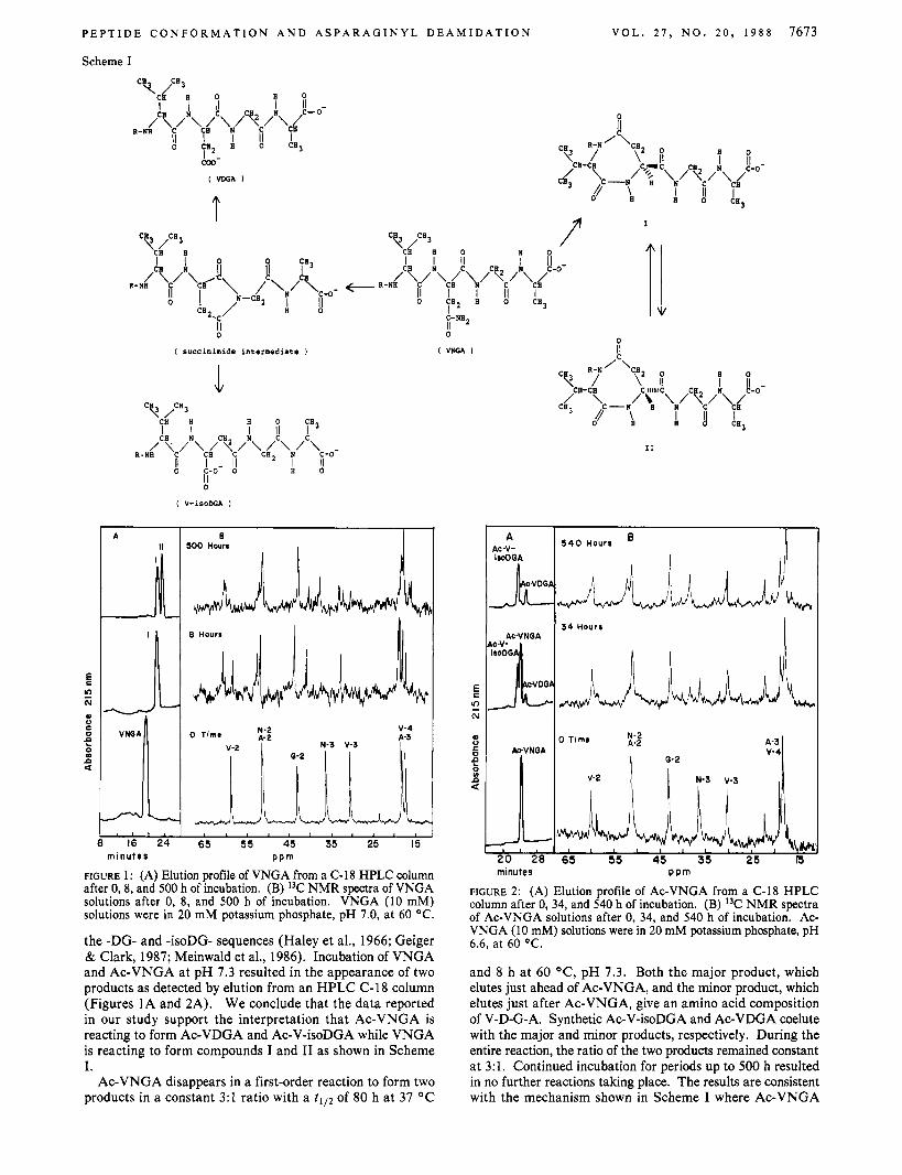
FIGURE 2: (A) Elution profile of Ac-VNGA from a C-18 HPLC column after 0, 34, and 540 h of incubation. (B) I3C NMR spectra of Ac-VNGA solutions after 0, 34, and 540 h of incubation. Ac- VNGA (10 mM) solutions were in 20 mM potassium phosphate, pH 6.6, at 60 OC.
and 8 h at 60 "C, pH 7.3. Both the major product, which elutes just ahead of Ac-VNGA, and the minor product, which elutes just after Ac-VNGA, give an amino acid composition of V-D-G-A. Synthetic Ac-V-isoDGA and Ac-VDGA coelute with the major and minor products, respectively. During the entire reaction, the ratio of the two products remained constant at 3:l. Continued incubation for periods up to 500 h resulted in no further reactions taking place. The results are consistent with the mechanism shown in Scheme I where Ac-VNGA
7674 B I O C H E M I S T R Y
forms a succinimide intermediate in the ratedetermining step, and this intermediate hydrolyzes in a 3:l ratio to Ac-V-isoD- GA and Ac-VDGA.
VNGA disappears in a first-order reaction with a t I l2 of 36 h at 37 "C and 3.5 h at 60 OC, pH 7.3, to form a single product (Figure 2A). Continued incubation results in the disappear- ance of this product (compound I in Scheme I) to form con- comitantly a second compound with a t I l2 of about 200 h (compound 11 in Scheme I). Amino acid analysis showed both products to have an amino acid composition of V-D-G-A. Neither product released any amino acids when sequenced from the N-terminus, and both products released only alanine upon digestion with carboxypeptidase Y. Fast atom bom- bardment mass spectrometry of compounds I and I1 showed that they both had an m/z = 341, consistent with the car- boxylate form of the compounds. The results suggest the sequential conversion of VNGA to compound I and compound 11. This relationship was confirmed by isolating both com- pounds I and I1 and showing that their incubation a t pH 10 resulted in the formation of the other compound. The rate of interconversion was very pH sensitive, having a tI l2 of only 0.5 h, 60 OC, at pH 11. The equilibrium constant of 2, how- ever, was pH insensitive in the range 7-11.
"C and ' H NMR Analysis of Reaction Products of VNGA and Ac-VNGA. Further characterization of the reaction products of VNGA was achieved by using "C NMR analysis of the reaction during the formation of compounds I and 11. Figure 1B shows the spectra of VNGA, compound I, and a mixture of compounds I and 11. The conversion of VNGA to compound I results in almost no change in the chemical shift of the signals for glycine and alanine. However, significant changes oocur in the resonance positions of V-3, V-2, N-3, and N-2 during formation of compound I, which are all shifted upfield. These results are consistent with the formation of the proposed seven-membered ring between valine and asparagine as shown in Scheme I. The conversion of compound I to compound I1 is characterized by only minor shifts in the signals for V-2, V-3, and N-3. These results are also consistent with compound I1 being similar in structure to compound I.
In contrast to the change in the "C NMR signals of as- paragine and valine in the formation of products from VNGA, with Ac-VNGA the products differ in chemical shift only at N-2 and N-3 (Figure 2B). The valine carbons seem to be unaffected. Also, a 3:l mixture of synthetic Ac-V-isoDGA and Ac-VDGA gives a spectrum that is virtually identical with the spectrum shown for Ac-VNGA after the 540-h incubation. This further confirms that the products of the Ac-VNGA reaction are the deamidated forms arising from the hydrolysis of the succinimide intermediate as shown in Scheme I.
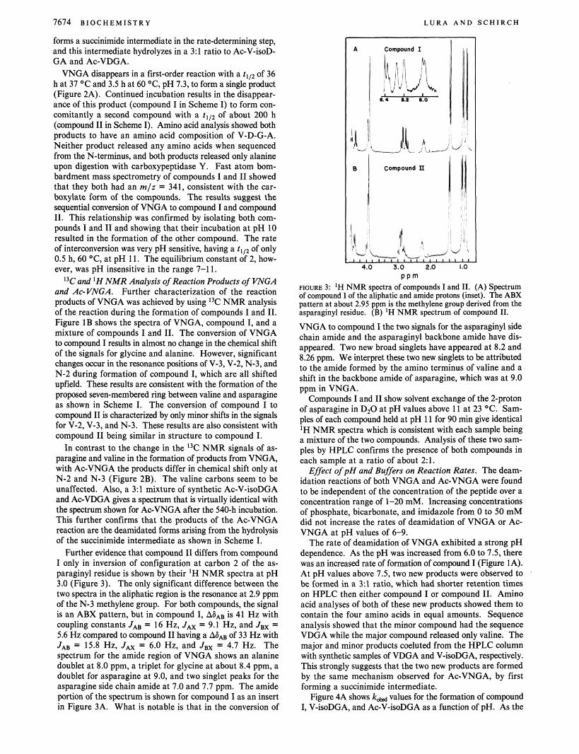
Further evidence that compound I1 differs from compound I only in inversion of configuration at carbon 2 of the as- paraginyl residue is shown by their 'H NMR spectra at pH 3.0 (Figure 3). The only significant difference between the two spectra in the aliphatic region is the resonance at 2.9 ppm of the N-3 methylene group. For both compounds, the signal is an ABX pattern, but in compound I, A6AB is 41 Hz with coupling constants JAB = 16 Hz, Jm = 9.1 Hz, and JBx = 5.6 Hz compared to compound I1 having a AS, of 33 Hz with JAB = 15.8 Hz, Jm = 6.0 Hz, and JBx = 4.7 Hz. The spectrum for the amide region of VNGA shows an alanine doublet at 8.0 ppm, a triplet for glycine at about 8.4 ppm, a doublet for asparagine at 9.0, and two singlet peaks for the asparagine side chain amide at 7.0 and 7.7 ppm. The amide portion of the spectrum is shown for compound I as an insert in Figure 3A. What is notable is that in the conversion of
L U R A A N D S C H I R C H
p p m ncme 3: 'H NMR spectra of "pounds I and 11. (A) Spectrum of compound I of the aliphatic and amide protons (inset). The ABX pattern at about 2.95 ppm is the methylene group derived from the asparaginyl residue. (B) 'H NMR spectrum of compound 11.
VNGA to compound I the two signals for the asparaginyl side chain amide and the asparaginyl backbone amide have dis- appeared. Two new broad singlets have appeared at 8.2 and 8.26 ppm. We interpret these two new singlets to be attributed to the amide formed by the amino terminus of valine and a shift in the backbone amide of asparagine, which was at 9.0 ppm in VNGA.
Compounds I and I1 show solvent exchange of the 2-proton of asparagine in D,O at pH values above 11 at 23 OC. Sam- ples of each compound held at pH 11 for 90 min give identical lH NMR spectra which is consistent with each sample being a mixture of the two compounds. Analysis of these two sam- ples by HPLC confirms the presence of both compounds in each sample at a ratio of about 21.
Effect of pH and Buffers on Reaction Rates. The deam- idation reactions of both VNGA and Ac-VNGA were found to be independent of the concentration of the peptide over a concentration range of 1-20 mM. Increasing concentrations of phosphate, bicarbonate, and imidazole from 0 to 50 mM did not increase the rates of deamidation of VNGA or Ac- VNGA at pH values of 6-9.
The rate of deamidation OS VNGA exhibited a strong pH dependence. As the pH was increased from 6.0 to 7.5, there was an increased rate of formation of compound I (Figure 1A). At pH values above 7.5, two new products were observed to be formed in a 3:l ratio, which had shorter retention times on HPLC then either compound I or compound 11. Amino acid analyses of both of these new products showed them to contain the four amino acids in equal amounts. Sequence analysis showed that the minor compound had the sequence VDGA while the major compound released only valine. The major and minor products coeluted from the HPLC column with synthetic samples of VDGA and V-isoDGA, respectively. This strongly suggests that the two new products are formed by the same mechanism observed for Ac-VNGA, by first forming a succinimide intermediate.
Figure 4A shows kM values for the formation of compound I, V-isoDGA, and Ac-V-isoDGA as a function of pH. As the
P E P T I D E C O N F O R M A T I O N A N D A S P A R A G I N Y L D E A M I D A T I O N V O L . 2 7 , N O . 2 0 , 1 9 8 8 7675
2m I v-4
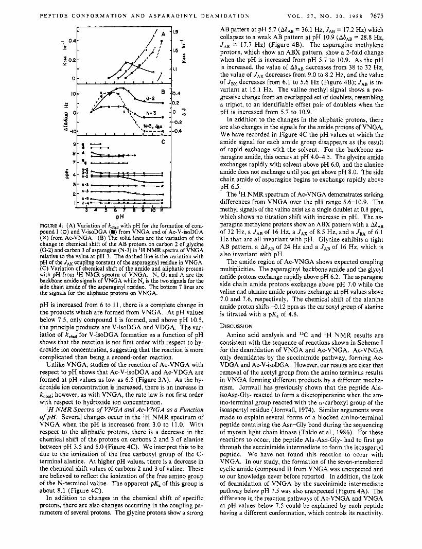
PH FIGURE 4: (A) Variation of kobd with pH for the formation of com- pound I (0) and V-isoDGA (m) from VNGA and of Ac-V-isoDGA (X) from Ac-VNGA. (B) The solid lines are the variation of the change in chemical shift of the AB protons on carbon 2 of glycine (G-2) and carbon 3 of asparagine (N-3) in IH NMR spectra of VNGA relative to the value at pH 3. The dashed line is the variation with pH of the JAx coupling constant of the asparaginyl residue in VNGA. (C) Variation of chemical shift of the amide and aliphatic protons with pH from 'H NMR spectra of VNGA. N, G, and A are the backbone amide signals of VNGA while N, is the two signals for the side chain amide of the asparaginyl residue. The bottom 7 lines are the signals for the aliphatic protons on VNGA.
pH is increased from 6 to 11, there is a complete change in the products which are formed from VNGA. At pH values below 7.5, only compound I is formed, and above pH 10.5, the principle products are V-isoDGA and VDGA. The var- iation of kobsd for V-isoDGA formation as a function of pH shows that the reaction is not first order with respect to hy- droxide ion concentration, suggesting that the reaction is more complicated than being a second-order reaction.
Unlike VNGA, studies of the reaction of Ac-VNGA with respect to pH shows that Ac-V-isoDGA and Ac-VDGA are formed at pH values as low as 6.5 (Figure 3A). As the hy- droxide ion concentration is increased, there is an increase in koM; however, as with VNGA, the rate law is not first order with respect to hydroxide ion concentration.
H N M R Spectra of VNGA and Ac- VNGA as a Function o f p H . Several changes occur in the 'H NMR spectrum of VNGA when the pH is increased from 3.0 to 11.0. With respect to the aliphatic protons, there is a decrease in the chemical shift of the protons on carbons 2 and 3 of alanine between pH 3.5 and 5.0 (Figure 4C). We interpret this to be due to the ionization of the free carboxyl group of the C- terminal alanine. At higher pH values, there is a decrease in the chemical shift values of carbons 2 and 3 of valine. These are believed to reflect the ionization of the free amino group of the N-terminal valine. The apparent pK, of this group is about 8.1 (Figure 4C).
In addition to changes in the chemical shift of specific protons, there are also changes occurring in the coupling pa- rameters of several protons. The glycine protons show a strong
AB pattern at pH 5.7 (A6- = 36.1 Hz, JAB = 17.2 Hz) which collapses to a weak AB pattern at pH 10.9 (A6AB = 28.8 Hz, J A B = 17.7 Hz) (Figure 4B). The asparagine methylene protons, which show an ABX pattern, show a 2-fold change when the pH is increased from pH 5.7 to 10.9. As the pH is increased, the value of A ~ A B decreases from 38 to 32 Hz, the value of JAx decreases from 9.0 to 8.2 Hz, and the value of JBx decreases from 6.1 to 5.6 Hz (Figure 4B); JAB is in- variant at 15.1 Hz. The valine methyl signal shows a pro- gressive change from an overlapped set of doublets, resembling a triplet, to an identifiable offset pair of doublets when the pH is increased from 5.7 to 10.9.
In addition to the changes in the aliphatic protons, there are also changes in the signals for the amide protons of VNGA. We have recorded in Figure 4C the pH values at which the amide signal for each amide group disappears as the result of rapid exchange with the solvent. For the backbone as- paragine amide, this occurs at pH 4.0-4.5. The glycine amide exchanges rapidly with solvent above pH 6.0, and the alanine amide does not exchange until you get above pH 8.0. The side chain amide of asparagine begins to exchange rapidly above pH 6.5.
The 'H NMR spectrum of Ac-VNGA demonstrates striking differences from VNGA over the pH range 5.6-10.9. The methyl signals of the valine exist as a single doublet at 0.8 ppm, which shows no titration shift with increase in pH. The as- paragine methylene protons show an ABX pattern with a A6, of 32 Hz, a JAB of 16 Hz, a JAx of 8.5 Hz, and a Jex of 6.1 Hz that are all invariant with pH. Glycine exhibits a tight AB pattern, a A6AB of 24 Hz and a JAB of 16 Hz, which is also invariant with pH.
The amide region of Ac-VNGA shows expected coupling multiplicities. The asparaginyl backbone amide and the glycyl amide protons exchange rapidly above pH 6.2. The asparagine side chain amide protons exchange above pH 7.0 while the valine and alanine amide protons exchange at pH values above 7.0 and 7.6, respectively. The chemical shift of the alanine amide proton shifts -0.12 ppm as the carboxyl group of alanine is titrated with a pK, of 4.8.
DISCUSSION Amino acid analysis and 13C and 'H NMR results are
consistent with the sequence of reactions shown in Scheme I for the deamidation of VNGA and Ac-VNGA. Ac-VNGA only deamidates by the succinimide pathway, forming Ac- VDGA and Ac-V-isoDGA. However, our results are clear that removal of the acetyl group from the amino terminus results in VNGA forming different products by a different mecha- nism. Jornvall has previously shown that the peptide Ala- isoAsp-Gly- reacted to form a diketopiperazine when the am- ino-terminal group reacted with the a-carboxyl group of the isoaspartyl residue (Jornvall, 1974). Similar arguments were made to explain several forms of a blocked amino-terminal peptide containing the Asn-Gly bond during the sequencing of myosin light chain kinase (Takio et al., 1986). For these reactions to occur, the peptide Ala-Asn-Gly- had to first go through the succinimide intermediate to form the isoaspartyl peptide. We have not found this reaction to occur with VNGA. In our study, the formation of the seven-membered cyclic amide (compound I) from VNGA was unexpected and to our knowledge never before reported. In addition, the lack of deamidation of VNGA by the succinimide intermediate pathway below pH 7.5 was also unexpected (Figure 4A). The difference in the reaction pathways of Ac-VNGA and VNGA at pH values below 7.5 could be explained by each peptide having a different conformation, which controls its reactivity.
L U R A A N D S C H I R C H
is increased to 8.0. The model also shows that in the howhoe conformation the positively charged amino-terminal group of valine can rotate around the bond connecting carbon atoms 1 and 2. This results in the charged amino terminus alternately being either next to the negatively charged carboxylate group or next to the side chain amide of the asparaginyl residue. Only this latter conformation is shown in Figure 5. The horseshoe conformation then would be stabilized by three interactions, Le., the electrostatic interaction of the amino terminus and the carboxy terminus, hydrogen bonding of the carboxy terminus with the asparagine backbone amide, and hydrogen bonding of the valine carbonyl with the alanine amide. Since the coupling constants for the amide protons are similar to those for amides in free rotation, the horseshoe conformation is viewed as not being a rigid structure but represents an energetically favorable conformation (Bystrov et al., 1973).
The proposed conformation for VNGA below pH 7.5 (Figure 5) is also supported by the aliphatic IH NMR spec- trum. The model predicts that the two aliphatic protons of glycine (Hs and HR) have different environments. Indeed, the spectrum shows a strong AB pattern for these glycine protons. The methylene protons of asparagine show an ABX pattern, and from the observed coupling constants, the relative dihedral angles can be calculated for the AX and BX portions to be 140’ and -127O. respectively (Kopple et al., 1973). These angles agree well with the peptide conformation shown in Figure 5 . Another feature of the model is that it places the valine amino group near the carbonyl side chain amide of asparagine, and thus would favor attack from this group rather than the glycine amide which is far removed from the as- paragine side chain. The two methyl groups on valine form an overlapping doublet, suggesting that their environments are very similar, consistant with our suggestion that there is ro- tation around carbon atoms 1 and 2 of valine.
When the pH is increased above pH 7.5, the valine ami- no-terminal group loses its positive charge, and there appears to be a concomitant change in the mechanistic pathway of deamidation and the conformation of the peptide. The glycine aliphatic protons collapse into a tight AB pattern, suggesting that this portion of the molecule now has much more rotational freedom. The two methyl groups on valine become more dissimiler and split into well-resolved doublets. This can be attributed to a decrease in rotational freedom of the isopropyl group when the amino terminus is fully titrated to the un- charged NH, group. The pair of free electrons can add electronic hulk, restricting the rotation of this group. The changes of the asparagine methylene protons with pH can be explained by an opening up of the horseshoe shape, resulting in a pivoting of the entire side chain and a rotation around the bond connecting carbon atoms 2 and 3. This conforma- tional change would explain both the change in the coupling constants for JAx and Jsx and the value of All of the changes observed in the proton NMR spectrum suggest that as the free amino group becomes neutral the horseshoe shape of the VNGA becomes more linear with increased freedom of rotation. The linear arrangement is much more favorable to forming the succinimide intermediate and, therefore, ex- plains the change in mechanistic pathway for deamidation with increasing pH.
The ‘H NMR spectrum of Ac-VNGA varies little with pH. One can observe the titration of the alanine carboxyl group at pH 4.5, but the chemical shift and coupling of the aliphatic protons suggest that at even this low pH, Ac-VNGA has a conformation similar to VNGA at high pH. One notable
7676 B I O C H E M I S T R Y
\ V a l cn,
ploURe 5: Stick and spacefilling model of an important mnformation of VNGA at pH values between 5.5 and 7.5.
Small tetrapeptides might be expected to have complete flexibility and not to assume any particular conformation. However, the ‘H NMR spectra of VNGA and Ac-VNGA suggest that at pH values below 7.5 these two peptides have different conformations in solution. Above pH 9.0, the ‘H NMR spectra of the two peptides are very similar, suggesting they have similar conformations. The model in Figure 5 represents our interpretation of an important conformation of VNGA below pH 7.5.
The amide region of the spectra may supply the greatest amount of information concerning the conformation of VNGA. Amide protons in peptides undergo rapid solvent exchange around pH 6 (Bundi & Wuthrich, 1979). With VNGA, this value is observed for the glycine amide proton, but the as- paraginyl backbone amide proton undergoes rapid exchange near pH 4.5, and the alaninyl amide proton undergoes rapid exchange at pH 8.0. The asparagine amide proton appears to undergo rapid exchange with the concomitant titration of the alanine carboxyl group. This suggests that the carboxylate group is in a position to interact with the asparagine amide proton and, therefore, facilitate its solvent exchange. As ob- served in the model in Figure 5, this requires a bending of the backbone chain into a horseshoe shape to bring the alanine carboxyl group to within hydrogen-bonding distance of the asaparagine amide proton. In this horseshoe conformation, the alanine amide proton is within hydrogen-bonding distance of the valine carbonyl group. Hydrogen bonding between these two groups would stabilize the alanine amide proton and ex- plain why it does not undergo solvent exchange until the pH
Biochemistry 1988, 27, 7677-7681 7677
difference is that the asparagine backbone amide exchanges protons with solvent 2 pH units higher in Ac-VNGA than in VNGA. This suggests that the asparagine backbone amide does not interact with the alanine carboxyl group as proposed for VNGA.
In conclusion, our results suggest that the deamidation of -NG- sequences in peptides is independent of general base catalysis by solvent buffer but is dependent on the confor- mation of the peptide backbone. This suggests that -NG- sequences in proteins will not all undergo deamidation at the same rate but will deamidate at rates which are controlled by the conformation of the segment of the protein containing the -NG- sequence. Our results also show that proteins which have a penultimate asparagine can undergo, at neutral pH, a deamidation reaction involving the amino terminus to form a cyclic amide. The driving force for this reaction to occur in VNGA appears to be the interaction of the negatively charged carboxyl group on the C-terminus with the positively charged N-terminus. REFERENCES Aswad, D. W. (1984) J. Biol. Chem. 259, 10714-10721. Battersby, A. R., Robinson, J. C. (1955) J. Chem. SOC.,
Breitmaier, E., & Voelter, W. (1987) Carbon-I3 N M R Spectroscopy, 3rd ed., pp 415-420, VCH, Weinheim, FDR.
Bundi, A., & Wuthrich, K. (1979a) Biopolymers 18,285-297. Bundi, A., & Wuthrich, K. (1 979b) Biopolymers 18,299-3 1 1 . Bystrov, V. F., Ivanov, V. T., Portnova, S . L., Balashova, T.
A., & Ovchinnikov, Yu. A. (1973) Tetrahedron 29,
259-269.
873-877.
Geiger, T., & Clarke, S. (1987) J. Biol. Chem. 262,785-794. Haley, E. E., Corcoran, B. J., Dorer, F. E., & Buchanan, D.
Jornvall, H. (1974) FEBS Lett. 38, 329-333. Kopple, K. D., Wiley, C. R., & Tauke, R. (1973) BiopoZymers
Martini, F., Angelaccio, S. , Pascarella, S . , Barra, D., Bossa, F., & Schirch, V. (1987) J. Biol. Chem. 262, 5499-5509.
Meinwald, Y. C., Stimson, E., & Scheraga, H. A. (1986) Znt. J . Pept. Protein Res. 28, 79-84.
Midelfort, C. F., & Mehler, A. H. (1972) Proc. Natl. Acad. Sci. U.S.A. 69, 1816-1819.
Murray, E. D., Jr., & Clarke, S. (1984) J. Biol. Chem. 259,
Robinson, A. B., & Rudd, C. J. (1974) Curr. Top. Cell. Regul.
Sandheimer, E., & Holley, R. W. (1954) J. Am. Chem. SOC.
Schirch, L., Gavilanes, F., Peterson, D., Bullis, B., Barra, D., &z Bossa, F. (1984) Chemical and Biological Aspects of Vitamin B6 Catalysis: Part A , pp 301-388, Alan Liss, New York.
Schirch, V., Schirch, D., Martini, F., & Bossa, F. (1986) Eur. J . Biochem. 161, 45-49.
Stewart, J. M., & Young, J. D. (1969) Solid Phase Peptide Synthesis, W. H. Freeman, San Francisco.
Takio, K., Blumenthal, D. K., Walsh, K. A., Titani, K., & Krebs, E. G. (1986) Biochemistry 25, 8049-8057.
Yuksel, K. U., & Gracy, R. W. (1986) Arch. Biochem. Bio-
L. (1966) Biochemistry 5, 3229-3235.
12, 627-636.
10722-10732.
8, 247-295.
76, 2467-2470.
phys. 248, 452-459.
Acceleration of Cleavage of the Carbon-Cobalt Bond of Sterically Hindered Alkylcobalamins by Binding to Apoprotein of Diol Dehydrase
Tetsuo Toraya* and Atsuhiko Ishida Department of Chemistry, College of Liberal Arts and Sciences, Kyoto University, Sakyo- Ku, Kyoto 606, Japan
Received March 25, 1988; Revised Manuscript Received June 2, 1988
ABSTRACT: Cleavage of the C-Co bond of sterically hindered alkylcobalamins bearing neither an adenine moiety nor functional groups, such as isobutylcobalamin, neopentylcobalamin, and cyclohexylcobalamin, was markedly accelerated by their interaction with apoprotein of diol dehydrase, although these cobalamins do not function as coenzyme. Acceleration of the conversion of alkylcobalamins to enzyme-bound hy- droxocobalamin was stoichiometric and obeyed first-order reaction kinetics. These results, together with strong competitive inhibition by these alkylcobalamins with respect to adenosylcobalamin, indicate that acceleration of the C-Co bond cleavage by the apoenzyme is due to labilization of their C-Co bond by binding to the active site of the enzyme. This labilization is considered to be caused by a steric distortion of the corrin ring which is induced by specific tight interaction of the cobalamin moiety with apoprotein. The importance of such a labilizing effect for activation of the C-Co bond of adenosylcobalamin in enzymatic reactions is discussed.
H o m o l y t i c cleavage of the C-Co bond of AdoCbl' is an essential early event in the AdoCbl-dependent enzymatic re-
actions involving diol dehydrase. Since the C-Co bond of AdoCbl is sufficiently weak (homolytic bond dissociation en- ergy ca. 26 kcal/mol) (Halpern et al., 1984), only a modest additional labilization by interaction with apoprotein would be required for homolysis. That is, the apoenzyme elicits the function of the coenzyme by activating (labilizing) this or- ganometallic bond'
During the course of our studies addressed to know the mechanism of activation of the C-Co bond of AdoCbl in