Role of Platelet Membrane Glycoproteins Ib/IX and llb/ll la, and of Platelet
s-Granule Proteins in Platelet Aggregation Induced by Human
Osteosarcoma Cells
P h i l i p p e C l e z a r d i n , 2 J e a n n e D r o u i n , M a r i e - C h r i s t i n e M o r e l - K o p p , M i c h e l H a n s s , B e a t e K e h r e l , C l a i r e - M a r i e Serre ,
C~c i l e K a p l a n , a n d P i e r r e D. D e l m a s
INSERM Research Unit 234, Pavilion F [P C., C-M. S., P. D. D.] and Pavilion E [M. H.], H@ital Edouard Herriot, Place d'Arsonval, 69437Lyon, France; Division of Hematology, Ottawa General Hospital, Ottawa, Ontario, Canada [J. D.]; Service lmmunologie Leuco-Plaquettaire, Institut National de TransJitsion Sanguine, Rue Alexandre-Cabanel, 75739 Paris, France [C. K., M-C. M-K.]; and ExperimenteUe Hiimostaseforschung, lnnere Medizin A, Universitiitskliniken Miinster, Germany [B. K.].
A B S T R A C T
We have previously shown that the platelet-aggregating activity of hu- man MG-63 and HOS osteosarcoma cells depends at least in part upon tumor cell surface-associated thrombospondin, and suggested that plate- let-osteosarcoma cell interactions could occur through interactions with specific platelet membrane receptors. In this study, the platelet-aggregat- ing activity of MG-63 and HOS ceils was studied by using a variety of platelet disorders. Both osteosarcoma cdl lines induced a biphasic platelet aggregation response when added to normal platelet-rich plasma, while the second phase of aggregation was absent when added to gray platelets (deficiency in a-granule proteins) and to aspirin-treated platelets. Platelets from two unrelated patients with type I Gianzmann's thrombasthenia (deficiency in glycoprotein (GP) GPIIb/IIIa) did not aggregate at all with osteosarcoma ceils. Using giant platelets from three patients with Bernard- Soulier syndrome (deficiency in GPIb/IX), the aggregation response in- duced by MG-63 and HOS cells was monophasic and reversible when compared to normal-sized platelets and to giant platelets from a patient with May-Hegglin anomaly (no membrane GP defect). Because GPIb serves as a receptor for von Willebrand factor during hemostasis, aggre- gation experiments were also conducted with the platelet-rich plasma of two patients with a low plasma von Wiilebrand factor concentration (type I yon Willebrand's disease) before and after the infusion of deamino-D- arginine vasopressin. MG-63 and HOS cells induced biphasic platelet aggregation both before and after deamino-D-arginine vasopressin treat- ment, while the ristocetin-dependent binding of von Willebrand factor to platelets only occurred after deamino-D-arginine vasopressin treatment. Preincubation of normal platelet-rich plasma with monocional antibody SZ-2 directed against the yon Wiilebrand binding domain of GPIb did not inhibit the platelet-aggregating activity of osteosareoma cells, whereas anti-GPIb antibody SZ.2 did inhibit ristocetin-induced platelet agglutina- tion. In addition, anti-GPIX antibodies did not affect platelet-osteosar- coma cell interactions. In conclusion, our data demonstrate that the first phase of the platelet-aggregating activity of human osteosarcoma cells is initiated by the interaction of these tumor cells with platelet membrane GPIIb/IIIa, whereas the second phase, even if plasma von Willebrand factor is deficient, involves platelet membrane GPIb and the participation of platelet a-granule proteins in membrane-mediated events, making ag- gregation irreversible.
I N T R O D U C T I O N
Metastasis is a complex phenomenon that involves tumor cell dis- semination, and tumor cell interactions with host cells (platelets, endo- thelial cells) and subendothelial matrix (For review see Ref. 1). The survival of circulating metastatic cells and the attachment of tumor
Received 5/10/93; accepted 7/27/93. The costs of publication of this article were defrayed in part by the payment of page
charges. This article must therefore be hereby marked advertisement in accordance with 18 U.S.C. Section 1734 solely to indicate this fact.
1 This study was supported by grants from INSERM/Medical Research Council (P. C., J. D.), la FEdEration Nationale des Centres de Lutte contre le Cancer (P. C.), the Groupe- ment des Entreprises Franqaises pour la Lutte contre le Cancer (P. C.), the Deutsche Forschungs Gemeinschaft (Ke397/1-2) (B. K.), and the Ottawa General Hospital Medical Research Fund (J. D.).
2 To whom requests for reprints should be addressed, at INSERM Unit Research 234, Pavilion F, H6pital Edouard Herriot, 3 Place d'Arsonval, 69437 Lyon CEdex 03, France.
cells to endothelial cells and subendothelial matrix largely depend on the interaction of tumor cells with platelets (1-3). Such an interaction of tumor cells with platelets involves a series of sequential events (1, 4). Initially, a few platelets adhere to tumor cells which activate them through expression of prothrombogenic activities, depending on the origin of the tumor cells. This results in the recruitment of addi- tional platelets, and formation of focal aggregates of activated, de- granulated platelets attached to the tumor cell surface. Subsequently, tumor cells respond to activated platelets with the appearance of cellular processes that interdigitate with platelet aggregates, suggest- ing tumor cell cytoskeletal alterations. Several cell surface\adhesive proteins have been implicated in linking platelets to tumor cE~ls. Among these adhesive proteins are fibronectin (5), von Willebrahd factor (5), fibrinogen (6), and thrombospondin (7, 8). During hemo- stasis, von Willebrand factor binds to platelet membrane glycoprotein complex GPIb/IX, 3 and the membrane glycoprotein complex GPIIb/ Ilia serves as a platelet receptor for fibrinogen, fibronectin, von Wil- lebrand factor, and thrombospondin (for review see Ref. 9). Adhesive proteins are also secreted from a-granules upon platelet activation and help to stabilize platelet aggregates (10). The functions of these major platelet glycoproteins have been elucidated by the study of platelets from patients with Bernard-Soulier syndrome, Glanzmann's throm- basthenia, and gray platelet syndrome which lack GPIb/IX, GPIIb/Illa and a-granule proteins, respectively (9, 11). Immunoinhibition has been widely used in evaluating the effects of antibodies against plate- let GPIIb/IIIa on the platelet-aggregating activity of tumor cells (5, 12-15). Pretreatment of platelets with anti-GPIIb/IIIa monoclonal antibodies inhibit both the binding of mouse CT26 and human HCT8 colon carcinoma cells to platelets (5), and the platelet-aggregating activity of different tumor cell lines, including human HMV-I mela- noma cells and M7609 colon carcinoma cells (12-15). By contrast, the significance of the results obtained with anti-GPIb monoclonal anti- bodies remains unclear. Anti-GPIb monoclonal antibodies have no effect on the binding of mouse CT26 and human HCT8 colon carci- noma cells to platelets (5) and on the platelet-aggregating activity of M3Dau melanoma cells (14), while they inhibit the platelet-aggregat- ing activity of human HMV-I melanoma cells and M7609 colon carcinoma cells (15). We have previously shown that the platelet- aggregating activity of human osteosarcoma cells depend at least in part upon tumor cell surface-associated thrombospondin, and sug- gested that platelet-osteosarcoma cell interactions could occur through interactions with specific platelet membrane receptors (8). However, it is not known whether platelet receptors such as GPIb/IX or GPIIb/IIIa are involved during platelet aggregation induced by osteosarcoma cells, or if platelet-released a-granule proteins play a role in this context. The present study investigates the role of platelet membrane glycoproteins GPIb/IX and GPIIb/IIIa, and of a-granule proteins dur- ing platelet aggregation induced by human osteosarcoma cells, using
3 The abbreviations used are: GP, glycoprotein; DDAVP, deamino-o-arginine vasopres- sin.
PLATELET GPs IN OSTEOSARCOMA CELL-INDUCED PLATELET AGGREGATION
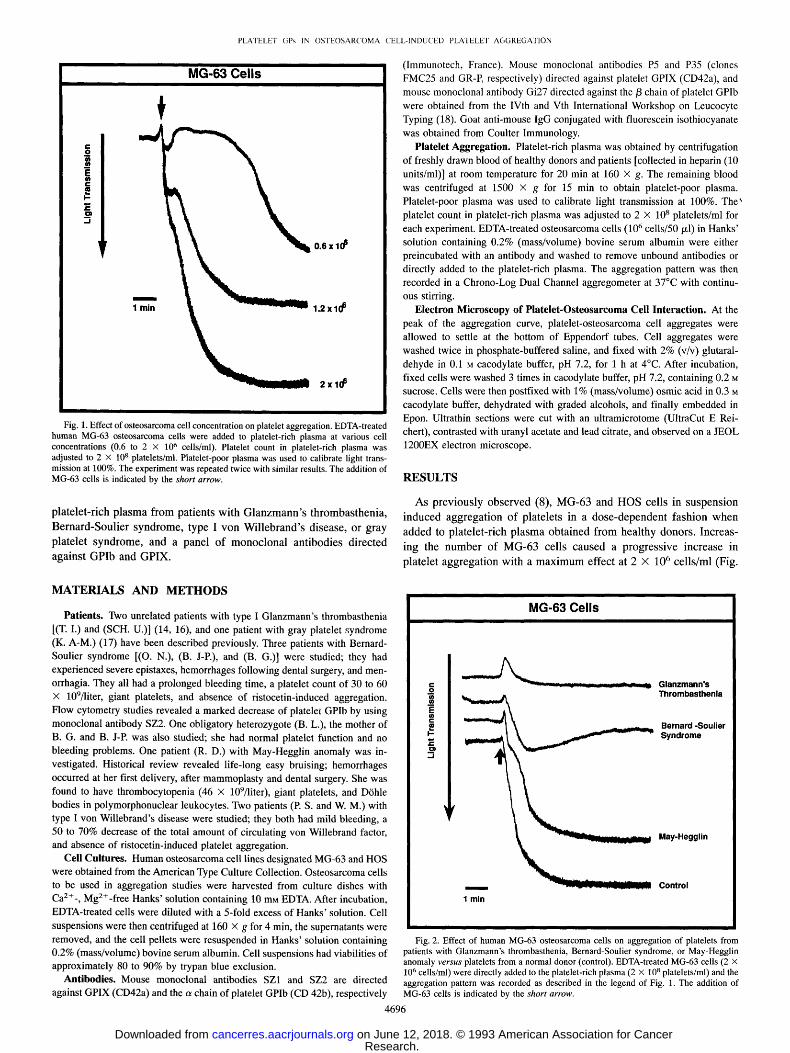
Fig. 1. Effect of osteosarcoma cell concentration on platelet aggregation. EDTA-treated human MG-63 osteosarcoma cells were added to platelet-rich plasma at various cell concentrations (0.6 to 2 x 10⁶ cells/ml). Platelet count in platelet-rich plasma was adjusted to 2 X 10⁸ platelets/ml. Platelet-poor plasma was used to calibrate light trans- mission at 100%. The experiment was repeated twice with similar results. The addition of MG-63 cells is indicated by the shor t arrow.
platelet-rich plasma from patients with Glanzmann 's thrombasthenia,
Bernard-Soulier syndrome, type I v o n Willebrand's disease, or gray
platelet syndrome, and a panel of monoclonal antibodies directed
against GPIb and GPIX.
(Immunotech, France). Mouse monoclonal antibodies P5 and P35 (clones FMC25 and GR-P, respectively) directed against platelet GPIX (CD42a), and mouse monoclonal antibody Gi27 directed against the/3 chain of platelet GPIb were obtained from the IVth and Vth International Workshop on Leucocyte Typing (18). Goat anti-mouse IgG conjugated with fluorescein isothiocyanate was obtained from Coulter Immunology.
Platelet Aggregation. Platelet-rich plasma was obtained by centrifugation of freshly drawn blood of healthy donors and patients [collected in heparin (10 units/ml)] at room temperature for 20 min at 160 X g. The remaining blood was centrifuged at 1500 X g for 15 min to obtain platelet-poor plasma. Platelet-poor plasma was used to calibrate light transmission at 100%. The' platelet count in platelet-rich plasma was adjusted to 2 x 10⁸ platelets/ml for each experiment. EDTA-treated osteosarcoma cells (106 cells/50 ~l) in Hanks' solution containing 0.2% (mass/volume) bovine serum albumin were either preincubated with an antibody and washed to remove unbound antibodies or directly added to the platelet-rich plasma. The aggregation pattern was then recorded in a Chrono-Log Dual Channel aggregometer at 37~ with continu- ous stirring.
Electron Microscopy of Platelet-Osteosarcoma Cell Interaction. At the peak of the aggregation curve, platelet-osteosarcoma cell aggregates were allowed to settle at the bottom of Eppendorf tubes. Cell aggregates were washed twice in phosphate-buffered saline, and fixed with 2% (v/v) glutaral- dehyde in 0.1 M cacodylate buffer, pH 7.2, for 1 h at 4~ After incubation, fixed cells were washed 3 times in cacodylate buffer, pH 7.2, containing 0.2 M sucrose. Cells were then postfixed with 1% (mass/volume) osmic acid in 0.3 M cacodylate buffer, dehydrated with graded alcohols, and finally embedded in Epon. Ultrathin sections were cut with an ultramicrotome (UltraCut E Rei- chert), contrasted with uranyl acetate and lead citrate, and observed on a JEOL 1200EX electron microscope.
R E S U L T S
As previously observed (8), MG-63 and HOS cells in suspension
induced aggregation of platelets in a dose-dependent fashion when
added to platelet-rich plasma obtained f rom healthy donors. Increas-
ing the number of MG-63 cells caused a progressive increase in
platelet aggregation with a m a x i m u m effect at 2 X 10⁶ cells/ml (Fig.
M A T E R I A L S A N D M E T H O D S
Patients. Two unrelated patients with type I Glanzmann's thrombasthenia [(T. I.) and (SCH. U.)] (14, 16), and one patient with gray platelet syndrome (K. A-M.) (17) have been described previously. Three patients with Bernard- Soulier syndrome [(O. N.), (B. J-P.), and (B. G.)] were studied; they had experienced severe epistaxes, hemorrhages following dental surgery, and men- orrhagia. They all had a prolonged bleeding time, a platelet count of 30 to 60 X 109/liter, giant platelets, and absence of ristocetin-induced aggregation. Flow cytometry studies revealed a marked decrease of platelet GPIb by using monoclonal antibody SZ2. One obligatory heterozygote (B. L.), the mother of B. G. and B. J-P. was also studied; she had normal platelet function and no bleeding problems. One patient (R. D.) with May-Hegglin anomaly was in- vestigated. Historical review revealed life-long easy bruising; hemorrhages occurred at her first delivery, after mammoplasty and dental surgery. She was found to have thrombocytopenia (46 x 109/liter), giant platelets, and D6hle bodies in polymorphonuclear leukocytes. Two patients (P. S. and W. M.) with type I von WiUebrand's disease were studied; they both had mild bleeding, a 50 to 70% decrease of the total amount of circulating von Willebrand factor, and absence of ristocetin-induced platelet aggregation.
Cell Cultures. Human osteosarcoma cell lines designated MG-63 and HOS were obtained from the American Type Culture Collection. Osteosarcoma cells to be used in aggregation studies were harvested from culture dishes with Ca 2§ MgZ+-free Hanks' solution containing 10 mM EDTA. After incubation, EDTA-treated cells were diluted with a 5-fold excess of Hanks' solution. Cell suspensions were then centrifuged at 160 x g for 4 min, the supernatants were removed, and the cell pellets were resuspended in Hanks' solution containing 0.2% (mass/volume) bovine serum albumin. Cell suspensions had viabilities of approximately 80 to 90% by trypan blue exclusion.
Antibodies. Mouse monoclonal antibodies SZ1 and SZ2 are directed against GPIX (CD42a) and the c~ chain of platelet GPIb (CD 42b), respectively
4696
Fig. 2. Effect of human MG-63 osteosarcoma cells on aggregation of platelets from patients with Glanzmann's thrombasthenia, Bernard-Soulier syndrome, or May-Hegglin anomaly versus platelets from a normal donor (control). EDTA-treated MG-63 cells (2 X 10⁶ cells/ml) were directly added to the platelet-rich plasma (2 x 10⁸ platelets/ml) and the aggregation pattern was recorded as described in the legend of Fig. 1. The addition of MG-63 cells is indicated by the shor t arrow.
PLATELET GPs IN OSTEOSARCOMA CELL-INDUCED PLATELET AGGREGATION
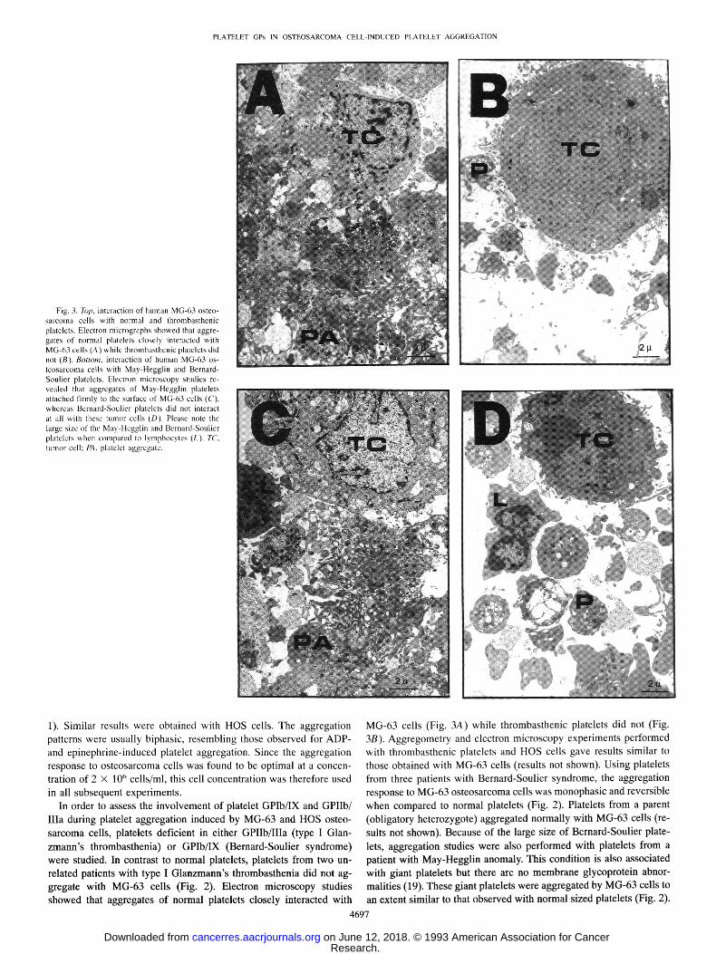
Fig. 3. Top, interaction of human MG-63 osteo- sarcoma cells with normal and thrombasthenic platelets. Electron micrographs showed that aggre- gates of normal platelets closely interacted with MG-63 cells (A) while thrombasthenic platelets did not (B). Bottom, interaction of human MG-63 os- teosarcoma cells with May-Hegglin and Bernard- Soulier platelets. Electron microscopy studies re- vealed that aggregates of May-Hegglin platelets attached firmly to the surface of MG-63 cells (C), whereas Bernard-Soulier platelets did not interact at all with these tumor cells (D). Please note the large size of the May-Hcgglin and Bernard-Soulier platelets when compared to lymphocytes (L). TC, tumor cell: PA, platelet aggregate.
1). Similar results were obtained with HOS cells. The aggregation patterns were usually biphasic, resembling those observed for ADP- and epinephrine-induced platelet aggregation. Since the aggregation response to osteosarcoma cells was found to be optimal at a concen- tration of 2 X 10 ~' cells/ml, this cell concentration was therefore used in all subsequent experiments.
In order to assess the involvement of platelet GPIb/IX and GPIIb/ IIIa during platelet aggregation induced by MG-63 and HOS osteo- sarcoma cells, platelets deficient in either GPIIb/IIIa (type I Glan- zmann's thrombasthenia) or GPIb/IX (Bernard-Soulier syndrome) were studied. In contrast to normal platelets, platelets from two un- related patients with type I Glanzmann's thrombasthenia did not ag- gregate with MG-63 cells (Fig. 2). Electron microscopy studies showed that aggregates of normal platelets closely interacted with
MG-63 cells (Fig. 3A) while thrombasthenic platelets did not (Fig. 3B). Aggregometry and electron microscopy experiments performed with thrombasthenic platelets and HOS cells gave results similar to those obtained with MG-63 cells (results not shown). Using platelets from three patients with Bernard-Soulier syndrome, the aggregation response to MG-63 osteosarcoma cells was monophasic and reversible when compared to normal platelets (Fig. 2). Platelets from a parent (obligatory heterozygote) aggregated normally with MG-63 cells (re- sults not shown). Because of the large size of Bernard-Soulier plate- lets, aggregation studies were also performed with platelets from a patient with May-Hegglin anomaly. This condition is also associated with giant platelets but there are no membrane glycoprotein abnor- malities (19). These giant platelets were aggregated by MG-63 cells to an extent similar to that observed with normal sized platelets (Fig. 2).
PLATELET GPs IN OSTEOSARCOMA CELL-INDUCED PLATELET AGGREGATION
ited in the presence of anti-GPIb/3 antibody Gi27 or antibodies (SZ-1, P5, and P35) directed against platelet GPIX.
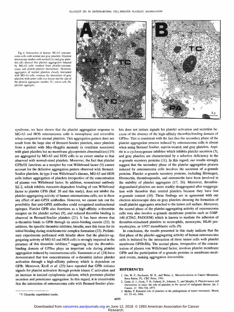
Platelets in gray platelet syndrome are characterized by a selective deficiency in the c~-granule secretory proteins (11). Using gray plate- lets, the second phase of the biphasic aggregation response to MG-63 cells was absent when compared to normal platelets (Fig. 5). Using platelet-rich plasma from a normal donor receiving aspirin (1 g/day during 3 days prior to the experiment), a monophasic aggregation response to MG-63 cells was also observed (Fig. 5). Electron micros- copy studies with normal and gray platelets showed that platelet aggregation induced by MG-63 osteosarcoma cells resulted from platelet-osteosarcoma and platelet-platelet interactions (Fig. 6). How- ever, aggregates of normal platelets closely interacted with MG-63 cells (Fig. 6A), while the interaction of gray platelets with osteosar- coma cells was looser and the size of the platelet aggregates was smaller (Fig. 6B). The aggregation response of gray platelets to HOS cells and the electron microscopic appearance of the platelet aggre- gates formed with HOS cells gave results similar to those obtained with MG-63 cells (results not shown).
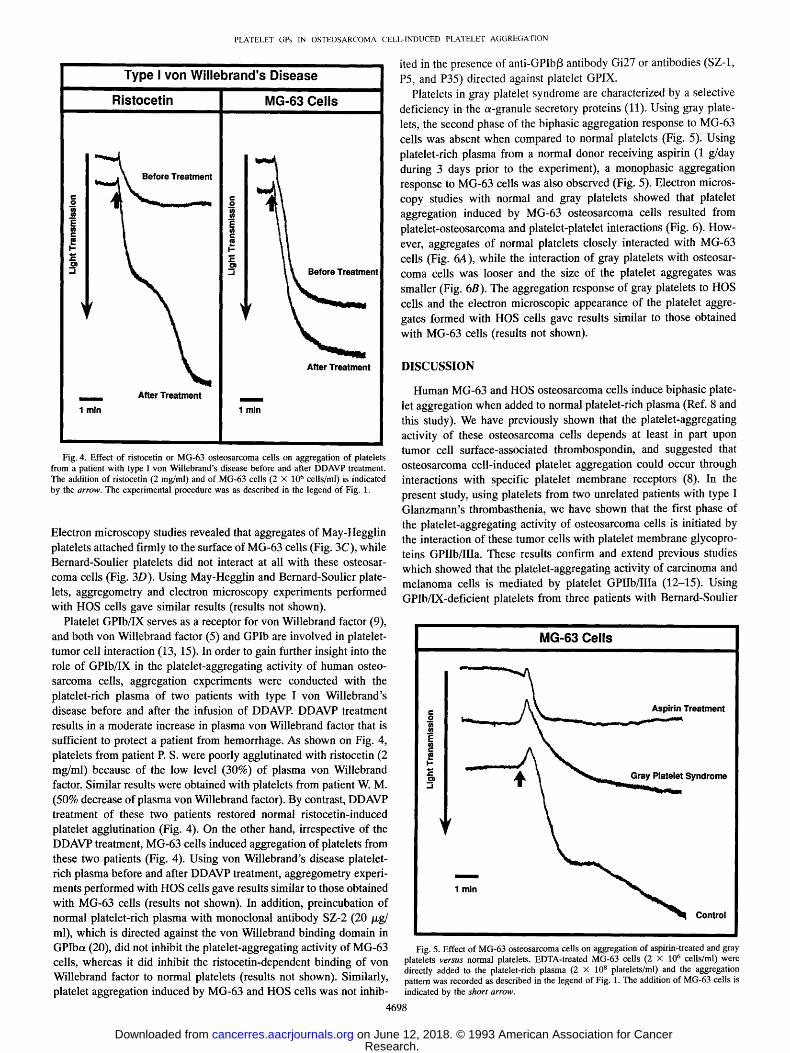
Fig. 4. Effect of ristocetin or MG-63 osteosarcoma cells on aggregation of platelets from a patient with type I v o n WiUebrand's disease before and after DDAVP treatment. The addition of ristocetin (2 mg/ml) and of MG-63 cells (2 x 10⁶ cells/ml) is indicated by the arrow. T h e experimental procedure was as described in the legend of Fig. 1.
Electron microscopy studies revealed that aggregates of May-Hegglin platelets attached firmly to the surface of MG-63 cells (Fig. 3C), while Bernard-Soulier platelets did not interact at all with these osteosar- coma cells (Fig. 3D). Using May-Hegglin and Bernard-Soulier plate- lets, aggregometry and electron microscopy experiments performed with HOS cells gave similar results (results not shown).
Platelet GPIb/IX serves as a receptor for von Willebrand factor (9), and both von Willebrand factor (5) and GPIb are involved in platelet- tumor cell interaction (13, 15). In order to gain further insight into the role of GPIb/IX in the platelet-aggregating activity of human osteo- sarcoma cells, aggregation experiments were conducted with the platelet-rich plasma of two patients with type I von Willebrand's disease before and after the infusion of DDAVP. DDAVP treatment results in a moderate increase in plasma von Willebrand factor that is sufficient to protect a patient from hemorrhage. As shown on Fig. 4, platelets from patient P. S. were poorly agglutinated with ristocetin (2 mg/ml) because of the low level (30%) of plasma von Willebrand factor. Similar results were obtained with platelets from patient W. M. (50% decrease of plasma von Willebrand factor). By contrast, DDAVP treatment of these two patients restored normal ristocetin-induced platelet agglutination (Fig. 4). On the other hand, irrespective of the DDAVP treatment, MG-63 cells induced aggregation of platelets from these two patients (Fig. 4). Using von Willebrand's disease platelet- rich plasma before and after DDAVP treatment, aggregometry experi- ments performed with HOS cells gave results similar to those obtained with MG-63 cells (results not shown). In addition, preincubation of normal platelet-rich plasma with monoclonal antibody SZ-2 (20/xg/ ml), which is directed against the von Willebrand binding domain in GPIbot (20), did not inhibit the platelet-aggregating activity of MG-63 cells, whereas it did inhibit the ristocetin-dependent binding of von Willebrand factor to normal platelets (results not shown). Similarly, platelet aggregation induced by MG-63 and HOS cells was not inhib-
4698
DISCUSSION
Human MG-63 and HOS osteosarcoma cells induce biphasic plate- let aggregation when added to normal platelet-rich plasma (Ref. 8 and this study). We have previously shown that the platelet-aggregating activity of these osteosarcoma cells depends at least in part upon tumor cell surface-associated thrombospondin, and suggested that osteosarcoma cell-induced platelet aggregation could occur through interactions with specific platelet membrane receptors (8). In the present study, using platelets from two unrelated patients with type I Glanzmann's thrombasthenia, we have shown that the first phase of the platelet-aggregating activity of osteosarcoma cells is initiated by the interaction of these tumor cells with platelet membrane glycopro- teins GPIIb/IIIa. These results confirm and extend previous studies which showed that the platelet-aggregating activity of carcinoma and melanoma cells is mediated by platelet GPIIb/IIIa (12-15). Using GPIb/IX-deficient platelets from three patients with Bernard-Soulier
Fig. 5. Effect of MG-63 osteosarcoma cells on aggregation of aspirin-treated and gray platelets versus normal platelets. EDTA-treated MG-63 cells (2 X 10⁶ cells/ml) were directly added to the platelet-rich plasma (2 x 10⁸ platelets/ml) and the aggregation pattern was recorded as described in the legend of Fig. 1. The addition of MG-63 cells is indicated by the shor t arrow.
PLATELET GPs IN OSTEOSARCOMA CELL-INDUCED PLATELET AGGREGATION
Fig. 6. Interaction of human MG-63 osteosar- coma cells with normal and gray platelets. Electron microscopy studics with normal (A) and gray plate- lets (B) showed that platelet aggregation induced by MG-63 cells resulted from platelet-osteosar- coma and platelct-platelet interactions. However, aggregates of normal platelets closely interacted with MG-63 cells, whereas the interaction of gray platelets with tumor cells was looser and the size of the platelet aggregates smaller. TC, tumor cell; PA, platelet aggregate.
syndrome, we have shown that the platelet aggregation response to MG-63 and HOS osteosarcoma cells is monophasic and reversible when compared to normal platelets. This aggregation pattern does not result from the large size of Bernard-Soulier platelets, since platelets from a patient with May-Hegglin anomaly (a condition associated with giant platelets but no membrane glycoprotein abnormalities) (19) are aggregated by MG-63 and HOS cells to an extent similar to that observed with normal-sized platelets. Moreover, the fact that platelet GPIb/IX functions as a receptor for von Willebrand factor (9) cannot account for the defective aggregation pattern observed with Bernard- Soulier platelets. In type Ivon Willebrand's disease, MG-63 and HOS cells induce aggregation of platelets irrespective of the concentration of plasma von Willebrand factor. In addition, monoclonal antibody SZ-2, which inhibits ristocetin-dependent binding of von Willebrand factor to platelet GPIb (Ref. 20 and this study), does not inhibit the platelet-aggregating activity of human osteosarcoma cells, nor is there any effect of anti-GPIX antibodies. However, we cannot rule out the possibility that anti-GPIX antibodies could recognized nonfunctional epitopes. Platelet GPIb also functions as a high-affinity a-thrombin receptor on the platelet surface (9), and reduced thrombin binding is observed in Bernard-Soulier platelets (21). It has been shown that a-thrombin binds to GPIb through its anion-binding exosite (22). In addition, the specific thrombin inhibitor, hirudin, uses this locus for its initial binding during stoichiometric complex formation (23). Prelimi- nary experiments performed with hirudin show that the platelet-ag- gregating activity of MG-63 and HOS cells is strongly impaired in the presence of this thrombin inhibitor, 4 suggesting that the thrombin- binding domain of GPIba plays an important role during platelet aggregation induced by osteosarcoma cells. Yamamoto et al. (24) have demonstrated that low concentrations of a-thrombin induce platelet activation through a high-affinity pathway which is dependent on GPIb. Moreover, Kroll et al. (25) have reported that GPIb initiates signals for platelet activation through protein kinase C activation and an increase in ionized cytoplasmic calcium, which promotes platelet secretion and potentiates aggregation. In this regard, it is conceivable that the interaction of osteosarcoma cells with Bernard-Soulier plate-
4 p. Clezardin, unpublished results.
lets does not initiate signals for platelet activation and secretion be- cause of the absence of the high-affinity thrombin-binding domain of GPIba. This is consistent with the fact that the secondary phase of the platelet aggregation process induced by osteosarcoma cells is absent when using Bernard Soulier, aspirin-treated, and gray platelets. Aspi- rin is a cyclooxygenase inhibitor which inhibits platelet secretion (3), and gray platelets are characterized by a selective deficiency in the a-granule secretory proteins (11). In this regard, our results strongly suggest that the secondary phase of the platelet aggregation process induced by osteosarcoma cells involves the secretion of a-granule proteins. Platelet a-granule secretory proteins, including fibrinogen, fibronectin, thrombospondin, and osteonectin have been involved in the stability of platelet aggregates (17, 26). Moreover, thrombin- degranulated platelets are more readily deaggregated after reaggrega- tion with thrombin than control platelets because they have lost a-granule content (10). These findings are in agreement with our electron microscopic data on gray platelets showing the formation of small platelet aggregates attached to the tumor cell surface. Moreover, the second phase of the platelet-aggregating activity of osteosarcoma cells may also involve a-granule membrane proteins such as GMP- 140 (CD62, PADGEM) which is known to mediate the adhesion of thrombin-stimulated platelets to neutrophils, monocytes, HL60 pro- myelocytes, or U937 monoblastic cells (9).
In conclusion, the results presented in this study indicate that the first phase of the platelet-aggregating activity of human osteosarcoma cells is initiated by the interaction of these tumor cells with platelet membrane GPIIb/IIIa. The second phase, irrespective of the concen- tration of plasma von Willebrand factor, involves platelet membrane GPIb and the participation of a-granule proteins in membrane-medi- ated events, making aggregation irreversible.
REFERENCES
1. Orr, W. E, Buchanan, M. R., and Weiss, L. Microcirculation in Cancer Metastasis. Boca Raton, FL: CRC Press, 1991.
2. Gasic, G. J., Gasic, T. B., Galanti, N., Johnson, T., and Murphy, S. Platelet-tumor cell interactions in mice: the role of platelets in the spread of malignant disease. Int. J. Cancer, 11: 704-718, 1973.
3. Mehta, P. Potential role of platelets in the pathogenesis of tumor metastasis. Blood, 63: 55-63, 1984.
PLATELET GPs IN OSTEOSARCOMA CELL-INDUCED PLATELET AGGREGATION
4. Chopra, H., Timar, J., Rong, X., Grossi, I. M., Hatfield, J. S., Fligiel, S. E. G., Finch, C. A., Taylor, J. D., and Honn, K. V. Is there a role for the tumor cell integrin OQib~3 3 and cytoskeleton in tumor cell-platelet interaction? Clin. Exp. Metastasis, 10: 125- 137, 1992.
5. Karpatkin, S., Pearlstein, E., Ambrogio, C., and Coller, B. S. Role of adhesive proteins in platelet-tumor interaction in vitro and metastasis formation in vivo. J. Clin. Invest., 81: 1012-1019, 1988.
6. Camez, A., Dupuy, E., Bellucci, J., Calvo, E, Bryckaert, M. C., and Tobelem, G. Invasion Metastasis, 6: 321-334, 1986.
7. Tuszynski, G. P., Gasic, T. B., Rothman, V. L., Knudsen, K. A., and Gasic, G. J. Thrombospondin, a potentiator of tumor cell metastasis. Science (Washington DC), 47: 4130-4133, 1987.
8. Clezardin, P., Serre, C. M., Trzeciak, M. C., Drouin, J., and Delmas, P. D. Throm- bospondin binds to the surface of human osteosarcoma cells and mediates platelet- osteosarcoma cell interaction. Cancer Res., 51: 2621-2627, 1991.
9. Kieffer, N., and Phillips, D. R. Platelet membrane glycoproteins: functions in cellular interactions. Annu. Rev. Cell Biol., 6: 32%357, 1990.
10. Kinlough-Rathbone, R. L., Packham, M. A., Perry, D. W., Mustard, J. E, and Catta- neo, M. Lack of stability of aggregates after thrombin-induced reaggregation of thrombin-degranulated platelets. Thromb. Haemostasis, 67: 453--457, 1992.
11. Gerrard, J. M., Phillips, D. R., Ran, G. H. R., Plow, E. E, Walz, D. A., Harker, A., and White, J. G. Biochemical studies of two patients with the gray platelet syndrome. Selective deficiency of platelet alpha granules. J. Clin. Invest., 66: 102-109, 1980.
12. Batisda, E., Almirall, L., and Ordinas, A. Tumor cell-induced platelet aggregation is a glycoprotein dependent and lipoxygenase-associated process. Int. J. Cancer, 39: 760-763, 1987.
13. Grossi, I. M., Fitzgerald, L. A., Kendall, A., Taylor, J. D., Sloane, B. E, and Honn, K. V. Inhibition of human tumor cell induced platelet aggregation by antibodies to platelet glycoproteins Ib and lib/Ilia. Proc. Soc. Exp. Biol. Med., 186: 378--383, 1987.
14. Boukerche, H., Berthier-Vergnes, O., Tabone, E., Dor6, J-F., Leung, L. L. K., and McGregor, J. L. Platelet-melanoma cell interaction is mediated by the glycoprotein Ilb-IIIa complex. Blood, 74: 658--663, 1989.
15. Kitawaga, H., Yamamoto, N., Yamamoto, K., Tanoue, K., Kosaki, G., and Yamazaki, H. Involvement of platelet membrane glycoprotein Ib and glycoprotein IIb/IIIa com- plex in thrombin-dependent and -independent platelet aggregations induced by tumor cells. Cancer Res., 49: 537-541, 1989.
16. Kehrel, B., Kokott, R., Stenzinger, W., and Balleisen, L. Analysis of platelet glyco- proteins in thrombocytopathias using the lectin-avidin-biotin-peroxidase (LABP) technique. Folia Haematol. (Leipz.), 115: 425--429, 1988.
17. Clezardin, P., Malaval, L., Morel, M-C., Guichard, J., Lecompte, T., Trzeciak, M-C., Dechavanne, M., Breton-Gofius, J., Delmas, E D., and Kaplan, C. Osteonectin is an a-granule component involved with thrombospondin in platelet aggregation. J. Bone Miner. Res., 6: 105%1070, 1991.
18. Knapp, W. Leucocyte Typing IV. White Cell Differentiation Antigens. New York: Oxford University Press, 1989.
19. Coller, B. S., and Zarrabi, M. H. Platelet membrane studies in the May-Hegglin anomaly. Blood, 58: 27%284, 1981.
20. Ruan, C., Du, X., Xi, X., Castaldi, P. A., and Bemdt, M. C. Amurine antiglycoprotein Ib complex monoclonal antibody, SZ 2, inhibits platelet aggregation induced by both ristocetin and collagen. Blood, 69: 570-577, 1987.
21. Jamieson, G. A., and Okumura, T. Reduced thrombin binding and aggregation in Bernard-Soulier platelets. J. Clin. Invest., 61: 861-864, 1978.
22. Bouton, M. C., Thurieau, C., Fauchere, J. L., Verbeuren, T., Guillin, M. C., and Jandrot-Perrus, M. Glycoprotein Iba peptide 26%287 binds to human a-thrombin and modulates enzyme activity, Thromb. Haemostasis, 69: 2323, 1993.
23. Stone, S. R., and Hofsteenge, J. Kinetics of the inhibition of thrombin by hirudin. Biochemistry, 25: 4622---4628, 1986.
24. Yamamoto, N., Green, N. J., Barnard, M. R., Tanoue, K., Yamazaki, H., Jamieson, G. A., and Michelson, A. D, Blood, 77: 1740-1748, 1991.
25. Kroll, M. H., Harris, T. S., Moake, J. L., Handin, R. I., and Schafer, A. I. yon Willebrand factor binding to platelet GPIb initiates signals for platelet activation. J. Clin. Invest., 88: 1568-1573, 1991.
26. Peerschke, E. I. B. Events occurring after thrombin-induced fibrinogen binding to platelets. Semin. Thromb. Hemostasis, 18: 34--43, 1992.