Role of the Central Metal Ion and Ligand Charge in the DNABinding and Modification by Metallosalen Complexes
Subhrangsu S. Mandal,† Umesh Varshney,‡ and Santanu Bhattacharya*,†
Department of Organic Chemistry and Center for Genetic Engineering, Indian Institute of Science,Bangalore 560012, India. Received November 14, 1996X
Several metal complexes of three different functionalized salen derivatives have been synthesized.The salens differ in terms of the electrostatic character and the location of the charges. The interactionsof such complexes with DNA were first investigated in detail by UV-vis absorption titrimetry. Itappears that the DNA binding by most of these compounds is primarily due to a combination ofelectrostatic and other modes of interactions. The melting temperatures of DNA in the presence ofvarious metal complexes were higher than that of the pure DNA. The presence of additional chargeon the central metal ion core in the complex, however, alters the nature of binding. Bis-cationic salencomplexes containing central Ni(II) or Mn(III) were found to induce DNA strand scission, especiallyin the presence of co-oxidant as revealed by plasmid DNA cleavage assay and also on the basis of theautoradiogram obtained from their respective high-resolution sequencing gels. Modest base selectivitywas observed in the DNA cleavage reactions. Comparisons of the linearized and supercoiled forms ofDNA in the metal complex-mediated cleavage reactions reveal that the supercoiled forms are moresusceptible to DNA scission. Under suitable conditions, the DNA cleavage reactions can be inducedeither by preformed metal complexes or by in situ complexation of the ligand in the presence of theappropriate metal ion. Also revealed was the fact that the analogous complexes containing Cu(II) orCr(III) did not effect any DNA strand scission under comparable conditions. Salens with pendantnegative charges on either side of the precursor salicylaldehyde or ethylenediamine fragments didnot bind with DNA. Similarly, metallosalen complexes with net anionic character also failed to induceany DNA modification activities.
INTRODUCTION1
Design and synthesis of small synthetic systems thatrecognize specific sites of DNA is an important area ofmuch current research (1). These could be at least inpart due to the formation of noncovalently associatedcomplexes by several such molecules with nucleic acids.Many times, such physical complexation may produceimportant pharmacological effects by interfering with thebiological processes in which DNA/RNAs take part. Suchinvestigations also sometimes provide insights for themechanism of action of naturally occurring antitumorantibiotics (2). Toward this direction, there is a continu-ing search for newmetal complexes that strongly interactwith DNA. These studies led to the development ofseveral new reagents. Examples include complexes e.g.iron-EDTA (3), copper- or rhodium-1,10-phenanthro-line (4, 5), manganese-porphyrin (6), platinum-diamine(7), nickel-cyclam (8), and several photonucleases (9),among others. Examination of the interactions of transi-tion metal complexes with DNA aids in the developmentof new reagents that are potentially useful in molecularbiology (10) and also in the design of putative drugs (11).Utilities of some metal complexes as structural probes(1, 12) for DNA and in drug/protein-DNA footprinting(10, 13) are also known. Sometimes, such studies also
offer useful leads in the elucidation of potential toxicitiesof various metal complex-based systems (14).Earlier investigations (16-19) have indicated that,
while some metallosalen derivatives bind DNA avidly(15), few others induce effective DNA scission (16-19)or inhibit lymphocyte proliferation (20). Therefore, subtlevariation in the structure of the salen unit and variationof the central metal ion influence the effect of theresulting complex on DNA in a widely unrelated fashion.Griffin carefully studied (16) some of these aspects ofDNA modification in detail with manganese-salen com-plexes. Burrows and Rokita synthesized (17) somenickel-salen derivatives which induced DNA cross-linking. More recently, Routier et al. examined theinteraction of DNA with a functionalized salen-copper-(II) complex which induced DNA strand scission in thepresence of a reducing agent (18). We also examined theDNA cleavage processes induced by some bis-cationicsalen analogues (19). Examination of the above reportsmakes it clear that much still remains to be understoodabout the role of salen structure and that of the centralmetal ion in the DNA binding/modification processes. Wethought that a progressive structural variation of thesalens with respect to charge on the aryl rings, at theimine portions, and systematic variation of the centralmetal ion under a given ligand (salen) environment mightprovide additional insight on the factors involved in thechemistry of binding and DNA modification by suchcomplexes. Herein, we report the synthesis of a numberof transition metal complexes based on a few differentwater-soluble salen derivatives, 1-5. The results of thebinding abilities of different metal complexes with nativeEscherichia coli genomic DNA at various salt concentra-tions and with alkali-denatured DNA and the effects ofsuch binding on DNAmelting are included in this report.
* To whom correspondence should be addressed. Also atChemical Biology Unit, Jawaharlal Nehru Centre for AdvancedScientific Research, Bangalore 560064, India.
† Department of Organic Chemistry.‡ Center for Genetic Engineering.X Abstract published in Advance ACS Abstracts, August 1,
1997.1 Caution. Although we did not experience any difficulty
while working with these systems, perchlorate salts are poten-tially explosive!
The DNA cleavage reactions and the nicking patternsproduced by different metal complexes are also presented.
EXPERIMENTAL PROCEDURES
General Aspects. Melting points were determinedin open capillaries and are uncorrected. Infrared spectrawere recorded with a Shimadzu FTIR 8101 infraredspectrophotometer equipped with a DR-8001 workstation.1H-NMR spectra were recorded with a Bruker AC-200(200 MHz) or a Jeol FX-90 (90 MHz) NMR spectrometerusing CDCl3 or the appropriate deuterated solvent.Elemental analyses were performed by the microanalyti-cal division of the Department of Organic Chemistry atthe Indian Institute of Science in Bangalore and wererun on a Carlo Erba model 1106 elemental analyzer.These are presented as percents. Electronic spectra wererecorded on a Shimadzu model 2100 UV-visible spec-trophotometer equipped with a TCC-60 temperaturecontroller unit or a Julabo F-10 water-circulating tem-perature-controlled bath.Materials. Silica gel (UV254) plates (Sigma) were used
for TLC. Column chromatography was performed onsilica gel (60-120 mesh) from Merck (Schuchardt, Ger-many). Commercial compounds used in the synthesisand other studies herein were purchased from bestknown suppliers and used as received; solvents werepurified and dried by standard methods. All othermaterials used for preparing buffers were of the highestquality commercially available. Millipore grade waterwas used for all the experiments. The enzymes used inthis study were obtained from Pharmacia.Calf thymus DNA (CT DNA) obtained from Sigma
Chemical Co. (St. Louis, MO) was purified by phenol-chloroform extraction followed by ethanol precipitationas described previously (21). Solutions of CT DNA in 10mM Tris-HCl, 1 mM EDTA buffer (pH 7.4) gave a ratioof the UV absorbances at 260 and 280 nm (A260/A280) of>1.9. The DNA concentrations were determined by usingan extinction coefficient of 6600 M-1 cm-1 at 260 nm andwere expressed in terms of base molarity (22). Meltingtemperature determinations and the spectroscopic bind-ing studies were performed with CT DNA in 10 mM Tris-HCl, 1 mM EDTA buffer (pH 7.4).E. coli genomic DNA was isolated and purified as
described (23). Briefly, E. coli cells (CA 274 strain) wereovergrown in LB media, and the cells were subjected tolysozyme treatment, followed by lysis with SDS andproteinase K treatment. The mixture was finally sub-jected to phenol-chloroform extraction two times, andthe DNA was precipitated using 2-propanol and washedwith 70% ethanol. Then, the DNA was dissolved in 10
mM Tris-HCl, 1 mM EDTA buffer (pH 7.4) which gavean A260/A280 of ∼1.9.The plasmid DNA pTZ19R used herein was grown in
E. coli cells and isolated in our laboratory as describedearlier (19a). The supercoiled plasmid was obtained byCsCl/ethidium bromide density gradient centrifugation.Synthetic Methods. 4-[(3-Bromopropyl)oxy]salicyl-
aldehyde (6). A mixture of 138 mg (1 mmol) of 2,4-dihydroxybenzaldehyde and 404 mg (2 mmol) of 1,3-dibromopropane was refluxed in the presence of 140 mg(1.4 mmol) of KHCO3 in 50 mL of dry acetone for 60 h.At the end of this period, the solvent from the reactionmixture was removed in a rotavapor, 5 mL of water wasadded to it, and then the solvent was extracted with 3 ×10 mL portions of CHCl3. The combined chloroformextracts were evaporated to dryness and then purifiedby column chromatography over silica gel (60-120 mesh)using 2-3% EtOAc in light petroleum (60-80 °C boilingfraction). Upon chromatographic purification, a whitecrystalline solid was obtained in ca. 60% yield: mp 62-64 °C; 1H-NMR (200 MHz, CDCl3) δ 2.3 (2H, m, CH2CH2-CH2Br), 3.6 (2H, t, CH2Br), 4.2 (2H, t, CH2CH2O), 6.45(1H, d, ArH3, Jm ) 2.0 Hz), 6.55 (1H, dd, ArH5, Jm ) 2Hz, J ) 9.5 Hz), 7.5 (1H, d, ArH6, J0 ) 9.5 Hz), 9.7 (1H,s, CHO), 11.5 (1H, s, ArOH); IR (Nujol) (cm-1) 1630 (s),1510, 1210, 1095. Anal. Calcd for C10H11BrO3: C, 46.35;H, 4.27. Found: C, 46.42; H, 4.26.4-[[3-(Trimethylammonio)propyl]oxy]salicylaldehyde Bro-
mide (6a). One hundred milligrams (0.38 mmol) of 6 wasdissolved in 20 mL of EtOAc in a screw-top pressure tube.Two milliliters of dry trimethylamine gas was passed intothis solution precooled at ∼0-5 °C. Then, the screw topof the tube was closed tightly and the tube allowed tostand for 6 h at ambient temperature. At the end of thisperiod, a brownish-white solid was obtained. The solidwas collected by filtration and was recrystallized from amixture of EtOAc/EtOH. Recrystallization afforded acolorless, crystalline, hygroscopic product in practicallyquantitative yield: mp 152-154 °C; 1H-NMR (90 MHz,D2O) δ 2.4 (2H, m, CH2CH2CH2N+Me3), 3.2 (9H, s, N+-(CH3)3), 3.6 (2H, t, CH2N+Me3), 4.23 (2H, t, ArOCH2), 6.52(1H, d, ArH3, Jm ) 2.0 Hz), 6.68 (1H, dd, ArH5, Jm ) 2Hz, J ) 9.5 Hz), 7.68 (1H, d, ArH6, J0 ) 9.5 Hz), 9.8 (1H,s, CHO); IR (Nujol) (cm-1) 1630 (s), 3400 (s). Anal. Calcdfor C13H20NO3Br‚H2O: C, 46.44; H, 6.59; N, 4.17.Found: C, 46.41; H, 6.30; N, 4.02.4-[[3-(Trimethylammonio)propyl]oxy]salicylaldehyde Per-
chlorate (6b). The above material, 4-[[3-(trimethyl-ammonio)propyl]oxy]salicylaldehyde bromide (6a, 425mg, 1.3 mmol), was dissolved in 1 mL of water, and theresulting aqueous solution was treated with 1 mL ofaqueous NaClO4 (327 mg, 2.6 mmol). This resulted in
Charge in the Metallosalen Complexes Bioconjugate Chem., Vol. 8, No. 6, 1997 799
the formation of the corresponding perchlorate salt whichprecipitated as a white solid. This was filtered and driedin a desiccator, ca. 95% yield: mp 140-142 °C; 1H-NMR(200 MHz, DMSO-d6) δ 2.1 (2H, m, CH2CH2CH2N+Me3),3.05 (9H, s, N+(CH3)3), 3.4 (2H, t, CH2N+Me3), 4.05 (2H,t, ArOCH2), 6.3 (1H, d, ArH3), 6.5 (1H, dd, ArH5), 7.5 (1H,ArH6, d), 9.5 (1H, s, CHO); IR (Nujol) (cm-1) 1630 (s),1460 (s), 1375 (s), 1230 (s), 1100 (s), 625 (s). Anal. Calcdfor C13H20NO7Cl: C, 46.23; H, 5.97; N, 4.15. Found: C,46.21; H, 6.01; N, 4.02.N,N′-Bis[4-[[3-(trimethylammonio)propyl]oxy]salic-
ylidene]ethylenediamine Diperchlorate (1). 6b, 4-[[3-(trimethylammonio)propyl]oxy]salicylaldehyde perchlo-rate (250 mg, 0.74 mmol), was dissolved in 10 mL of dryMeOH by careful warming. To this was added 22.2 mg(0.37 mmol) of freshly distilled ethylenediamine, and theresulting mixture was warmed to ∼60 °C for 10 minwhich upon cooling to ambient temperature produced ayellow precipitate. The yellow residue was isolated andwashed with cold MeOH and dried (yield ca. 80%): mp258-260 °C; 1H-NMR (90 MHz, DMSO-d6) δ 2.1 (4H, m,CH2CH2CH2NMe3), 3.05 (18H, s, N(CH3)3), 3.4 (4H, t,CH2NMe3), 3.9 (4H, s, dNCH2CH2Nd), 4.05 (4H, t,ArOCH2), 6.3-6.45 (4H, m, ArH3, ArH5), 7.2 (2H, d,ArH6), 8.45 (2H, s, ArHCdN); IR (Nujol) (cm-1) 1630 (s),1610 (s), 1520 (s), 1460 (s), 1370 (s), 1210 (s), 1095 (s),620 (s). Anal. Calcd for C28H44N4O12Cl2: C, 48.06; H,6.34; N, 8.0. Found: C, 47.62; H, 6.33; N, 7.90.N,N′-Bis[4-[[3-(trimethylammonio)propyl]oxy]salic-
ylidene]ethylenediamine-Nickel (II) Bisperchlorate (2a).1 (55 mg, 0.079 mmol) was suspended in 10 mL of dryMeOH. NiCl2‚6H2O (15 mg, 0.08 mmol) was added inthe form a methanolic solution into this suspension, andthe resulting mixture was warmed at 60 °C for 30 min.This gave a deep-red solution which was first allowed tocool to room temperature and then kept in a refrigeratorfor 48 h. A red-colored, hygroscopic, crystalline precipi-tate separated out which was collected after filtration,recrystallized from dry MeOH, and dried (yield ca.42%): UV-vis [λmax (nm), ε] 1:1 (CH3CN/CH3OH) 348(25 000), 304 (29 000), 236 (14 000), 211 (20 000); IR (KBrpellet) (cm-1) 600 (m), 730 (m), 920 (m), 1040 (m), 1120(s), 1200 (s), 1215 (s), 1300 (m), 1420 (m), 1480 (s), 1490(s), 1530 (s), 1600 (s). Anal. Calcd for C28H42N4-O12Cl2Ni‚2.5H2O: C, 41.94; H, 5.28; N, 6.99. Found: C,42.01; H, 5.49; N, 6.99.N,N′-Bis[4-[[3-(trimethylammonio)propyl]oxy]salic-
ylidene]ethylenediamine-Copper(II) Bisperchlorate (2b).The ligand 1 (40 mg, 0.05 mmol) was suspended in 10mL of dry MeOH, to which a methanolic solution of 16mg (0.08 mmol) of Cu(OAc)2‚4H2O was added, and theresulting mixture was warmed for 30 min at 60 °C. Thesolution slowly turned into a bluish pink color which uponcooling gave a pink crystalline precipitate. The residuewas collected by filtration and repeatedly recrystallizedfrom dry MeOH to give a pink hygroscopic solid (yieldca. 60%): UV-vis [λmax (nm), ε] (H2O) 365.5 (10 000),282.5 (20 000), 249.5 (30 000), 226 (29 000); IR (KBrpellet) (cm-1) 620 (s), 840 (m), 1090 (s), 1110 (s), 1210(s), 1400 (m), 1540 (m), 1610 (s). Anal. Calcd for C28H42-N4O12CuCl2‚1H2O: C, 43.10; H, 5.43; N, 7.18. Found: C,42.89; H, 5.54; N, 7.08. EPR: AII 190.1 G, gII 2.2.N,N′-Bis[4-[[3-(trimethylammonio)propyl]oxy]salic-
ylidene]ethylenediamine-Cobalt(II) Bisperchlorate as aMonomeric Oxygen Adduct (2c). 1 (30 mg, 0.043 mmol)was suspended in 10 mL of dry MeOH. To this wasadded a methanolic solution of 17.5 mg (0.07 mmol) ofCo(OAc)2‚4H2O, and the resulting mixture was warmedat 60 °C for 30 min. This gave a brownish-red solutionwhich gradually changed into a deep dark brown color
upon standing at ambient temperature in exposure to air.It was kept for 1 week in this manner, from which a darkbrown complex was precipitated upon addition of dryether (yield ca. 44%): UV-vis [λmax (nm), ε] (H2O) 361(7100), 289 (broad hump), 258 (48 000); FTIR (KBr pellet)(cm-1) 625 (s), 940 (m), 1000 (w), 1090 (s), 1125 (s), 1145(s), 1200 (m), 1430 (s), 1550 (s), 1600 (s), 1620 (s). Anal.Calcd for C28H42N4O12CoCl2O2‚2H2O: C, 40.97; H, 5.62;N, 6.79. Found: C, 40.94; H, 5.63; N, 6.35 (24).N,N′-Bis[4-[[3-(trimethylammonio)propyl]oxy]salic-
ylidene]ethylenediamine-Manganese(III) Trisperchlorate(2d). The ligand 1 (66 mg, 0.09 mmol) was suspendedin 15 mL of dry MeOH to which was added a methanolicsolution of 26.7 mg (0.2 mmol) of Mn(OAc)3‚4H2O andheated at 60 °C for 30 min. The initial yellowish colorof the solution of the Schiff base soon changed into agrayish-black solution from which 70% of the MeOH wasremoved under vacuum. Upon addition of dry ether tothis, a grayish-black solid separates out followed bycooling in a refrigerator. The solid was repeatedlyrecrystallized from dry MeOH to give a hygroscopicmaterial of 44% yield: UV-vis [λmax (nm), ε] (CH3CN)343 (15 400), 303 (21 400), 237 (12 300), 208 (17 400);FTIR (KBr pellet) (cm-1 630 (s), 850 (w), 1000 (w), 1095(s), 1110 (s), 1150 (s), 1300 (w), 1400 (w), 1490 (w), 1530(w), 1600 (s). Anal. Calcd for C28H42N4O16Cl3Mn‚2.5H2O: C, 37.49; H, 5.28; N, 6.24. Found: C, 37.32; H,5.17; N, 6.15.N,N′-Bis(salicylidene)-3,4-diaminobenzoic Acid-Nick-
el(II) (3). To a solution of 400 mg (3.3 mmol) of salicyl-aldehyde in 10 mL of MeOH was added with stirring amethanolic solution of 250 mg (1.6 mmol) of freshlyrecrystallized 3,4-diaminobenzoic acid. From this solu-tion after some time, an orange-colored crystalline solidprecipitated out, which was filtered, dried, and thenrecrystallized from MeOH. This orange Schiff base (100mg) was dissolved in hot MeOH; to this was added amethanolic solution of 46.7 mg (0.28 mmol) of the NiCl2,and the resulting mixture was heated at 60 °C for 30 min.This produced a transparent deep red-colored solutionwhich upon on standing at ambient temperature gavean orange-red-colored precipitate which was filtered andrecrystallized from a mixture of MeOH and CHCl3 in 80%yield. Anal. Calcd for C21H14N2O4Ni‚0.5CHCl3: C, 53.16;H, 3.22; N, 5.76. Found: C, 53.38; H, 3.45; N, 6.05.[(6-Oxo-6-ethoxyhexyl)oxy]benzaldehyde (7). A mixture
of 1.198 g (8 mmol) of 2,4-dihydroxybenzaldehyde and 2g of ethyl 6-bromohexanoate was refluxed in dry acetonefor 2 days in the presence of 800 mg (8 mmol) ofanhydrous KHCO3. Then, the solvent form this reactionmixture was removed, and 5 mL of water was added intothe residue. The resulting mixture was extracted with3 × 30 mL of chloroform. The chloroform extract wasevaporated to dryness and then purified by columnchromatography over silica gel (60-120 mesh) using 2%EtOAc in light petroleum (60-80 °C boiling fraction).Upon concentration of the eluted fraction, a highlyviscous liquid was obtained in ca. 50% yield: IR (neat)(cm-1) 3250-3400 (br), 2900 (s), 1715 (s), 1620 (s), 1495(s), 1200 (s), 1100 (s), 1000 (s); 1H-NMR (90 MHz, CDCl3)δ 1.2 (3H, t, CH2CH3), 1.5-2.0 (6H, m, OCH2(CH2)3CH2),2.4 (2H, t, CH2COO), 4.05-4.2 (4H, m, ArOCH2, +COOCH2CH3), 6.45 (1H, d, ArH3, Jm ∼ 1.5 Hz), 6.55 (1H,dd, ArH5, Jm ∼ 1.5 Hz, J0 ∼ 9.4 Hz), 7.4 (1H, d, ArH6, Jo∼ 9.4 Hz), 9.8 (1H, s, CHO), 11.5 (1H, s, ArOH).[(6-Hydroxy-6-oxohexyl)oxy]benzaldehyde (7a). Com-
pound 7 was saponified under refluxing conditions using2% aqueous NaOH. The resulting reaction mixture wascooled in ice and neutralized by dropwise addition ofconcentrated HCl under ice-cold conditions. This gave
a white precipitate which was filtered, and the residuewas washed with cold water, dried, and then purified bycolumn chromatography over silica gel (60-120 mesh)using 2% MeOH in CHCl3. The purified material wasrecrystallized from EtOAc to afford a solid in 80% yield:mp 124-125 °C; IR (Nujol) (cm-1) 3320 (s), 1710 (s), 1610(s), 1460 (s), 1370 (s), 1300 (s), 1200 (s), 1050 (s), 860 (s);1H-NMR (90MHz, CDCl3) δ 1.8 (6H, m, OCH2(CH2)3CH2),2.2 (2H, t, CH2COO), 4.0 (2H, t, ArOCH2), 6.45 (1H, d,ArH3, Jm ∼ 1.5 Hz), 6.6 (1H, dd, ArH5, Jm ) 1.5 Hz, Jo ∼9.4 Hz), 7.3 (1H, d, ArH6, Jo ∼ 9.4 Hz), 9.8 (1H, s, CHO),11.45 (1H, s, ArOH). Anal. Calcd for C13H16O5, 0.4-EtOAc: C, 62.38; H, 6.83. Found: C, 62.71; H, 6.83.N,N′-Bis[4-[(6-hydroxy-6-oxohexyl)oxy]salicylidene]Eth-
ylenediamine-Nickel(II) (4). 7a (252 mg, 1 mmol) wassuspended in 5 mL of dry MeOH. Freshly distilledethylenediamine (30 mg, 0.5 mmol) was added into this.The resulting yellowing solution was stirred and warmedfor 5 min which on prolonged standing at ambienttemperature gave a yellow-colored precipitate, 7b. Thiswas filtered, washed with cold MeOH, and dried (yieldof 90%). The Schiff base 7b (100 mg, 0.18 mmol) wassuspended in 5 mL of a mixture of dry MeOH and CHCl3.A methanolic solution containing 31 mg (0.18 mmol) ofNiCl2 was added to this, and the mixture was warmedfor 30 min at 60 °C. During warming, the yellowsuspension got slowly converted to a deep red solutionwhich on cooling and standing at ambient temperaturefor several hours gave a red-colored crystalline precipi-tate. It was filtered, and the residue was washed withice-cold, dry MeOH and then recrystallized from amixture of MeOH and CHCl3 to yield a material of ca.70% yield: UV-vis [λmax (nm), ε] [5 mM Tris-HCl buffer(pH 7.4) 376 (7000), 258 (41 000); FTIR (KBr pellet)(cm-1) 700 (s), 760 (w), 800 (w), 830 (m), 1110 (s), 1200(s), 1220 (s), 1540 (s), 1600 (s), 1700 (s). Anal. Calcd forC28H34N2O8Ni‚0.75CHCl3: C, 51.17; H, 5.19; N, 4.15:Found: C, 50.84; H, 5.30; N, 4.37.N,N′-Bis(salicylidene)ethylenediamine-Manganese-
(III) Acetate (5). This was prepared by following aliterature procedure (16a). Briefly, 569 mg (2.1 mmol)ofN,N′-bis(salicylidene)ethylenediamine was dissolved in10 mL of dry MeOH, to which was added a methanolicsolution of 442 mg (2.2 mmol) of Mn(OAc)3‚4H2O, andthe resulting mixture was warmed at 60 °C for 30 min.Then, it was allowed to cool to room temperature. Uponaddition of dry ether to this, a gray-black precipitate wasobtained. This compound showed all the properties asreported previously (16a) (yield of 70%): IR (cm-1) 1630(s), 1595 (s), 1560 (s), 1460 (s), 1440 (s), 1385 (s), 1330(s), 1290 (s), 1200 (m), 1140 (m), 1130 (s), 1080 (m), 1030(m), 960 (m), 900 (s), 800 (s), 770 (s); UV-vis [λmax (nm),ε] (ethanol) 400 (6100), 350 (8000), 310 (16 000), 283(21 000), 220 (41 000). Anal. Calcd for C18H17N2O4Mn:C, 56.85; H, 4.50; N, 7.37. Found: C, 56.82; H, 4.47; N,7.11.Absorption Titration. Absorption titrations were
carried out by keeping the concentration of the probeconstant while adding a concentrated solution of the E.coli genomic DNA in 1 mM Tris-HCl, 0.1 mM EDTAbuffer (pH 7.4) in increasing amounts in both the cuvettesuntil the saturations in hypochromism were observed.The saturation in hypochromism is quantitatively shownby plotting the A0/A vs [DNA], where A0 and A are theabsorption intensities of the individual metal complexesin the absence and presence of various concentrations ofDNA, respectively. The intrinsic binding constants forthe different metal complexes with DNAwere determinedby the half-reciprocal plot method as given in theliterature (25). The intrinsic binding constant (K) for a
given complex with DNA was then obtained from a plotof D/∆εap vs D according to eq 1, D/∆εap ) D/∆ε + 1/(∆εK)(1) where D is the concentration of DNA in base molarity,∆εap ) |εa - εf|, and ∆ε ) |εb - εf|, where εb and εf arerespective extinction coefficients of the complex in thepresence and absence of DNA, respectively. The appar-ent extinction coefficient, εa, was obtained by calculatingAobsd/[complex]. The data were fitted to eq 1, with a slopeequal to 1/∆ε and a y-intercept equal to 1/(∆εK). Theintrinsic binding constants (K) were determined from theratio of the slope to the y-intercept.Effect of the Addition of Salt on DNA Binding.
To find out the effect of the variation in the ionic strengthon the binding abilities of metal complexes with CT DNA,we studied the effects of addition of various salts to asolution containing DNA-2b complexes by adding in-creasing amounts of different salt solutions (30 and 45mM NaCl or 15 mM MgCl2) to the 2b-DNA complex.The effects of such additions were followed by UV-visabsorption spectroscopy (26).DNA Denaturation and Binding of Metallosalen.
The comparative binding efficiencies of the Ni complex(2a) toward double-stranded and alkali-denatured CTDNA were studied by using UV-vis absorption spectros-copy. The denatured CT DNA was obtained by incubat-ing the DNA in 1 M NaOH at room temperature for 10min. The spectrum of the Ni complex (2a) was recordedfirst; then, 5 µL of alkali-denatured CT DNA was addedto this, and the changes in the spectra were then recordedagain. The pH of the resulting solution was then slowlydecreased by the addition of increasing amounts of HCluntil the saturation in binding was reached.Melting Temperatures. Melting temperatures for
DNA in the absence and in the presence of various metalcomplexes were measured by following the changes inthe UV-vis absorption spectra at 260 nm as a functionof temperature in a Shimadzu model UV-2100 UV-visspectrophotometer. The absorption intensities at 260 nmwere plotted against individual temperatures in thepresence of each metal complex, and the midpoints of theinflection regions in the temperature vs A260 curves weretaken as the corresponding Tm values (27). The temper-atures of the cuvettes were maintained using a Julabomodel F-10 water-circulating bath. Each melting tem-perature determination employed 72 µM (base molarity)CT DNA in 5 mM Tris-HCl buffer (pH 7.4).DNA Cleavage Experiments. The DNA cleavage or
modification reactions induced by different metal com-plexes in the absence and in the presence of co-oxidantswere performed using the supercoiled plasmid pTZ19R(2.9 kbp, Pharmacia) in 20 mM Tris-HCl buffer (pH 7.4).In a typical experiment, the plasmid DNA (0.25 µg/reaction) was incubated in a reaction mixture (10 µL)containing various concentrations of individual metalcomplexes (either preisolated or prepared in situ) in 20mM Tris-HCl buffer (pH 7.4) at 37 °C. Reactions wereinitiated by the addition of MMPP (0.5 mM) and werestopped after 5 min by the addition of a terminatingagent (5 µL) containing 10 mM â-mercaptoethanol, 20%glycerol, 25 mM EDTA, and 0.05% bromophenol blue/xylene cyanol (1:1). Then, this was kept on ice. Thesamples were loaded on 1% neutral agarose gel and weresubjected to electrophoresis in a horizontal slab gelapparatus using 0.5 × TBE as the buffer (at 100 V for ∼1.5 h). After electrophoresis, the gels were stained witha solution of 0.5 µg/mL ethidium bromide for ∼60 minand then destained. Bands of DNA were then visualizedunder UV light (photodyne transilluminator, 312 nm) andphotographed (Canon SLR camera with an orange filter)in a darkroom.
Charge in the Metallosalen Complexes Bioconjugate Chem., Vol. 8, No. 6, 1997 801
Sequencing and Autoradiogram. The sequence orbase selectivities in the DNA modification reactionseffected by different salen complexes were determinedin the following manner. The supercoiled plasmid pTZ19Rwas linearized with EcoRI. After complete digestion ofthe plasmid with EcoRI (confirmed by agarose gel), theDNA was subjected to ethanol precipitation and drying.The DNA prepared in the above manner was used forthe different reagent-induced DNA cleavage experiments.The DNA cleavage reaction conditions for the sequenc-
ing experiments were similar to those employed in theagarose gel assays (described above) except that a totalreaction volume of 50 µL and 2 µg of plasmid/reactionwere used. After incubation for 5 min, 10 µL of thereaction mixture was aliquoted and analyzed by gelelectrophoresis on agarose to confirm the reagent-inducedDNA cleavage. The residual reaction mixture was firstsubjected to ethanol precipitation and then dried undervacuum. The dry DNA pellets were then resuspendedin water and used as templates for the primer extensionreactions. The primer extension reactions were carriedout by the adaptation of a literature procedure (28).Briefly, ∼3 pmol of the 32P-end-labeled primer (300 000counts, a 19-mer oligonucleotide) and 4 µL of 0.01 MNaOH were added into a solution containing ∼0.5 µg ofthe DNA templates. Then, the volume of the resultingreaction mixture was made up to 40 µL which afterthorough mixing was kept at 80 °C for 2 min and finallyleft on ice for an additional 5 min. Into this ice-coldreaction mixture was added 5 µL of the primer extensionbuffer [0.5 mM Tris-HCl (pH 7.4), 0.1 M MgSO4, and 2mM DTT], and the resulting mixture was again incu-bated at 45 °C for 3 min. After this, the mixture waskept on ice for a period of 30 min. Then, an aliquot of 5µL of the dNTP mix (dATP, dTTP, dCTP, and dGTP eachat 5 mM) was added to this mixture, and primer exten-sion reactions for each reaction were initiated by theaddition of 1 unit of Klenow DNA polymerase at 50 °cfor 10 min. Finally, the reactions were quenched by thesuccessive addition of 10 µL of 4 mM ammonium acetate/20 mM EDTA and 180 µL of absolute EtOH. Theresulting mixture was then kept at -20 °C for 1 h andpelleted down by centrifugation. The pellets thus ob-tained were air-dried and resuspended in 8 µL of 80%formamide-denaturing gel loading dye, heated for 2 minat 92 °C, then loaded onto a 7% polyacrylamide-bisacrylamide/8 M urea denaturing sequencing gel, andfinally electrophoresed at 1500 V until the bromophenolblue touched the foot of the gel (∼2 h). Then, the gelwas autoradiogrammed.
RESULTS AND DISCUSSION
Choice of the Ligand. The tetradentate chelatorsalen [N,N′-ethylene bis(salicylideneaminato)] has beenwidely employed for various purposes in the chemicalliterature (29) over a long period of time. The utilitiesof several metal-salen complexes in effecting catalytic,regioselective alkene epoxidation have also been demon-strated (30). Recently, Griffin et al. examined in detailthe DNA scission activities of an array of Mn(III)-salenderivatives (16). Preferential cleavages of DNA at A‚Trich sites by the Mn(III)-salen reagents such as 5 wereobserved in the presence of the co-oxidant, magnesiummonoperoxyphthalate (MMPP). Due to our continuinginterest in the studies of metal complex systems that bindDNA and induce DNA scission (19), we sought to examinesystematically the interactions that exist between indi-vidual transition metal-salen with DNA under compa-rable conditions. Since salen itself is not soluble in water,we first chose to functionalize the parent salen unit by
covalent appendages with two NMe3+ residues (ligand,1). This renders the ligand (1) and its different metalcomplexes water-soluble irrespective of the pH of thesolution. This also allows these compounds to comecloser to the DNA surface due to the favorable electro-static disposition. It should be mentioned, however, thatthe idea of the introduction of the cationic residues intosalen is not new. Burrows and Rokita (17) developed abis-cationic Ni(II)-salen derivative which was shown toinduce interstrand cross-linking in DNA. Subsequently,Sato et al. used a different cationic salen type Cu(II)complex to examine their DNA binding abilities (15).More recently, Bailly and Bernier examined the interac-tions and the DNA-cleaving properties of the salen-Cu(II) complex containing a pendant alkyl ammoniumresidue (18). To find the effects of the role of other ligandcharges around the metal complexes, two anionic ligandsand their corresponding metal complexes (3 and 4) havebeen also synthesized. The syntheses of the ligands anddifferent metal complexes are summarized in Scheme 1.Synthesis. The synthesis of the ligand 1 began with
the functionalization of 4-hydroxybenzaldehyde with 1,3-dibromopropane in the presence of KHCO3 in dry acetoneunder refluxing conditions. The product, 4-[(3-bromo-propyl)oxy]salicylaldehyde (6), thus obtained was quat-ernized quantitatively upon reaction with Me3N inEtOAc. The counterion of the quaternary salt waschanged to perchlorate by treatment with NaClO4, andthen the perchlorate salt was converted to the corre-sponding salen (1) in 80% yield upon coupling withethylenediamine in MeOH. Different metal complexesof this ligand were synthesized on treatment with ap-propriate metal salts in MeOH. The ligand 3 wasprepared by coupling 3,4-diaminobenzoic acid withsalicylaldehyde in MeOH, and the corresponding Ni(II)complex was synthesized upon treatment with NiCl2 inMeOH. For the preparation of salens with pendantnegatively charged residues, 4-hydroxysalicylaldehydewas coupled with ethyl 6-bromohexanoate in acetone inthe presence of KHCO3. This gave 7 in ∼50% yield uponchromatographic purification. 7 was then saponified tothe corresponding acid (80%) which upon conversion intoimine with ethylenediamine gave the bis-anionic salen7b in 90% yield. This on complexation with NiCl2 in amixture of MeOH/CHCl3 afforded the salen 4 in ca. 70%yield. All the final compounds and the intermediateswere characterized by UV-vis, FTIR, and 1H-NMRspectroscopy and by elemental analysis. The details ofthe synthesis and characterization of each of the newcompounds are given in Experimental Procedures.Absorption Titration. DNA appears to be a conve-
nient target for different metal complex-based reagents(31) as most of the metal complexes contain a variety ofpotential DNA binding loci. The presence of the nucleo-bases with ligating abilities and phosphodiester linkagesoffers scope for direct coordination with a central metalion in a complex. Furthermore, the coordinatively “un-saturated” sites on the metal ion centers of these com-plexes may also promote cross-linking at different regionswithin a long DNA strand or between different strands.In addition to these, other modes of interaction such asion pairing or hydrogen bonding to minor or majorgrooves of the DNA and the intercalation of the planararomatic subunits of some of these complexes into thestacked base pairs may also be possible. The aboveconsiderations make it clear that in principle it shouldbe possible to modulate the binding and reactivities ofthe transition metal complexes with DNA by changing
the central metal ion, its oxidation state, and theelectrostatic character of the ligands of the metal com-plexes.In the present study, we investigated the interactions
between the newly synthesized salen complexes withDNA. The binding of salen derivatives (1) and its variousmetal complexes to duplex DNA led to decreases in theabsorption intensities with a small amount of red shiftsin the UV-vis absorption spectra of the salen species.Figure 1 shows the absorption spectra of the bis-cationicsalen analogue 1 and its various metal complexes in thepresence and absence of varying concentrations of E. coligenomic DNA. Absorption titrations with a given com-pound were carried out by adding increasing amounts ofE. coli genomic DNA of a known concentration into asolution [Tris-HCl buffer (pH 7.4)] containing a fixedconcentration of the metal complex or the ligand byfollowing changes in the UV-vis spectroscopy after eachaddition. The addition of DNA was continued until asaturation in the observed spectral changes for a givensystem was reached. The absorption titration of differentmetal complexes or the ligand with E. coli genomic DNAallowed us to obtain estimates of the binding constantsof the individual metal complexes with DNA.Curve 1 in Figure 1A shows the UV-vis spectrum due
to an aqueous solution (pH 7.4 Tris buffer) containing2.14 × 10-6 M ligand 1 alone in the absence of DNA.Curves 2 and 3 show spectra due to 1 manifested uponaddition of increasing amounts of E. coli genomic DNA.It is clear that the progressive addition of DNA leads tostrong hypochromism in the absorption intensity of 1finally reaching a saturation at P/D ) 2.3, where P is
the concentration of the DNA in phosphate molarity andD is the concentration of the compound used in the study(ligand, 1). The saturation plot obtained by plotting Ao/Aagainst [DNA] due to this titration is also given (inset ofFigure 1A).Panels B-D of Figure 1 show the results of the UV-
visible absorption titration of bis-cationic metal com-plexes 2a-c, respectively, in the presence of increasingamounts of E. coli genomic DNA. Spectral trace 1 in allof these panels represents the UV-vis absorption spectraof metal complexes 2a-c, respectively, in pH 7.4 Trisbuffer in the absence of any DNA. In each instance, theaddition of progressively increasing amounts of DNAresulted in gradual hypochromism in the absorptionintensities of the respective metal complexes whichfinally reached saturation. The insets in panels B-D ofFigure 1 show the corresponding saturation plots.The results of the absorption titration in the presence
of progressively increasing amounts of DNA with eithertricationic Mn complex 2d or monocationic Mn complex5 are shown in panels E and F of Figure 1, respectively.In either of these cases, spectrum 1 (in the absence ofDNA) gradually changed to traces 2 and then to 3 uponaddition of increasing amounts of DNA. Continuousdecreases in the intensities of absorption due to 2d or 5were followed by saturation at high concentrations ofDNA (insets in panels E and F of Figure 1, respectively).Isosbestic points are clearly observed in panels A (∼290
and ∼305 nm), B (∼330 nm), C (∼290 nm), D (∼300 nm),and F (∼260 and ∼330 nm) of Figure 1 for binding ofrespective salens with DNA. The presence of the isos-bestic points in these titrations suggests that chemical
Scheme 1
Charge in the Metallosalen Complexes Bioconjugate Chem., Vol. 8, No. 6, 1997 803
equilibria exist between the bound and the free metalcomplexes or the ligand with no spectroscopically detect-
able intermediate states in the presence of DNA in eachof the above instances. Strong hypochromism and spec-
Figure 1. Absorption titration spectra of the ligand and different metal complexes in the presence of E. coli DNA. The absorptiontitrations were carried out by keeping the concentration of the complex constant while adding increasing amounts of E. coli genomicDNA until the saturation was reached. In all cases, spectrum 1 represents probe alone in the absence of any DNA; spectra 2 and3 were obtained upon addition of increasing concentrations of DNA while keeping the probe concentration fixed. (A) 1 vs DNA. [1]) 2.14 × 10⁻⁶ M. (B) 2a vs DNA. [2a] ) 5.67 × 10⁻⁶ M. (C) 2b vs DNA. [2b] ) 2.6 × 10⁻⁶ M. (D) 2c vs DNA. [2c] ) 3.9 × 10⁻⁶
M. (E) 2d vs DNA. [2d] ) 2.56 × 10⁻⁶ M. (F) 5 vs DNA. [5] ) 2.1 × 10⁻⁶ M. (G) 3 vs DNA. [3] ) 5.7 × 10⁻⁶ M. (H) 4 vs DNA.[4] ) 7.19 × 10⁻⁶ M. (Insets) Respective saturation plots. The saturation in absorption intensity hypochromism is indicated by theplot of Ao/A vs [DNA] in phosphate molarity, where Ao and A are the absorption intensities in the absence and in the presence ofvarying concentrations of DNA, respectively.
tral broadening in absorption intensity indicate intenseinteraction between the electronic states of the complexchromophore with that of DNA bases.Panels G and H of Figure 1 show the UV-visible
absorption titration spectra for themonoanionic and thedianionic Ni complexes 3 and 4, respectively, with DNA.Interestingly, in either of these instances, small or nosignificant spectral changes were observed upon additionof increasing amounts of DNA to solutions containing theabove anionic metal complex 3 or 4.Experimentally determined parameters from the ab-
sorption titration for the different free and DNA-boundmetal complexes as well as the metal free ligand arecollected in Table 1. The data clearly demonstrate thatthe electronic spectra of all the cationic metal complexesas well as those of the bis-cationic, water-soluble, metalfree ligand are indeed significantly affected upon bindingto DNA. This is in marked contrast with the absorptiontitration involving the anionic metal complexes in thepresence of increasing amounts of DNA (panels G and Hof Figure 1).The structural characteristics of compounds 1 and
2a-d are such that each of them is dicationic in thebivalent oxidation state of the central metal ion. Ligandsin such complexes have positively charged residuesthrough a short spacer chain [(CH2)3NMe3+]. Thesependant NMe3+ residues remain positively charged ir-respective of the pH and anionic strength of the media.Thus, the overall positively charged character of themetal complexes provides the basis for electrostaticbinding of these compounds with polyanionic DNA.Under our experimental conditions, the bis-cationic
metal complexes, including that of Cu(II), showed stronginteraction with DNA as shown by the pronouncedhypochromism and red shift. This is in contrast to theobservation reported in the DNA binding studies of theCu(II)-salen analogue by Sato et al. It is noteworthythat the ionic strength (50 mM) used by Sato et al. wasmuch higher than the one used in the present study.Binding of several other metal chelates with DNA waspreviously shown to depend on the ionic strength of themedia in which the titration experiments were conducted.Binding Constants of Salen-DNA Association.
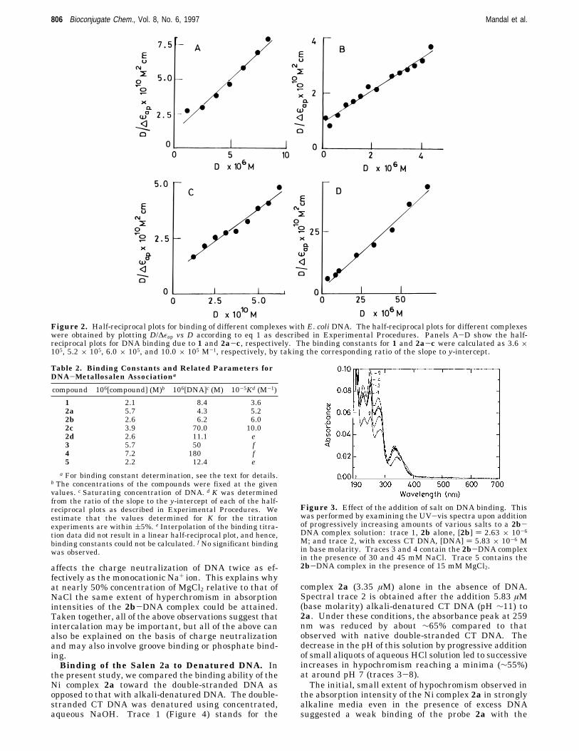
The association constants of different metal complexesor the ligand with DNA were calculated by employingthe method (half-reciprocal plot) using eq 1 as describedpreviously (cf. Experimental Procedures). The half-reciprocal plots for different compounds were constructedusing the most affected wavelength maxima as deter-
mined from the respective absorption titration experi-ments. Importantly, the plots of D/∆εap vs D resulted instraight lines (Figure 2) for the absorption titration of 1and 2a-c with DNA. Individual association constantsdue to DNA binding with 1 and 2a-c are given in Table2. It is important to note that under comparable condi-tions, however, when the absorption titration data ofeither 2d or 5 in the presence of DNA were plotted, theresulting fits did not give linear (straight) half-reciprocalplots. Thus, the saturation plots corresponding to Mncomplexes 2d and 5 (insets of panels E and F of Figure1, respectively) differed from the other saturation plotsgiven in the insets of panels A-D of Figure 1 with othermetal complexes or the ligand 1. These findings suggestthat the binding mode of either of these manganese-basedmetal complexes (2d and 5) with DNA relative to that ofthe free ligand or its Co(II), Cu(II), or Ni(II) complexcounterparts could be different, although all of the salencomplexes showed strong absorption hypochromicities aslong as the compounds contained net cationic charge.Similar differences were also apparent while the effectsof inclusion of these metal complexes on the DNAmeltingwere examined (see below).Effect of the Addition of Salt on DNA Binding.
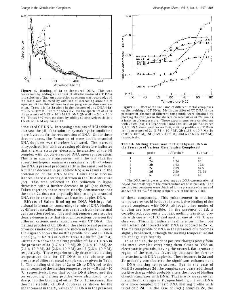
To elucidate the influence of the ionic strength on theDNA binding abilities of the above metal complexes, wethen studied the effects of the addition of increasingamounts NaCl and MgCl2 on the DNA binding of one ofthe above metal complexes, e.g. 2b. This experiment wasperformed by progressive addition of several aliquots ofconcentrated solutions of either NaCl or MgCl2 into asolution containing fully DNA-bound 2b. In Figure 3,spectral trace 1 respresents the UV-vis absorptionspectrum due to the solution containing 2b alone (2.63µM), in the absence of DNA. Spectrum 2 in Figure 3shows the UV-vis absorption spectra of fully DNA-bound2b obtained upon addition of excess CT DNA to thesolution containing 2b. Traces 3 and 4 were obtainedupon addition of 30 and 45 mM NaCl to the 2b-DNAcomplex. Thus, addition of increasing amounts of NaClinto the 2b-DNA complex led to an apparent hyper-chromism in the absorption intensities of 2b finallyreaching a saturation. While further addition of NaCldid not lead to any spectral changes, addition of aconcentrated aqueous solution of MgCl2 into the aboveled to further hyperchromism (trace 5, [MgCl2] ) 15 mM).In a separate experiment, we also examined the effect
of the addition of several aliquots of aqueous solutionsof MgCl2 to a solution containing the 2b-DNA complex(figure not shown). Upon addition of increasing amountsof MgCl2, the apparent hyperchromism in the absorptionintensities of the metal complex was observed, finallyreaching a saturation. To obtain the same extent ofhyperchromism in absorption intensities of the DNA-bound 2b, nearly half the concentration of MgCl2 wasrequired compared to that of NaCl. Notably, the satura-tion in the hyperchromism in the absorption intensitiesof the DNA-bound 2b obtained by the addition of excessMgCl2 could not be altered by further addition of NaCl.The hyperchromism observed upon addition of increas-
ing amounts of salt to salen-bound DNA could beexplained by considering the enhanced charge neutral-ization of the negatively charged DNA in the presenceof the metal ion of the salt (26). This charge neutraliza-tion minimizes the intra and interstrand repulsionbetween the DNA double helix, promoting the formationof more “compact” DNA structure. Under these circum-stances, the average distance between base pairs isdecreased. This makes the accommodation of 2b into theduplex DNA difficult. The dicationic nature of Mg2+ ion
Table 1. UV-Vis Absorption Spectral Properties ofMetallosalens and Metal Free Ligand in the Presenceand Absence of E. coli Genomic DNAa
a See the text for the details of the experimental conditions.b Upon binding with DNA, the λmax due to the bound complexwhich was used for the construction of half-reciprocal plots isitalicized. c E. coli genomic DNA was purified by the phenol-chloroform extraction as described in the text. d No significantspectral changes were observed.
Charge in the Metallosalen Complexes Bioconjugate Chem., Vol. 8, No. 6, 1997 805
affects the charge neutralization of DNA twice as ef-fectively as the monocationic Na+ ion. This explains whyat nearly 50% concentration of MgCl2 relative to that ofNaCl the same extent of hyperchromism in absorptionintensities of the 2b-DNA complex could be attained.Taken together, all of the above observations suggest thatintercalation may be important, but all of the above canalso be explained on the basis of charge neutralizationand may also involve groove binding or phosphate bind-ing.Binding of the Salen 2a to Denatured DNA. In
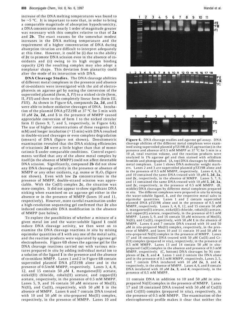
the present study, we compared the binding ability of theNi complex 2a toward the double-stranded DNA asopposed to that with alkali-denatured DNA. The double-stranded CT DNA was denatured using concentrated,aqueous NaOH. Trace 1 (Figure 4) stands for the
complex 2a (3.35 µM) alone in the absence of DNA.Spectral trace 2 is obtained after the addition 5.83 µM(base molarity) alkali-denatured CT DNA (pH ∼11) to2a. Under these conditions, the absorbance peak at 259nm was reduced by about ∼65% compared to thatobserved with native double-stranded CT DNA. Thedecrease in the pH of this solution by progressive additionof small aliquots of aqueous HCl solution led to successiveincreases in hypochromism reaching a minima (∼55%)at around pH 7 (traces 3-8).The initial, small extent of hypochromism observed in
the absorption intensity of the Ni complex 2a in stronglyalkaline media even in the presence of excess DNAsuggested a weak binding of the probe 2a with the
Figure 2. Half-reciprocal plots for binding of different complexes with E. coli DNA. The half-reciprocal plots for different complexeswere obtained by plotting D/∆εap vs D according to eq 1 as described in Experimental Procedures. Panels A-D show the half-reciprocal plots for DNA binding due to 1 and 2a-c, respectively. The binding constants for 1 and 2a-c were calculated as 3.6 ×10⁵, 5.2 × 10⁵, 6.0 × 10⁵, and 10.0 × 10⁵ M-1, respectively, by taking the corresponding ratio of the slope to y-intercept.
Table 2. Binding Constants and Related Parameters forDNA-Metallosalen Associationa
a For binding constant determination, see the text for details.b The concentrations of the compounds were fixed at the givenvalues. c Saturating concentration of DNA. d K was determinedfrom the ratio of the slope to the y-intercept of each of the half-reciprocal plots as described in Experimental Procedures. Weestimate that the values determined for K for the titrationexperiments are within (5%. e Interpolation of the binding titra-tion data did not result in a linear half-reciprocal plot, and hence,binding constants could not be calculated. f No significant bindingwas observed.
Figure 3. Effect of the addition of salt on DNA binding. Thiswas performed by examining the UV-vis spectra upon additionof progressively increasing amounts of various salts to a 2b-DNA complex solution: trace 1, 2b alone, [2b] ) 2.63 × 10⁻⁶
M; and trace 2, with excess CT DNA, [DNA] ) 5.83 × 10⁻⁶ Min base molarity. Traces 3 and 4 contain the 2b-DNA complexin the presence of 30 and 45 mM NaCl. Trace 5 contains the2b-DNA complex in the presence of 15 mM MgCl2.
denatured CT DNA. Increasing amounts of HCl additiondecrease the pH of the solution by making the conditionsmore favorable for the renaturation of DNA. Under thesecircumstances, the formation of more double-strandedDNA duplexes was therefore facilitated. The increasein hypochromism with decreasing pH therefore indicatesthat there is stronger electronic interaction of the Nicomplex with double-stranded DNA upon renaturation.This is in complete agreement with the fact that theabsorption hypochromism was maximal at pH ∼7 wherethe DNA is present predominantly in the renatured form.A further decrease in pH (below 6.5) also results in theprotonation of the DNA bases. Under these circum-stances, there is a strong distortion in the DNA structure(32). This was reflected in the reduction in hypo-chromism with a further decrease in pH (not shown).Taken together, these results clearly demonstrate thatthe salen 2a does not physically bind to single-strandedDNA to the extent it does with native duplex DNA.Effects of Salen Binding on DNA Melting. Ad-
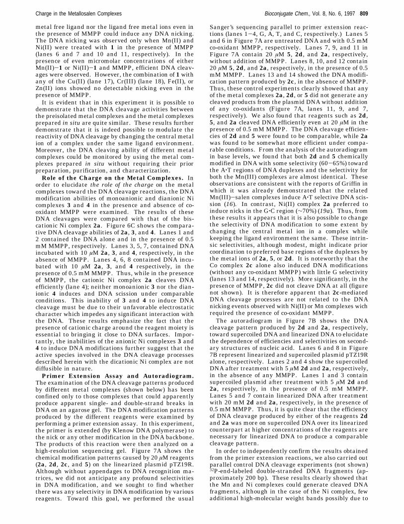
ditional information concerning the role of DNA bindingby different metallosalens was available from the thermaldenaturation studies. The melting temperature studiesclearly demonstrate that strong interactions between thedifferent cationic metal complexes with CT DNA. Themelting profiles of CT DNA in the absence and presenceof various metal complexes are shown in Figure 5. Curve1 in Figure 5 shows the melting profile of 72 µMCT DNAalone (Tm ∼ 62 °C) in 5 mM Tris-HCl buffer (pH 7.4).Curves 2-6 show the melting profiles of the CT DNA inthe presence of 2a (1.7 × 10⁻⁵ M), 2b (1.6 × 10⁻⁵ M), 2c(2.1 × 10⁻⁵ M), 2d (2.6 × 10⁻⁵ M), and 5 (2.6 × 10⁻⁵ M),respectively. These experimentally determined meltingtemperature data for CT DNA in the absence andpresence of different metal complexes are given in Table3. The binding of either of 2a or 2b to DNA led to theenhancement of the melting temperature by∼18 and∼10°C, respectively, from that of the DNA alone, and thecorresponding melting profiles were also found to besharp. So the binding of either 2a or 2b increased thethermal stability of DNA duplexes as shown by theenhancement in the Tm values of CT DNA in the presence
of these compounds. This enhancement in meltingtemperatures could be due to intercalative binding of themetal complexes with DNA, although other modes ofbinding are also possible. In the presence of 2d, acomplicated, apparently biphasic melting transition pro-file with one at ∼51 °C and another one at ∼79 °C wasobserved. This might indicate the differences in the modewith which 2d interacts with DNA as opposed to 2a-c.The melting profile of DNA in the presence of 5 becomesslightly broadened, although the melting temperature didnot change significantly.In 2a and 2b, the pendant positive charges (away from
the metal complex core) bring them closer to DNA onelectrostatic grounds, leaving the neutral, flat, aromaticregion of the complex favorably disposed for furtherinteraction with DNA duplexes. These features in 2a and2b probably contribute to the significant enhancementin DNA melting temperatures. But in the case ofMn(III) complexes 2d, the complex core bears additionalpositive charge which probably alters the mode of bindingof such complexes with DNA. That is why we see eitherinsignificant changes in Tm upon binding of 5 with DNAor a more complex biphasic DNA melting profile withtricationic 2d. In the case of Co(II) complex 2c, the
Figure 4. Binding of 2a to denatured DNA. This wasperformed by adding an aliquot of alkali-denatured CT DNAinto solution of 2a. An absorption spectrum was recorded, andthe same was followed by addition of increasing amounts ofaqueous HCl to this mixture to allow progressive slow renatur-ation. Trace 1 is for 2a alone in the absence of any DNA; [2a]) 3.35 × 10⁻⁶ M. Trace 2 shows UV-vis the spectrum of 2a inthe presence of 5.83 × 10⁻⁶ M CT DNA ([NaOH] ) 5.0 × 10⁻³
M). Traces 3-7 were obtained by adding successively each time1.5 µL of 0.6 M aqueous HCl.
Figure 5. Effect of the inclusion of different metal complexeson the melting of CT DNA. Melting profiles of CT DNA in thepresence or absence of different compounds were obtained byplotting the changes in the absorption intensities at 260 nm asa function of temperature. These experiments were carried outwith 72 µM (bM) CT DNA with 5 mM Tris-HCl at pH 7.4: curve1, CT DNA alone; and curves 2-6, melting profiles of CT DNAin the presence of 2a (1.74 × 10⁻⁵ M), 2b (1.63 × 10⁻⁵ M), 2c(2.09 × 10⁻⁵ M), 2d (2.59 × 10⁻⁵ M), and 5 (2.63 × 10⁻⁵ M),respectively.
Table 3. Melting Transitions for Calf Thymus DNA inthe Presence of Various Metallosalen Complexesa
entry probe 105[probe]b Tm (°C)c
1 d d 622 2a 1.74 803 2b 1.63 72.54 2c 2.09 67.55 2d 2.59 79, 516 5 2.63 62
a The DNA melting was carried out at a DNA concentration of72 µM (base molarity). b The concentration of the salen used. c Themelting temperatures were obtained in the presence of salen andare within (1 °C. d Melting temperature of the DNA alone.
Charge in the Metallosalen Complexes Bioconjugate Chem., Vol. 8, No. 6, 1997 807
increase of the DNA melting temperatures was found tobe ∼5 °C. It is important to note that, in order to bringa comparable magnitude of absorption hypochromicity,a DNA concentration nearly 1 order of magnitude greaterwas necessary with this complex relative to that of 2aand 2b. The exact reasons for the somewhat modestincreases in the DNA melting temperature and therequirement of a higher concentration of DNA duringabsorption titration are difficult to interpret adequatelyat this time. However, it could be (i) due to the abilityof 2c to promote DNA scission even in the absence of co-oxidants and (ii) owing to its high oxygen bindingcapacity (24) the resulting complex may also adopt anonplanar shape. This deviation from planarity couldalter the mode of its interaction with DNA.DNA Cleavage Studies. The DNA cleavage abilities
of different metal complexes in the presence and absenceof co-oxidants were investigated with the aid of electro-phoresis on agarose gel by noting the conversion of thesupercoiled plasmid (form, I, FI) to a nicked circle (form,II, FII) and then to the completely linear form (form III,FIII). As shown in Figure 6A, compounds 2a, 2d, and 5were able to induce oxidative cleavages of DNA. Incuba-tion of the plasmid DNA pTZ19R at 37 °C for 5 min with10 µM 2a, 2d, and 5 in the presence of MMPP causedappreciable conversion of form I to the nicked circularform II (lanes 9, 7, and 5, respectively, in Figure 6A).The use of higher concentrations of these reagents (0.1mM) and longer incubation (>15 min) with DNA resultedin double-strand cleavages or even complete degradation(smears) of DNA (figure not shown). Densitometricexamination revealed that the DNA nicking efficienciesof tricationic 2d were a little higher than that of mono-cationic 5 under comparable conditions. Control experi-ments verified that any of the above metal complex byitself (in the absence of MMPP) could not affect detectableDNA scission. Significantly, compound 2b did not showany DNA cleavage activity in the presence or absence ofMMPP or any other oxidants, e.g. oxone or H2O2 (figurenot shown). Even with low 2a concentrations in thepresence of MMPP, double-strand scission was appre-ciable. With the Co(II) complex 2c, the situation wasmore complex. It did not appear to show significant DNAnicking when examined on an agarose gel either in theabsence or in the presence of MMPP (lanes 10 and 11,respectively). However, more careful examination undera high-resolution sequencing gel confirmed that 2c alsoinduced considerable DNA modification in the absenceof MMPP (see below).To explore the possibilities of whether a mixture of a
given metal ion and the water-soluble ligand 1 couldinduce DNA cleavage activity, we then went on toexamine the DNA cleavage reactions in situ by mixingequimolar quantities of 1 with any one of the metal salts,and the reaction products were separated by agarose gelelectrophoresis. Figure 6B shows the agarose gel for theDNA cleavage reactions carried out with various mix-tures prepared in situ by adding individual metal ion toa solution of the ligand 1 in the presence and the absenceof co-oxidant MMPP. Lanes 1 and 2 in Figure 6B containsupercoiled plasmid DNA pTZ19R alone and in thepresence of 0.5/mM MMPP, respectively. Lanes 3, 4, 8,12, and 15 contain 50 µM 1, manganese(II) acetate,nickel(II) chloride, cobalt(II) acetate, and copper(II)acetate, respectively, in the presence of 0.5 mM MMPP.Lanes 5, 9, and 16 contain 50 µM mixtures of Mn(II),Ni(II), and Cu(II), respectively, with 50 µM 1 in theabsence of MMPP. Lanes 6 and 7 contain DNA treatedwith 10 and 50 µM in situ-prepared Mn(II) complex,respectively, in the presence of MMPP. Lanes 10 and
11 contain DNA in addition to 10 and 50 µM in situ-prepared Ni(II) complex in the presence of MMPP. Lanes17 and 18 contained DNA treated with 50 µM of Cu(II)and Cr(III) complex (prepared in situ), respectively, inthe presence of 0.5 mM MMPP. The examination of theelectrophoreetic profile makes it clear that neither the
Figure 6. DNA cleavage studies and agarose gel assay. DNAcleavage abilities of the different metal complexes were exam-ined using supercoiled plasmid pTZ19R (0.25 µg/reaction) in thepresence and absence of 0.5 mM MMPP at 37 °C for 5 min in a10 µL total reaction volume, and the reaction products wereanalyzed in 1% agarose gel and then stained with ethidiumbromide and photographed. (A, top) DNA cleavages by differentmetal complexes. Lane 1 shows DNA molecular weight mark-ers. Lanes 2 and 3 are supercoiled plasmid pTZ19R alone andin the presence of 0.5 mM MMPP, respectively. Lanes 4, 6, 8,and 10 contained the same DNA treated with 10 µM 5, 2d, 2a,and 2c, respectively, in the absence of MMPP. Lanes 5, 7, 9,and 11 contained the same DNA treated with 10 µM 5, 2d, 2a,and 2c, respectively, in the presence of 0.5 mM MMPP. (B,middle) DNA cleavages by different metal complexes preparedin situ. The different complexes were prepared in situ by mixingthe water-soluble ligand, 1, and the respective metal salts inequimolar quantities. Lanes 1 and 2 contain supercoiledplasmid DNA pTZ19R alone and in the presence of 0.5 mMMMPP, respectively. Lanes 3, 4, 8, 12, and 15 contain 50 µM1, manganese(II) acetate, nickel(II) chloride, cobalt(II) acetate,and copper(II) acetate, respectively, in the presence of 0.5 mMMMPP. Lanes 5, 9, and 16 contain 50 µM mixtures of Mn(II),Ni(II), and Cu(II), respectively, with 50 µM 1 in the absence ofMMPP. Lanes 6 and 7 contain DNA treated with 10 and 50µM in situ-prepared Mn(II) complex, respectively, in the pres-ence of MMPP, and lanes 10 and 11 contain 10 and 50 µM insitu-prepared Ni(II) complex in the presence of MMPP. Lanes17 and 18 contained DNA treated with 50 µM Cu(II) and Cr-(III) complex (prepared in situ), respectively, in the presence of0.5 mM MMPP. Lanes 13 and 14 contain 50 µM in situ-prepared Co(II) complex in the absence and presence of 0.5 mMMMPP, respectively. (C, bottom) DNA cleavages by Ni com-plexes of 2a, 3, and 4. Lanes 1 and 2 contain the DNA aloneand in the presence of 0.5 mMMMPP, respectively, Lanes 3, 5,and 7 contain DNA incubated with 10 µM 2a, 3, and 4,respectively, in the absence of MMPP. Lanes 4, 6, and 8 containDNA incubated with 10 µM 2a, 3, and 4, respectively, in thepresence of 0.5 mM MMPP.
metal free ligand nor the ligand free metal ions even inthe presence of MMPP could induce any DNA nicking.The DNA nicking was observed only when Mn(II) andNi(II) were treated with 1 in the presence of MMPP(lanes 6 and 7 and 10 and 11, respectively). In thepresence of even micromolar concentrations of eitherMn(II)-1 or Ni(II)-1 and MMPP, efficient DNA cleav-ages were observed. However, the combination of 1 withany of the Cu(II) (lane 17), Cr(III) (lane 18), Fe(II), orZn(II) ions showed no detectable nicking even in thepresence of MMPP.It is evident that in this experiment it is possible to
demonstrate that the DNA cleavage activities betweenthe preisolated metal complexes and the metal complexesprepared in situ are quite similar. These results furtherdemonstrate that it is indeed possible to modulate thereactivity of DNA cleavage by changing the central metalion of a complex under the same ligand environment.Moreover, the DNA cleaving ability of different metalcomplexes could be monitored by using the metal com-plexes prepared in situ without requiring their priorpreparation, purification, and characterization.Role of the Charge on the Metal Complexes. In
order to elucidate the role of the charge on the metalcomplexes toward the DNA cleavage reactions, the DNAmodification abilities of monoanionic and dianionic Nicomplexes 3 and 4 in the presence and absence of co-oxidant MMPP were examined. The results of theseDNA cleavages were compared with that of the bis-cationic Ni complex 2a. Figure 6C shows the compara-tive DNA cleavage abilities of 2a, 3, and 4. Lanes 1 and2 contained the DNA alone and in the presence of 0.5mM MMPP, respectively. Lanes 3, 5, 7, contained DNAincubated with 10 µM 2a, 3, and 4, respectively, in theabsence of MMPP. Lanes 4, 6, 8 contained DNA incu-bated with 10 µM 2a, 3, and 4 respectively, in thepresence of 0.5 mMMMPP. Thus, while in the presenceof MMPP, the cationic Ni complex 2a cleaves DNAefficiently (lane 4); neither monoanionic 3 nor the dian-ionic 4 induces and DNA scission under comparableconditions. This inability of 3 and 4 to induce DNAcleavage must be due to their unfavorable electrostaticcharacter which impedes any significant interaction withthe DNA. These results emphasize the fact that thepresence of cationic charge around the reagent moiety isessential to bringing it close to DNA surfaces. Impor-tantly, the inabilities of the anionic Ni complexes 3 and4 to induce DNA modifications further suggest that theactive species involved in the DNA cleavage processesdescribed herein with the dicationic Ni complex are notdiffusible in nature.Primer Extension Assay and Autoradiogram.
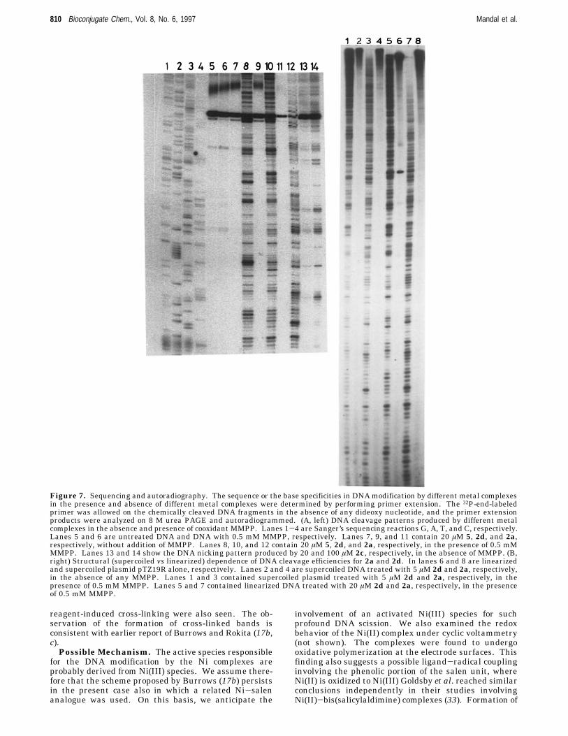
The examination of the DNA cleavage patterns producedby different metal complexes (shown below) has beenconfined only to those complexes that could apparentlyproduce apparent single- and double-strand breaks inDNA on an agarose gel. The DNA modification patternsproduced by the different reagents were examined byperforming a primer extension assay. In this experiment,the primer is extended (by Klenow DNA polymerase) tothe nick or any other modification in the DNA backbone.The products of this reaction were then analyzed on ahigh-resolution sequencing gel. Figure 7A shows thechemical modification patterns caused by 20 µM reagents(2a, 2d, 2c, and 5) on the linearized plasmid pTZ19R.Although without appendages to DNA recognition ma-trices, we did not anticipate any profound selectivitiesin DNA modification, and we sought to find whetherthere was any selectivity in DNAmodification by variousreagents. Toward this goal, we performed the usual
Sanger’s sequencing parallel to primer extension reac-tions (lanes 1-4, G, A, T, and C, respectively.) Lanes 5and 6 in Figure 7A are untreated DNA and with 0.5 mMco-oxidant MMPP, respectively. Lanes 7, 9, and 11 inFigure 7A contain 20 µM 5, 2d, and 2a, respectively,without addition of MMPP. Lanes 8, 10, and 12 contain20 µM 5, 2d, and 2a, respectively, in the presence of 0.5mM MMPP. Lanes 13 and 14 showed the DNA modifi-cation pattern produced by 2c, in the absence of MMPP.Thus, these control experiments clearly showed that anyof the metal complexes 2a, 2d, or 5 did not generate anycleaved products from the plasmid DNA without additionof any co-oxidants (Figure 7A, lanes 11, 9, and 7,respectively). We also found that reagents such as 2d,5, and 2a cleaved DNA efficiently even at 20 µM in thepresence of 0.5 mM MMPP. The DNA cleavage efficien-cies of 2d and 5 were found to be comparable, while 2awas found to be somewhat more efficient under compa-rable conditions. From the analysis of the autoradiogramin base levels, we found that both 2d and 5 chemicallymodified in DNA with some selectivity (60-65%) towardthe A‚T regions of DNA duplexes and the selectivity forboth the Mn(III) complexes are almost identical. Theseobservations are consistent with the reports of Griffin inwhich it was already demonstrated that the relatedMn(III)-salen complexes induce A‚T selective DNA scis-sion (16). In contrast, Ni(II) complex 2a preferred toinduce nicks in the G‚C region (∼70%) (19a). Thus, fromthese results it appears that it is also possible to changethe selectivity of DNA modification to some extent bychanging the central metal ion in a complex whilekeeping the ligand environment the same. These intrin-sic selectivities, although modest, might indicate priorcoordination to preferred base regions of the duplexes bythe metal ions of 2a, 5, or 2d. It is noteworthy that theCo complex 2c alone also induced DNA modifications(without any co-oxidant MMPP) with little G selectivity(lanes 13 and 14, respectively). More significantly, in thepresence of MMPP, 2c did not cleave DNA at all (figurenot shown). It is therefore apparent that 2c-mediatedDNA cleavage processes are not related to the DNAnicking events observed with Ni(II) or Mn complexes wichrequired the presence of co-oxidant MMPP.The autoradiogram in Figure 7B shows the DNA
cleavage pattern produced by 2d and 2a, respectively,toward supercoiled DNA and linearized DNA to elucidatethe dependence of efficiencies and selectivities on second-ary structures of nucleic acid. Lanes 6 and 8 in Figure7B represent linearized and supercoiled plasmid pTZ19Ralone, respectively. Lanes 2 and 4 show the supercoiledDNA after treatment with 5 µM 2d and 2a, respectively,in the absence of any MMPP. Lanes 1 and 3 containsupercoiled plasmid after treatment with 5 µM 2d and2a, respectively, in the presence of 0.5 mM MMPP.Lanes 5 and 7 contain linearized DNA after treatmentwith 20 mM 2d and 2a, respectively, in the presence of0.5 mMMMPP. Thus, it is quite clear that the efficiencyof DNA cleavage produced by either of the reagents 2dand 2a was more on supercoiled DNA over its linearizedcounterpart at higher concentrations of the reagents arenecessary for linearized DNA to produce a comparablecleavage pattern.In order to independently confirm the results obtained
from the primer extension reactions, we also carried outparallel control DNA cleavage experiments (not shown)32P-end-labeled double-stranded DNA fragments (ap-proximately 200 bp). These results clearly showed thatthe Mn and Ni complexes could generate cleaved DNAfragments, although in the case of the Ni complex, fewadditional high-molecular weight bands possibly due to
Charge in the Metallosalen Complexes Bioconjugate Chem., Vol. 8, No. 6, 1997 809
reagent-induced cross-linking were also seen. The ob-servation of the formation of cross-linked bands isconsistent with earlier report of Burrows and Rokita (17b,c).Possible Mechanism. The active species responsible
for the DNA modification by the Ni complexes areprobably derived from Ni(III) species. We assume there-fore that the scheme proposed by Burrows (17b) persistsin the present case also in which a related Ni-salenanalogue was used. On this basis, we anticipate the
involvement of an activated Ni(III) species for suchprofound DNA scission. We also examined the redoxbehavior of the Ni(II) complex under cyclic voltammetry(not shown). The complexes were found to undergooxidative polymerization at the electrode surfaces. Thisfinding also suggests a possible ligand-radical couplinginvolving the phenolic portion of the salen unit, whereNi(II) is oxidized to Ni(III) Goldsby et al. reached similarconclusions independently in their studies involvingNi(II)-bis(salicylaldimine) complexes (33). Formation of
Figure 7. Sequencing and autoradiography. The sequence or the base specificities in DNAmodification by different metal complexesin the presence and absence of different metal complexes were determined by performing primer extension. The 32P-end-labeledprimer was allowed on the chemically cleaved DNA fragments in the absence of any dideoxy nucleotide, and the primer extensionproducts were analyzed on 8 M urea PAGE and autoradiogrammed. (A, left) DNA cleavage patterns produced by different metalcomplexes in the absence and presence of cooxidant MMPP. Lanes 1-4 are Sanger’s sequencing reactions G, A, T, and C, respectively.Lanes 5 and 6 are untreated DNA and DNA with 0.5 mM MMPP, respectively. Lanes 7, 9, and 11 contain 20 µM 5, 2d, and 2a,respectively, without addition of MMPP. Lanes 8, 10, and 12 contain 20 µM 5, 2d, and 2a, respectively, in the presence of 0.5 mMMMPP. Lanes 13 and 14 show the DNA nicking pattern produced by 20 and 100 µM 2c, respectively, in the absence of MMPP. (B,right) Structural (supercoiled vs linearized) dependence of DNA cleavage efficiencies for 2a and 2d. In lanes 6 and 8 are linearizedand supercoiled plasmid pTZ19R alone, respectively. Lanes 2 and 4 are supercoiled DNA treated with 5 µM 2d and 2a, respectively,in the absence of any MMPP. Lanes 1 and 3 contained supercoiled plasmid treated with 5 µM 2d and 2a, respectively, in thepresence of 0.5 mM MMPP. Lanes 5 and 7 contained linearized DNA treated with 20 µM 2d and 2a, respectively, in the presenceof 0.5 mM MMPP.
ligand-radical should in part be responsible for the DNAcross-linking.With regard to the chemistry of the Mn complexes, we
feel that the DNA cleavages in these instances areaffected in the following manner. The combination of thesalen-Mn3+ and MMPP produces direct DNA nicksthrough pathways that do not require dioxygen. DNAcleavage releases free, unmodified nucleobases. Suchobservations are consistent with a plausible mechanisminvolving deoxyribose CH activation potentiated by anoxidatively activated species such as [salen-Mn(V)O]+.The above suggestions are indeed supported in literaturefrom the independent work of Griffin and others (16b,34).The mechanistic pathways by which the Co(II)-salen-
O2-mediated DNAmodification occurs is currently underinvestigation. Preliminary studies indicate that a su-peroxide radical-mediated active species is probablyresponsible for the observed DNA modification in thisinstance. In the presence of MMPP, the complex isoxidized to a Co(III) species which does not have abilityto activate dioxygen, and hence, this does not possessDNA cleaving ability.Concluding Remarks. In summary, we have devel-
oped several salen-based transition metal complexeswhich interact with or modify DNA. The binding andDNA modifications by such complexes can be modulatedby the charge on the salen unit and more profoundly bythe selection of the central metal ion. Only the salenswith net cationic electrostatic character bind DNA, andthe physical binding of the salen complexes does not occurto denatured (predominantly single-stranded) DNA to theextent that it does with native double-stranded DNA.Thus, probably a combination of the electrostatic andintercalative/groove binding interactions with DNA raisethe duplex melting temperatures significantly. Theanionic salens neither bind DNA nor induce strandscission irrespective of the central metal ion selected. Thesupercoiled forms of the plasmid DNA were found to bemore susceptible to scission compared to their linearizedforms by the cationic salens. Thus, to obtain comparabledegrees of DNA cleavage, higher concentrations of thereagents were necessary with the linearized DNA.The present findings demonstrate that modulation of
the reactivities of the salen-based reagents toward DNAis possible and form a basis for further exciting studies.The newly introduced salen derivatives when tetheredto suitable nucleic acid structural recognition matricesshould be attractive as sequence specific reagents. Elu-cidations of the exact reaction conditions and mechanisticpathways for such important applications and also forthe mapping of DNA tertiary structures are currentlyunderway in our laboratory.
ACKNOWLEDGMENT
We are grateful to the referees for useful comments.S.S.M. thanks CSIR for a senior research fellowship. Thiswork was supported by the funding from DST (INDO-HUNG to S.B.) and by the DBT and DST (to U.V.).
LITERATURE CITED
(1) (a) Sigman, D. S., Bruice, T. W., Mazumder, A., and Sutton,C. L. (1993) Acc. Chem. Res. 26, 98. (b) Barton, J. K., andPyle, A. M. (1990) Prog. Inorg. Chem. 38, 413. (c) Burkhoff,A. M., and Tullius, T. D. (1988)Nature 331, 455. (d) Riordan,C. G., and Wei, P. J. (1992) J. Am. Chem. Soc. 116, 2189.
(2) (a) Tan, J. D., Hudson, S. E., Brown, S. J., Olmted, M. M.,and Mascharak, P. K. (1992) J. Am. Chem. Soc. 114, 3841.(b) Nagai, K., Carter, B. J., Xu, J., and Hecht, S. M. (1991) J.
Am. Chem. Soc. 113, 5099. (c) Nicolau, K. C., Maligres, P.,Shin, J., de Leon, E., and Rideout, D. (1992) J. Am. Chem.Soc. 114, 7825.
(3) Dervan, P. B. (1992) Nature 359, 87.(4) Sigman, D. S., Mazumder, A., and Perrin, D. M. (1993)Chem. Rev. 93, 2295.
(5) Sardesai, N. Y., Zimmermann, K., and Barton, J. K. (1994)J. Am. Chem. Soc. 116, 7502.
(6) Pratviel, G., Bernadou, J., and Meunier, B. (1995) Angew.Chem., Int. Ed. Engl. 34, 746.
(7) Keck, M.-V., and Lippard, S. J. (1992) J. Am. Chem. Soc.114, 3386.
(8) Burrows, C. J., and Rokita, S. E. (1994) Acc. Chem. Res. 27,295.
(9) (a) Breslin, D. T., and Schuster, G. B. (1996) J. Am. Chem.Soc. 118, 2311. (b) Breiner, K. M., Daugherty, M. A., Das, T.G., and Thorp, H. H. (1995) J. Am. Chem. Soc. 117, 11673.(c) Croke, D. T., Perrouslt, L., Sari, M. A., Battionti, J. P.,Mansuy, D., Helene, C., and Doon, T. L. (1993) J. Photochem.Photobiol. 18, 41. (d) Nielsen, P. E. (1990) J. Mol. Recognit.3, 1. (e) Bhattacharya, S., and Mandal, S. S. (1996) J. Chem.Soc., Chem. Commun., 1515.
(10) (a) Papavassiliou, A. G. (1995) Biochem. J. 305, 345. (b)Siltani, A., Long, E. C., Pyle, A. M., and Barton, J. K. (1992)J. Am. Chem. Soc. 114, 2303.
(11) (a) Guajardo, R. J., Hudson, S. E., Brown, S. J., andMascharak, P. K. (1993) J. Am. Chem. Soc. 115, 7971. (b)Stubbe, J., and Kozarich, J. W. (1987) Chem. Rev. 87, 1107.(c) Uhlmann, E., and Reyman, A. (1990) Chem. Rev. 90, 543.
(12) Jaeger, J., Santalucia, J., and Tinoco, I. (1993) Annu. Rev.Biochem. 62, 255.
(13) Taylor, S. J., Schultz, P. G., and Dervan, P. B. (1984)Tetrahedron 40, 457.
(14) (a) Kasprazak, K. S. (1991) Chem. Res. Toxicol. 4, 604. (b)Tullius, T. D., Ed. (1989) Metal-DNA Chemistry, ACSSymposium Series 402, American Chemical Society, Wash-ington, DC. (c) Vahter, M. E. (1988) in Biological Monitoringof Toxic Metals (Clarkson, T. W., Friberg, L., Nordberg, G.F., and Sanger, P. R., Eds.) pp 303-321, Plenum, New York.(d) Yamanaka, K., and Okada, S. (1994) Environ. HealthPerspect. 102 (Suppl. 3), 37.
(15) Sato, K., Chikira, M., Fujii, Y., and Komatsu, A. (1994) J.Chem. Soc., Chem. Commun., 625.
(16) (a) Gravert, D. J., and Griffin, J. H. (1993) J. Org. Chem.58, 820. (b) Gravert, D. J., and Griffin, J. H. (1996) Inorg.Chem. 35, 4837.
(17) (a) Woodson, S. A., Muller, J. G., Burrows, C. J., andRokita, S. E. (1993) Nucleic Acids Res. 21, 5524. (b) Muller,J. G., Paikoff, S. J., Rokita, S. E., and Burrows, C. J. (1994)J. Inorg. Biochem. 54, 199. (c) Burrows, C. J., and Rokita, S.E. (1994) Acc. Chem. Res. 27, 295.
(18) Routier, S., Bernier, J.-L., Waring, M. J., Colson, P.,Houssier, C., and Baily, C. (1996) J. Org. Chem. 61, 2326.
(19) (a) Mandal, S. S., Vinaykumar, N., Varshney, U., andBhattacharya, S. (1996) J. Inorg. Biochem. 63, 265. (b)Bhattacharya, S., and Mandal, S. S. (1997) Biochem. Biophys.Acta 1323, 29. (c) Mandal, S. S., Renuka, K., Guru Row, T.N., and Bhattacharya, S. (1996) J. Chem. Soc., Chem.Commun. 2725.
(20) Rajaram, R., Balachandran, U. N., and Ramasami, T.(1994) Biochem. Biophys. Res. Commun. 205, 327.
(21) Muller, W., and Crothers, D. M. (1975) Eur. J. Biochem.54, 267.
(22) Reichmann, Y., Rice, S. A., Thomas, C. A., and Doty, P.(1954) J. Am. Chem. Soc. 76, 3047.
(23) Maniatis, T., Fritsch, E. F., and Sambrook, S. (1982)Molecular Cloning, p 458, Cold Spring Harbor LaboratoryPress, Plainview, NY.
(24) Busetto, C., Cariati, F., Fusi, A., Gullotti, M., Marazzoni,F., Pasini, A., Ugo, R., and Valenti, V. (1973) J. Chem. Soc.,754.
(25) Pyle, A. M., Rehmann, J. P., Meshoyrer, R., Kumar, C. V.,Turro, N. J., and Barton, J. K. (1989) J. Am. Chem. Soc. 111,3051.
(26) Meehan, T., Gamper, H., and Becker, J. F. (1982) J. Biol.Chem. 257, 10479.
Charge in the Metallosalen Complexes Bioconjugate Chem., Vol. 8, No. 6, 1997 811
(27) Cory, M., McKee, D. D., Kagan, J., Henry, D. W., andMiller, J. A. (1985) J. Am. Chem. Soc. 107, 2528.
(28) Sasse-Dwight, S., and Gralla, J. D. (1988) J.Mol. Biol. 202,107.
(29) Sheldon, R. A., and Kochi, J. K. (1981) Metal-CatalyzedOxidation of Organic Compounds, Academic, New York.
(30) (a) Koola, J. D., and Kochi, J. K. (1987) Inorg. Chem. 26,908. (b) Chen, D., and Martell, A. E. (1987) Inorg. Chem.26, 1026. (c) Yoon, H., and Burrows, C. J. (1988) J. Am.Chem. Soc. 110, 4087.
(31) (a) Cheng, C.-C., Goll, J. G., Neyhart, G. A., Welch, T. W.,Singh, P., and Thorp, H. H. (1995) J. Am. Chem. Soc. 117,2970. (b) Groves, J. T., and Kady, I. O. (1993) Inorg. Chem.
32, 3868. (c) Pamatong, F. V., Detmer, C. A., III, andBocarsly, J. R. (1996) J. Am. Chem. Soc. 118, 5339. (d) Keek,M. V., and Lippard, S. J. (1992) J. Am. Chem. Soc. 114, 3386.(e) Carter, M. T., Rodriguez, M., and Bard, A. J. (1989) J.Am. Chem. Soc. 111, 8901.
(32) Singleton, S. F., and Dervan, P. B. (1992) Biochemistry 31,1099.
(33) Goldsby, K. A., Blaho, J. K., and Hoferkamp, L. A. (1989)Polyhedron 8, 113.
(34) Srinivasan, K., Michand, P., and Kochi, J. K. (1986) J. Am.Chem. Soc. 108, 2309.